Effect of Triple Antibiotic Paste on Bond Strength of Composite to
Dentin: An In Vitro Study
Maryam Zare Jahromi1 , Parvin Mirzakouchaki2, Sanaz Mirsatari3, Shadi Kazemi4.
1Assistant Professor, Department of Endodontics, School of Dentistry, Islamic Azad University Isfahan (Khorasgan) Branch,
Isfahan, Iran
2Assistant Professor, Department of Operative Dentistry, School of Dentistry, Islamic Azad University Isfahan (Khorasgan)
Branch, Isfahan, Iran
3Dentist, Private Office, Isfahan, Iran
4Postgraduate Student, Department of Endodontics, School of Dentistry, Islamic Azad University, Isfahan (Khorasgan) Branch,
Isfahan, Iran
Corresponding author: M. Zare Jahromi, Assistant Professor, Department of Endodontics, School of Dentistry, Islamic Azad University Isfahan (Khorasgan) Branch, Isfahan, Iran
m.zare@khuisf.ac.ir
Received: 20 Oct 2014 Accepted: 28 May 2015
Abstract
Background and Aim: Considering the increasing use of antibiotics in root canal therapy and the possible effects of intracanal medicaments on the bond strength of composite restorations, the aim of this study was to evaluate the effect of triple antibiotic paste (TAP) on shear bond strength of three types of composite resins to dentin.
Materials and Methods: In this in-vitro, experimental study, the enamel of 60 extracted premolars was ground parallel to the longitudinal axis of the teeth in order to produce flat dentin surfaces (5 mm2minimum surface area). The samples were divided into six
groups. In the control group, dentin surfaces were stored in saline solution and in the experimental groups dentin surfaces were exposed to TAP for 10 days. After washing and drying, Single Bond was applied for dentin bond to Z100 and Z350 composites. Adhesive resin was used for bond to P90 composite. Composites were applied on the dentin surfaces in six groups and cured in four directions. The shear bond strength was measured by Instron machine. The data were analyzed by t-test, one-way ANOVA and Tukey’s test.
Results: The highest mean shear bond strength was found in Z350 composite group following the use of TAP (38.75 MPa) and the lowest mean strength was found in Z100 composite group following the use of TAP (14.64 MPa). The mean shear bond strength of P90 and Z100 composites (in contrast to Z350 composite) was higher in the saline compared to the TAP groups (p=0.1). The differences between saline and antibiotic groups were not significant (p=0.959) but the difference in this regard among the three types of composites was significant.
Conclusion: The results of this study showed that use of TAP has no effect on the shear bond strength of composite to dentin but the type of composite significantly affects the shear bond strength.
Key Words: Triple Antibiotic Paste, Composite, Shear Bond
Journal of Islamic Dental Association of IRAN (JIDAI) Spring 2015 ;27, (2)
Introduction
Success of endodontic treatment depends on several factors and attempts are made to find methods to decrease the failure rate [1,2].
Restoration of endodontically treated teeth is very important [3]. Changes in the enamel and dentin structure as the result of use of root canal treatment
materials can affect the quality of tooth-colored restorations [3]. Failure at the dentin-restoration interface depends on the general characteristics of the primary substrate and adhesive, time and environmental factors [4]. At present, composite resins are increasingly used for tooth restoration, and production of composites with higher bond
strength and antibacterial effects may significantly increase the durability of restorations [5-7]. Moreover, use of more efficient restorative techniques such as total etch and self etch systems that improve the resin-dentin bond can also enhance the quality and longevity of restorations [8,9]. Several studies have evaluated the effect of factors such as the cavity preparation design, irrigants, adhesives, solvents and aseptic agents on bond strength of composite to dentin with variable results [10-15].
Intracanal medicaments have long been used as an adjunct in root canal therapy and play a significant role in success of these treatments [16]. Antibiotics have also been proposed for use in endodontic treatments, and prevention of antibiotic resistance and efficient elimination of microorganisms are among the major goals of endodontic treatments. Investigations are ongoing to find effective antibiotics and an aseptic treatment technique for elimination of microorganisms [17-19]. Use of a single antibiotic does not seem to be adequate for an aseptic treatment.
Santos et al, in their in vitro study in 2009 showed that doxycycline was more efficient for smear layer removal at the cervical and middle thirds of the root canal compared to the combination of tetracycline and sodium hypochlorite; sodium hypochlorite alone was not effective either [20]. A consensus has been reached that combined antibiotic therapy is superior to the use of a single antibiotic in order to perform an aseptic technique [21, 22]. The TAP is among the most efficient antibiotic mixtures, which contains metronidazole, ciprofloxacin and minocycline [17]. It has been used to eliminate the bacteria in carious dentin and pulp, root canal therapy and apexification with favorable results [17].
Aseptic restorative treatments can significantly improve the bond strength and durability of composite-dentin bond. Stainislawczuk et al, in 2011 evaluated the effect of modified tetracycline on etch and rinse adhesive bond to dentin and showed that 2% minocycline and 2% chlorhexidine as an hydrating agent after acid etching had no effect on resin-dentin bond strength while 2% doxycycline negatively affected the bond strength [23].
composite restoration of endodontically treated teeth and the increasing use of TAP, this study aimed to assess the effect of TAP on bond strength of three types of composites to dentin.
Materials and Methods
This in-vitro, experimental study was conducted on 60 human premolars extracted within the past nine months. The collected teeth were immersed in 0.2% thymol solution (Merck Co., Frankfort, Germany) for 48 hours for disinfection. The teeth were then rinsed and stored in saline until the experiment.
The teeth were mounted in cylindrical molds containing auto polymerizing acrylic resin (Acropars, Kaveh, Tehran, Iran). The teeth were mounted to the level of their cementoenamel junction. To prevent the adverse effect of heat generated during polymerization, as soon as the resin obtained its primary consistency, the specimens were placed in a container filled with saline. The teeth surfaces were cleaned and the enamel of the buccal and palatal/lingual surfaces was ground to expose dentin using a fissure bur (Tizkavan, Tehran, Iran)and high-speed handpiece under water and air spray (NSK, Japan). A smooth dentin surface was exposed as such. This surface had a minimum of 5mm2surface area in all groups.
Vertical position of dentin surface relative to the horizon was ensured using a surveyor. The specimens were divided into 6 groups of 10. Each specimen was coded and stored in a separate container filled with saline.
In groups 1-3, dentin surfaces were covered with a cotton pellet dipped in saline. In groups 4-6, dentin surfaces were covered with TAP, which was a mixture of 250mg ciprofloxacin (Aria-daru, Tehran, Iran), 250mg metronidazole (Parsdaru, Iran) and 100mg minocycline (Teopharma Co., Italy) with equal weight percentages.
Each specimen was then covered with cellophane to prevent dehydration and placed in a small screw-top container. Dentin surfaces were exposed to the study materials for 10 days. During this period, the samples were stored in an incubator (Behdad, Tehran, Iran) at 37°C. To prevent dehydration of dentin surfaces, small amount of saline was added to each container. Also, a beaker
humid environment. After three days, the specimens were removed from the incubator and TAP was refreshed. The specimens were placed in an incubator again. After 10 days, the exposed dentin was rinsed with saline for 5 seconds. Samples were divided into 6 groups of 10.
In groups 1 and 4 (exposed to saline and TAP, respectively), P90 primer (3M ESPE, USA) was gently applied to dentin surfaces by an applicator according to the manufacturer’s instructions. This layer was then dried and evenly distributed over the surface by gentle dry air spray and cured for 10 seconds using a light-curing unit (LED Turbo Co., USA) with an intensity of 360mW/cm2. The
light-curing unit was calibrated by a light meter prior to curing. Bonding agent (3M ESPE, USA) was then applied by an applicator and gently air dried and cured for 20 seconds. Clear plastic molds (Tygon tubes) with an internal diameter of 2mm and height of 3mm were filled with A2 shade of P90 composite. The composite surface in the mold was formed convex so that first the center of composite surface would contact the dentin in order to avoid void formation. The molds filled with composite were placed on the surfaces and the excess material was removed by a scalpel (Juya, Iran). Light curing was performed at four areas (one at the center and three lateral spots) and each area was cured for 20 seconds (total of 80 seconds). Clear plastic molds were then gently cut by a scalpel and separated from the composite. Dentin surfaces in groups 2 and 5 were exposed to saline and TAP, respectively. According to the manufacturer’s instructions, dentin surfaces were etched with 37% phosphoric acid (Ultra Etch, USA) and rinsed with water for 15 seconds followed by 5 seconds of gentle air drying in such a way that the dentin surface remained moist. Next, a layer of Single Bond (3M ESPE, USA) was gently applied by an applicator, air dried and cured for 20 seconds. Plastic molds were filled with A2 shade of Z100 composite and the rest of the procedure was similar to that performed for groups 1 and 4.
Dentin surfaces in groups 3 and 6 were exposed to saline and TAP, respectively. Dentin surfaces were etched with 37% phosphoric acid (3M ESPE,
USA), primed and bonded as in groups 2 and 5 and A2 shade of Z350 composite was used as described above.
The samples were sent to a laboratory where shear bond strength was measured by an Instron machine (Dartec, Stourbridge, UK) with 5000 N capacity (minimum of 1 and maximum of 10 mm/minute). Shear load was applied at a crosshead speed of 1mm/minute vertically by a blade with 0.5mm thickness at the closest possible area to the bond interface. Load at fracture was read on the monitor and recorded. By dividing the load in N by the surface area of the interface in mm2, shear bond
strength in MPa was calculated. The mean shear bond strengths of each composite in the two groups of saline and TAP were compared using t-test. One-way ANOVA was used for the comparison of three composites in each of the saline and TAP groups. Tukey’s test was applied for pairwise comparison of composites.
Results
The results showed that Z350had the highest bond strength in saline group, followed by P90 and Z100, respectively. The same results were obtained for the TAP group. The mean shear bond strengths of P90 and Z100 composites were higher in saline than in TAP group; whereas, the mean shear bond strength of Z350 was higher in TAP compared to saline group. These differences between saline and TAP groups were not significant (p>0.05) (Table 1).
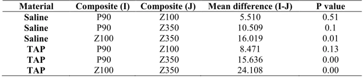
ANOVA showed strongly significant differences within the saline (p=0.01) and TAP (p=0.00) groups among the composites. Tukey’s test revealed significant differences in the TAP group among Z100, P90 and Z350 and in the saline group among Z100 and Z350 composites (Table 2)
Discussion
The results showed that the mean bond strength of P90 and Z100 decreased following the use of TAP compared to saline. In other words, TAP decreased the shear bond strength of P90 and Z100 to dentin; although not significantly. Also, TAP increased the bond strength of Z350 to dentin but not significantly. Thus, in general, TAP had no adverse
Table 1. Comparison of the mean shear bond strength of three types of composites in the saline and TAP groups in MPa
*T-test;p<0.05 level of significance
Table 2. Comparison of the mean shear bond strength of three types of composites when saline was used as an intracanal medicament (n=6×10)
*Post-hoc Tukey’s test; p<0.05 level of significance
effect on bond strength of methacrylate-based (Z350 and Z100) and silorane-based (P90) composites to dentin.
Studies on the shear bond strength of tooth-colored restorative materials following the use of antibiotics are scarce. Some studies, similar to ours, have shown positive effects of antibiotics on bond strength. However, this may vary depending on the type of antibiotic, and further investigations are required on this topic. Mortazavi et al. Reported that combination of tetracycline, 1.3% sodium hypochlorite and detergent according to a protocol did not significantly decrease the composite bond to dentin, which was in contrast to the effect of 35% phosphoric acid with the self-etch technique [24]. Elkassas et al. compared different disinfectants and reported no significant effect of antibiotics on tensile bond strength. They used 5.25% sodium hypochlorite, 2% chlorhexidine, 0.1% benzalkonium chloride and 3% doxycycline. Their results showed that sodium
strength followed by the three disinfectants with no significant difference with the control group; these results are in line with our findings regarding no adverse effect of antibiotics [25]. Stanislawczuk et al. reported variable results based on the type of antibiotics for tensile bond strength. In their study, 2% doxycycline decreased the bond strength while 2% minocycline and 2% chlorhexidine had no negative effect on microtensile bond strength of composite to dentin after acid etching [23]. Our findings showed reduction in bond strength of P90 and Z100 due to the effect of TAP, which may be due to the presence of minocycline (considering the composition of matrix and type of bonding agent used); because antibiotics in the tetracycline family bond to calcium in dentin and prevent resin monomer penetration into dentinal tubules and consequently, decrease the bond strength of tooth-colored restorations [26].
In use of TAP, maximum shear bond strength Composite Material Number of samples Mean Standard deviation P value
P90 Saline 10 23.98 5.61 0.71
TAP 10 23.12 4.61
Z100 Saline TAP 10 10 18.47 14.64 11.46 10.58 0.45
Z350 Saline TAP 10 10 34.49 38.75 14.24 11.49 0.47
Material Composite (I) Composite (J) Mean difference (I-J) P value
Saline P90 Z100 5.510 0.51
Saline P90 Z350 10.509 0.1
Saline Z100 Z350 16.019 0.01
TAP P90 Z100 8.471 0.13
TAP P90 Z350 15.636 0.00
TAP Z100 Z350 24.108 0.00
Z100 and the differences between Z100 and Z350 and also Z350 and P90 were statistically significant. Difference in shear bond strength of Z350 and P90 may be due to the type of bonding agent used, application of 37% phosphoric acid, and filler percentage of Z350. The bonding agent for Z350 needs to be rinsed after acid etching. But P90 is used with a self-etch bonding agent that does not require rinsing after the application. Moreover, 37% phosphoric acid used with Z350 more efficiently eliminates the smear layer compared to the primer used with P90. On the other hand, high filler content of P90 results in a harder material and increases its modulus of elasticity. A harder restorative material can affect the gap at the tooth-restoration interface [27]. In the control (saline) group, maximum shear bond strength belonged to Z350 group followed by P90 and Z100 groups; the difference between Z350 and Z100 was significant. Z100 is a hybrid composite with a filler size of 0.01 to 3.5µ and filler volume percent of 66%. Z350 however, is a nano-hybrid composite with a mean filler cluster size of 0.6 to 10µ (combination of 20nm silica and 4-11nm zirconia particles) and a mean volume percent of 63.3%. Both these composites are bonded to dentin using a fifth generation bonding agent (etch and rinse).
The only difference between these two composites is in the size and shape of their fillers, which may
affect their shear bond strength to
dentin. In our study, this resulted in lower shear bond strength of Z100 to dentin. In other words, the smaller size of fillers and their cluster formation increase the shear bond strength to dentin [28, 29].
Results of McLeod et al. [30] are in accord with our findings. They bonded Filtek Z350 using 35% phosphoric acid with an approximate pH of 0.6 and Single Bond. But, Filtek P90 was used with two-step self-etch primer with a pH of 2.7. Since the self-etch primer has less acidity than the phosphoric acid, it partially dissolves the smear layer; whereas, 35% phosphoric acid completely eliminates the smear layer and thus hydrophilic primer and hydrophobic bonding agent are in direct contact with the tooth structure.
Thus, despite less shrinkage of Filtek P90, it may
cause higher shrinkage stresses than
methacrylate-based composites due to less acidity of its primer and consequently decrease the bond strength. Similarly, Tanno et al [31]. Found no significant difference between low shrinkage and methacrylate-based composites in the tooth-restoration gap formation.
They attributed the lack of a significant difference to the harder nature of silorane-based composites due to their higher filler content and believed that harder nature of these composites may compromise the marginal fit of composite restorations. Not as-sessing the bacterial flora after antibiotic interven-tion, short-term assessment of samples and not si-mulating oral conditions were among the limita-tions of this study, which might have affected the results.
Conclusion
The results showed that use of TAP in endodontic treatment does not affect the shear bond strength of Z350, P90 and Z100 composites to dentin exposed to this paste. In fact, the type of composite used determines the shear bond strength value.
References
1. Torabinejad M, Anderson P, Bader J, Brown LJ, Chen LH, Goodacre CJ, et al. Outcomes of root canal treatment and restoration, implant-supported single crowns, fixed partial dentures, and extraction without replacement: A systematic review. J Prosthet Dent. 2007 Oct;98(4):285-311. 2. Basmadjian-Charles CL, Farge P, Bourgeois DM, Lebrun T. Factors influencing the long-term results of endodontic treatment: a review of the literature. Int Dent J. 2002 Apr; 52(2):81-6.
3. Cohen S, Burns RC. Pathways of the pulp. 8 th ed. St Louis: Mosby; 2002, p 103.
4. Craig RG, Powers JM. Restorative dental materials. 11th ed. St Louis: Mosby; 2002.
5. Sehgal V, Shetty VS, Mogra S, Bhat G, Eipe M, Jacob S, et al. Evaluation of antimicrobial and physical properties of orthodontic composite resin modified by addition of antimicrobial agents-an in-vitro study. Am J Orthod Dentofac Orthop. 2007 Apr;131(4):525-9.
6. Scannapieco FA, Torres G, Levine MJ. Salivary alpha-amylase: Role in dental plaque and caries formation. Crit Rev Oral Biol Med. 1993; 4(3-4): 301-7.
7. Imazato S, Torii M, Tsuchitani Y, McCabe JF, Russell RR. Incorporation of bacterial inhibitor into resin composite. J Dent Res. 1994 Aug; 73 (8):1437-43.
8. Knobloch LA, Gailey D, Azer S, Johnston WM, Clelland N, Kerby RE. Bond strengths of one-and two-step self-etch adhesive systems. J Prosthet Dent. 2007 Apr;97(4):216-22.
9. Proença JP, Polido M, Osorio E, Erhardt MC, Aguilera FS, García-Godoy F, et al. Dentin regional bond strength of self-etch and total-etch adhesive systems. Dent Mater. 2007 Dec; 23(12): 1542-8.
10. Lopes GC, Cardoso PC, Vieira LC, Baratieri LN, Rampinelli K, Costa G. Shear bond strength of acetone-based one-bottle adhesive systems. Braz Dent J. 2006 May; 17(1):39-43.
11. Simões DM, Basting RT, Amaral FL, Turssi CP, França FM. Influence of chlorhexidine and/or ethanol treatment on bond strength of an etch- and-rinse adhesive to dentin: an in vitro and in situ study. Oper Dent [Internet]. 2014 Jan-Feb; 39(1): 64-71. Available from: http://www. ncbi. nlm. nih. gov/pubmed/23675741.
12. Zhou J, Tan J, Yang X, Cheng C, Wang X, Chen L. Effect of chlorhexidine application in a self-etching adhesive on the immediate resin-dentin bond strength. J Adhes Dent. 2010 Feb; 12 (1):27-31.
13. Ercan E, Erdemir A, Zorba YO, Eldeniz AU, Dalli M, Ince B, et al. Effect of different cavity disinfectants on shear bond strength of composite resin to dentin. J Adhes Dent. 2009 Oct; 11(5): 343-6.
14. Barbizam JV, Trope M, Teixeira EC, Tanomaru-Filho M, Teixeira FB. Effect of calcium hydroxide intracanal dressing on the bond strength of a resin-based endodontic sealer. Braz Dent J. 2008;19(3):224-7.
15. Nassar M, Awawdeh L, Jamleh A, Sadr A, Tagami J. Adhesion of epiphany self-etch sealer to dentin treated with intracanal irrigating solutions. J Endod. 2011 Feb; 37(2):228-30.
16. Walton RE, Torabinejad M. Principles and Practice of Endodontics, 2nd ed. W.B. Saunders: Philadelphia, 1996; 201-22.
17.Mohammadi Z. Strategies to manage permanent non-vital teeth with open apices: A clinical update.
18. Athanassiadis B, Abbott PV, Walsh LJ. The use of calcium hydroxide, antibiotics and biocides as antimicrobial medicaments in endodontics. Aust Dent J. 2007 Mar;52(1 suppl):S64-S82.
19. Jungermann GB, Burns K, Nandakumar R, Tolba M, Venezia RA, Fouad AF. Antibiotic resistance in primary and persistent endodontic infections. J Endod. 2011 Oct; 37(10):1337-44. 20. Santos MC, Vitor C, Cristina S, Tadeu W. Removal of intracanal smear layer by doxycycline: SEM analysis. Aust Endod J. 2010 Aug;36(2):64-9.
21. Windley W, Teixeira F, Levin L, Sigurdsson A, Trope M. Disinfection of immature teeth with a triple antibiotic paste. J Endod. 2005 June; 31 (6):439-43.
22.Vijayaraghavan R, Mathian VM, Sundaram AM, Karunakaran R, Vinodh S. Triple antibiotic paste in root canal therapy. J Pharm Bioallied Sci [Internet]. 2012 Aug;4 (Suppl 2):S230-3.http: //www. pubmedcentral. nih. gov/articlerender. fcgi? Artid = 3467921 & tool = pmcentrez & rendertype= abstract.
23. Stanislawczuk R, Costa JA, Polli LG, Reis A, Loguercio AD. Effect of tetracycline on the bond performance of etch-and-rinse adhesives to dentin. Brazil Oral Res. 2011 Sept-Oct; 25(5): 459-65. 24. Mortazavi V, Khademi A, Khosravi K, Fathi M, Ebrahimi–Chaharom M, Shahnaseri S, et al. Effect of MTAD on the shear bond strength of self-etch adhesives to dentin. Dent Res J. 2012 Jan-Mar; 9(1):24-30.
25. Elkassas DW, Fawzi EM, El Zohairy A. The effect of cavity disinfectants on the micro-shear bond strength of dentin adhesives. Europ J Dent. 2014 Apr-Jun; 8(2):184-90.
26. Al Wazzan KA. Effect of three endodontic materials on the bond strength of two composite core materials to dentin. J Prosthodont. 2002 Jun; 11(2):92-7.
27. Papadogiannis D, Kakaboura A, Palaghias G, Eliades G. Setting characteristics and cavity adaptation of low-shrinking resin composites. Dent Mater. 2009 Dec; 25(12):1509-16.
28.Chen MH. Update on dental nanocomposites. J Dent Res. 2010 June; 89(6):549-60.
29. Li Y, Swartz ML, Phillips RW, Moore BK, Roberts TA. Effect of filler content and size on
(12):1396-401.
30. Mcleod ME, Price RB, Felix CM. Effect of configuration factor on shear bond strengths of self-etch adhesive systems to ground enamel and dentin. Oper Dent. 2010 Jan-Feb; 35(1):84-93.
31. Tanno K, Hiraishi N, Otsuki M, Tagami J. Evaluation of cavity adaptation of low-shrinkage composite resin. Asian Pac J Dent. 2011 Jan-June; 11(2):27-33.