ABSTRACT
Polyploidy, a common characteristic of cancer cell lines, puts extra pressure on
the regulatory components of mitosis. Specifically, the process of chromatin
condensation becomes especially critical due to the challenge of accurately aligning and
segregating a multifold increase of DNA on the mitotic spindle. The regulator of
chromosome condensation 1 (RCC1) is a protein bound to DNA throughout the cell
cycle, which we have shown to be necessary for chromosome size scaling in C.
elegans. We hypothesize that RCC1 levels increase in polyploid cells to compensate for this excess DNA content and promote adequate DNA
condensation. We isolated populations of highly polyploid HeLa human cancer cells and found that RCC1 levels scale with DNA content in both highly polyploid and control
HeLa cells. We believe that this scaling relationship is what affords highly polyploid cells
the ability to maintain effective chromatin condensation and perhaps mitotic
progression. Highly polyploid HeLa cells that have been stably transfected with a
non-DNA-binding RCC1 mutant maintain their polyploidy through more rounds of division
than do control polyploid populations. Analysis of metaphase chromosomes has
revealed that these cells contain over-condensed chromosomes. We now believe that
the loss of RCC1’s DNA-binding ability, which allows over-condensation of
chromosomes, is what allows these highly polyploid cells to maintain effective rounds of
mitosis, as smaller chromosomes are likely more easily segregated in highly polyploid
INTRODUCTION
Cancer is a complex and constantly adapting disease that has captured the
interest of researchers for decades. Its most notable feature is unchecked growth, which
occurs from the ability of cancer cells to ignore checkpoints on its way through the cell
cycle, allowing them to divide with little to no checkpoint regulation. This is the basis for
the majority of chemotherapy drugs in use, which are most effective against cells that
proliferate rapidly. However, this causes a number of side effects for many somatic
tissues whose cells are also unintentionally inhibited (Niu et al., 2015).
Another defining characteristic of cancer is abnormally high amounts of DNA,
which is a result of mitotic defects that lead to cells with extra chromosomes, also
known as aneuploidy (Weaver et al., 2007). This phenomenon has long been known
and has even been proposed as the cause of cancer (Boveri et al., 1914). However,
aneuploidy is unique to cancer cells, and thus offers a novel approach to selectively
target cancerous cells without producing the harmful side effects seen in contemporary
chemotherapies.
Upon entry into mitosis nuclear DNA must condense into highly ordered
structures called chromosomes. This process is called chromosome condensation and
is a necessary checkpoint to proceed through mitosis (Koshland et al., 1996). Errors in
chromosome condensation and subsequent problems with the mitotic spindle can lead
to improper segregation of chromosomes and aneuploidy (Hegyi et al., 2012). Although
cancer cells progress through mitosis in an anarchic way, we believe that chromosome
The regulator of chromatin condensation 1 (RCC1) was first discovered in a cell
line that carried a temperature sensitive RCC1 mutant (tsBN2), which induced
premature chromatin condensation at the nonpermissive temperature (Ohtsubo et al,
1987). It was later discovered that RCC1 is a nuclear-localized protein that is DNA
bound (Ohtsubo et al, 1989). RCC1 also has a known function as a Ran guanine
nucleotide exchange factor (Ran-GEF). RCC1 exchanges GDP for GTP on Ran
creating a Ran-GTP/GDP gradient, which drives nuclear import of various cargoes
(Klebe et al., 1995 & Clarke et al., 2008). During assembly of the mitotic spindle, the
Ran-GTP/GDP gradient attracts growing microtubules to the metaphase plate
(Carazo-Salas et al., 1999). Other than this phenotypic evidence, RCC1’s exact mechanism in
regulating chromosome condensation remains a mystery. In this study, we use a
non-DNA-bound RCC1 mutant (RCC1KRR) to explore how RCC1 regulates chromosome
condensation in a polyploid system.
Changes in chromosome condensation have been well characterized in multiple
developmental systems. This phenomenon is known as chromosome size scaling,
where rapid divisions create smaller cells, which necessitate smaller chromosomes to
maintain effective divisions. This has been observed in both C. elegans (Ladouceur et al., 2015) and X. laevis (Kieserman et al., 2011). In C. elegans, RCC1 is required for this scaling relationship (Ladouceur et al., 2015). We want to explore how chromosome
size is regulated in polyploid human cancer cells. We hypothesize that RCC1 levels
increase in polyploid human cancer cells to compensate for excess DNA and promote
METHODS Cell Culture
The HeLa cell line and its derivatives, which expresses RCC1-GFP and
triple-mutant-RCC1-GFP, were grown in Dulbecco's modified Eagle's medium (DMEM)
supplemented with 10% (v/v) fetal bovine serum and penicillin-streptomycin (Invitrogen)
at 37°C in a humidified 5% CO2, 95% air incubator. For shRNA digestions, cells were
grown to 70-80% confluency. 100 μL Opti-MEM (ThermFisher Scientific) and 6 μL
Lipofectamine (Invitrogen) were incubated with 1 μg of both shRNA plasmids targeted
for the 3’ UTR of RCC1 for 15 minutes. 100 μL of this was added dropwise to each plate
and incubated for 24 hours at 37°C before immunostaining or metaphase chromosome
spreads were performed. Stable cell lines were created by following the preceding
transfection process with a plasmid containing the wildtype or mutated RCC1 with a
GFP tag and a kanamycin resistance gene. Cultures were grown in g418 (Gibco) and
sorted to monoclonal cell lines using the FACSAria II (BD Biosciences) with help from
the UNC Flow Cytometry Core.
Flow Cytometry
HeLa cells were grown in standard culture medium described above in 10-cm cell
culture dishes to 80-90% confluency. On the day of the experiment, cells are detached
using Trypsin-EDTA 0.05% and spun down to a pellet. Cells were resuspended in
phosphate buffered saline (PBS) containing 2% (v/v) fetal bovine serum. DNA was
incubated with Hoechst 33342 (Thermo Scientific) at a working concentration of 5
μg/mL. Fluorescence-activated cell sorting was done with the help of the UNC Flow
done on an LSRII (BD Biosciences). Flow Cytometry data were collected using FlowJo
X.0.7 software.
Immunostaining
HeLa cells, plated on a EtOH-cleaned coverslip, to be stained for fixed cell
imaging were fixed at 4oC with a 1:1 solution of methanol:acetone for 20 minutes. They
were then permeabilized with a solution of 0.5% Triton-X in PBS for 5 minutes. The
coverslips were then blocked in Abdil-milk solution (0.1% Triton-X, 0.1% NaN3 and 2%
milk in PBS) for 30 minutes. Primary antibodies used include anti-RCC1 N-19 (Santa
Cruz) and anti-alpha tubulin (LSBio), both at a dilution of 1:300 in the blocking solution
for 2 hours. Fluorophore-conjugated secondary antibodies were diluted at 1:300 and
were allowed to react for 45 minutes. Coverslips were then incubated with 0.5 μg/mL
DAPI in PBS for 15 minutes. Coverslips were then mounted onto slides with mounting
media (90% glycerol and 5 mg/mL n-propyl gallate in diH2O) and sealed using nail
polish.
Metaphase Chromosome Spreads
Chromosome spreads were prepared according to the protocol described by
Earnshaw et al. 1989. Cells plated on a coverslip were swollen in a hypotonic solution
(75 mM KCl) for 25 minutes. Equal volume fixative (3:1 Methanol:Glacial Acetic Acid)
was added to the hypotonic solution for 2 minutes. The liquid was aspirated and fixative
was added again for 5 minutes. Fixative was aspirated and the coverslips were blown
dry using a cut 1000 μL pipet tip. Chromosomes were rehydrated in PBS Azide (1 mM
EGTA and 0.01% NaN3 in PBS) for 10-15 minutes. The spreads were then incubated in
mounting media (90% glycerol and 5 mg/mL n-propyl gallate in diH2O) and sealed using
nail polish.
Microscopy
Imaging was performed at room temperature on a DeltaVision microscope using
Softworx software (Applied Precision) equipped with a CoolSnap HQ2 camera
(Photometrics) at 1 × 1 binning and a ×60 Nikon oil-immersion objective using the 45
μm pinhole setting. All images were deconvolved using Softworx software (Applied
Precision).
Data Analysis
All image analysis was done using Fiji (ImageJ) and statistics were analyzed with
Microsoft Excel (Microsoft) and Prism (CenturyLink). Figures we created using Adobe
Illustrator.
RESULTS
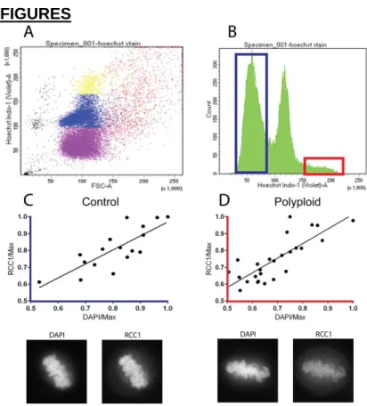
RCC1 scales with DNA content in polyploid HeLa cells
RCC1 is a Ran-GEF, and is necessary for nuclear import by loading Ran with
GTP, which initiates cargo release (Edens et al., 2012). We originally hypothesized that
maintaining extra DNA content would require more nuclear import, which would be
dependent on increased activity or expression of RCC1. Furthermore, previous
published work from our lab has demonstrated that RCC1 is necessary to maintain
chromosome size scaling with cell and nuclear size in early C. elegans embryonic development (Ladouceur et al., 2015). Since RCC1 is also a DNA bound protein, we
first hypothesized that RCC1 protein levels would scale with the total amount of DNA.
distribution of DNA content in a population of HeLa cells using flow cytometry (Figure
1B). We were able to use fluorescence-activated cell sorting (FACS) to gate the
population as shown in Figure 1B into control and polyploid populations, which we
stably maintained in culture. To determine the relationship between DNA content and
RCC1 protein levels, we fixed cells from both gated populations and probed with an
RCC1 antibody, co-staining with DAPI to quantify DNA content. We found that RCC1
levels scaled with the amount of DNA content in both our control and polyploid sorted
populations (Figure 1C-D).
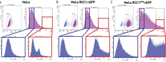
Loss of RCC1’s DNA-binding activity maintains polyploidy levels within a population
We wanted to determine RCC1’s role in polyploid HeLa cells and decouple the
protein from its scaling relationship with DNA. We achieved this by creating a triple-point
mutant RCC1 protein, whose DNA-binding residues (Makde et al., 2010) were mutated
to alanines (Figure 3A, RCC1KRR::GFP). This protein was tagged with GFP and no
longer localized to the metaphase plate during mitosis (Figure 3B). This construct, along
with a RCC1::GFP construct, were stably transfected into HeLa cells. These HeLa cell
lines were again sorted into control and polyploid populations, while gating for
GFP-positive cells only (Figure 2). Following the experimental protocol established for the
first FACS experiments, these populations were grown out for two passages and
analyzed using flow cytometry to determine how the distribution of DNA content had
changed. Non-transfected polyploid HeLa cells almost immediately returned to the
wildtype distribution of DNA content (Figure 2A). Polyploid HeLa cells that were
but centered around a lower chromosome number (Figure 2B). The polyploid population
that was isolated from HeLa cells transfected with RCC1KRR::GFP maintained a large
number of polyploid cells compared to isolated populations of HeLa cells and HeLa cells
transfected with RCC1::GFP (Figure 2C).
To explore this phenomenon on a cellular level, we performed fixed-cell staining
of our isolated control and polyploid HeLa cell lines. We also used shRNA directed to
the 3’ UTR of RCC1 to deplete the endogenous protein and leave only our transfected
constructs. Depletion of RCC1 using two different constructs directed at the 3’ UTR of
RCC1 led to an approximate 75% knockdown of endogenous RCC1 protein levels as
shown by Western blot analysis (Figure 4C-D). Interestingly, metaphase plate lengths
increase when endogenous RCC1 is depleted in all cell lines (Figure 5).
Disruption of RCC1’s DNA binding ability decreases chromosome area
We wanted to explore how HeLa cells containing the RCC1KRR::GFP construct
were able to maintain their polyploidy. Previous work from our lab has shown that RCC1
depletion decreased chromosome size in C. elegans (Ladouceur et al., 2015). Therefore, to determine how chromosome size is regulated by our RCC1KRR::GFP
construct, metaphase chromosome spreads were analyzed to measure how
chromosome size scaling is disrupted. Chromosome area was measured consistent
with the quantification method outlined in Kieserman et al., 2011. We found that
chromosome area decreases in response to RCC1 depletion in HeLa cells (Figure 4).
We also found that chromosome area significantly decreases in HeLa cells that carry
the non-DNA-binding RCC1, even without depletion of the endogenous RCC1 (Figure
DISCUSSION
We have demonstrated that RCC1 protein levels scale in response to increased
DNA content, or polyploidy. However, contrary to our predictions, RCC1KRR::GFP was
able to afford maintenance of ploidy better than wildtype RCC1, as evidenced by the
maintenance of a large population of polyploid cells in sorted RCC1KRR::GFP HeLa cell
lines. Our lab previously reported that upon partial depletion of RCC1 in C. elegans,
chromosomes were significantly shorter (Ladouceur et al., 2015). We observed that in
HeLa cells also, chromosome size decreased in response to both RCC1 depletion and
disruption of the DNA-binding ability of RCC1. This contradicts our hypothesis predicting
that the scaling relationship between RCC1 and DNA provides a mechanism to ensure
effective chromatin condensation. In response to this confounding data we hypothesize
that decreased chromosome size in RCC1KRR::GFP cells explains their ability to
maintain effective rounds of mitosis despite the abnormally large amount of DNA, as
smaller chromosomes are likely more efficiently and accurately segregated in polyploid
cells. This could explain how highly polyploid HeLa cells that contain the RCC1KRR::GFP
maintain the polyploidy through more rounds of division than do HeLa cells containing
the wildtype RCC1.
An interesting observation that we made that would seem opposed to this theory
is that metaphase plate lengths increase in response to RCC1 depletion. However, we
believe that this is due to irregular spindle assembly. RCC1KRR::GFP likely creates a
mislocalized RanGTP gradient that is not located at the metaphase plate. Therefore,
growing microtubules will not be accurately directed to chromosomes, creating a larger
However, our data still poses many questions about how exactly RCC1 is
regulating chromatin condensation. The disruption of DNA binding facilitates the
decrease in chromosome size, but RCC1 has a role in nuclear transport, which could
also explain how it affects condensation. Potentially, RCC1 could regulate the import of
other factors into the nucleus, such as epigenetic modifiers or other known components
of the condensation machinery. Further study of chromosome structure using super
resolution microscopy could elucidate how exactly RCC1 regulates chromosome size.
Further work needs to be down to determine how our RCC1KRR::GFP protein disrupts
this regulation. Potentially, the residues that we have mutated could have a wider range
of effects on RCC1’s function than just disrupting DNA binding. It is easy to imagine that
non-DNA-bound RCC1 cannot properly facilitate the import of critical condensation
factors, due to loss of Ran-GEF ability; however, this is currently unknown. We believe
that our RCC1KRR::GFP is a separation of function mutation, and that nuclear import is
still occurring at near-wildtype efficiency, which is supported by the data presented here.
Further study of our RCC1KRR mutant will be vital to understanding RCC1’s exact role in
chromatin condensation and also chromosome size scaling.
ACKNOWLEDGEMENTS
I would like to thank Anne-Marie Ladouceur for her unrelenting mentorship and
guidance over the last two years, Paul Maddox for the opportunity to work on interesting
and relevant biological problems, Lydia Smith for constant editing and answering of
questions, no matter how dumb, and the rest of Maddox Labs for their advice and
from the UNC Flow Cytometry Core and Tony Perdue from the Microscopy Core for
providing their expertise.
FIGURES
Figure 2. Non-DNA-bound RCC1 facilitates maintenance of polyploid cells through more rounds of division than control cells. (Top Panel) Sorted populations of control (blue box) and polyploid (red box) cells from HeLa cells (A) and HeLa cells stably transfected with RCC1::GFP (B) or RCC1KRR::GFP (C). (Bottom Panel) Populations were maintained in culture for one week and the DNA content profiles were measured using flow cytometry.
Figure 3. RCC1KRR::GFP (K232A-R234A-R237A)
Figure 4. Chromosome area decreases upon RCC1 depletion and in HeLa cells transfected with RCC1KRR::GFP. (A) Chromosome area (chromosome length x chromosome width) is plotted
based on control treatment (blue dots) and RCC1-depletion (red dots). (B) A representative structural illumination microscopy (SIM) image of a metaphase chromosome spread. (C) Western blot showing the depletion of RCC1 protein levels upon treatment with two separate (or combined) shRNA constructs directed to the 3’ UTR of the human RCC1 gene. Asterisks indicate treatment with MNase. (D) Graphical representation of RCC1 depletion as graphed by the ratio of RCC1 intensity to H2B (control) intensity.
REFERENCES
Boveri T. Gustav Fischer, Jena. (1914). English translation The Origin of Malignant Tumors by Boveri. M. Williams and Wilkins; Baltimore: 1929. Zur Frage der Entstehung maligner Tumoren.
Carazo-Salas R. E., Guarguaglini G., Gruss O. J., Segref A., Karsenti E., & Mattaj I. W. (1999). Generation of GTP-bound Ran by RCC1 is required for
chromatin-induced mitotic spindle formation. Nature, 400 (6740), 178 - 181.
Clarke P. R., & Zhang C. (2008). Spatial and temporal coordination of mitosis by Ran GTPase. Nature, 9, 464–477.
Earnshaw W. C., Ratrie III H., & Stetten G. (1989). Visualization of centromere proteins CENP-B and CENP-C on a stable dicentric chromosome in cytological spreads.
Chromosoma, 98, 1 - 12.
Edens L. J., White K. H., Jevtic P., Li X., & Levy D. L. (2012). Nuclear size regulation: from single cells to development and disease. Trends in Cell Biology, 23 (4), 151 - 159.
Goehring N. W., & Hyman A. A. (2012). Organelle growth control through limiting pools of cytoplasmic components. Current Biology, 22, R330 - R339.
Hegyi K., & Mehes G. (2012). Mitotic Failures in Cancer: Aurora B Kinase and its Potential Role in the Development of Aneuploidy. Pathology & Oncology Research,18 (4), 761 - 769.
Hirano T. (2006) At the heart of the chromosome: SMC proteins in action. Nature Review Molecular Cell Biology, 7, 311–322.
Kieserman E. K., & Heald R. (2011). Mitotic chromosome size scaling in Xenopus. Cell Cycle, 10 (22), 3863 - 3870.
Klebe C., Prinz H., Wittinghofer A., & Goody R. S. (1995). The kinetic mechanism of Ran--nucleotide exchange catalyzed by RCC1. Biochemistry, 34 (39), 12543 - 12552.
Koshland D., & Strunnikov A. (1996). Mitotic Chromatin Condensation. Cell and Developmental Biology, 12, 305–333.
Ladouceur A.-M., Dorn J. F., & Maddox P. S. (2015). Mitotic chromosome length scales in response to both cell and nuclear size. Journal of Cell Biology, 209 (5), 645 - 652.
Makde R. D., England J. R., Yennawar H. P., & Tan S. (2010). Structure of RCC1 chromatin factor bound to the nucleosome core particle. Nature, 467, 562 - 566. Niu N., & Wang L. (2015). In vitro human cell line models to predict clinical response to
anticancer drugs. Pharmacogenetics 16 (3), 273 - 285.
cDNA of the human cell cycle gene (RCC1) involved in the regulation of onset of chromosome condensation. Genes & Development, 1(6), 585 - 593.
Ohtsubo M., Okazaki H., & Nishimoto T. (1989). The RCC1 protein, a regulator for the onset of chromosome condensation locates in the nucleus and binds to DNA. J Cell Biol, 109 (4), 1389 -1397.