Sex differences in anxiety and fear learning behavior
following genetic manipulation of the 5-HT1A receptor
in the bed nucleus of the stria terminalis (BNST)
By
Cayce Dorrier
Senior Honors Thesis
Chemistry Department
University of North Carolina at Chapel Hill
April 10, 2015
Approved:
_____________________________
Thomas Kash, Thesis Advisor
Brian Hogan, Reader
Abstract
The bed nucleus of the stria terminalis (BNST) is a critical node in the fear and anxiety circuitry
of the brain that plays a key role in an organism’s ability to respond to stress. Serotonin 1A
(5-HT1A) receptor signaling in the BNST has been implicated in anxiety and fear-related behavior
using acute pharmacological manipulations that target pre- and post-synaptic receptors (Levita et
al., 2004; Gomes et al., 2012). The goal of this project is to elucidate the role of post-synaptic
5-HT1A receptors within the BNST in anxiety, fear, and depressive-like behavior using a genetic
deletion approach that selectively targets postsynaptic 5-HT1A receptors. Viral vectors containing
the gene for Cre recombinase and a fluorescent tag (AAV5-Cre-GFP) or a control vector
(AAV5-GFP) were infused into the BNST of genetically modified male and female 5-HT1A
receptor flox mice. One month later, behavior on the elevated plus maze (EPM), open field,
forced swim, novelty-suppressed feeding and fear conditioning assays were assessed. The results
of this project indicate that the genetic deletion of 5-HT1ARs in the BNST has an anxiolytic-like
effect in the EPM but enhances fear consolidation and contextual fear following a tone-shock
protocol in male mice. These seemingly discrepant results may reflect the fact that under low
stress conditions (e.g. the EPM), deletion of the 5-HT1A receptor disinhibits BNST output
neurons which are anxiolytic (Kim et al., 2013; Jennings et al., 2013). In high stress (e.g.
footshock) conditions, local neurons are recruited which are both anxiogenic and fear-enhancing
(Levita et al, 2004). Deletion of 5-HT1A receptors from these neurons potentiates their fear
enhancing actions, resulting in an increase in fear consolidation and contextual fear. The
behavioral phenotypes of the female mice following this genetic manipulation were less
pronounced, which could be due to the anatomical differences of the BNST between male and
Introduction
Anxiety disorders afflict an estimated 18% of adults in the United States and pose a significant
public health burden, costing more than $42 billion annually. The bed nucleus of the stria
terminalis (BNST) is a critical relay station in the brain that has been implicated in anxiety-like
behaviors. As shown in Figure 1, it projects to and receives projections from the amygdala,
hippocampus, ventral tegmental area, dorsal raphe, and periaqueductal gray. Of particular
interest to this project is the serotonin (5-hydroxytryptamine; 5-HT) projection of serotonin from
the dorsal raphe (DR) to the BNST.
Electrical stimulation of the BNST causes behavioral responses similar to that of stressful stimuli
(Casada and Dafny, 1991). The goal of the current study is to elucidate the role of 5-HT1A
receptor signaling in the BNST in these aversive behavioral responses using a targeted genetic
deletion approach.
Many anti-anxiety drugs work by preventing the reuptake of the neurotransmitter HT. The
5-HT receptors are classified into seven families based on sequence homology, pharmacological
characteristics and effector coupling (5-HT1-7) and these can further subdivided into subtypes
(5-HT1A, 5-HT1B, etc.). Here we will focus on the 5-HT1A receptor subtype, a Gi/o-coupled GPCR
that inhibits adenylyl cyclase activity and closes G protein-coupled inwardly-rectifying
potassium channels (GIRKs). For this reason, activation of 5-HT1ARs has a hyperpolarizing
effect on cells and is generally inhibitory. It has been implicated as the primary subtype of
serotonin responsible for the inhibitory action of serotonin in the BNST (Guo et al., 2009), and
activation of the 5-HT1A receptor in the BNST using a 5-HT1AR agonist has been shown to have
anxiolytic effects in male rats (Levita et al., 2004).
Additionally, activation of 5-HT1A receptors in the BNST decreased contextual fear learning
(Gomes el al., 2012). Together, these studies indicate that 5-HT1A receptor signaling in the
BNST attenuates aversive behaviors. In agreement with these acute pharmacological studies,
global deletion of the 5-HT1AR using a genetic knock-out approach has been shown to increase
anxiety-like and depressive behaviors in both male and female mice (Ramboz et al., 1998; Parks
et al., 1998).
5-HT1A also plays a role in many other behavioral phenotypes such as feeding and motor control.
Fluoxetine, a selective serotonin reuptake inhibitor, has been shown to suppress food intake in
intensifies this effect (Dominic et al., 1998). A 5-HT1AR antagonist has been shown to decrease
locomotor activity, although this effect was most pronounced with microinjections into the
hippocampus (Belcheva et al., 1997). Conversely, a non-selective 5-HT1B/A agonist has been
shown to increase forward locomotion (Aronsen et al., 2014). These phenotypes in addition to
fear and anxiety will be studied using a BNST 5-HT1AR knock-out model to determine if they
are modulated through the BNST.
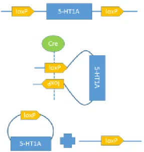
The receptor will be deleted using genetically
modified 5-HT1A flox mice, in which 2 loxP
segments surround the gene for the 5-HT1A
receptor. This 34 base pair sequence is
recognized and bound by the enzyme cre
recombinase. In the 5-HT1A flox mice, a single
recombinase will bind to each loxP segment. The
two enzymes will then come together, forming a
tetramer and bringing the two loxP sites together
in a recombination event that will excise the
segment of the gene corresponding to the 5-HT1A
receptor (Nagy et al., 2000). This recombination event essentially knocks out this receptor, as
depicted in Figure 2. Injecting a virus containing the gene for cre recombinase into the BNST of
these mice will insert this gene into the DNA of BNST neurons. The cell’s machinery is then
able to produce this enzyme in only BNST neurons, leading to the knock-out of 5-HT1A receptors
in this specific brain region.
Behavioral testing will be used to determine if the absence of this receptor leads to an increase an
anxiety- and depressive-like behaviors, feeding behaviors, and fear learning in these mice.
Specifics tests include elevated plus maze (EPM), open field, forced swim, novelty-suppressed
feeding and fear conditioning. The body weight of all mice was recorded after surgery and
throughout the cohort of behavioral experiments to look for any evidence of changes in feeding
behaviors.
All experiments were conducted on male and female mice in order to look for any sex-specific
differences in behavioral outcomes associated with 5-HT1A receptor deletion in the BNST.
Without any drug treatment or genetic manipulation, male and female mice respond differently
to many behavioral tests and paradigms. For example, female mice appear less anxious than
males in the elevated plus maze, show less immobility in the forced swim test and more often fail
to adapt to repeated stress procedures than male mice (Blanchard et al., 1991). In response to
5-HT1A, female mice have been shown to be more sensitive to the 5-HT1A agonist 8-OH-DHAP
(Blanchard et al., 1991).
While both sexes have been studied in the context of a global 5-HT1A receptor deletion, the
studies on 5-HT1A receptors in the BNST have not included female animals. Additionally, while
many brain regions such as the hippocampus implicated in the global knockout lack drastic
anatomical sex differences, the volume of the medial posterior region of the BNST is
significantly larger in male rats than in females (del Abril et al., 1987). This difference has been
correlated to a greater number of apoptotic nuclei in the principle nucleus of the BNST in female
rats sacrificed on postnatal day 12 (Chung et al., 2000). This anatomical difference could lead to
differences in behavior when the BNST is the only targeted brain region. It is important to study
women are approximately twice as likely to be afflicted with anxiety and depression as men and
are thus more likely to be prescribed drugs such as SSRIs that target 5-HT systems (Kessler et
al., 1993, Kessler et al., 2005).
Based on the previous studies cited above, our hypothesis is that the deletion of the 5-HT1A
receptor in the BNST will increase anxiety and depressive-like behavior and fear learning in
mice with the potential for sex differences in behavior.
Methods
Mice
All animals were group housed on a 12 hour light cycle with ad libitum access to rodent chow
and water, unless described otherwise. 5-HT1A flox mice were obtained from Eric Delpire,
Vanderbilt University Department of Anesthesiology. Three separate cohorts of these mice were
run through the experiments explained below, each containing mice receiving the virus harboring
the gene for cre recombinase Cre-GFP, designated as cre) and a control virus
(AAV5-GFP, designated as control). Both of the AAV viruses were produced by the Gene Therapy
Center Vector Core at the University of North Carolina at Chapel Hill. The first cohort of mice
contained 15 male mice, n=8 cre and n=7 control, the second 12 female mice, n=7 cre and n=5
control, and the third 6 male mice, n=3 cre and n=3 control. The behavioral data from the two
cohorts of male mice was pooled to create an overall male total of n=11 cre and n=10 control.
Stereotaxic Surgery
Twenty-four hours prior to surgery, 5-HT1A flox mice were given a solution of acetaminophen in
their drinking water (5 ml/200 ml v/v). The mice were then anesthetized with isoflurane and
placed in a stereotaxic frame (Kopf Instruments, Tujunga, CA). 32 gauge needles connected to a
1 µL Hamilton syringe were used to bilaterally inject 400 nl either Cre-GFP or
AAV5-GFP into the BNST (coordinates from Bregma: ± 1.0 ML, 0.4 AP, -4.35 DV). Mice were
allowed to recover for 6 weeks after surgery.
Body Weight Measurements
The body weight of each mouse was recorded approximately every other day from the day of
surgery through the end of the behavioral experiments to look for changes in body weight over
time.
Behavior
EPM
Mice were placed in the center of a standard
elevated plus maze, depicted in Figure 3, facing an
open arm and allowed to explore the maze for 5 min.
The number of open arm entries, probability of
entering the open arm, the time spent in the open
arm, and the total distance traveled in the maze were
scored using EthoVision XT 7. In this test, mice
exhibiting anxiety-like behavior avoid the open arms
(Pellow et al., 1985).
Figure 3. A standard EPM
Open Field
Mice were placed into the corner of a white Plexiglas open field arena (25 x 25 x 25 cm) and
were allowed to explore the arena for 30 min. The total distance travelled, time spent in the
central 25% of the box, and the latency to enter the center of the box were scored using
EthoVision XT 7. Avoidance of the center of the box reflects heightened anxiety (Choleris et al.,
2001).
Forced Swim
Mice were placed in a clear, plexiglass container that was filled ¾ full with water at 25°C for 6
min. Immobility during the last 4 minutes were hand-scored using The Observer XT 10. This
test has been used to compare the effects of various antidepressants as it puts mice in a situation
where they can lose hope of escaping a stressful environment (Can et al., 2012). Mice who
exhibit more behavioral despair are more likely to spend a higher percentage of their time in an
immobile state (Pellow et al., 1895).
Novelty Suppressed Feeding
Mice were introduced to Froot Loops (Kellogg’s) 48 hours before experimentation and food
deprived for 24 hours prior to experimentation. 45 min before experimentation mice were placed
into new cages without food. The open field box was modified for this experiment by adding
bedding and taping colored paper to the walls of the box. A single Froot Loop was placed on a
piece of filter paper in the center of the box. The mice were then placed on the box until they
began to eat the Froot Loop, and their latency to feed was recorded. They were then immediately
transferred to their home cage with a pre-determined weight of Froot Loops (around 1 g.) for 10
back into their home cages. The latency to feed is a measure of anxiety, with a higher latency to
feed reflective of more anxiety- like behaviors, and the food consumption post-test reflects
feeding behaviors. (Samuels and Hen, 2001).
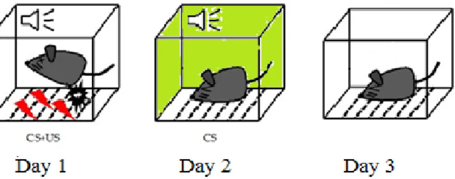
Fear Conditioning
Fear recall and extinction was assessed through a 3 day protocol, presented in Figure 4. In Day 1
the mice were placed in a hood and carried straight to the room containing a fear conditioning
chamber (Med Associates) that was cleaned with a 1% vanilla/19.5 % ethanol/ 78.5% water
solution. After an initial 3 min baseline period, 5 tone/shock pairings occurred in intervals
ranging from 60-120 s. After another 2 min period the mice were removed from the box. In Day
2, which tested cued fear learning, the mice were placed in a cabinet and carried around a
hallway to the behavior room. The mice were placed in a different fear conditioning chamber
(Med Associates) that contained a striped pattern on the walls and was cleaned and scented with
a 70% EtOH solution. After an initial 3 min baseline period, a tone lasting 30 s was repeated 10
times with 5 s between each tone. After another 2 min period the mice were moved from the box.
Day 3 tested contextual fear recall by placing mice in the same box with the same scent as day 1
for 10 minutes without a tone or shock. Percent freezing, defined as a lack of all movement
except for respiration, was used as the behavioral parameter in these experiments with a higher
percent freezing indicative of an increase in fear learning (Seidl et al., 2000). Each mice was
Figure 4. Fear conditioning protocol. A three-day protocol was used to test for fear learning in the mice, where CS represents the conditioned stimulus (a tone) and US represents the
unconditioned stimulus (a footshock).
Immunohistochemistry
Immunohistochemistry was performed using anti-green fluorescent protein (anti-GFP) primary
antibodies to label BNST cell bodies containing either the cre or the control virus. Slices 45 µm
in thickness containing the BNST were collected using a Leica VT1000S vibratome (Leica
Microsystems, Nussloch, Germany) and stored in a 50% glycerol solution at −20 °C until
immunohistochemistry was performed. Slices were washed in PBS three times for 5 min
followed by a 30 min incubation in 50% methanol, a 5 min incubation in 3% H2O2 in PBS, three
10 min PBS washes, a 30 min incubation in 0.5% Triton X-100 in PBS, and an additional 10 min
PBS wash. The slices were then incubated for 60 min in a blocking solution of 0.1% Triton/10%
Normal Donkey Serum (Jackson ImmunoResearch, West Grove, PA) in PBS and transferred to a
primary solution of the blocking solution containing a 1:500 dilution of anti-Green Fluorescent
Protein (Aves Labs, Tigard, Oregon) where they incubated overnight at room temperature.
Slices were then washed with PBS three times for 10 min before incubating for 2 hrs at room
temperature in a secondary solution containing a 1:200 dilution of Alexa Fluor 488 Donkey
four times for 10 min, mounted on slides and allowed to air dry, and covered with Vecta-Shield
Mounting Medium (Vector Laboratories, Burlingame, CA). Images of the BNST were collected
on an Olympus FV1000 confocal microscope with FluoView 1000 Software using the 20x
objectives and a Zeiss AXIO Zoom V16 microscope.
Immunohistochemistry was also performed using anti-GFP and anti-tryptophan hydroxylase
(anti-TPH) primary antibodies in SERT-cre mice to verify the serotonin projection from the DR
to the BNST. This immunohistochemistry followed the same protocol as above with the addition
of a 1:500 dilution of anti-TPH (Sigma-Aldrich, St. Louis, Mo) at the primary step and a 1:200
dilution of Alexa Fluor 647 donkey anti-mouse (Jackson ImmunoResearch, West Grove, PA) at
the secondary step.
Statistical Analysis
Statistical differences between cre and control mice were examined separately in males and
females using a Student’s t-tests with the α level set to 0.05. A two-way ANOVA was also
performed with the male and female data pooled together, resulting in a data set with two
independent variables. Bonferroni posttests were used to make between group comparisons. All
data were analyzed with GraphPad Prism software.
Results
Anatomical verification of 5-HT projection from the DR to the BNST
Transgenic mice expressing Cre recombinase under the control of a serotonin transporter (SERT)
(ChR2)-eYFP virus into the DR. GFP immunohistochemistry was performed to amplify the YFP
signal which enabled the visualization of 5-HT neurons and their axon fiber tracts
(Narbox-Neme et al., 2008). The presence of axon fibers in the BNST in Figure 5(A) verifies this 5-HT
DR→BNST projection.
In the DR, a GFP, tryptophan hydroxylase (TPH) double label was performed to verify that cell
bodies expressing ChR2-eYFP were in fact serotonergic. TPH converts the amino acid
tryptophan into 5-hydroxytryptophan, a reaction constituting the rate-limiting step in the
production of serotonin, and is present only in serotonergic neurons. The overlap of green (GFP)
and red (TPH) cell bodies in Figure 5(B) verify that ChR2-eYFP expression is restricted to 5-HT
neurons in the DR. The presence of cell bodies containing GFP in the DR also serves to confirm
that the injection of ChR2 was a hit.
A. B.
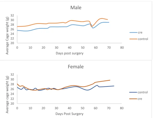
Body weight
For this and all further experiments, the mice were separated into two groups, those that received
the AAV5-Cre-GFP virus (cre) or those that received the AAV5-GFP virus (control). The body
weight of neither the male nor female cre mice varied significantly from the control mice
throughout surgery and the course of the behavioral experiments, suggesting that the 5-HT
DR→BNST projection is not involved in feeding behavior. The drops in body weight of both the
cre and control mice for both sexes occur during the first two weeks post-surgery and the day of
the novelty-suppressed feeding test, in which the mice had been food deprived for 24 hours. The
average body weight of representative cages of both male and female cre and control mice are
presented in Figure 6.
Figure 6. Body Weight. The body weights of a representative 6 male mice (n=3 cre, n=3 control) and 6 female mice (n=3 cre, n=3 control) post-surgery and throughout the behavioral experiments did not vary between the cre and the control groups.
20 22 24 26 28 30 32
0 10 20 30 40 50 60 70 80
Av era ge Cage w eight (g)
Days post surgery
Male
cre control 20 22 24 26 28 30 320 10 20 30 40 50 60 70 80
Av era ge cage we ight (g)
Days Post Surgery
Female
control
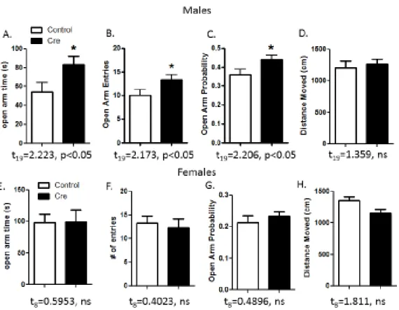
Behavioral Experiments
In the EPM test, the cre male mice spent a significantly greater amount of time in the open arms
and were more likely to enter them than the control mice, which suggests that the 5-HT1AR
BNST deletion led to a decrease in anxiety-like behavior. There was no significant difference
between the cre and control mice in locomotor activity in the EPM for the duration of the test.
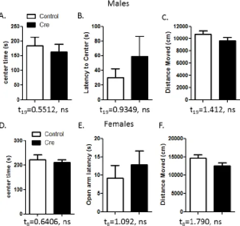
The female cre mice did not exhibit any significant anxiolytic phenotypes. In another test of
anxiety, the open field test, neither male nor female cre and control mice exhibited any
significant differences in anxiety-like behaviors or locomotor activity. The results for both of
these tests are presented in Figures 7 and 8.
Figure 8. Open Field.(A-F) There were no significant differences between the cre and the control mice in both sexes in the amount of time spent in the center of the box, the latency to the center of the box, and the total distance moved in the box.
There were also no significant differences in food consumption or latency to feed between the
cre and control mice for both males and females in the novelty-suppressed feeding test. In the
forced swim test there were no significant differences between percent immobility between the
Figure 9. Novelty-suppressed feeding and forced swim.(A, B, C, D) The 5-HT1AR BNST
deletion did not have any effect in the latency to feed and food consumption for both males and females in the novelty-suppressed feeding test. (E, F) The forced swim test also failed to differentiate between the cre and control mice of both sexes.
As shown in Figure 10, there were no differences between male cre and control mice in fear
learning or cued fear. Male cre mice showed a significant increase in cued fear consolidation but
not contextual fear learning, although the p value was close to significant at 0.0591. The
significant interaction between treatment (cre) and tone in female mice on day 1 suggests that cre
females exhibited delayed fear learning. Cre female mice appeared to have an opposite, but not
significant, effect in cued fear consolidation than the male cre mice when compared to control
Figure 10. Fear Conditioning. (A,B) The deletion of 5-HT1AR in the BNST did not lead to
differences in fear learning or cued fear in male mice. (C,D) This deletion did lead to a significant increase in cued fear consolidation in male mice but not in contextual fear recall.
(E,F) Female cre mice exhibited a delay in fear leaning but no significant effects in cued fear.
(G,H) The deletion led to an opposite, but not significant, effect in cued fear consolidation in female mice than male mice and no differences in contextual fear recall.
The data from the two-way ANOVA tests are shown in Figure 11 (EPM) and Figure 12
(contextual fear). This data indicates that significant sex differences were present in the open arm
time and probability of entering the open arms for the EPM test and contextual fear learning.
Female mice spent more time in the open arms but were less likely to enter them than male mice.
Male mice had a significantly higher percentage of freezing on the third day of the fear
Figure 11. Two-way ANOVA data for EPM.(A) There was a significant difference between male and female mice but not cre and control mice in the amount of time spent in the open arms of the EMP apparatus. (B) This effect was also reflected in the probability of the mice entering the open arms.
Virus Placement Verification
GFP immunohistochemistry was then used to verify the placement of the virus and the ablation
of the 5-HT1A receptor in the BNST. Mice that did not have adequate expression of the virus in
this brain region were removed from the data set. Representative pictures of a mouse that
remained at the data set and a mouse that was removed are presented in Figure 13. Overall, only
2 female cre mice were removed from the data set, making the female cohort n=5 cre and n=5
control.
A.
B.
Figure 13. BNST virus placement verification. Images of the BNST following GFP immunohistochemistry. (A) Images of a mouse that remained in the data set. (B) Images of a mouse that was taken out of the data set. The images on the left were taken with a Zeiss AXIO Zoom V16 microscope and the images on the right were taken with an Olympus
Discussion
Deletion of 5-HT1AR in the BNST leads to a decrease of anxiety-like behavior in male mice
The data above indicates several behavioral phenotypes differ between mice lacking the 5-HT1A
receptor in the BNST and control mice. The EPM data suggests that for male mice this deletion
leads to a decrease in anxiety-like behavior, indicating that this receptor may be involved in
anxiogenic pathways. This is the opposite effect reported in previous studies with a global
5-HT1A deletion. Given that 5-HT1ARs are primarily inhibitory, deletion of this receptor would tend
to disinhibit neurons in which they are expressed. This suggests to us that 5-HT1ARs in the
BNST are primarily expressed in a subset of neurons that mediate anxiolysis, such as BNST
output neurons to the ventral tegmental area (VTA) and lateral hypothalamus (LH) (Kim et al.,
2013; Jennings et al., 2013). Deletion of the 5-HT1A receptor in these BNST outputs would
result in the disinhibition of these neurons and hence anxiolysis, which is congruent with our
results. While these effects differ from those reported in the Levita paper, the only behavioral
experiment presented in that paper is the acoustic startle test, which presents mice with a stressor
(a loud tone) before testing for any anxiety-related phenotypes. The EPM test does not introduce
any stressors and therefore only tests basal anxiety levels. We hypothesize that the presence of a
stressor may recruit a different subset of local BNST neurons that promote anxiety-like behavior
and enhance fear recall.
The lack of this effect in females could be attributed to the lesser number of overall neurons in
the BNST of female mice. Having a smaller BNST volume could be indicative that changes to
Lack of feeding or depressive effects in both sexes
The lack of significant affects in both the body weight measurements and food consumption in
the novelty-suppressed feeding test indicate that this receptor in the BNST does not play a role in
feeding behavior. This lack of effect is not surprising as serotonin’s feeding-related phenotypes
have been attributed to projections to the hypothalamus. (Magalhães et al., 2010). The forced
swim results indicate that this receptor also does not play a role in depressive-like behaviors.
While BNST lesions have been shown to increase immobility in the forced swim test for both
male and female rats, this effect has not been attributed to serotonin and the data presented here
indicate that 5-HT1A is not responsible for the effect (Pezuk et al., 2008).
Increase in fear consolidation for male mice
While there were no significant effects of 5-HT1AR deletion on fear learning or cued fear for
male mice, there was a significant elevation in freezing for male cre mice in the 2-min period
after the tones were stopped on day 2 (i.e. consolidation). The BNST was previously shown to
play a critical role in fluoxetine enhancement of cued fear in rats (Ravinder et al., 2012), but this
is likely mediated by 5-HT2C rather than 5-HT1A receptors (Burghardt et al., 2013). We
hypothesize that this increase in freezing could be due to the cre mice retaining their fear in the
context of Day 2. This is consistent with the trend that contextual fear recall (Day 3 of the
protocol) was higher for the cre mice than the control mice of both sexes, although not
significant. This trend aligns with the finding that the introduction of a 5-HT1A agonist into the
BNST decreases contextual fear recall (Gomes et al., 2012) and our hypothesis that the presence
of a stressor recruits local BNST neurons which are both anxiogenic and fear enhancing, and that
Female cre mice exhibited a delay in fear learning for the intermediate tones on Day 1 that was
extinguished by the last tone, indicating that both the cre and control mice were able to form the
aversion to a shock that was paired with a tone. Effects on cued fear and consolidation were
insignificant in females. Increasing the n of both groups could lead to significant effects in this
type of learning.
Sex differences in EPM and Contextual Fear
The data presented in the two-way ANOVA analysis showed significant differences between
male and female mice in manifestations of anxiety-like behavior and fear learning, but not
between cre and control mice. This could be due to the BNST size differences between males
and females.
Any significant data between the cre and control mice using a simple T-test was no longer
significant using this method, as the threshold for significance was higher due to the increase in
independent variables and the low n of the female mice.
Conclusion
Serotonin modulates behaviors including fear, anxiety, depression, and feeding, and treatments
preventing the reuptake of serotonin are used every day to treat major psychiatric disorders.
Serotonin projections from the DR reach many brain regions, one of them the BNST, a critical
relay station in the brain. The experiments above study the 5-HT1A receptor, a receptor for a
subtype of serotonin, in the BNST and suggests that the activation of 5-HT1A signaling in the
BNST of male mice leads to an increase in anxiety under basal stress levels and a decrease in
phenotypes were reported, which could indicate that the BNST 5-HT1A outputs differ in males
and females. Repeating the above experiments with more female mice could lead to significant
values for the tests in that gender.
While current treatments in humans cannot target a specific brain region such as the BNST,
understanding the specific pathways that lead to certain behaviors could be important for future
therapeutics. The data above indicates that serotonin in the BNST is a potential target for treating
anxiety and fear disorders.
Acknowledgements
I would like to thank Dr. Thomas Kash and Dr. Catherine Marcinkiewcz for their mentorship and
direction throughout this project and the entirety of the Kash lab for their constant assistance.
Thanks also to Dan Perron for completing the SERT-cre surgeries. Additionally, I would like to
thank my family, roommates, and friends for their support and understanding throughout my
References
Aronsen, D., et al., RU 24969-produced adipsia and hyperlocomotion: Differential role of 5HT1A
and 5HT1B receptor mechanisms. Pharmacology Biochemistry and Behavior.2014. 124, 1-4.
Belcheva, I., et al., Behavorial responses to the 5-HT1A receptor antagonist NAN190 injected into rat CA1 hippocampal area. General Pharmacology: The Vascular System.1997. 28(3), 435-441.
Blanchard, C., et al., Sex Effects in Defensive Behavior: Baseline Differences and Drug
Interactions. Neuroscience & Behavioral Reviews. 1991. 15, 461-468.
Burghardt N.S., et al. Acute and chronic effects of selective serotonin reuptake inhibitor treatment on fear conditioning, implications for underlying fear circuits. Neuroscience. 2013,
247, 253–272.
Can, A., et al. The Mouse Forced Swim Test. Journal of visualized experiments.2012, 59, 3638-3645.
Casada JH, Dafny N. Restraint and stimulation of bed nucleus of the stria terminalis produce similar stress-like behaviors. Brain Res Bull. 1991, 27, 207–212.
Choleris, E., et al.A detailed ethological analysis of the mouse open field test: effects of diazepam, chlordiazepoxide and an extremely low frequency pulsed magnetic field. Neurosci Biobehav Rev. 2001, 25, 235–260.
Chung, W., et al. Apoptosis during Sexual Differentiation of the Bed Nucleus of the Stria Terminalis in the Rat Brain. J Neurobio. 2000, 43, 234-243.
Del Abril, A., et al., The bed nucleus of the stria terminalis in the rat: regional sex differences controlled by gonadal steroids early after birth. Dev Brain Res. 1987. 32(2), 295-300.
Dominic, L., et al., 5HT receptor antagonists enhance the functional activity of fluoxetine in a 1A mouse model of feeding. Brain Research. 1998, 781, 121-128.
Gomes, F., et al. Cannabidiol injected into the bed nucleus of the stria terminalis reduces the expression of contextual fear conditioning via 5-HT 1A receptors. J Psychopharmacol. 2012,
26(104), 104-113.
Guo Y., et al. Evaluation of the antipsychotic effect of bi-acetylated l-stepholidine (l-SPD-A), a novel dopamine and serotonin receptor dual ligand. Schizophr Res. 2009, 115, 41–49.
Jennings J. H., et al. Distinct extended amygdala circuits for divergent motivational states.
Nature. 2013. 496, 224–228.
Kessler, R. C., et al., Sex and depression in the National Comorbidity Survey I: lifetime prevalence, chronicity and recurrence. J. Affect. Disord.1993,29, 85–96.
Kessler, R. C., et al., Lifetime prevalence and age-of-onset distributions of DSM-IV disorders in the National Comorbidity Survey Replication. Arch. Gen. Psychiatry. 2005, 62, 593–602.
Kim S. Y., et al. Diverging neural pathways assemble a behavioural state from separable features in anxiety. Nature. 2013,496, 219–223.
Levita, L., et al. 5-Hydroxytryptamine1A-like Receptor Activation in the Bed Nucleas of the Stria Terminalis: Electrophysiological and Behavioral Studies. Neuroscience. 2004,128, 583-596.
Magalhães, M., et al., Modulatory role of serotonin on feeding behavior. Nutritional Neuroscience. 2010, 13, 246-255.
Nagy, A. Cre Recombinase: The Universal Reagent for Genome Tailoring. Genesis. 2000, 26,
99-109.
Narbox-Neme, N., et al. Serotonin transporter transgenic (SERTcre) mouse line reveals developmental targets of serotonin specific reuptake inhibitors (SSRIs). Neuropharm. 2008,
55(6), 994-1005.
Palanza, P. Animal models of anxiety and depression: how are females different? Neuroscience & Biobehavioral Reviews. 2001, 25(3), 219-233.
Parks, C., et al., Increased anxiety of mice lacking the serotonin1A receptor. Proc Natl Acad Sci U.S.A.1998, 95(18), 10734-10739.
Pellow, S., et al. Validation of open: closed arm entries in an elevated plus-maze as a measure of anxiety in the rat. J Neurosci Methods.1985, 14, 149–167.
Pezuk, P., et al. Effects of BNST lesions on forced swimming and navigational learning. Brain Research.2008, 1228, 199-207.
Ramboz, S., et al. Serotonin receptor 1A knockout: An animal model of anxiety-related disorder.
Proc Natl Acad Sci U.S.A.1998, 95(24), 14476-14481.
Ravinder S., et al. A role for the extended amygdala in the fear-enhancing effects of acute selective serotonin reuptake inhibitor treatment. Transl. Psychiatry. 2013, 3.