MODULATION OF P53 SIGNALING IN an APT
121INDUCED PROSTATE CANCER
MODEL
Wenqi Pan
A dissertation submitted to the faculty of the University of North Carolina at
Chapel Hill in partial fulfillment of the requirements for the degree of Doctor of
Philosophy in the Curriculum of Genetics and Molecular Biology.
Chapel Hill
2011
Approved by
Advisor: Yanping Zhang
Reader: Beverly H. Koller
Reader: Adrienne D. Cox
Reader: Norman E. Sharpless
© 2011
Wenqi Pan
ABSTRACT
Wenqi Pan: Modulation of p53 signaling in an APT
121induced prostate cancer
model
(Under the Direction of Dr. Yanping Zhang)
somatic deletion of p53 accelerates tumor progression and confirms that the
stromal tumors observed in the previous report are indeed caused by paracrine
signaling from epithelial cells.
In the second part of our study, we focused on novel p53 signaling which
has been reported to play a critical role in maintaining cell homeostasis after
ribosomal stress. Disruption of the binding of ribosomal proteins (RP) and
Mdm2 (a primary inhibitor of p53) in the Mdm2 point-mutation mouse model
Mdm2
C305F, has been shown to attenuate p53 signaling in Myc-induced
lymphoma. This suggests that RP-Mdm2-p53 signaling could be a general
tumor suppressor pathway activated in response to a variety of oncogenic
stresses. To investigate this possibility, we crossed Mdm2
C305Fmice with
APT
121 mice. Our results showed that disruption of the RP-Mdm2-p53 pathwaydoes not accelerate prostate tumorigenesis induced by APT
121. Instead, the
p19Arf-Mdm2-p53 pathway is the major player in response to Rb inhibition.
To all those I love and those love me,
TABLE OF CONTENTS
List of Tables
... viii
List of Figures
...ix
List of Abbreviations
...xi
CHAPTER ONE Introduction
... 1
Prostate Cancer in Humans
... 1
APT
121Mouse Model of Prostate Cancer
... 2
p53
ERMouse Model
... 8
RP-Mdm2-p53 Signaling Pathway
... 11
Mdm2
C305FKnock-in Mouse Model
... 15
CHAPTER TWO Targeted p53 deletion in APT121-initiated epithelial cells
accelerates prostate cancer development that
can be reversed by p53 restoration
... 29
Abstract
... 29
Introduction
... 31
Results
... 33
Discussion
... 51
Materials and methods
... 54
Abstract
... 56
Introduction
... 58
Results
... 61
Discussion
... 79
Materials and methods
... 82
CHAPTER FOUR Summary and Future Directions
... 86
Role of p53 in APT
121-initiated Prostate Cancer Model
... 86
RP-Mdm2-p53 Pathway in Signaling Oncogenic Stress
... 89
List of Tables
Table
Table 1. Early onset of prostate cancer in APT 121 ; p53cf/f ; Pb-Cre mice... 36
Table 2. Restoration of p53 function in APT 121 ; p53TAM/- mice delays the onset of
prostate tumors.... 50
Table 3. Summary of prostate tumor stages in 6 month-old Mdm2+/+, Mdm2C305F/C305F,
APT121;Mdm2+/+ , and APT121; Mdm2C305F/C305F mice... 65
Table 4. Summary of prostate tumor stages in 5 month-old APT121;p19Arf+/+ and
List of Figures
Figure
Figure 1. APT121 mouse prostate cancer model... 4
Figure 2. Progression of epithelial tumor and stromal tumor in APT121;p53-/- mice. 6
Figure 3. A diagram of the p53ERm allele... 9
Figure 4. Diagram of RP-Mdm2-p53 signaling. ... 13
Figure 5. A diagram showing that a point mutation of Mdm2 disrupts its binding to ribosomal proteins L11 and L5... 16
Figure 6. Mdm2C305F mutant protein does not bind to L11 and L5... 18
Figure 7. Mdm2C305F MEFs exhibit attenuated p53 response to ribosome biogenesis stress... 21
Figure 8. Myc-induced lymphomagenesis is accelerated by Mdm2C305F mutation
... 24
Figure 9. Myc-induced tumorigenesis is accelerated by loss of ARF... 27
Figure 10. Deletion of p53 in prostate epithelium accelerates both adenocarcinoma and stromal tumors in APT121 mice... 37
Figure 11. APT121; p53cf/f; Pb-Cre cancer cells do not derive from neuroendocrine
cells... 41
Figure 12. p53 deletion in ATP121 prostate induced adenocarcinomas are the
results of increased proliferation but not apoptosis... 44
Figure 13. Restoration of p53 expression in APT121; p53ER/- epithelial cells prevents
prostate malignant progression... 48
Figure 15. Mdm2 C305F mutation decreases proliferation but does not affect apoptosis of APT121-induced prostate cancer... 68
Figure 16. Effects of p19Arf loss on tumor progression in APT121-induced prostate
cancer... 72
List of Abbreviations
1. ARR2PB compostite probasin promoter
2. CDK cyclin dependent kinase
3. CP cytoplasm
4. H&E hematoxylin-eosin
5. IHC immunohistochemistry
6. KO knock out
7. LOH loss of heterozygosity
8. NO nucleolus
9. NP nucleoplasm
10. PB rat small probasin promoter
11. RP ribosomal protein
12. SV40 simian virus 40
13. T
121a truncated T-Ag comprised of the N-terminal 121 amino acids
14. TRAMP Transgenic Adenocarcinoma of the Mouse Prostate
15. TUNEL terminal deoxynucleotidyl transferase mediated dUTP nick end
labeling
CHAPTER ONE
INTRODUCTION
Prostate Cancer in Humans
Prostate cancer is the second leading cause of cancer death in American men, behind
only lung cancer. According to the American Cancer Society, about 217,730 new cases of
prostate cancer were diagnosed in the United States in 2010, and 32,050 men died of
prostate cancer. About 1 man in 6 will be diagnosed with prostate cancer during his lifetime
(American cancer society: Cancer facts and figures 2010). Although the majority of patients
initially respond well to androgen ablation, eventually nearly all men with advanced prostate
cancer progress to hormone-refractory prostate cancer (HRPC). At this stage, chemotherapy
may be used to extend survival, yet no curable therapeutic option is available (1). Despite the
magnitude of the statistics, the underlying causes of prostate cancer remain elusive mainly
due to the heterogeneity of genetic alterations found in the tumors (2). Several tumor
suppressor genes have been implicated including Rb (Retinoblastoma) (3) and p53 (also
APT121 Mouse Model of Prostate Cancer
Rb and its family members p107, and p130 control the G1 to S-phase transition of the
cell cycle primarily by interacting with the E2F family of transcription factors(5). Rb recruits
chromatin-remodeling proteins, such as histone deactylases and histone methyl-transferases
to repress the transcriptional activation of E2F1 (6-8). It was reported that 27% of human
prostatic adenocarcinomas lost one RB allele (9) and both reduced expression of Rb mRNA
and pRb protein have been reported (10-11).
Mice lacking the Rb gene die at around 13 days of gestation before the prostate forms
(12). Mice heterozygous for Rb or chimeric for Rb develop only pituitary or thyroid tumors
(13-15). Somatic deletion of Rb in mouse epithelial cells only leads to early stage of prostate
cancer (16). This appears to be because of compensation by p107 and/or p130 in many
murine cell types (17).
To bypass the redundancy of p107 and p130 in mice while focusing on the function of the
pRb family alone in the development of prostate cancer, the APT121prostate cancer mouse
model was generated by Dr. Terry Van Dyke’s lab. APT121is a truncated SV40 large T antigen
(maintains the first 121 NH2-terminal amino acids; T121) under the probasin promoter. The
transgene binds Rb and its family members, p130 and p107, and is specifically expressed in
prostate epithelium (Figure 1A). When Rb is suppressed, E2F1 is activated and cells become
tumorigenic. As early as 8 weeks of age, the prostates of APT121 mice exhibit dysplasia.
APT121mice show mouse prostatic intraepithelial neoplasia (mPIN) by 12 weeks old,
develop micro-invasive adenocarcinomas by 16 weeks old, characterized as epithelial cells
breaking through the basementmembrane into the stroma (Figure 1B) (18)
When APT121mice are crossed with p53-/- mice, stromal tumors, identified as the
presence and introductal growth patterns of stromal cells (Figure 2A and 2B), were greatly
accelerated by p53 haploinefficiency. Stromal tumors occur as early as 5 months old in
APT121;p53+/- mice. In contrast, stromal tumors occur only randomly and much later in
Figure 1. APT121 mouse prostate cancer model (18).
A. A diagram of APT121 mouse model: a truncated SV40 large T antigen under probasin
promoter. The expression of the transgene is targeted to mouse prostate epithelial cells. The
transgene is studied in the B6D2F1 mouse.
B. Histological pictures demonstrated the progression of tumors in APT121mice at 2 months
A
Figure 2. Progression of epithelial tumor and stromal tumor in APT121;p53-/- mice (18).
A. A diagram of p53 status and tumor progression in APT121 mice. p53 heterozygousity does
not affect the onset of epithelial tumors, yet it accelerates the onset of stromal tumors with
tumors appearing in mice by 5 month of age. APT121;p53+/- mice die around 7 months due to
the enlargement of tumors. Both epithelial and stromal tumors are greatly advanced in
APT121;p53-/- mice and mice die around 5 months of age.
B. Large tumors in APT121;p53 -/- mice are comprised of extensive stromal component. Star
A
p53ER Mouse Model
p53ER mice were generated by Gerard Evan lab (19). The hormone-binding domain of
the modified estrogen receptor, ERTAM (20) was fused to p53 at the its’ 3'-end (Figure 3A). In
the absence of 4-hydroxytamoxifen, p53m/m mice are similar to p53 null mice. Both develop
tumors of the similar incidence and spectrum, though the onset of tumors in the p53ER/ER mice
Figure 3. A diagram of the p53ERm allele (19)
The ligand binding domain of estrogen receptor is fused to the 3’ end of p53 allele. Without
RP-Mdm2-p53 Signaling Pathway
p53 is well known as the guardian of the genome. Under stress such as DNA damage,
oncogenic stress and hypoxia, p53 is stabilized and activated, resulting in the induction of a
variety of downstream signaling pathways, including cell cycle arrest, apoptosis, DNA repair
and senescence (21). Under normal condition, the level of p53 is kept low mainly by its
inhibitor, Mdm2 (mouse double minute 2). The C-terminus of Mdm2 has an intrinsic E3 ligase
activity which promotes the ubiquitiation and degradation of p53. The N-terminus of Mdm2
binds to the transactivation domain of p53 and inhibits the recruitment of co-activators.
Additionally, Mdm2 is directly transactivated by p53, thereby forming an Mdm2-p53 feedback
loop to maintain cellular homeostasis (22). p53 has been found to be mutated in about 50%
of human tumors, indicative of its importance in tumorigenesis (23). In addition to mutation of
p53 directly, overexpression of Mdm2 can also cause tumors (24).
The ribosome is the ‘factory’ for protein synthesis. The mammalian 80S ribosome is
comprised of a large 60S and a small 40S ribosomal subunit. The 60S large subunit contains
5S, 5.8S, and 28S rRNAs and around 47 large ribosomal proteins (RPL). The 40S small
subunit contains 18S rRNA and around 32 small ribosomal proteins (RPS). Ribosomal
subunits are made in the nucleolus and exported to the cytoplasm for the assembly of
ribosomes. Ribosomal biogenesis requires balanced expression between rRNAs and
ribosomal proteins, processing of rRNA precursors into mature rRNAs and assembly of 40S
and 60S ribosomal subunits into 80S ribosome (25). When any of these steps are disrupted,
or 5-fluorouracil (5-FU) (28), ribosomal stress is induced. In addition to drug treatment,
changing cell culture conditions, such as serum starvation and contact inhibition(29), or
overexpression of nucleostemin or the dominant negative mutant Bop1(30) can also induce
ribosomal stress.
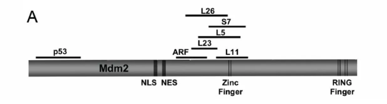
Under ribosomal stress, free forms of RPs (RPL and RPS), including L11 (31-32), L23
(33-34) , and L5 (27), S7 (35) enter the nucleoplasm to interact with the zinc finger domain of
Mdm2. This binding by L11, L23 or L5 inhibits MDM2's E3 ligase function, resulting in p53
accumulation and activation (Figure 4). Knockdown of L11 and L5 does not induce a p53
response, whereas knockdown of L23 does induce a p53 response. This indicates that these
ribosomal proteins function similarly as well as distinctly to transmit ribosomal stress to the
Mdm2-p53 pathway.
S7 forms a ternary complex with Mdm2 and p53. Mdm2 can target S7 for ubiquitination,
and MdmX (homolog of Mdm2) enhances the inhibition of Mdm2 by S7 (36). L26 also binds
the zinc finger domain of mdm2 and besides that, L26 binds to the 5′ untranslated region
(UTR) of p53 mRNA and augments its translation. The binding between Mdm2 and L26
promotes the ubiquitination and degradation of L26 and therefore, weakens p53
Figure 4. Diagram of RP-Mdm2-p53 signaling (38).
Under normal conditions, large and small ribosomal subunits are assembled in the nucleolus
(NO) and then exported to the cytoplasm (CP) for protein synthesis. However, under
nucleolar stress, the balance between rRNA and ribosomal proteins (RPL and RPS) is
disrupted. Free forms of ribosomal proteins are released from the NO to the nucleoplasm (NP)
and bind to Mdm2. p53 is thereby activated and stabilized. Excess free forms of ribosomal
proteins can also be released from cytoplasmic ribosomes and then enter the NP, interacting
Mdm2C305F Knock-in Mouse Model
In vitro data have shown that mutation of the zinc-coordinating cysteine (C) residues to
phenylalanine (F) disrupts Mdm2’s interaction with L11 and L5 while retaining interaction with
L23 (Figure 5). As a result, The MDM2C305F mutant is attenuated in mediating p53
degradation after ribosomal stress (39). Based on in vitro data, our lab generated the
Figure 5. A diagram showing that a point mutation of Mdm2 disrupts its binding to
ribosomal proteins L11 and L5 (40)
The Mdm2C305F mutation disrupts L5 and L11 binding (40)
Previous study also investigated the binding activity of the Mdm2C305F mutant protein.
Mouse embryonic fibroblasts (MEFs) were used for an immunoprecipitation-coupled western
blot (IP-western) assay (40). When cell lystates were immunoprecipitated with anti-Mdm2
antibody and then immunoblotted with anti-L5 and anti-L11 antibody, L5 and L11 were
present in the Mdm2+/+ but not in the Mdm2C305F/C305F immunoprecipitates (Figure 7A).
Mdm2C305F was still able to bind p53. Because the anti-Mdm2 antibody (clone 2A10) affects the binding between Mdm2 and L23, we cannot evaluate the interaction of L23 with Mdm2
using immunoprecipitation. We can however immunoprecipitate with anti-L23 antibody and
then detect Mdm2 by western blotting. As shown in Figure 7B, L23 still binds toMdm2C305F.
Figure 6. Mdm2C305F mutant protein does not bind to L11 and L5 (40)
A. Mdm2+/+ and Mdm2m/mMEFs were treated with 5 nM Act D. Cell lysates were
immunoprecipitated with anti-Mdm2 antibody and to detect Mdm2, p53, L5 and L11 as
indicated. 5% of total IP lysate is used as loading control.
Mdm2C305F mutation attenuates p53 response to ribosome biogenesis stress (40)
Ribosomal stress induces releasing free forms of PRs, such as L11 and L5. L11 and L5
can bind to Mdm2 and thereby, leads to p53 stabilization and activation. Since the Mdm2C305F
mutant protein cannot bind to L5 and L11 as shown in figure 6, it is expected that the p53
response would be attenuated after ribosomal stress. To assess this, Mdm2+/+, Mdm2+/C305F,
and Mdm2C305F/C305F MEFs were either untreated or treated with a low dose of actinomycin D
( 5nM Act D). Mdm2+/+ MEFs exhibited the highest induction of p53, compared to Mdm2+/C305F
showing intermediate induction of p53, and Mdm2C305F/C305F with the lowest induction of p53
(Figure 9A). Two other drug, 5-FU and MPA, are also known to induce ribosomal stress. MEF
cells were treated with these two drugs to further confirm that p53 induction was attenuated
in Mdm2C305F/C305F MEFs. Figure 9B show that again the p53 response is attenuated in a gene dosage dependent manner. Taken together, the data show that the p53 response to
Figure 7. Mdm2C305F MEFs exhibit attenuated p53 response to ribosome biogenesis
stress (40).
A. MEFs were either mock treated or treated with 5 nM Act D for 12 hr and then harvested for
western blotting analysis.
B. MEFs were treated with 1 μM 5-FU or 2 μM MPA for 12 hr and examined for p53 protein
A
Myc-induced lymphomagenesis is accelerated by Mdm2C305F mutation (40)
Translocation (t8;14) of Myc into or close to Ig loci is often observed in Burkitt’s B cell
lymphoma (41). To mimic this in the mouse model, the Myc gene was coupled to an Emu
immunoglobulin heavy chain enhancer, resulting in the overexpression of Myc in the B cell
lineage (42). Eμ-myc mice die of B cell lymphoma and the median survival is 20 weeks. The
Eμ-Myc;Mdm2+/C305F mice had a median survival of about 15 weeks, while the survival of Eμ-Myc;Mdm2C305F/C305F mice was reduced to a median of 9 weeks (Figure 10A). Tumors
from the Eμ-Myc;Mdm2C305F/C305F mice were morphologically similar to those from
Eμ-Myc;Mdm2+/+ mice (Figure 10B) (40). Given that Myc upregulates ribosomal biogenesis by interacting with all three RNA polymerases (43), these findings established the
Figure 8. Myc-induced lymphomagenesis is accelerated by Mdm2C305F mutation (40) .
A. Survival of Eμ-Myc transgenic mice. Eμ-Myc;Mdm2+/+ (+/+, n = 24), Eμ-Myc;Mdm2+/m (+/m,
n = 27), and Eμ-Myc;Mdm2m/m (m/m, n = 18) mice.
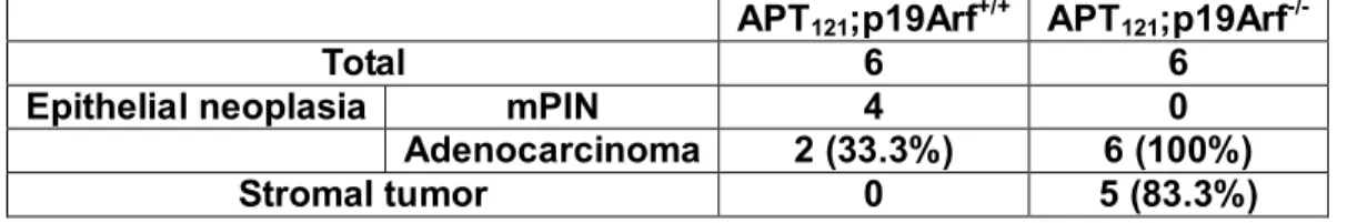
RP-Mdm2-p53 functions in a pattern similar to p19Arf-Mdm2-p53 in response to Myc
Eμ–myc transgenics that were wild-type for p19Arf displayed a mean survival of 20
weeks (42), while Eμ–myc transgenics heterozygous for p19Arf displayed a mean survival of
11 weeks. Eμ–myc;p19Arf −/− mice die soon after birth in some offspring, and of those that
survived initially, all died of lymphoma by 8 weeks of age (44)(Figure 8A). Tumors from
Eμ–myc;p19Arf −/− mice were phenotypically indistinguishable from those from wild-type
Eμ–myc transgenics. Noticeably, this pattern of tumor acceleration is very similar to the
accelerated tumorigenesis observed in mice harboring the Mdm2C305 mutation in the Eμ–myc
Figure 9. Myc-induced tumorigenesis is accelerated by loss of ARF (44).
Survival of Eμ-Myc transgenic mice. Eμ-Myc; ARF+/+ (ARF+/+, n = 31), Eμ-Myc; ARF+/−
(ARF+/−, n = 85), and Eμ-Myc; ARF−/−(ARF−/−, n = 20) mice. Lymphoma was documented in
CHAPTER TWO
Targeted p53 deletion in APT
121-initiated epithelial cells accelerates prostate
cancer development that can be reversed by p53 restoration
Abstract
In the APT121 mouse model, Rb and its family members, p107 and p130, are
inactivated specifically in prostate epithelial cells by expression of APT121, an SV40 T
antigen N-terminal fragment placed under regulation of the prostate
epithelium-specific probasin promoter. It has been shown that APT121 transgenic mice
develop prostatic intraepithelial neoplasia (mPIN), which further progresses into
adenocarcinomas, and that this phenotype is accelerated by additional germline
mutation of p53. However, prostate cancer is a disease more commonly associated
with somatic mutations. Thus, we sought to investigate the role of somatic p53
deletion in the development of prostate epithelial tumors. Here, we generated
APT121;p53cf/f;Pb-Cre mice, in which the expression of APT121 and deletion of p53 occur
simultaneously and specifically in the prostate of compound male mice at 6 weeks of
age, as a preclinical animal model to both genetically and biologically emulate human
prostate cancer. We show that deletion of p53 significantly accelerated prostate
mechanism that favors cell proliferation without affecting apoptosis. In addition, we
took advantage of the previously generated p53ER mouse model where tamoxifen
treatment re-establishes wild-type p53 function and developed APT121;p53ER/- mice to
evaluate the consequence of restoration of p53 in prostate cancer cells. We show that
reinstating p53 function by treating APT121;p53ER/- mice with tamoxifen resulted in
decreased proliferation and increased apoptosis in the prostate, and significantly
reduced tumor progression. Together, our data suggest that p53 plays a crucial role in
prostate cancer initiation and progression and that therapies focusing on restoring
p53 activity in epithelial cells could be an attractive approach to not only prevent
malignant progression but also to delay the onset of prostate stromal tumors.
Acknowledgement
We thank Paula L. Miliani de Marval and Hilary Clegg for modifying the manuscript of
Introduction
Prostate cancer is the second most common cancer among men. 25 to 50% of human
prostate adenocarcinomas harbor aberrations in the Retinoblastoma (Rb) pathway. Rb is a
tumor suppressor that binds to and represses the transcriptional activity of E2F family
transcription factors that control the G1-S cell cycle transition. Phosphorylation of Rb by
Cdk4/6-Cyclin D or Cdk2-Cyclin E releases Rb from the E2F complex, allowing
transactivation of E2F target genes to occur. Rbinactivation is common in prostate cancer
and generally precedes somatic alterations affecting the tumor suppressor gene p53 (45).
Functional interactions between the two major tumor suppressor genes appear to directly
influence tumor development in the mouse prostate (46). In particular, mutations of the p53
gene are frequently associated with metastasis and an androgen depletion–independent
phenotype in prostate cancer (47).
Targeted inactivation of p53 in a murine model of prostate cancer was found to induce
mouse Prostatic Intraepithelial Neoplasia (mPIN) but failed to progress to invasive carcinoma,
indicating that loss of p53 may be a synergistic rather than an initiative event in promoting
prostate tumorigenesis (48). On the other hand, deletion of both the Rb and p53 genes in
mice resulted in rapid development of invasive and metastatic prostate cancer (49). Further
studies indicated that the cell origin of prostate cancer associated withp53 and Rb deficiency
islikely to be stem/progenitor cells that control luminal and neuroendocrinedifferentiation
(50).
experienced a great susceptibility to the development of oversized tumors comprised of
adenocarcinomas and a large amount of abnormal stromal hyperproliferation (18, 51).
However, germline mutation in the p53 gene does not seem to be an early event in prostate
tumorigenesis; rather, it seems to be associated with the development of malignant
progression. Thus, we proposed to investigate whether somatic inactivation of p53 in the
prostate epithelium of APT121 mice alters the onset of adenocarcinomas and whether it
Results
Prostate epithelial-specific deletion of p53 accelerates adenocarcinoma development
and induces stromal tumors in APT121-induced prostate cancer
Prostate cancer is mostly associated with somatic gene mutations rather than germline
gene mutations. In this context, results obtained from various mouse models have shown that
complete ablation of p53 in mice may influence tumor development by altering the cell
microenvironment. Thus, we sought to provide a preclinical system that could better simulate
the human disease by generating a mouse model with prostate-specific somatic deletion of
p53. We generated APT121compound mice carrying a probasin-driven Cre/loxP system (52)
to obtain conditional ablation of p53 expression. We bred the previously developed p53cf/f
conditional knockout mice (53) with Probasin-Cre transgenic mice (Pb-Cre) (48), which
express the Cre transgene in the epithelial cells of the prostate beginning at the onset of
puberty around 6 weeks of age. The p53cf/f;Pb-Cre mice were then crossed with APT121 mice
to generate APT121;p53cf/f;Pb-Cre compound mice. Thus, expression of APT121 and deletion
of p53 occur simultaneously and in the same cellular compartment in the compound mice at 6
weeks of age.
We established tumor appearance by harvesting prostates from wild type, APT121, and
APT121;p53cf/f;Pb-Cre mice every four weeks beginning at 8 weeks of age. To evaluate tumor
progression, hematoxylin and eosin staining (H&E) of tissue sections was used.All groups of
mice were monitored daily for signs of morbidity and prostate cancer development. None of
However, consistent with previous reports (18), the APT121 micedeveloped mPIN at 12 weeks
(75% incidence), which further transformed into adenocarcinomas by 4 months of age (75%
incidence) (Table 1). The APT121;p53cf/f;Pb-Cre mice displayed a shorter latency for tumor
development as reflected by the appearance of mPIN at 8 weeks with a 66% incidence. At 3
months, APT121;p53cf/f;Pb-Cre mice had already developed adenocarcinomas with an 87.5%
incidence, and by 5 months we observed 100% incidence of stromal neoplasias, consistent
with a previous study of the APT121; p53-/- mouse prostate model (51). Interestingly, in the
presence of wild type p53 APT121 prostates did not showstromal tumors until later in life, after
11 months, and with a low incidence (30%) (Table 1). The survival rate correlated with the
malignancy and progression of the tumors, as all of the APT121;p53cf/f;Pb-Cre mice died or
had to be sacrificed by 5-6 months due to the tumor burden (Table 1 and Figure 10B). This
phenotype was not apparent in mice harboring wild-type p53 (APT121 and WT mice) (Table 1
and Figure 10A). H&E staining of wild type prostates from 5-month old animals exhibited
normal prostate acini architecture arranged in a lobular configuration, consisting of a single
layer of luminal cells and a layer of epithelial cells surrounded by a basement membrane
separating the prostate gland from the stroma. The stroma, in turn, is formed by two to three
layers of smooth muscle with loose connective tissue found between the glands (Figure 10C).
APT121 prostates showed adenocarcinomas with the classic “acinar type” phenotype in which
the tumor is thought to arise from or recapitulate prostatic acini (Figure 10D). The acinar type
is characterized by back-to-back proliferation of small- to intermediate-sized tumor acini with
at this age. In contrast, the APT121;p53cf/f;Pb-Cre compound mice displayed
poorly-differentiated adenocarcinomas with infiltration of the prostate acina into the
fibromuscular stroma and multiple areas where the basal cell layer was absent, highlighting
the invasive and malignant nature of these tumors (Figure 10E). Furthermore, the enlarged
tumors were comprised of a large portion of stromal neoplastic cells that expanded into the
glands (Figure 10F).
Altogether, these data indicate that deletion of p53 in the epithelium of APT121prostates
results in early tumor onset with features of malignancy, invasion, and the occurrence of
stromal neoplasia, the latter of which is likely induced by a paracrine effect from the abnormal
epithelial cells to the surrounding mesenchymal tissue. This is consistent with the hypothesis
that in APT121;p53+/- prostates, the oncogenic stress in prostate epithelium signals a
mitogenic stimulation to the mesenchyme leading to the development of stromal tumors (51).
Table 1. Early onset of prostate cancer in APT 121 ; p53cf/f ; Pb-Cre mice
Genotype mPIN Adenocarcinoma Stromal tumor Survival
Wild-type None (0/4) None (0/5) None (0/5) >18 months
APT121 3 months (3/4) 4 months (6/8) >11 months (3/10) >15 months
APT121; p53cf/f;
Figure 10. Deletion of p53 in prostate epithelium accelerates both adenocarcinoma
and stromal tumors in APT121 mice.
Gross anatomy of prostates from APT121 (A) and APT121;p53cf/f; Pb-Cre (B) prostate
gland and associated seminal vesicles.. APT121; p53cf/f; Pb-Cre prostate displays solid tumors,
dramatically enlarged with important vasculature. Hematoxilin and Eosin (H&E) staining of
wild-type prostate (C) shows normal gland architectures.and the inset illustrates the single
layer of epithelial cells in the gland. (D) APT121 prostate shows well-differentiated
adenocarcinoma, and the inset points out to the intact basement membrane. (E)
APT121;p53cf/f; Pb-Cre prostate displays poorly-differentiated adenocarcinoma. The arrow
indicates the increased proliferation and loss of basement membrane that resulted in
invasion to the stroma. (F) A representative stromal tumor found in APT121;p53cf/f; Pb-Cre
prostate, the (*) demarks the stromal neoplasia and the inset shows the expansion of stromal
cells inside of the epithelial gland and the lack of organization of the mesenchymal cells. All
APT
121;p53
cf/f;Pb-Cre
WT
*
APT
121;p53
cf/f;Pb-Cre
APT
121Seminal vesicle
Prostate
APT
121Prostate
APT
121;p53
cf/f;Pb-Cre
A
B
C
D
Adenocarcinomas developed in APT121;p53cf/f;Pb-Cre compound mice are not derived
from neuroendocrine cells
Some prostate cancer mouse models develop tumors classified as adenocarcinomas,
while other models form neuroendocrine carcinomas. It has been previously demonstrated
that in some mouse models of prostate cancer, the origin of the tumor and the progression of
the disease are influenced by the mouse strain background. One example is the widely used
transgenic adenocarcinoma mouse prostate (TRAMP) model. While TRAMP mice with a
C57BL/6J background develop adenocarcinomas, TRAMP mice with an FVB background
have a significantly shorter lifespan than TRAMP-C57BL/6J mice due to the rapid
progressionof neuroendocrine carcinomas forming frombipotential progenitor cells at early
stages of prostate tumorigenesis(57). Consistent with these studies, the APT121;p53cf/f;Pb-Cre
mice, which have a genetic background similar to that of the FVB-TRAMP mice, have a
shorter lifespan due to the size and the malignancy of the prostate tumors. We performed a
series of immunohistochemistry (IHC) stainings to define the APT121;p53cf/f;Pb-Cre prostate
tumor cell origin. We first confirmed that the transgene T121 expression was confined to the
epithelial compartment (Figure 11A), ruling out the possibility of leaky expression of the
transgene. IHC staining showed that the APT121;p53cf/f;Pb-Cre neoplasias were positive for
the androgen-receptor (AR), which is a marker expressed in both prostate epithelial and
mesenchymalconnective cells but not in neuroendocrine cells (Figure 11B). Figure 11C
epithelium of prostate tumors, as expected. Lastly, synaptophysin (SYN), a specific marker
for neuroendocrine cells, was negative in all samples tested (Figure 11D), thus ruling out the
possibility that the tumors detected in APT121;p53cf/f;Pb-Cre prostates were of neuroendocrine
origin as was reported for the FVB-TRAMP model (57). Together, these evidences indicate
that the tumors formed in APT121;p53cf/f;Pb-Cre mice can be classified as adenocarcinomas.
Additionally, we assessed the invasive capacity of the adenocarcinomas by staining for
α-smooth actin (SMA), a marker found at the fibromuscular stromal cell layer that surrounds
the tumors. We observed a loss of this marker at a large portion of the area surrounding the
APT121;p53cf/f;Pb-Cre tumors, indicating that the stromal layer was disrupted in this area,
enhancing the tumors’ ability to migrate (Figure 11E and 11F). This loss of α-smooth actin
Figure 11. APT121; p53cf/f; Pb-Cre cancer cells do not derive from neuroendocrine
cells.
(A) T121 IHC staining: Using the SV40 T antigen mouse mAb, the expression of T121 is
restricted to the prostate epithelial cells of APT121; p53cf/f; Pb-Cre mice. (B) The
Androgen-receptor (AR) IHC staining illustrates that the expression of this marker in the
glandular epithelium and the stroma is conserved. (C) Immunofluorescence for cytokeratins
18/8 (CK18/8) shows adequate distribution of luminal epithelia cells. (D) Synaptophysin (SYN)
specifically identifies neuroendocrine cells, which were not detected in any of the compound
mice tumors. (E) α-smooth actin (SMA) is a marker of fibromuscular cells and underlies the
basement membrane of the glandular tissue. (F) As demarked by the arrow and the pointed
line, the fibromusular cells distribution becomes lose with areas of complete absence of the
APT
121;p53
cf/f;Pb-Cre
APT
121A
SMA
E
APT
121D
CK18/8
C
AR
B
SMA
Prostate epithelial-specific deletion of p53 does not affect apoptosis but enhances
proliferation in APT121-induced prostate cancer
The data presented here demonstrate that specific deletion of p53 in prostate epithelial
cells results in reduced tumor latency and increased malignancy. In order to understand the
probable mechanism leading to increased tumor growth in APT121;p53cf/f;Pb-Cre prostate
neoplasms, we quantified the rates of proliferation and apoptosis by immunohistochemical
staining. In APT121 prostates, the quantity of proliferating epithelial cells was 49%, as
measured by Ki67 staining (a marker of cell proliferation) (Figure 12A and 12C). The
APT121;p53cf/f;Pb-Cre prostate epithelial cells were significantly more proliferative than the
APT121 cells, with 73% of cells staining positive for Ki67 (Figure 12B and 12C). To measure
the rate of apoptosis in the prostate tumor cells, a TUNEL assay (terminal deoxynucleotidyl
transferase–mediated dUTP-biotinnick end labeling) was used. No significant difference in
apoptosis was observed between APT121;p53cf/f;Pb-Cre prostates and APT121 prostates
(14.2% and 12.2% apoptotic cells, respectively), (Figure 12D-F). These data indicate that
p53 function in the prostate epithelial tissue is important in the regulation of cell proliferation
Figure 12. p53 deletion in ATP121 prostate induced adenocarcinomas are the results of
increased proliferation but not apoptosis.
(A-B) Representative Ki67 staining from 5 month-old mice of the indicated genotype. Brown
staining indicates proliferating cells. (C) Average % proliferating cells (Ki67-positive) ± SEM
from 5 month-old mice of the indicated genotype. n=5 for each genotype. At least five
independent fields consisting of a total of at least 1,000 cells from each prostate sample were
counted. *p < 0.05 as assessed by Student’s t test. (D-E) Representative TUNEL assay from
5 month-old mice of the indicated genotype. Apoptotic cells are stained purple. (F) Average
% apoptotic cells (TUNEL-positive) ± SEM from 5 month-old mice of the indicated genotype.
n=5 for each genotype. At least five independent fields consisting of a total of at least 1,000
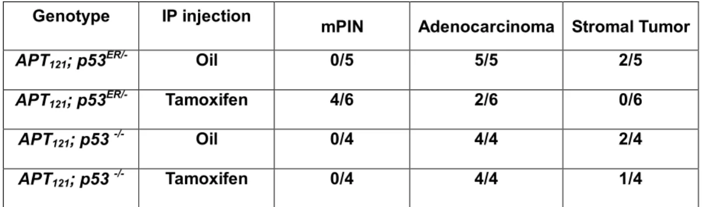
Restoration of p53 function in APT121; p53ER/- mice delays the onset of APT121-induced
prostate cancer by reducing proliferation and increasing apoptosis
We have demonstrated that p53 plays a pivotal role in tumor progression of
APT121-induced prostate cancer. To further investigate the function of p53 in tumor
suppression, we took advantage of an inducible-p53 mouse model, p53ER (19). These mice
have inactive p53 in the absence of tamoxifen, and upon inoculation with this drug, the
activity of wild-type p53 is reinstated. We generated and selected APT121;p53ER/-males to
evaluate whether the malignant tumor phenotype could be reversed or delayed by restoring
p53 function. Without tamoxifen injection, APT121;p53ER/-miceresemble APT121;p53-/- mice.
After tamoxifen injection, the APT121;p53ER/- prostate epithelial cells become heterozygous for
p53 (APT121;p53+/-). Considering that APT121;p53ER/- mice die around 4 months of age due to
oversized prostate tumors, we began tamoxifen treatments at 2 months. Tamoxifen or oil
(mock treatment) were introduced intraperitoneally (i.p) every 3 days during a 30-day period
(a total of 10 applications). The mice were euthanized within 3 days after the last treatment.
Prostate samples were formalin-fixed and paraffin-embedded for histological examination. As
shown in Table 2, oil-injected APT121;p53ER/- mice exhibited a 100% incidence of
adenocarcinomas. In contrast, the tamoxifen-injected APT121;p53ER/- mice exhibited a
dramatic drop in the incidence of adenocarcinomas; instead, the majority of these mice (4 out
of 6) showed only mPIN (Table 2). Notably, all APT121;p53-/- mice developed
adenocarcinomas (measured at the same time), regardless of tamoxifen treatment.
adenocarcinoma, whereas the tamoxifen-treated APT121;p53ER/- mice displayed benign,
well-differentiated mPIN (Figure 13A and 13B). The control APT121;p53-/- mice displayed, as
expected, prostate adenocarcinoma with all treatments (Figure 13C and 13D). To examine
how reinstating p53 expression in the prostate cells affects tumor onset, prostate sections
were assessed by immunohistochemistry for the expression of Ki67, a proliferation marker,
and with a TUNEL assay. Restoration of p53 expression by tamoxifen injection resulted in a
significant decrease in proliferation and increase in apoptosis in the prostate of APT121;p53
ER/-mice: Proliferating cells decreased from 44% to 28% of total cells, and apoptotic cells
increased from 8% to 18% (Figure 4M and 4N, open bars). This effect was not observed in
control APT121;p53-/- mice (Figure 4M and 4N, filled bars). Representative IHC images were
shown in Figure 13E-H for Ki67 staining and in Figure 13I-L for TUNEL staining. In summary,
our results indicate that the expression of wild-type p53 is essential for preventing malignant
initiation and progression in prostate tumors, and that restoration of p53 function in these
tumor cells can effectively reverse malignant prostate adenocarcinoma progression and
Figure 13. Restoration of p53 expression in APT121; p53ER/- epithelial cells prevents
prostate malignant progression
Representative H&E staining of prostate sections from mice of the indicated genotypes and
treatments. (A) APT121; p53ER/- prostate shows adenocarcinomas upon Oil injection. APT121;
p53ER/- prostate shows mPIN upon tamoxifen inoculation that results in restoration of p53
expression (B). Both oil-injected and tamoxifen-injected APT121; p53-/- prostates show
adenocarcinoma with stromal tumor phenotype.(C and D). Representative Ki67 staining
from mice of the indicated treatment. Brown staining indicates proliferating prostatic cells.of
oil or tamoxifen-treated APT121; p53ER/- prostates (E and F) and that of oil or
tamoxifen-treated APT121; p53-/- prostates (G and H). Representative TUNEL staining from
mice of the indicated treatment. Purple staining indicates apoptotic cells of oil or
tamoxifen-treated APT121; p53ER/- prostates (I and J) and that of oil or tamoxifen-treated
APT121; p53-/- prostates (K and L).
(M) Average % Ki67-positive cells ± SEM from mice of the indicated treatment. n=5-6 for
each genotype. At least five independent fields consisting of a total of at least 1,000 cells
from each prostate sample were counted. **p < 0.01 as assessed by Student’s t test. (N)
Average % TUNEL-positive cells ± SEM from mice of the indicated treatment. n=5-6 for each
genotype. At least five independent fields consisting of a total of at least 1,000 cells from
Table 2. Restoration of p53 function in APT 121 ; p53TAM/- mice delays the onset of
prostate tumors.
Genotype IP injection
mPIN Adenocarcinoma Stromal Tumor
APT121; p53ER/- Oil 0/5 5/5 2/5
APT121; p53ER/- Tamoxifen 4/6 2/6 0/6
APT121; p53 -/- Oil 0/4 4/4 2/4
Discussion
Prostate cancer is a devastating disease that affects adult men and is the second most
common cancer among this population. It is predicted that 1 out of 6 men will be diagnosed
with prostate cancer during their lifetime (58). Over the last few years, many research groups
have developed animal models to analyze the stages and progression of the disease.
However, the heterogeneous nature of prostate cancer has proven difficult to recapitulate.
One useful model to study the progression of prostate cancer is the APT121 transgenic mouse.
In this model, the expression of Rb and its family members, p107 and p130, is inhibited upon
expression of the N-terminal fragment of the SV40 T antigen, which is regulated in this model
by the prostate epithelial-specific probasin promoter. APT121 mice have normal expression of
p53, making these mice suitable for studying the role of Rb as a cancer initiator to which
other mutations can be added in order to understand the multistage nature of the disease.
In this study, we have deleted p53 somatically in prostate epithelial cells in order to better
recapitulate the progression of human prostate cancer. In particular, we took advantage of
the APT121 mouse model to mimic the initial aberrant event in the development of prostate
neoplasia and then induced somatic inactivation of p53 by generating APT121;p53cf/f;Pb-Cre
compound animals. Here, we were able to demonstrate that APT121;p53cf/f;Pb-Cre mice,
similar to the previously reported APT121;p53-/- mice, develop adenocarcinomas much earlier
and at a higher incidence than APT121 transgenic mice. Furthermore, the
APT121;p53cf/f;Pb-Cre tumors also recapitulated the appearance of stromal tumors, which
rare, are a clinical challenge during the development of human prostate malignancies, due to
the uncommon pattern of proliferation that results in various histological presentations. These
tumors, also known as Stromal Tumors of Uncertain Malignant Potential (STUMP), present a
unique pattern of proliferation in these stromal cells, which are behaviorally and histologically
distinct from benign hyperplasias and whose behavior cannot be predicted by histological
appearance. In this case, we found that the APT121;p53cf/f;Pb-Cre tumors were greatly
enlarged, in part due to uncontrolled proliferation and expansion of the mesenchymal
compartment (Figure 1E-F). These results indicate that p53 deletion in prostate epithelium
with a compound loss of Rb function is sufficient to drive prostate tumorigenesis and
stromal-associated malignant transformation. Furthermore, the data presented here suggest
that the stromal tumors observed in a previous study (18, 51) in APT121;p53+/- and
APT121;p53-/- prostates could be the result of paracrine signals induced by the epithelial cells
to the stroma, rather than a consequence of cell-autonomous loss of p53 in the stroma.
However, whether this is actually true remains to be determined.
Another interesting finding from this study is that tumors derived from
APT121;p53cf/f;Pb-Cre mice display increased proliferation compared to APT121 tumors, but
without noticeable changes in apoptotic levels. p53-independent apoptosis in prostate tumors
has been demonstrated in the APT121 model (18). Here, we found evidence that p53 ablation
enhances the over-proliferative phenotype induced by loss of Rb function in prostate
epithelial cells, which may cause the early onset of prostate cancer. Furthermore, we
ER/-mouse model. In this case, restoration of p53 activity by administration of tamoxifen resulted
in decreased proliferation and increased apoptosis in the prostate and significantly reduced
tumor progression. In this scenario, p53-dependent apoptosis clearly play a role in the
prevention of the malignant phenotype, which is different from the results obtained with
APT121;p53cf/f;Pb-Cre mice. A possible explanation is that in APT121;p53cf/f;Pb-Cre mice,
prostate cells are able to adapt to the absence of p53, whereas in tamoxifen-treated
APT121;p53ER/- mice, rapid restoration of p53 induces apoptosis. It is likely that epithelial
deletion of the p53 gene is sufficient to drive malignant transformation and the development
of surrounding stromal neoplasias. As the cells undergo aberrant proliferation, there may be
selection pressure for cells that have acquired further mutations leading to genomic instability,
resulting in the loss of other genes involved in apoptosis including p53. In summary, our
results emphasize the relevance of p53 in mediating prostate cancer and its collaboration
with Rb inactivation. Our data suggest that delivery of p53 in the epithelial prostate gland
Materials and methods
Mouse breeding strategies
To study the somatic deletion of p53 in APT121mice, APT121 females were crossed with p53cf/f
males to create and select for APT121;p53 cf/+ mice; p53cf/f females were then crossed with
Pb-Cre males to generate and select for Pb-Cre;p53cf/+ males. The APT121;p53cf/+ females
were crossed with Pb-Cre;p53cf/+ males to generate APT121;p53cf/f;Pb-Cre mice and littermate
controls without Pb-Cre. Similarly, to study the effect of p53 restoration in APT121 mice, the
mice were mated with p53ER/- and with p53-/- mice. The offspring generated by the breeding,
APT121;p53ER/- and APT121;p53-/- mice, were intraperitoneally injected with oil or tamoxifen
(1mg/mouse) once every 3 days for 30 days to restore p53 activity. Prostate tissues were
harvested for analysis of tumor development and for measuring proliferation and apoptosis.
Histopathology
Prostatesamples were fixed overnight in 10% phosphate-buffered formalin,transferred to
70% ethanol, and then embedded into paraffin. Sampleswere sectioned for 10 successive
layers at 5-µm intervalsand stained with H&E for histopathologic examination.
Immunohistochemistry
Immunohistochemical analysis was performed on formalin-fixed, paraffin embedded tumor
sections. Detectionof antibodies for Ki67 (550609, BD Pharmigen, San Diego, CA) , T121
Deutschland, Germany), α-smooth muscle actin (A2547, Sigma-Aldrich, St. Louis, MO),
androgen receptor (PG-21, Upstate, Temecula, CA) and synaptophysin (611880, BD
Pharmigen, San Diego, CA) was done using the Vector ABC Elite kit and a Vector
3,3'-Diaminobenzidine kit for substrate detection (Vector Laboratories, Burlingame, CA).
Apoptotic levels were assessed usingthe terminal deoxynucleotidyl transferase–mediated
dUTP-biotin nick end labeling (TUNEL) assay (ApopTaq Peroxidease in situ Kit from
Chemicon) following the manufacturer’s instructions. Cells from 5 random areas of each
prostate tumor sample were counted, and the percentage of positively stained cells
compared to the total number of cells was calculated. Differences in proliferation or apoptosis
levels among mice of all genotypes were evaluated using a t-test (P < 0.05 was consideredof
CHAPTER THREE
The in vivo Role of the RP-Mdm2-p53 Pathway in Signaling Oncogenic Stress
Induced by pRb Inactivation and Ras Overexpression
Abstract
The Mdm2-p53 tumor suppression pathway plays a vital role in regulating cellular
homeostasis by integrating a variety of stressors and eliciting effects on cell growth
and proliferation. Recent studies have demonstrated an in vivo signaling pathway
mediated by ribosomal protein (RP)-Mdm2 interaction that responds to ribosome
biogenesis stress and evokes a protective p53 reaction. It has been shown that mice
harboring a Cys-to-Phe mutation in the zinc finger of Mdm2 that specifically disrupts
RP L11-Mdm2 binding are prone to accelerated lymphomagenesis in an oncogenic
c-Myc driven mouse model of Burkitt's lymphoma. Because most oncogenes when
upregulated simultaneously promote both cellular growth and proliferation, it
therefore stands to reason that the RP-Mdm2-p53 pathway might also be essential in
response to oncogenes other than c-Myc. Using genetically engineered mice, we now
show that disruption of the RP-Mdm2-p53 pathway by an Mdm2C305F mutation does not
accelerate prostatic tumorigenesis induced by inactivation of the pRb family proteins
(pRb/p107/p130). In contrast, loss of p19Arf greatly accelerates the progression of
prostate cancer induced by inhibition of pRb family proteins. Moreover, using
ectopically expressed oncogenic H-Ras we demonstrate that p53 response remains
which is considered a general oncogenic response pathway, the RP-Mdm2-p53
pathway appears to specifically suppress the tumorigenesis induced by oncogenic
c-Myc.
Acknowledgement
We thank Sameer Issaq for modifying the manuscript of chapter three. This chapter is
Introduction
p53 is a critical tumor suppressor gene which is mutated in about 50% of all human
tumors (23). It is often referred to as the guardian of the genome because under various
cellular stress conditions such as DNA damage, oncogenic insult, and hypoxia, p53 is
stabilized and activated, inducing cell cycle arrest, apoptosis, DNA damage repair,
senescence, and a variety of other protective responses (21). Under normal conditions, p53
levels are kept low, mainly through inhibition by Mdm2 (mouse double minute 2). The
C-terminus of Mdm2 has an intrinsic E3 ligase activity, which promotes the ubiquination and
degradation of p53. The N-terminus of Mdm2 binds to the transactivation domain of p53 and
inhibits the recruitment of co-activators. Mdm2 is also directly transactivated by p53,
therefore forming an Mdm2-p53 feedback loop to maintain cellular homeostasis (22).
Recently several ribosomal proteins, including L11 (59), L5 (34) and L23 (33, 60) have
been shown to bind Mdm2 at its zinc finger domain. Under normal conditions, these proteins,
along with rRNAs, form the large and small subunits of ribosomes in the nucleolus (25).
However, under conditions of ribosome stress, free forms of ribosomal proteins are released
into the nucleoplasm and bind to Mdm2, leading to p53 stabilization and activation (61). A
cancer-associated cysteine-to-phenylalanine point mutation in the zinc finger domain of
Mdm2 causes disruption of L11 and L5 binding to Mdm2 (62), and based on this in vitro data,
we previously generated a knock-in mouse with the Mdm2 C305F mutation. Mdm2C305F
mutant mice maintain a normal p53 response to DNA damage, but are deficient in p53
induction in response to induced ribosomal stress (40).
Intriguingly, the Mdm2 C305F mutation was recently shown to significantly accelerate B cell
lymphomagenesis in an Eμ-Myc induced mouse model of B cell lymphoma (40). The ability of
Myc to promote cell growth and proliferation is closely linked to its role in regulating ribosomal
biogenesis. Myc facilitates the recruitment of Pol I to rDNA promoters (63-64), promotes the
transcription of 5S rRNA and tRNA (69). In the case of Eμ-myc-induced lymphoma, ribosomal
proteins L11 and L5 are unable to bind and suppress Mdm2C305F in Eμ-Myc;Mdm2C305F/C305F
mice, and as a result activation of p53 is attenuated and B cell lymphomagenesis is
accelerated (40). These findings established the RP-Mdm2-p53 pathway as a genuine barrier
to Myc-induced tumorigenesis.
Another well-studied pathway suppressing Myc-induced B cell lymphoma is
ARF-Mdm2-p53 signaling. Loss of p19Arf results in a similar acceleration of Eμ-Myc induced
lymphomagenesis to that caused by Mdm2 C305F mutation (40, 44). ARF can physically
interact with Mdm2, and therefore, releasing p53 from Mdm2-mediated degradation and
transactivation silencing (70-73). Besides Myc, ARF can also induce p53 in response to E2F1
and Ras. E2F1 directly activates human p14Arf at transcriptional level(74).Overexpression of
Ras transforms p19 null mouse embryo fibroblasts (MEFs) via bypassing p53-mediated
checkpoint control (75). Ras induces a cell cycle arrest in wild type murine keratinocytes,
which mediated by an increased expression of p19Arf (76). While ARF-Mdm2-p53 signaling
acts downstream of a variety of oncogenes, that ARF-independent induction of p53 can also
occur upon oncogenic stress. For instance, when expressing T121, a transgene inhibiting
pRb and therefore activating E2F1, in choroid plexus (CP) epithelial cells, p19Arf is
dispensable for p53-mediated tumor suppression and apoptosis (77). Ras induction of
p53-dependent cell cycle arrest in murine keratinocytes also does not rely on ARF (78). The
alternative pathway leading to p53 activation is unclear. Given that oncogenes promote cell
proliferation and/or growth associated with elevated protein synthesis, ribosomal biogenesis
might be generally disrupted in response to oncogenic stress. Therefore, RP-Mdm2-p53
signaling may play a general role in responding to oncogenic stress and suppressing
tumorigenesis like it does in Myc-induced B cell lymphoma.
E2F1 has been reported to bind rRNA promoter and enhance its activity (79). Similarly, in
Ifh1(transcriptional co-activator) binding to RP gene promoters, a network linking cell growth
to ribosomal biogenesis (80). In the mammalian cell, maRas-PI3K-Akt-mTOR is well-known
to promote protein translation and cell growth (81). All these cellular process may induce
ribosomal stress, which leads to activation of RP-Mdm2-p53 signaling. Hence, the current
study focuses on examining whether the RP-Mdm2-p53 pathway may act as a general
response to oncogenic stress by utilizing models of pRb inactivation and Ras activation.
Specifically, to investigate whether disruption of RP-Mdm2-p53 signaling accelerates
tumorigenesis induced by inactivation of pRb, we crossed Mdm2C305 mice with a
well-characterized mouse prostate cancer model called APT121, in which a truncated SV40
large T antigen under the probasin promoter leads to pRb inactivation in prostate epithelium
(18, 82), to see if tumor progression is accelerated by Mdm2C305.To investigate whether
disruption of RP-Mdm2-p53 signaling accelerates tumorigenesis induced by Ras activation,
we used normal mouse keratinocytes and mouse embryonic fibroblasts (MEFs) systems to
Results
Mdm2 C305F mutation causes reduced prostate size and slows the progression of
APT121-induced prostate cancer
Inactivation of p53 alone in the murine prostate leads to the development of prostatic
intraepithelial neoplasia (PIN) with no progression to invasive carcinoma, suggesting that
loss of p53 may be a complementary rather than initiating event in promoting prostate
tumorigenesis (83). Previous findings have also shown that attenuation of p53 signaling
through loss of one allele of p53 does not accelerate the onset of epithelial tumors in an
APT121-induced mouse model of prostate cancer, but induces a stromal tumor phenotype,
which is characterized by extensive stromal cell presence and intraductal growth patterns
(51). The Mdm2 C305Fmutation, which disrupts the binding of ribosomal proteins L11 and L5
to Mdm2 (40), causes an attenuation of p53 signaling, suggesting that the Mdm2 C305F
mutation may alter the progression, rather than initiation, of prostate tumorigenesis in a
similar way as p53 heterozygosity.
To examine the importance of the RP-Mdm2-p53 pathway in APT121-induced prostate
cancer, we generated APT121;Mdm2+/+and APT121;Mdm2C305F/C305F mice and non-tumorigenic
control Mdm2+/+ and Mdm2C305F/C305F mice. The progression of tumorigenesis was then
compared among these mice to see if disruption of RP-Mdm2-p53 signaling altered the
development of cancer.
APT121;Mdm2+/+and APT121;Mdm2C305F/C305F mice did not exhibit noticeable differences
from mice at 6 months of age. Surprisingly, the prostates from Mdm2C305F/C305F mice were
generally smaller than those from Mdm2+/+ mice, and consistent with this finding, the
prostates from APT121;Mdm2C305F/C305F mice were smaller than those from APT121;Mdm2+/+
mice (Figure 14A). The average weight of 11 Mdm2C305F/C305F prostates was 0.088 grams
while that of 12 Mdm2+/+ prostates was 0.117 grams. The average weight of 13
APT121;Mdm2C305F/C305F prostates was 0.172 grams and that of 9 APT121;Mdm2+/+ prostates
was 0.221 grams (Figure 14B). The differences in weight were statistically significant, with *p
< 0.05 and **p < 0.01 respectively.
We next examined prostate histology by hematoxylin and eosin (H&E) staining on
paraffin-embedded prostate samples isolated from 6 month-old mice. None of the
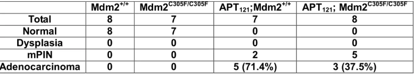
Mdm2C305F/C305F or Mdm2+/+ mice exhibited abnormality in their prostates (Figure 14C). Prostate adenocarcinoma, defined as penetration of malignant prostate epithelial cells
through the basement membrane of the prostate gland into the surrounding stroma, was
often observed in APT121;Mdm2+/+ mice, while the majority of the APT121;Mdm2C305F/C305F mice
only developed mPIN (mouse prostatic intraepithelial neoplasia), with few examples of
well-differentiated adenocarcinoma (Figure 14C). As shown in Table 3, 71.4% of
APT121;Mdm2+/+ mice developed adenocarcinomas compared with only 37.5% of
APT121;Mdm2C305F/C305F mice. Thus the progression from mPIN to adenocarcinoma is
Figure 14. Mdm2 C305F mutation causes reduced prostate size and slows the
progression of APT121-induced prostate cancer.
A. Photographs showing representative prostates from 6 month-old mice of the indicated
genotypes.
B. Average prostate mass ± SD from 6 month-old mice of the indicated genotypes. Mdm2+/+
(n=12), Mdm2C305F/C305F (n=11), APT121;Mdm2+/+ (n=9), and APT121;Mdm2C305F/C305F (n=13) . *
p < 0.05 and ** p < 0.01 as assessed by Student’s t test.
C. Representative H&E staining of prostate sections from 6 month-old mice of the indicated
genotypes demonstrating histology associated with the indicated stages of tumor progression.
Scale bar was shown in the first picture and all pictures were taken at the same
A
B
Table 3. Summary of prostate tumor stages in 6 month-old Mdm2+/+, Mdm2C305F/C305F,
APT121;Mdm2+/+ , and APT121; Mdm2C305F/C305F mice.
Mdm2+/+ Mdm2C305F/C305F APT121;Mdm2+/+ APT121; Mdm2C305F/C305F
Total 8 7 7 8
Normal 8 7 0 0
Dysplasia 0 0 0 0
mPIN 0 0 2 5