Cover Page
The handle http://hdl.handle.net/1887/66110 holds various files of this Leiden University dissertation.
Author: Flach, K.D.
Title: Holding the balance; the equilibrium between ERα-activation, epigenetic
alterations and chromatin integrity
Holding the balance;
the equilibrium between
ERα-activation, epigenetic
alterations and chromatin
integrity
ISBN number: 978-94-6375-038-7
Cover: The cover depicts Theo Flach ice skating. Anke Giesen®
and Koen Flach
Layout: Koen Dorus Flach
Printing: Ridderprint BV | www.ridderprint.nl
The work described in this thesis was performed at the Division
of Molecular Pathology and the Division of Oncogenomics at the
Netherlands Cancer Institute (NKI-AVL), Amsterdam,
Nether-lands. The work was supported by grants from Koningin
Wilhelmi-na Fonds voor de Nederlandse Kankerbestrijding (KWF),
Neder-landse Organisatie voor Wetenschappelijk Onderzoek (NWO) and
A Sister’s Hope.
the equilibrium between
ERα-activation, epigenetic alterations
and chromatin integrity
Proefschrift
ter verkrijging van
de graad van Doctor aan de Universiteit Leiden,
op gezag van Rector Magnificus prof.mr. C.J.
J.M. Stolker, volgens besluit van het College
voor Promoties
te verdedigen op dinsdag 25 september 2018
klokke 13.45 uur
door
Koen Dorus Flach
geboren te Amsterdam
Promotor
Prof. Dr. J.J.C. Neefjes
Copromotor
Dr. W.T. Zwart (NKI-AVL)
Promotiecommissie
Prof. dr. V.T.H.B.M. Smit
Prof. dr. H. Ovaa
Prof. dr. B. van de Water
Prof. dr. P.J. van Diest (UMCU)
Prof. dr. J Jonkers (LACDR/ NKI-AVL)
Prof. dr. S.C. Linn (UU/ NKI-AVL)
Thesis outline
Chapter 1
Introduction
The first decade of Estrogen Receptor cistromics in
breast cancer.
Koen D Flach and Wilbert Zwart. Journal
of Endocrinology, 2016.
Chapter 2
Posttranslational modification of ERα
– part 1
PKA phosphorylation redirects ERα to promoters of
a unique gene set to induce tamoxifen resistance.
Adapted
from Renée de Leeuw, Koen Flach, Cristiane Bentin
Toal-do, Xanthippi Alexi, Sander Canisius, Jacques Neefjes,
Rob Michalides, Wilbert Zwart. Oncogene, 2013.
Chapter 3
Posttranslational modification of ERα
- part2
Interaction of 14-3-3 proteins with the estrogen
re-ceptor alpha F domain provides a drug target interface.
Chapter 4
Composition of ERα’s transcriptional
complex – part 1
Co-regulated gene expression by estrogen receptor-α
and liver receptor homolog-1 is a feature of the estrogen
response in breast cancer cells.
Chun-Fui Lai, Koen D.
Flach, Xanthippi Alexi, Stephen P. Fox, Silvia Ottaviani,
Paul T.R. Thiruchelvam, Fiona J. Kyle, Ross S. Thomas,
Rosalind Launchbury, Hui Hua, Holly B. Callaghan,
Ja-son S. Carroll, R. Charles Coombes, Wilbert Zwart, Laki
Buluwela, and Simak Ali. Nucleic Acids Research, 2013.
Chapter 5
Composition of ERα’s transcriptional
complex – part 2
Estrogen Receptor DNA-damage/methylation cycle
as drug interface in tamoxifen resistant breast cancer by
FEN1 blockade.
Koen Dorus Flach, Manikandan
Peri-yasamy, Ajit Jadhav, Theresa E. Hickey, Mark Opdam,
Hetal Patel, Sander Canisius, David M. Wilson III,
Dor-jbal Dorjsuren, Marja Nieuwland, Roel Kluin, Alexey V.
Zakharov, Jelle Wesseling, Lodewyk Frederik Ary Wessels,
Sabine Charlotte Linn, Wayne D. Tilley, Anton Simeonov,
Simak Ali, Wilbert Zwart. In submission
Chapter 6
ERα Cofactor phosphorylation
SRC3 phosphorylation at Serine 543 is a positive
in-dependent prognostic factor in ER positive breast cancer.
Shaw, Ian O. Ellis, R. Charles Coombes, Jason S.
Car-roll, Simak Ali, and Carlo Palmieri. * authors contributed
equally. Clinical Cancer Research, 2016.
Chapter 7
Discussion
Addendum
Nederlandse samenvatting
English
summary
Nederlands
Curriculum
Vitae
English
Curriculum
Vitae
Holding the balance;
the equilibrium between
ERα-activation, epigenetic
alterations and chromatin
integrity
General introduction
In this thesis we reflect on the effects differential DNA binding of the estrogen receptor α (ERα) can have on the behavior of breast cancer and which factors
can contribute to this. Approximately 70% of all breast tumors are derived from the inner lining of cells in the mammary ducts (also known as luminal
tumors) and their proliferation is dependent on the activity of (ERα). During the development and homeostasis of the female reproductive organs ERα plays a key role. In ERα-positive breast tumors however, ERα has a causative role in carcinogenesis (1). ERα can be activated by its natural ligand estradiol
(2), a steroidal estrogen, which can induce the formation of an activated
ERα-dimer. After a conformational change (3), this dimer “opens up” the
activator-binding pocket (4) resulting in the recruitment of essential
co-factors (5) leading to the assembly of the ERα–transcriptional complex. One of the mainstay endocrine treatment options for ERα-positive breast cancer patients is tamoxifen (6-8), which is an ERα antagonist and competitively inhibits the interaction of ERα and estrogen, thereby repressing ERα activity (9-11). Alternatively aromatase inhibitors can be used to block the synthesis of estrogen, rendering ERα inactive. Despite these therapeutic options, still a significant proportion of patients develop a recurrence.
Although cross-resistance between the different therapeutic options does occur, a proportion of patients that relapse on one type of therapy can still
benefit from a different treatment modality. This illustrates the existence of
multiple resistance mechanisms which can be treatment selective. A better
understanding of ERα-biology and the development of drug resistance, cannot
Thesis outline
Chapter 1 provides a general overview of ERα-biology and more specifically the mechanistic insights the genome-wide interrogation of ERα-chromatin
binding has brought us. Furthermore we discuss where future developments might take us.
Chapter 2 describes a well-known mechanism of
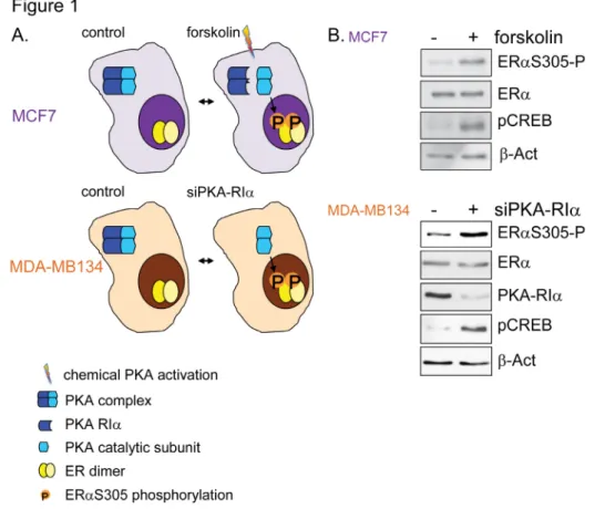
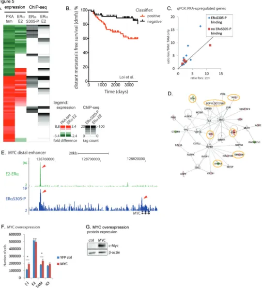
tamoxifen-resistance; the PKA-induced phosphorylation of ERα at Serine residue 305 (ERαS305-P). We report on the implications this phosphorylation has on the binding repertoire of ERα and describe the discovery of an altered gene expression profile capable of predicting the outcome of patients treated with
tamoxifen.
In Chapter 3 we provide experimental evidence for a previously
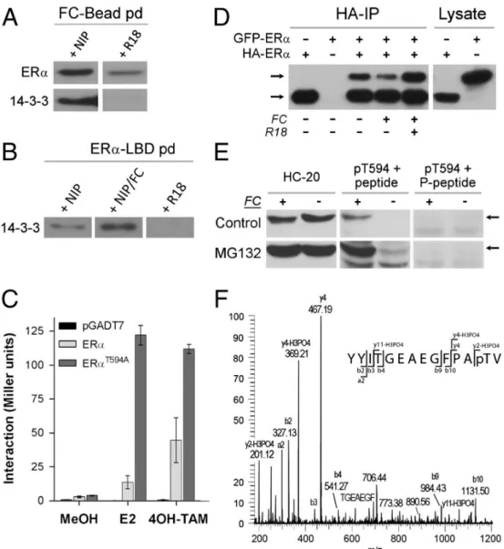
unknown ERα-phosphorylation (T594P) and demonstrate how this greatly diminished ERα’s binding capacity. Additionally we show that by stabilizing this phosphorylation by administering fusicoccin, ERα-mediated gene
transcription was reduced and tumor cell growth was inhibited.
In Chapter 4 we report on the fact that ERα-function can also be
altered by other nuclear receptors and demonstrate that LRH-1 knockdown
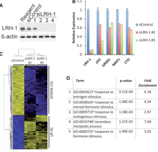
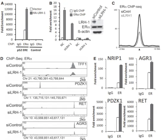
led to an altered gene expression profile. Additionally we revealed that there is a large overlap between chromatin binding sites of LRH-1 and ERα, and
that at these overlapping regions a synergistic stimulation is present.
In Chapter 5 we computationally refined a 111-gene classifier towards a single gene classifier, revealing that FEN1 levels are predictive of outcome in ERα-positive patients treated with tamoxifen. We describe our findings on the complex regulatory interplay between FEN1 and ERα, and postulate three manners by which FEN1 may modulate ERα activity. Additionally we performed a drug screen which led to the discovery of a FEN1-specific and potent inhibitor. We demonstrate a clear sensitivity of ERα-positive breast cancer cell lines to this inhibitor when compared to ERα-negative cell lines,
and an even greater sensitivity of tamoxifen-resistant cell lines. The latter
suggesting FEN1 inhibition might be a useful novel therapeutic option in the
case of tamoxifen resistant breast.
Chapter 6 contains our findings on the phosphorylation of essential ERα-cofactor SRC3 (SRC3-pS543). This phosphorylation leads to an altered
deposition of SRC3 at the chromatin, making it very likely to also alter the gene expression of breast cancer and thereby its phenotype. Furthermore a
with a functional ERα pathway, rendering it a promising novel biomarker for tamoxifen efficacy.
In Chapter 7 I discuss what the implications of our findings are with regards to a better understanding of ERα-biology and describe the new
questions this may invoke. Additionally I discuss how the novel biomarkers
described in this thesis could aid in patient stratification and what type of
research is still required to successfully implement such a biomarker. With
regards to the potential novel drug targets we identified, I describe what the
References
1. Hayashi SI, Eguchi H, Tanimoto K, Yoshida T, Omoto Y, Inoue A, et al. The
expression and function of estrogen receptor alpha and beta in human breast cancer and its
clinical application. Endocr Relat Cancer. 2003;10(2):193-202.
2. Dahlman-Wright K, Cavailles V, Fuqua SA, Jordan VC, Katzenellenbogen JA, Korach KS, et al. International Union of Pharmacology. LXIV. Estrogen receptors. Pharmacol
Rev. 2006;58(4):773-81.
3. Paige LA, Christensen DJ, Gron H, Norris JD, Gottlin EB, Padilla KM, et al. Estrogen receptor (ER) modulators each induce distinct conformational changes in ER alpha and ER beta. Proc Natl Acad Sci U S A. 1999;96(7):3999-4004.
4. Shiau AK, Barstad D, Loria PM, Cheng L, Kushner PJ, Agard DA, et al. The structural basis of estrogen receptor/coactivator recognition and the antagonism of this
interaction by tamoxifen. Cell. 1998;95(7):927-37.
5. Glass CK, Rose DW, Rosenfeld MG. Nuclear receptor coactivators. Curr Opin Cell Biol. 1997;9(2):222-32.
6. Osborne CK, Schiff R. Growth factor receptor cross-talk with estrogen receptor as a mechanism for tamoxifen resistance in breast cancer. Breast. 2003;12(6):362-7.
7. Tamoxifen for early breast cancer: an overview of the randomised trials. Early
Breast Cancer Trialists’ Collaborative Group. Lancet. 1998;351(9114):1451-67.
8. Gradishar WJ. Tamoxifen--what next? Oncologist. 2004;9(4):378-84.
9. Jordan VC, Murphy CS. Endocrine pharmacology of antiestrogens as antitumor agents. Endocr Rev. 1990;11(4):578-610.
10. Katzenellenbogen BS, Miller MA, Mullick A, Sheen YY. Antiestrogen action in
breast cancer cells: modulation of proliferation and protein synthesis, and interaction with estrogen receptors and additional antiestrogen binding sites. Breast Cancer Res Treat. 1985;5(3):231-43.
Chapter 1
Introduction and general discussion
The first decade of Estrogen Receptor cistromics in breast cancer
Koen D Flach and Wilbert Zwart Division of Molecular Pathology, the Netherlands Cancer Institute,
Amsterdam, the Netherlands
Abstract
The advent of genome-wide transcription factor profiling has revolutionized the field of breast cancer research. Estrogen Receptor alpha (ERα), the major
drug target in hormone receptor-positive breast cancer, has been known as
a key transcriptional regulator in tumor progression for over 30 years. Even though this function of ERα is heavily exploited and widely accepted as an
Achilles heel for hormonal breast cancer, only since the last decade we are beginning to understand how this transcription factor is functioning on a
genome-wide scale. Initial ChIP-on-chip analyses have taught us that ERα is
an enhancer-associated factor binding to many thousands of sites throughout the human genome, and revealed the identity of a number of directly interacting
transcription factors that are essential for ERα action. More recently, with the development of massive parallel sequencing technologies and refinements thereof in sample processing, a genome-wide interrogation of ERα has
become feasible and affordable with unprecedented data quality and richness.
These studies have revealed numerous additional biological insights in ERα
behaviour in cell lines and especially in clinical specimens. So what have we
Introduction
Breast cancer is the most prevalent form of cancer in women, with approx-imately 1.7 million annual new diagnoses (1). Despite the improvement of breast cancer treatment, still over half a million women die of this disease
every year (1). Approximately 70% of breast tumors are estrogen receptor α (ERα) positive and tumor cell proliferation is thought to be dependent on the
activity of this hormone-mediated transcription factor (2, 3).
The first evidence for a link between estrogens (produced in the ova -ries) and breast cancer was reported by George Thomas Beatson in 1896 with a case report describing a premenopausal breast cancer patient with metastat-ic disease (4). Although not aware of the exact mechanisms of hormonal ac-tion in human physiology, Beatson was familiar with a procedure performed in cattle where lactation after giving birth can be extended by removal of the ovaries. Inspired by this phenomenon, Beatson performed a bilateral oopho-rectomy on his patient, which initially resulted in a complete remission of the
disease (4, 5). The protein responsible for this clinical benefit was to be found almost 80 years later, with the seminal discovery of the Estrogen Receptor in 1973 by Elwood Jensen (6). In 1986, a complementary DNA clone of the
translated mRNA of the estrogen receptor from MCF-7 human breast cancer cells was sequenced and upon expression gave rise to a functional protein (7).
Today, ERα is recognized as the major drug target in hormonal breast can
-cer. In the adjuvant treatment of ERα-positive disease, receptor-inhibition is achieved by either a direct blockage of ERα-activation through competitive
inhibition of estradiol association using tamoxifen (8-10) or by preventing estrogen synthesis using aromatase inhibitors (11). Despite the extensive use
of these treatment modalities in adjuvant therapy, a significant number of
patients still develop a recurrence (12). Although cross-resistance between the different endocrine therapy options can occur, patients that relapse on one
type of endocrine therapy can still benefit from a different treatment modality
(13-15), suggesting that multiple resistance mechanisms can exist that may be treatment selective. In order to directly administer the right drug to the right
patient, it is vital to increase our knowledge about ERα-functioning as well as
its selective responses to prolonged exposure to hormonal agents.
Even though ERα-inhibitors are being used in the clinic since the early 1980’s, the direct mode of ERα’s genomic action on a genome-wide
scale has remained elusive for many years. With the initial development of ChIP-on-chip (chromatin immunoprecipitation coupled with tiling array) technologies, this situation changed dramatically with the interrogation of
ERα action for the first time on a human chromosome-wide scale (16). With
the development of massive parallel high-throughput sequencing techniques,
a full genome-coverage of ERα became possible (and importantly affordable) through ChIP-seq (17). Now, ten years after the first unbiased and systemic assessment of ERα binding sites in human cell lines, we will discuss what we have learned from the cistromics of ERα and where future developments
might take us.
Estrogen Receptor complex formation and its mode-of-action
ERα is activated through the association of its natural ligand estra -diol with the receptors’ ligand-binding domain, which enables dissociation
from chaperone protein Hsp90 (18-20) and facilitates ERα/chromatin inter
-actions (21). Initial ChIP-on-chip experiments have shown ERα to mainly
bind enhancer regions (16). Computational DNA sequence motif analyses
of ERα binding sites resulted in the identification of a number of upstream transcription factors that facilitate the binding of ERα to the chromatin, in -cluding pioneer factor FOXA1 (16, 22) and putative pioneer factors PBX1
(23) and AP-2γ (24) (Figure 1). Pioneer factors can associate with compacted chromatin and trigger enhancer competency by de-condensing the chroma-tin, facilitating the binding of additional chromatin binding factors (25, 26).
Additionally, ERα-cooperating transcription factor GATA3 is capable of me
-diating enhancer accessibility at ERα regulatory regions and has properties similar to FOXA1 (27, 28). Besides binding directly to the DNA, ERα can
also associate to the chromatin via other transcription factors, also known as tethering, including RUNX1 (29) and AP-1 (30-32).
After activation, ERα undergoes a conformational change (33), form -ing a co-activator-bind-ing pocket at the receptors’ carboxy-terminus (34). This interaction surface subsequently leads to the recruitment of the mem-bers of the p160 co-activator family; SRC1 (NCOA1) (35), SRC2 (NCOA2,
TIF2, GRIP1) (36, 37) and SRC3 (NCOA3, p/CIP, AIB1, ACTR) (38-41).
The binding of these SRCs to the co-activator-binding pocket of activated
ERα has been described to occur both in a competitive manner (exclusive
recruitment of one type of SRC) (34, 42, 43) as well as in a joint manner, possibly through hetero-dimerization (44). Reports on the exact
stoichiome-try within the p160/ERα complex are conflicting, describing a single p160 to associate with an ERα-dimer (43) or two SRCs per active ERα-complex (44,
45), although both situations might occur side-to-side (44). Recently it was
where two ligand-bound ERα-monomers individually recruit one SRC3 pro
-tein, after which an ERα-dimer (binding two SRC3 molecular) associates to single p300 protein (45). The p160 composition of the ERα transcriptional complex influences its genomic binding preferences on a genome-wide scale,
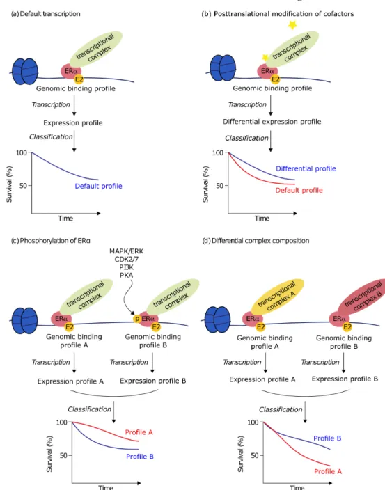
consequently resulting in an altered transcriptional repertoire (46) and altered phenotypic behavior (Figure 2).
After ERα binding, p160 proteins can subsequently recruit other es -sential proteins for transcriptional regulation, including p300 and CBP (47), which can modify chromatin accessibility through their acetyltransferase ac-tivity (48). In order to further modify the chromatin towards a transcription
favourable landscape, histone modifiers CARM1 (49, 50) and JMJD2B (51, 52) and members of the SWI/SNF chromatin remodelling complex, including
1
Figure 1: The Estrogen Receptor α transcriptional complex pathway. When acti
-vated by its natural ligand estradiol or by direct phosphorylations, ERα binds to
BAF57, are recruited (53) (Chapter 5).
With the recent discovery of estradiol-induced enhancer RNAs
(eR-NAs) at a set of ~1200 ERα-bound enhancer elements (54, 55), an additional layer of ERα biology was revealed. This eRNA production is not just limited to ERα-bound enhancers but is for example also apparent for the Androgen
Receptor (AR) (56) and p53 (57). DNAse I sensitivity assays demonstrated that eRNAs are capable of regulating genomic access of the
transcription-al complex to regulatory regions (58). eRNAs found at ERα binding sites
strongly correlated with the enrichment of a number of genomic features as-sociated with enhancers and enhancer looping to target gene promoters (54).
The physiological relevance of eRNAs in ERα-biology was further stipulated by the observation that knockdown of a subset of eRNAs (e.g. GREB1 en -hancer) reduced the transcription of coding gene transcripts, as well as reduc-ing promoter-enhancer interactions as shown by chromosome conformation
capture (3C) (55), although conflicting 3C results have also been described (54). Hah et al. found that inhibition of eRNA production by flavopiridol, a
CDK9 inhibitor blocking transcriptional elongation, did not affect other indi-cators of enhancer activity or estradiol-dependent promoter-enhancer looping (54), leaving the exact role of eRNAs somewhat elusive. These eRNA-asso-ciated promoter-enhancer interactions, also known as chromosomal looping
structures, have been described to promote ERα-regulated gene transcription
and seem to be stabilized by cohesion (55, 59, 60). Recently it was discovered that RNA binding to CBP stimulates its histone acetyl transferase activity, resulting in increased transcription of associated genes (61), providing an ad-ditional layer of possible eRNA function. Although these observations hint
towards an important role for eRNAs in ERα-regulated transcription, only a subset of eRNAs has yet been investigated thoroughly, with conflicting roles
in chromosomal looping, leaving the exact physiological roles for them cur-rently elusive.
After ERα has recruited its co-factors, an active transcriptional com -plex can be formed by RNA polymerase II (Pol II) recruitment and transcrip-tion of responsive genes can be initiated (62) (Figure 1). When treated with
tamoxifen, the ligand-binding-domain of ERα adopts an alternative confor
-mation, impairing the docking of p160 proteins to ERα, preventing the cor -rect assembly of the transcriptional complex (34).
The genome-wide kinetics with which the ERα-complex assembles on the chromatin is not yet fully understood. By using ChIP at three ERα re
1
Figure 2: ERα transcriptional complex composition, genomic profile and transcrip -tional output.
Illustration of ERα-induced transcription, where the genomic binding profile of ERα’s transcriptional complex leads to induced transcription and an expression pro
-of its coactivators associate on these estrogen responsive promoters in a
cy-clic fashion and that these cycles of ERα-complex assembly are followed by transcription (63). This cyclic recruitment of ERα and its coregulators could be confirmed by others, who reported cofactor recruitment to be preceded
by histone deacetylases and nucleosome-remodeling complexes at the TFF1
promoter (64). These data imply that transcriptional activation of ERα-re -sponsive genes may require both activating as well as repressive epigenetic
processes. Although both papers state that ERα-induced transcriptional acti -vation occurs in a cyclic fashion, both papers only investigated the dynamic
nature of ERα on a couple of sites and a comprehensive overview of ERα
dynamics on a genome-wide scale is currently lacking. Furthermore, whether
this cyclic ERα-complex assembly occurs only on promoters, as studied in both papers, or whether it is also apparent at ERα-bound enhancers remains
unclear (Chapter 5).
ERα cistromics in breast cancer cell lines
Initially, most reports on ERα chromatin interactions, its dynamics
and recruitment of coregulators were centred on single binding site-based analyses, often limited to the TFF1 promoter. With the technological
develop-ment of tiling arrays, ERα genomic interactions could reliably be assessed on
a chromosome-wide scale (16). As technology progressed, this approach was quickly succeeded by massive parallel sequencing technologies, enabling the
interrogation of ERα sites on a genome-wide scale, in a cost-effective man -ner (17). These initial reports resulted in a huge paradigm-shift, completely
changing the way we think about ERα genomics. These studies illustrated that even though most pioneering studies on ERα-genomics exclusively in
-terrogated promoters, this genomic behaviour of ERα clearly represents an exception. In fact, only a small proportion of about 5% of ERα binding sites
was found at gene promoters; a characteristic feature that has been validated by others (16, 46) and is also apparent for other nuclear receptors, including AR (65) and Glucocorticoid Receptor (GR) (66). Approximately 95% of all
ERα binding sites are found at distal cis-regulatory elements (hence designat -ed as ‘cistromics’ (67)) that were later recogniz-ed as enhancer regions. These regions are putative regulatory elements and might not all be functional.
Re-cently a CRISPR-Cas9 screen was used to functionally asses ERα enhancers
fications of cofactors (b), phosphorylations on ERα itself (c) and the composition of
elements and their effect on cell proliferation (68). Out of the 99 ERα bind -ings sites that were targeted, the deletion of only four of them affected cell
proliferation, further illustrating that only a subset of ERα bindings sites at
cis-regulatory elements might actually be functionally involved in cell prolif-eration processes.
The discovery of enhancer preference for ERα binding repositioned the classical promoter-centred ERα studies considerably on the level of phys -iological extrapolation. Chromatin interaction analysis by paired-end tag
sequencing (ChIA-PET) analyses, which enables the identification of
long-range chromatin interactions, illustrated that the distal enhancer-associated
ERα-bindings sites were found to loop to anchor genes through connections with proximal ERα-binding sites, suggesting that ERα functions by bringing
genes together for coordinated transcriptional regulation by extensive
chro-matin looping (59). At the GREB1 and TFF1 locus, this chrochro-matin looping was dependent on ERα expression and was inducible by estradiol stimula -tion (59, 69). Probing the three-dimensional architecture of the genome by coupling proximity-based ligation with massively parallel sequencing
(Hi-C) (70) yielded similar ERα mediated enhancer-promoter interactions (71).
These sites of chromatin looping highly correlated with CTCF-binding sites,
suggesting CTCF to play a key role defining the boundaries of chromosomal territories and influence gene expression through cross-talk between promot
-ers and regulatory elements (72-74). Besides for ERα, these chromatin loops
have also been observed for other nuclear transcription factors, including AR (75) and GR (76).
On the transcriptomic level, the use of global nuclear run-on and
se-quencing (GRO-seq) (77) analysis increased our understanding of ERα-reg -ulated transcription by identifying primary and immediate estrogen induced effects as opposed to steady-state transcript level analyses (78). GRO-seq demonstrated that estrogen is able to regulate the activity of all three RNA polymerases and led to the discovery of previously undetected
estrogen-reg-ulated intergenic transcripts (78). Transcription profiling by GRO-seq could
be used for the prediction of de novo enhancers across various cell types (54). In combination with RNA-seq, GRO-seq was able to annotate long noncod-ing RNAs (lncRNAs) and characterized the lncRNA transcriptome in MCF-7 breast cancer cells, including over 700 previously unannotated lncRNAs (79).
Furthermore, GRO-seq analysis at ERα enhancers revealed the existence of
estradiol-induced unidirectional and bidirectional eRNAs, that were strongly correlated with enhancer-promoter looping (54). The described role of these
intergenic transcripts in enhancer-promoter looping (55, 59, 60) and the fact that one promoter can be involved in multiple enhancer-associated loops (59, 71), might explain the seemingly large discrepancy between the number of
ERα-regulated genes (approximately 2,000 (46)) in relation to the number of ERα-binding sites in the same cell line (>10,000 (17, 22)).
Due to technical limitations in the ChIP-seq protocol, the resolution of DNA binding analyses is typically quite limited with events being mapped
with ±300 base pairs. Further refinement of the ChIP-seq procedure has led to
the implementation of lambda exonuclease digestion in the protocol
(ChIP-exo), enabling high resolution mapping of chromatin binding and identifica
-tion of unique transcrip-tion factor binding sites that could not be identified
by ChIP-seq (80-82). The addition of exonucleases also results in the deg-radation of contaminating DNA, effectively lowering the required depth of sequencing coverage.
Apart from forming the foundations of cis-regulatory gene regulation,
chromatin looping and eRNA action, genome-wide profiling analyses of ERα sites can also lead to the identification of additional transcription factor motifs often co-enriched at ERα sites and proximal to estrogen response elements (ERE). These motif analyses revealed the presence of Forkhead binding mo
-tifs at roughly 50% of ERα bindings sites (16). This observation led to the discovery that FOXA1 is essential for chromatin accessibility at ERα-sites and crucial for ERα binding and functionality (16, 22). More recently, this same approach was used to identify other pioneer factors for ERα, including PBX1 which can guide ERα to a specific subset of sites (23). When inves
-tigating the motifs of ERα-bindings sites identified by ChIA-PET, Tan et al.
found that approximately 40% of these binding sites contained the AP-2 motif
(24). They next demonstrated that transcription factor AP-2γ can bind to these ERα-bindings sites in a ligand-independent manner and there is a functional interplay between AP-2γ and FOXA1 (24).
Besides the interplay between ERα and its pioneer factors and coreg -ulators, it is becoming increasingly apparent that a complex interplay exists between different steroid hormone receptor family members. The androgen receptor (AR), a transcription factor classically known for its oncogenic role
in prostate cancer, is expressed in 84-95% of the ERα-positive breast cancers (83-85) and is usually associated with a favourable outcome (86-88). Exoge
-nous overexpression of AR inhibits ERα-transactivation activity and estrogen
induced cell growth (86, 89), which may be explained by a direct competition
was further strengthened by ChIP analysis showing AR recruitment to the progesterone receptor promoter in T47D cells (86).
Another steroid hormone receptor family member known for its
co-expres-sion and favourable outcome in ERα-positive breast cancer, is the proges -terone receptor (PR) (90, 91). Proges-terone induces the association of PR
with ERα, thereby regulating ERα-chromatin interactions and transcriptional
activity, providing mechanistic insights behind the clinical implications of
PR-status in ERα-positive tumors (92).
The Glucocorticoid Receptor (GR), in the presence of
dexametha-sone, is able to associate to similar binding regions as ERα, and GR-stimu
-lation leads to reduced transcription of key ERα-target genes (93, 94). This direct protein-protein interaction between GR and ERα can play an important role in the GR-mediated growth inhibition of ERα positive cells (93). Besides this general inhibitory role of GR, gene specific regulation with both cooper -ation and antagonism has also been described (95). Apart from direct physical interactions between nuclear receptors, nuclear receptors can also inhibit each
other’s activity through cross-interference (“squelching”), where direct com -petition for cofactor recruitment can inhibit nuclear receptor activity without associating to the same genomic regions (96, 97).
Cistromics of ERα coregulators
To date, several studies have compiled an overview of ERα co-regu -lators and interacting proteins, with numbers varying around 17 (98) to 108 (99). p160 protein family members are reproducibly and consistently
identi-fied as part of the ERα complex, for which a level of mutual exclusivity has been described for ERα binding (34, 42, 43). With the recent finding that an activated ERα dimer can bind one p300 protein (45) and p300 and CBP
have a substantial overlap of ~70% in binding sites (46), it is not unlikely that a level of mutual exclusivity between p300 and CBP also exists. As a
direct consequence thereof, the composition of ERα complexes can differ
between different sites on a genome-wide scale, with potentially far-reaching
consequences on gene expression profiles (Chapter 4). Cistromic analyses of the p160 family members illustrated that even though most genomic sites are shared between SRC1, SRC2 and SRC3, distinct subsets of sites were
identified where gene expression was selectively responsive to one specific p160 protein, as part of the ERα-complex (46). Interestingly, the gene-profile under the control of ERα with exclusively SRC3 binding (devoid of SRC1 or SRC2) had prognostic potential, and enabled identification of breast can
cer patients with a poor outcome after tamoxifen treatment (46). This link between SRC3 gene targets and tamoxifen treatment is in line with previous reports describing increased SRC3 expression, in combination with increased
ERBB2 expression, to correlate with a poor tamoxifen response (100-103). Another ERα interacting protein that can affect ERα-complex formation and
gene expression, is the transcriptional regulator RIP140 (104). Genes under
the specific control of RIP140 (identified by siRNA experiments) could be
used to classify tamoxifen-treated patients on clinical outcome (104). Both RIP140 and the p160 family members further stipulate the observation that the composition of the transcriptional complex may differ on a genome-wide scale, which could have direct physiological consequences on the level of transcriptional output and clinical response (Figure 2).
ERα phosphorylations and genome-wide effects on ERα action
Besides the composition of the transcriptional complex,
phosphoryla-tions on ERα can also regulate the transcriptional activity of the receptor and
play a crucial role in endocrine resistance (105, 106) (Chapter 2, 3). These
phosphorylation-events mainly revolve around serine residues 104/106 (107),
118 (108), 167 (109), 236 (110) and 305 (111) (Chapter 2). The kinases
in-volved in phosphorylation on ERα at s104/106 include CDK2 and ERK1/2 (107, 112); for s118 ERK1/2, EGFR and IGF1R (113, 114); for s167 AKT
and CK2 (115, 116); for s236 PKA (117) and for s305 PAK1 and PKA (111, 118). The clinical implications of these phosphorylations remain not fully understood, where higher expression of s118 and s167 phosphorylations are generally but not uniformly associated with a favorable outcome in patients on tamoxifen therapy (109, 119-122), whereas the s305 phosphorylation is associated with a poor clinical outcome (122, 123). Furthermore, s118 phos-phorylation expression appears to be a predictive biomarker for tamoxifen re-sponse (108, 119). Recently, the phosphorylation on the 594 threonine (t594)
residue of ERα was found to play a key role in the regulatory interaction of ERα with 14-3-3 proteins (124). This t594 phosphorylation resulted in de
-creased estradiol-stimulated ERα dimerization, reduced ERα-chromatin in -teractions and reduced gene expression (124) (Chapter 3).
The spectrum of ERα phosphorylation-events appears able to dictate differential transcriptional programs of ERα, as exemplified by the PKA-in
2). Additionally, it was found that stimulation of ERα by the epidermal growth factor (EGF), which induces s118 phosphorylation (125), led to a distinct
cistromic landscape and induced a unique set of genes, when compared to
estradiol stimulation (126). Stimulation of ERα by AKT, capable of inducing 167 phosphorylation (115), also mediated changes in ERα chromatin binding and altered its transcriptional output (127), further indicating that specific phosphorylations on ERα may yield distinct genomic actions and may target
unique locations throughout the genome (Figure 2). Although the binding
patterns of some of the phosphorylated ERα forms are known, a complete
and comparative overview is still lacking. Furthermore, multiple reports have
studied ERα cistromics upon activation of a specific cellular signalling cas
-cade, including the previously mentioned AKT or EGF, where it still remains elusive which specific variable is actually responsible for the altered ERα
behaviour.
Besides the effect direct ERα phosphorylations can have on ERα’s genomic landscape and transcriptional activity, posttranslational modifica
-tions of coregulators can also influence ERα action. Where ERα-bound SRC3
binding is predominantly enhancer-bound, phosphorylated SRC3 at Ser543
(pSRC3) was selectively found at promoters of ERα-regulated genes (128)
(Chapter 6). pSRC3 functioned as an independent prognostic factor as well as a predictive marker for tamoxifen treatment, potentially enabling the
iden-tification of patients with a good clinical outcome without receiving adjuvant
therapy (128). Additionally, SRC2 can be phosphorylated at Ser736 through the MAPK pathway, increasing SRC2 interactions with p300 and CBP, further
facilitating SRC2 recruitment to the ERα complex (129). These posttransla
-tional modifications on coregulators further illustrate the intrinsic complexity and flexibility of ERα transcription complex formation, where multiple cell signaling cascades converge to collaboratively fine-tune ERα action on a ge -nome-wide scale (Figure 2).
Cistromic analyses in clinical samples and potential clinical applications
Over recent years, the transition is being made from studying ERα cistromics in cell lines towards genomic interrogation of ERα sites in clin -ical specimens. Obviously, in contrast to cell lines, clin-ical samples cannot be readily manipulated and represent heterogeneous populations of multiple
cell types. Even with this difference between tumors and cell lines, the cistro -mic information obtained from both settings yields quite similar conclusions.
When looking at ERα, most well-described ERα binding sites found in
7 cells (16, 17) such as enhancer regions proximal to RARA, GREB1, XBP1 and TFF1, are also observed in tumor specimens (130). Not only for ERα, but
also for its coregulators the overlap of chromatin binding in cell lines versus clinical specimens was considerably high. For example, SRC3-pS543 ChIP-seq analyses showed 51% overlap in binding sites between MCF-7 cells and
an ER+/PR+ breast tumor, being in the same order of magnitude as found
between 2 tumor samples (61% overlap) (128) (Chapter 6).
The first analyses of ERα binding patterns in clinical samples directly illustrated the added value of assessing ERα binding in clinical specimens (130), where differential ERα binding sites found between tumors could stratify patients on outcome (130). A more recent study identified ERα chro -matin binding patterns in primary breast tumors that enabled patient
classi-fication on their response to aromatase inhibition in the metastatic setting (131). This same report analysed profiles for H3K27me3, resulting in a gene classifier that seemed to outperform other prognostic classifiers, including OncotypeDX (132) and PAM50 (133). Since the classification potential of
these genes was only partially preserved in a cohort of tamoxifen-treated
pa-tients, this suggests some treatment selectivity for patient classification. Both studies demonstrate clear advantages of studying ERα cistromic analyses in
clinical specimens, with the potential to facilitate tailored therapy selection
and enable patient stratification on outcome.
Although these cistromic classifiers made use of associated gene-pro
-files, it remains largely unknown which genes in these classifiers are now the
driving force behind any prognostic or predictive effect. Fine-tuning these
classifiers towards optimized gene sets and further biological investigation of
these genes could reveal the biologically most relevant genes for disease
pro-gression and might lead to novel biological insights in ERα biology as well as
potentially novel drug targets (Chapter 5).
Since the main function of ERα is to activate its downstream target genes involved in tumor progression, ERα cistromic analyses may yield nov -el drug targets. A key example for this line of thought can be found in Myc,
representing one of the best-studied ERα responsive genes (134-136) and
widely accepted as a potent novel drug target in cancer (137, 138).
Besides targeting ERα-regulated genes to inhibit its stimulatory ef
-fect, ERα-cofactors also receive increasing attention as potential drug targets.
Small molecule inhibitors against both SRC1 and SRC3 (139, 140) or SRC3 alone (141), as well as a stimulator for SRC3 activity (142) were recently
in vitro as well as in xenograft mouse models. Such novel therapeutic options could revolutionize endocrine therapeutic drug design, not aiming at block-ing the receptor itself, but targetblock-ing the proteins required for receptor action.
Since in case of endocrine therapy resistance ERα can still remain a driver
(13-15), such novel inhibitors have the potency to remain effective after pro-gression on currently available endocrine therapies (Chapter 5).
Even though promising, at the moment there are no cistromic classifi -ers being used in the clinic. One of the major practical limitations is the typ-ically low amounts of available tumor tissue. Although initially challenging, continuing technical developments, including single-tube linear DNA
ampli-fication method (LindDA) (143) and the combination of a high-sensitivity
ChIP-assay with new library preparation procedures (144), have now greatly increased the applicability of ChIP-seq on limited amounts of tissue. Another example of these developments is the incorporation of carrier chromatin that
can be removed before library preparation, improving ChIP efficiency while
limiting background signal (145). Furthermore, a great promise for the future of ChIP-seq on limited tumor material might be found in the combination of
microfluidics, DNA barcoding and sequencing, which recently enabled the
generation of ChIP-seq data at a single-cell resolution (146).
Discussion
Within 10 years, ERα genomics has gone from single-locus to genome-wide and towards single-cell. Initial reports on ERα cistromics in breast cancer have revolutionized the way we think about ERα action and ERα-respon
-sive genes. By far, most transcriptional effects found regulated by ERα are represented as eRNAs. With conflicting reports about the role of eRNAs in
chromosomal looping, a comprehensive overview of eRNA action, and with
this to a certain degree a functional overview of ERα-enhancer action, is cur
-rently lacking. Since ERα seems to function mostly through chromatin loops, it is not unlikely that ERα enhancers and a subset of responsive eRNAs are
functionally involved in such looping structures.
In ERα-positive breast cancer cell lines and tumors, many thousands of ERα binding sites can be found, of which a large number is shared between
them. This could imply a selection pressure throughout human evolution for
the maintenance of these ERα sites throughout the human genome. As tech -nological development continues, future studies will further elucidate the
functional relevance of all these ERα sites and identify the genomic regions
responsible for proliferative potential.
Clearly, our knowledge on ERα genomic regulation in breast cancer has in -creased exponentially over the last decade. A major factor in this, is the par-allel development of novel technologies and computational tools, which not only enable us to generate genomic data with an unprecedented level of data richness and detail, but also with the tools that enable us to process and under-stand the data. Now, with novel technologies on genome editing (e.g. CRISPR Cas9) and single-cell ChIP-seq analyses, the second decade of cistromics in breast cancer will no doubt unveil another layer of unprecedented complexity in breast cancer and may lead us towards a comprehensive understanding of the disease with its full genomic complexity and diversity.
Acknowledgements
The authors would like to thank the Dutch Cancer Society KWF, Alpe
d’HuZ-es, the Netherlands Organisation for Scientific Research (NWO) and A Sis
-ter’s Hope for financial support.
Disclosure
References
1. Ferlay J, Soerjomataram I, Dikshit R, Eser S, Mathers C, Rebelo M, et al. Cancer incidence and mortality worldwide: sources, methods and major patterns in GLOBOCAN 2012. International journal of cancer Journal international du cancer. 2015;136(5):E359-86.
2. Hayashi SI, Eguchi H, Tanimoto K, Yoshida T, Omoto Y, Inoue A, et al. The expres-sion and function of estrogen receptor alpha and beta in human breast cancer and its clinical application. Endocr Relat Cancer. 2003;10(2):193-202.
3. Dahlman-Wright K, Cavailles V, Fuqua SA, Jordan VC, Katzenellenbogen JA, Ko-rach KS, et al. International Union of Pharmacology. LXIV. Estrogen receptors. Pharmacol Rev. 2006;58(4):773-81.
4. Beatson G. On Treatment of Inoperable Cases of Carcinoma of the Mamma: Sug-gestions for a New Method of Treatment, with Illustrative Cases. Lancet. 1896;2(3802):104-7.
5. Thomson A. Analysis of Cases in which Oophorectomy was Performed for Inopera-ble Carcinoma of the Breast. British medical journal. 1902;2(2184):1538-41.
6. Jensen EV, DeSombre ER. Estrogen-receptor interaction. Science. 1973;182(4108):126-34.
7. Greene GL, Gilna P, Waterfield M, Baker A, Hort Y, Shine J. Sequence and expres -sion of human estrogen receptor complementary DNA. Science. 1986;231(4742):1150-4. 8. Jordan VC, Murphy CS. Endocrine pharmacology of antiestrogens as antitumor agents. Endocr Rev. 1990;11(4):578-610.
9. Katzenellenbogen BS, Miller MA, Mullick A, Sheen YY. Antiestrogen action in breast cancer cells: modulation of proliferation and protein synthesis, and interaction with estrogen receptors and additional antiestrogen binding sites. Breast Cancer Res Treat. 1985;5(3):231-43.
10. Arpino G, De Angelis C, Giuliano M, Giordano A, Falato C, De Laurentiis M, et al. Molecular mechanism and clinical implications of endocrine therapy resistance in breast cancer. Oncology. 2009;77 Suppl 1:23-37.
11. Fabian CJ. The what, why and how of aromatase inhibitors: hormonal agents for treatment and prevention of breast cancer. International journal of clinical practice. 2007;61(12):2051-63.
12. Early Breast Cancer Trialists’ Collaborative G, Davies C, Godwin J, Gray R,
Clarke M, Cutter D, et al. Relevance of breast cancer hormone receptors and other factors to
the efficacy of adjuvant tamoxifen: patient-level meta-analysis of randomised trials. Lancet.
2011;378(9793):771-84.
13. Vergote I, Amant F, Leunen K, Van Gorp T, Berteloot P, Neven P. Metastatic breast cancer: sequencing hormonal therapy and positioning of fulvestrant. International journal
of gynecological cancer : official journal of the International Gynecological Cancer Society.
2006;16 Suppl 2:524-6.
14. Wang J, Jain S, Coombes CR, Palmieri C. Fulvestrant in advanced breast cancer following tamoxifen and aromatase inhibition: a single center experience. The breast jour-nal. 2009;15(3):247-53.
15. Yoo C, Kim SB, Ahn JH, Jung KH, Ahn Y, Gong G, et al. Efficacy of fulvestrant in
heavily pretreated postmenopausal women with advanced breast cancer: a preliminary re-port. Journal of breast cancer. 2011;14(2):135-9.
16. Carroll JS, Liu XS, Brodsky AS, Li W, Meyer CA, Szary AJ, et al. Chromosome-wide mapping of estrogen receptor binding reveals long-range regulation requiring the forkhead protein FoxA1. Cell. 2005;122(1):33-43.
17. Welboren WJ, van Driel MA, Janssen-Megens EM, van Heeringen SJ, Sweep FC,
Span PN, et al. ChIP-Seq of ERalpha and RNA polymerase II defines genes differentially
responding to ligands. The EMBO journal. 2009;28(10):1418-28.
18. Catelli MG, Binart N, Jung-Testas I, Renoir JM, Baulieu EE, Feramisco JR, et al.
The common 90-kd protein component of non-transformed ‘8S’ steroid receptors is a
heat-shock protein. The EMBO journal. 1985;4(12):3131-5.
19. Pratt WB, Toft DO. Steroid receptor interactions with heat shock protein and immu-nophilin chaperones. Endocr Rev. 1997;18(3):306-60.
20. Devin-Leclerc J, Meng X, Delahaye F, Leclerc P, Baulieu EE, Catelli MG. Interac-tion and dissociaInterac-tion by ligands of estrogen receptor and Hsp90: the antiestrogen RU 58668 induces a protein synthesis-dependent clustering of the receptor in the cytoplasm. Molecular endocrinology. 1998;12(6):842-54.
21. Kumar V, Chambon P. The estrogen receptor binds tightly to its responsive element as a ligand-induced homodimer. Cell. 1988;55(1):145-56.
22. Hurtado A, Holmes KA, Ross-Innes CS, Schmidt D, Carroll JS. FOXA1 is a key determinant of estrogen receptor function and endocrine response. Nature genetics. 2011;43(1):27-33.
23. Magnani L, Ballantyne EB, Zhang X, Lupien M. PBX1 genomic pioneer func-tion drives ERalpha signaling underlying progression in breast cancer. PLoS genetics. 2011;7(11):e1002368.
24. Tan SK, Lin ZH, Chang CW, Varang V, Chng KR, Pan YF, et al. AP-2gamma regu-lates oestrogen receptor-mediated long-range chromatin interaction and gene transcription. The EMBO journal. 2011;30(13):2569-81.
25. Zaret KS, Carroll JS. Pioneer transcription factors: establishing competence for gene expression. Genes & development. 2011;25(21):2227-41.
27. Theodorou V, Stark R, Menon S, Carroll JS. GATA3 acts upstream of FOXA1 in me-diating ESR1 binding by shaping enhancer accessibility. Genome research. 2013;23(1):12-22.
28. Kong SL, Li G, Loh SL, Sung WK, Liu ET. Cellular reprogramming by the conjoint action of ERalpha, FOXA1, and GATA3 to a ligand-inducible growth state. Molecular sys-tems biology. 2011;7:526.
29. Stender JD, Kim K, Charn TH, Komm B, Chang KC, Kraus WL, et al. Genome-wide
analysis of estrogen receptor alpha DNA binding and tethering mechanisms identifies Runx1
as a novel tethering factor in receptor-mediated transcriptional activation. Molecular and cellular biology. 2010;30(16):3943-55.
30. Cheung E, Acevedo ML, Cole PA, Kraus WL. Altered pharmacology and distinct coactivator usage for estrogen receptor-dependent transcription through activating pro-tein-1. Proceedings of the National Academy of Sciences of the United States of America. 2005;102(3):559-64.
31. Kushner PJ, Agard DA, Greene GL, Scanlan TS, Shiau AK, Uht RM, et al. Estro-gen receptor pathways to AP-1. The Journal of steroid biochemistry and molecular biology. 2000;74(5):311-7.
32. Umayahara Y, Kawamori R, Watada H, Imano E, Iwama N, Morishima T, et al. Estrogen regulation of the insulin-like growth factor I gene transcription involves an AP-1 enhancer. The Journal of biological chemistry. 1994;269(23):16433-42.
33. Paige LA, Christensen DJ, Gron H, Norris JD, Gottlin EB, Padilla KM, et al. Estro-gen receptor (ER) modulators each induce distinct conformational changes in ER alpha and ER beta. Proceedings of the National Academy of Sciences of the United States of America. 1999;96(7):3999-4004.
34. Shiau AK, Barstad D, Loria PM, Cheng L, Kushner PJ, Agard DA, et al. The struc-tural basis of estrogen receptor/coactivator recognition and the antagonism of this interac-tion by tamoxifen. Cell. 1998;95(7):927-37.
35. Onate SA, Tsai SY, Tsai MJ, O’Malley BW. Sequence and characterization of a co -activator for the steroid hormone receptor superfamily. Science. 1995;270(5240):1354-7. 36. Hong H, Kohli K, Garabedian MJ, Stallcup MR. GRIP1, a transcriptional coactiva-tor for the AF-2 transactivation domain of steroid, thyroid, retinoid, and vitamin D recepcoactiva-tors. Molecular and cellular biology. 1997;17(5):2735-44.
37. Voegel JJ, Heine MJ, Zechel C, Chambon P, Gronemeyer H. TIF2, a 160 kDa tran-scriptional mediator for the ligand-dependent activation function AF-2 of nuclear receptors. The EMBO journal. 1996;15(14):3667-75.
38. Torchia J, Rose DW, Inostroza J, Kamei Y, Westin S, Glass CK, et al. The tran-scriptional co-activator p/CIP binds CBP and mediates nuclear-receptor function. Nature. 1997;387(6634):677-84.
39. Anzick SL, Kononen J, Walker RL, Azorsa DO, Tanner MM, Guan XY, et al.
AIB1, a steroid receptor coactivator amplified in breast and ovarian cancer. Science.
1997;277(5328):965-8.
40. Chen H, Lin RJ, Schiltz RL, Chakravarti D, Nash A, Nagy L, et al. Nuclear receptor coactivator ACTR is a novel histone acetyltransferase and forms a multimeric activation complex with P/CAF and CBP/p300. Cell. 1997;90(3):569-80.
41. Suen CS, Berrodin TJ, Mastroeni R, Cheskis BJ, Lyttle CR, Frail DE. A transcrip-tional coactivator, steroid receptor coactivator-3, selectively augments steroid receptor tran-scriptional activity. The Journal of biological chemistry. 1998;273(42):27645-53.
42. Carraz M, Zwart W, Phan T, Michalides R, Brunsveld L. Perturbation of estrogen receptor alpha localization with synthetic nona-arginine LXXLL-peptide coactivator binding inhibitors. Chem Biol. 2009;16(7):702-11.
43. Margeat E, Poujol N, Boulahtouf A, Chen Y, Muller JD, Gratton E, et al. The human
estrogen receptor alpha dimer binds a single SRC-1 coactivator molecule with an affinity
dictated by agonist structure. J Mol Biol. 2001;306(3):433-42.
44. Zhang H, Yi X, Sun X, Yin N, Shi B, Wu H, et al. Differential gene regulation by the SRC family of coactivators. Genes & development. 2004;18(14):1753-65.
45. Yi P, Wang Z, Feng Q, Pintilie GD, Foulds CE, Lanz RB, et al. Structure of a biologi-cally active estrogen receptor-coactivator complex on DNA. Molecular cell. 2015;57(6):1047-58.
46. Zwart W, Theodorou V, Kok M, Canisius S, Linn S, Carroll JS. Oestrogen
recep-tor-co-factor-chromatin specificity in the transcriptional regulation of breast cancer. The
EMBO journal. 2011;30(23):4764-76.
47. McKenna NJ, Xu J, Nawaz Z, Tsai SY, Tsai MJ, O’Malley BW. Nuclear receptor co -activators: multiple enzymes, multiple complexes, multiple functions. The Journal of steroid biochemistry and molecular biology. 1999;69(1-6):3-12.
48. Ogryzko VV, Schiltz RL, Russanova V, Howard BH, Nakatani Y. The transcriptional coactivators p300 and CBP are histone acetyltransferases. Cell. 1996;87(5):953-9.
49. Chen D, Ma H, Hong H, Koh SS, Huang SM, Schurter BT, et al. Regulation of tran-scription by a protein methyltransferase. Science. 1999;284(5423):2174-7.
50. Stallcup MR, Chen D, Koh SS, Ma H, Lee YH, Li H, et al. Co-operation between protein-acetylating and protein-methylating co-activators in transcriptional activation. Bio-chemical Society transactions. 2000;28(4):415-8.
51. Kawazu M, Saso K, Tong KI, McQuire T, Goto K, Son DO, et al. Histone demeth-ylase JMJD2B functions as a co-factor of estrogen receptor in breast cancer proliferation and mammary gland development. PloS one. 2011;6(3):e17830.
carcino-genesis. Proceedings of the National Academy of Sciences of the United States of America. 2011;108(18):7541-6.
53. Belandia B, Orford RL, Hurst HC, Parker MG. Targeting of SWI/SNF chromatin re-modelling complexes to estrogen-responsive genes. The EMBO journal. 2002;21(15):4094-103.
54. Hah N, Murakami S, Nagari A, Danko CG, Kraus WL. Enhancer transcripts mark active estrogen receptor binding sites. Genome research. 2013;23(8):1210-23.
55. Li W, Notani D, Ma Q, Tanasa B, Nunez E, Chen AY, et al. Functional roles of enhanc-er RNAs for oestrogen-dependent transcriptional activation. Nature. 2013;498(7455):516-20.
56. Wang D, Garcia-Bassets I, Benner C, Li W, Su X, Zhou Y, et al.
Reprogram-ming transcription by distinct classes of enhancers functionally defined by eRNA. Nature.
2011;474(7351):390-4.
57. Melo CA, Drost J, Wijchers PJ, van de Werken H, de Wit E, Oude Vrielink JA, et al. eRNAs are required for p53-dependent enhancer activity and gene transcription. Molecular cell. 2013;49(3):524-35.
58. Mousavi K, Zare H, Dell’orso S, Grontved L, Gutierrez-Cruz G, Derfoul A, et al. eRNAs promote transcription by establishing chromatin accessibility at defined genomic loci.
Molecular cell. 2013;51(5):606-17.
59. Fullwood MJ, Liu MH, Pan YF, Liu J, Xu H, Mohamed YB, et al. An oestrogen-re-ceptor-alpha-bound human chromatin interactome. Nature. 2009;462(7269):58-64.
60. Schmidt D, Schwalie PC, Ross-Innes CS, Hurtado A, Brown GD, Carroll JS, et
al. A CTCF-independent role for cohesin in tissue-specific transcription. Genome research.
2010;20(5):578-88.
61. Bose DA, Donahue G, Reinberg D, Shiekhattar R, Bonasio R, Berger SL. RNA Bind-ing to CBP Stimulates Histone Acetylation and Transcription. Cell. 2017;168(1-2):135-49 e22.
62. Glass CK, Rose DW, Rosenfeld MG. Nuclear receptor coactivators. Curr Opin Cell Biol. 1997;9(2):222-32.
63. Shang Y, Hu X, DiRenzo J, Lazar MA, Brown M. Cofactor dynamics and sufficiency
in estrogen receptor-regulated transcription. Cell. 2000;103(6):843-52.
64. Metivier R, Penot G, Hubner MR, Reid G, Brand H, Kos M, et al. Estrogen recep-tor-alpha directs ordered, cyclical, and combinatorial recruitment of cofactors on a natural target promoter. Cell. 2003;115(6):751-63.
65. Sharma NL, Massie CE, Ramos-Montoya A, Zecchini V, Scott HE, Lamb AD, et al. The androgen receptor induces a distinct transcriptional program in castration-resistant prostate cancer in man. Cancer cell. 2013;23(1):35-47.
66. Reddy TE, Pauli F, Sprouse RO, Neff NF, Newberry KM, Garabedian MJ, et al.
Genomic determination of the glucocorticoid response reveals unexpected mechanisms of gene regulation. Genome research. 2009;19(12):2163-71.
67. Lupien M, Brown M. Cistromics of hormone-dependent cancer. Endocr Relat Can-cer. 2009;16(2):381-9.
68. Korkmaz G, Lopes R, Ugalde AP, Nevedomskaya E, Han R, Myacheva K, et al. Functional genetic screens for enhancer elements in the human genome using CRISPR-Cas9. Nature biotechnology. 2016;34(2):192-8.
69. Pan YF, Wansa KD, Liu MH, Zhao B, Hong SZ, Tan PY, et al. Regulation of estro-gen receptor-mediated long range transcription via evolutionarily conserved distal response elements. The Journal of biological chemistry. 2008;283(47):32977-88.
70. Lieberman-Aiden E, van Berkum NL, Williams L, Imakaev M, Ragoczy T, Telling A, et al. Comprehensive mapping of long-range interactions reveals folding principles of the human genome. Science. 2009;326(5950):289-93.
71. Mourad R, Hsu PY, Juan L, Shen C, Koneru P, Lin H, et al. Estrogen induc-es global reorganization of chromatin structure in human breast cancer cells. PloS one. 2014;9(12):e113354.
72. Botta M, Haider S, Leung IX, Lio P, Mozziconacci J. Intra- and inter-chromo-somal interactions correlate with CTCF binding genome wide. Molecular systems biology. 2010;6:426.
73. Handoko L, Xu H, Li G, Ngan CY, Chew E, Schnapp M, et al. CTCF-mediated func-tional chromatin interactome in pluripotent cells. Nature genetics. 2011;43(7):630-8. 74. Splinter E, Heath H, Kooren J, Palstra RJ, Klous P, Grosveld F, et al. CTCF
medi-ates long-range chromatin looping and local histone modification in the beta-globin locus.
Genes & development. 2006;20(17):2349-54.
75. Wang Q, Carroll JS, Brown M. Spatial and temporal recruitment of androgen recep-tor and its coactivarecep-tors involves chromosomal looping and polymerase tracking. Molecular cell. 2005;19(5):631-42.
76. Hakim O, John S, Ling JQ, Biddie SC, Hoffman AR, Hager GL. Glucocorticoid receptor activation of the Ciz1-Lcn2 locus by long range interactions. The Journal of biolog-ical chemistry. 2009;284(10):6048-52.
77. Core LJ, Waterfall JJ, Lis JT. Nascent RNA sequencing reveals widespread pausing and divergent initiation at human promoters. Science. 2008;322(5909):1845-8.
78. Hah N, Danko CG, Core L, Waterfall JJ, Siepel A, Lis JT, et al. A rapid, exten-sive, and transient transcriptional response to estrogen signaling in breast cancer cells. Cell. 2011;145(4):622-34.
80. Rhee HS, Pugh BF. Comprehensive genome-wide protein-DNA interactions detect-ed at single-nucleotide resolution. Cell. 2011;147(6):1408-19.
81. Rhee HS, Pugh BF. ChIP-exo method for identifying genomic location of DNA-bind-ing proteins with near-sDNA-bind-ingle-nucleotide accuracy. Current protocols in molecular biology / edited by Frederick M Ausubel [et al]. 2012;Chapter 21:Unit 21 4.
82. Serandour AA, Brown GD, Cohen JD, Carroll JS. Development of an Illumi-na-based ChIP-exonuclease method provides insight into FoxA1-DNA binding properties. Genome biology. 2013;14(12):R147.
83. Niemeier LA, Dabbs DJ, Beriwal S, Striebel JM, Bhargava R. Androgen receptor in breast cancer: expression in estrogen receptor-positive tumors and in estrogen
receptor-neg-ative tumors with apocrine differentiation. Modern pathology : an official journal of the
United States and Canadian Academy of Pathology, Inc. 2010;23(2):205-12.
84. Qi JP, Yang YL, Zhu H, Wang J, Jia Y, Liu N, et al. Expression of the androgen recep-tor and its correlation with molecular subtypes in 980 chinese breast cancer patients. Breast cancer : basic and clinical research. 2012;6:1-8.
85. Chia K, O’Brien M, Brown M, Lim E. Targeting the androgen receptor in breast
cancer. Current oncology reports. 2015;17(2):4.
86. Peters AA, Buchanan G, Ricciardelli C, Bianco-Miotto T, Centenera MM, Harris JM, et al. Androgen receptor inhibits estrogen receptor-alpha activity and is prognostic in breast cancer. Cancer research. 2009;69(15):6131-40.
87. Hu R, Dawood S, Holmes MD, Collins LC, Schnitt SJ, Cole K, et al. Androgen recep-tor expression and breast cancer survival in postmenopausal women. Clinical cancer research
: an official journal of the American Association for Cancer Research. 2011;17(7):1867-74.
88. Castellano I, Allia E, Accortanzo V, Vandone AM, Chiusa L, Arisio R, et al.
Andro-gen receptor expression is a significant prognostic factor in estroAndro-gen receptor positive breast
cancers. Breast Cancer Res Treat. 2010;124(3):607-17.
89. Ando S, De Amicis F, Rago V, Carpino A, Maggiolini M, Panno ML, et al. Breast can-cer: from estrogen to androgen receptor. Molecular and cellular endocrinology. 2002;193(1-2):121-8.
90. Blows FM, Driver KE, Schmidt MK, Broeks A, van Leeuwen FE, Wesseling J, et al. Subtyping of breast cancer by immunohistochemistry to investigate a relationship between subtype and short and long term survival: a collaborative analysis of data for 10,159 cases from 12 studies. PLoS medicine. 2010;7(5):e1000279.
91. Pichon MF, Pallud C, Brunet M, Milgrom E. Relationship of presence of progester-one receptors to prognosis in early breast cancer. Cancer research. 1980;40(9):3357-60. 92. Mohammed H, Russell IA, Stark R, Rueda OM, Hickey TE, Tarulli GA, et al. Proges-terone receptor modulates ERalpha action in breast cancer. Nature. 2015;523(7560):313-7. 93. Karmakar S, Jin Y, Nagaich AK. Interaction of glucocorticoid receptor (GR) with
estrogen receptor (ER) alpha and activator protein 1 (AP1) in dexamethasone-mediated in-terference of ERalpha activity. The Journal of biological chemistry. 2013;288(33):24020-34. 94. Meyer ME, Gronemeyer H, Turcotte B, Bocquel MT, Tasset D, Chambon P. Ster-oid hormone receptors compete for factors that mediate their enhancer function. Cell. 1989;57(3):433-42.
95. Bolt MJ, Stossi F, Newberg JY, Orjalo A, Johansson HE, Mancini MA. Coactivators
enable glucocorticoid receptor recruitment to fine-tune estrogen receptor transcriptional re -sponses. Nucleic acids research. 2013;41(7):4036-48.
96. Cahill MA, Ernst WH, Janknecht R, Nordheim A. Regulatory squelching. FEBS let-ters. 1994;344(2-3):105-8.
97. Lopez GN, Webb P, Shinsako JH, Baxter JD, Greene GL, Kushner PJ. Titration by estrogen receptor activation function-2 of targets that are downstream from coactivators. Molecular endocrinology. 1999;13(6):897-909.
98. Foulds CE, Feng Q, Ding C, Bailey S, Hunsaker TL, Malovannaya A, et al. Pro-teomic analysis of coregulators bound to ERalpha on DNA and nucleosomes reveals coregu-lator dynamics. Molecular cell. 2013;51(2):185-99.
99. Mohammed H, D’Santos C, Serandour AA, Ali HR, Brown GD, Atkins A, et al. Endogenous purification reveals GREB1 as a key estrogen receptor regulatory factor. Cell
reports. 2013;3(2):342-9.
100. Osborne CK, Schiff R. Growth factor receptor cross-talk with estrogen receptor as a mechanism for tamoxifen resistance in breast cancer. Breast. 2003;12(6):362-7.
101. Hurtado A, Holmes KA, Geistlinger TR, Hutcheson IR, Nicholson RI, Brown M, et al. Regulation of ERBB2 by oestrogen receptor-PAX2 determines response to tamoxifen. Nature. 2008;456(7222):663-6.
102. Shou J, Massarweh S, Osborne CK, Wakeling AE, Ali S, Weiss H, et al. Mechanisms of tamoxifen resistance: increased estrogen receptor-HER2/neu cross-talk in ER/HER2-pos-itive breast cancer. J Natl Cancer Inst. 2004;96(12):926-35.
103. Zhao W, Zhang Q, Kang X, Jin S, Lou C. AIB1 is required for the acquisition of epi-thelial growth factor receptor-mediated tamoxifen resistance in breast cancer cells. Biochem Biophys Res Commun. 2009;380(3):699-704.
104. Rosell M, Nevedomskaya E, Stelloo S, Nautiyal J, Poliandri A, Steel JH, et al. Com-plex formation and function of estrogen receptor alpha in transcription requires RIP140. Cancer research. 2014;74(19):5469-79.
105. de Leeuw R, Flach K, Bentin Toaldo C, Alexi X, Canisius S, Neefjes J, et al. PKA phosphorylation redirects ERalpha to promoters of a unique gene set to induce tamoxifen resistance. Oncogene. 2013;32(30):3543-51.