Case Report
Migration of an intrauterine
contraceptive device to the sigmoid
colon: a case report
Ü. S. nceboz, H. T. Özçakir, Y. Uyar and H. Ça]lar
Celal Bayar University School of Medicine, Department of Obstetrics and Gynecology, Manisa, Turkey
A B S T R A C T Background Copper T intrauterine devices (IUDs) remain the mainstay of family planning measures in developing countries, but have been associated with serious complications such as bleeding, perforation and migration to adjacent organs or omentum. Although perforation of the uterus by an IUD is not uncommon, migration to the sigmoid colon is extremely rare. Here, we report a case of migration of an IUD to the sigmoid colon.
Case report A 40-year-old woman who had an IUD (Copper T), inserted 1 month after delivery, presented, 7 months later, with secondary amenorrhea and transient pelvic cramps. Clinical findings and ultrasonographic examinations of the patient revealed an 8-week pregnancy, while laboratory tests were normal. Transvaginal ultrasonography also visualized the IUD located outside the uterus, near the sigmoid colon, as if it were attached to the bowel. The pregnancy was terminated at the patient’s wish; a diagnostic laparoscopy was performed concomitantly, which showed bowel perforation owing to the migration of the IUD. The device, which was partially embedded in the sigmoid colon, was removed via laparoscopy; however, because of bowel perforation, laparotomy was performed to open colostomy.
Conclusion This case report highlights the continuing need for intra- and postinsertion vigilance, since even recent advances in IUD technique and technology do not guarantee risk-free insertion.
K E Y W O R D S Intrauterine contraceptive device, Migration, Sigmoid colon
I N T R O D U C T I O N
The intrauterine device (IUD) is an effective and relatively safe contraceptive, with a continuation rate of up to 75% after 1 year1. The reported morbidity associated with long-term IUD use is low, although the incidence of specific complications varies with each device. For example, the Copper T remains the
mainstay of family planning measures in developing countries but is associated with serious complications such as bleeding, pregnancy (both intrauterine or ectopic), perforation and migration to adjacent organs or omentum. Patients with a misplaced IUD may present with pregnancy or lost strings, or may remain
asymptomatic for years. In their series, Barsaul and associates2found 324 cases with a misplaced IUD, in which the IUD was found in the uterine cavity in 258 (79.3%) cases; in 47 cases (14.51%) it was removed from the cervical canal. In only 18 cases (5.56%) was the IUD translocated.
Although perforation of the uterus by an IUD is not uncommon, translocation to the peritoneal cavity of this type of IUD may provoke peritoneal or omental adhesions, volvulus, uterocutaneous fistula and bowel perforation, which involves significant morbidity. Here, we report a case of migration of an IUD to the sigmoid colon.
C A S E R E P O R T
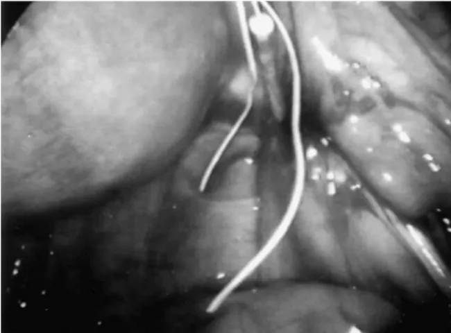
A 40-year-old woman (gravida 6, para 3) who had a Copper T IUD inserted 1 month after delivery, presented, after 7 months, with secondary amenorrhea and transient pelvic cramps. During the vaginal exami-nation, the IUD string was not visible at the external os and the cervix was cyanotic and softened, which suggested the probable manifestations of pregnancy. Ultimately, ultrasonographic examination of the patient revealed an 8-week intrauterine pregnancy, while other clinical findings and laboratory tests were normal. Transvaginal ultrasonography also visualized an extrauterine device located outside the uterus, near the sigmoid colon as if it were attached to the bowel (Figure 1). Consequently, following termination of the pregnancy at the patient’s request, a diagnostic laparoscopy was performed concomitantly, which showed bowel perforation because of the migration of the IUD next to the pregnant uterus (Figures 2 and 3). The device, which was partially embedded in the sigmoid colon, was removed via laparoscopy. How-ever, laparotomy was performed to open colostomy afterwards because of bowel perforation. Colostomy reversal was performed successfully 2 weeks later and the patient was discharged without complication.
D I S C U S S I O N
IUDs have been in use for many years, and migration from the uterus to the pelvic cavity has been reported2–4. However, a review of the literature
Sigmoid migration of an IUD Inceboz et al.
230 The European Journal of Contraception and Reproductive Health Care Figure 1 Transvaginal ultrasonography demonstrates an extrauterine device located near the sigmoid colon as if it were attached to the bowel. RIA, intrauterine device (IUD); R, rectum; U, uterus; GS, gestational sac
Figure 2 Laparoscopic view of the intrauterine contra-ceptive device migration to the sigmoid colon next to the pregnant uterus
Figure 3 Laparoscopic view of the intrauterine contra-ceptive device after removal
revealed very few cases of perforation of the recto-sigmoid by an IUD as in our case5–8. An IUD may perforate through the uterine wall into the pelvic or abdominal cavity, or into adjacent organs. Numerous factors may affect perforation. IUD-related factors include the design and structural characteristics of the device, as well as the nature and rigidity or plasticity of the inserter. With regard to the patient, uterine size and position, inherent anatomic configuration and timing of the insertion relative to delivery or abortion are all important determinants of potential perforation. Although the incidence of uterine perforation varies with the type of IUD, the incidence of IUD perfora-tion has been estimated to be 0.87 per 1000 inserperfora-tions9. Although some patients have signs and symptoms suggestive of perforation, i.e. difficulty with insertion resulting in pain or bleeding, many are apparently asymptomatic at the time the diagnosis of perforation is made. Perforation is often suspected or diag-nosed when the IUD string is no longer visible at the external os. The patient who has sustained a perforation is not protected against pregnancy, since the IUD is not in the proper location, and occasionally pregnancy is the condition that suggests that perfora-tion may have taken place as in our case.
Once the diagnosis of an extrauterine IUD has been made, the decision must be made whether to leave it alone or remove it. There is still controversy among researchers about such a situation. Markovitch and associates9 suggest that, whilst surgical procedures to remove a misplaced IUD must be performed on
symptomatic patients, asymptomatic patients, under certain circumstances, may benefit from conservative management. However, Demir and colleagues10 concluded that, in cases of extrauterine but intra-abdominal IUD, laparoscopic removal of the IUD must be the first choice of therapy. Grimaldo and co-workers11also suggested immediate removal of the device from the peritoneal cavity by either laparoscopy or laparotomy with the utilization of prophylactic antimicrobials for colon preparation before elective surgery because this variety of IUD translocated to the peritoneal cavity may provoke peritoneal or omental adhesions, volvulus, uterocutaneous fistula and bowel perforation, which involves a significant morbidity.
It is our opinion that an extrauterine IUD should be removed as soon as possible after diagnosis, although not necessarily on an emergency basis. Endoscopy thus helps in the localization and retrieval of mis-placed IUDs.
In conclusion, this case report highlights the con-tinuing need for intra- and postinsertion vigilance as even recent advances in IUD technique and tech-nology do not guarantee risk-free insertion. Regular follow-up of IUDs for visible threads would help in the earlier detection of misplaced IUDs. Proper train-ing of paramedical staff is mandatory in developtrain-ing countries to provide safe and better family planning services.
Conflict of interest Nil.
Source of funding Nil.
R E F E R E N C E S
1. The Medical Device and Drug Advisory Committees on Obstetrics and Gynecology. Second Report on Intrauterine Contraceptive Devices. DHEW, Food and Drug Administration. Washington: Government Printing Office, 1978
2. Barsaul M, Sharma N, Sangwan K. 324 cases of misplaced IUCD – a 5-year study. Trop Doct 2003; 33:11–12
3. Cuillier F, Ben Ghalem S, Haffaf Y. Intrauterine device appendicitis: an exceptional complication. J Gynecol Obstet Biol Reprod2003;32:55–7
4. Sarkar P. Translocation of a copper 7 intra-uterine contraceptive device with subsequent penetration of the caecum: case report and review. Br J Fam Plann
2000;26:161
5. Sogaard K. Unrecognized perforation of the uterine and rectal walls by an intrauterine contraceptive device.
Acta Obstet Gynecol Scand1993;72:55–6
6. Browning JJ, Bigrigg MA. Recovery of the intra-uterine contraceptive device from the sigmoid colon. Three case reports. Br J Obstet Gynaecol1988; 95:530–2
7. Hays D, Edelstein JA, Ahmad MM. Perforation of the sigmoid colon by an intrauterine contraceptive device.
Contraception1986;34:413–16
8. Sepulveda WH. Perforation of the rectum by a Copper-T intra-uterine contraceptive device; a case report.Eur J Obstet Gynecol Reprod Biol1990;35:275–8 9. Markovitch O, Klein Z, Gidoni Y, Holzinger M, Beyth
Y. Extrauterine mislocated IUCD: is surgical removal mandatory?Contraception2002;66:105–8
10. Demir SC, Cetin MT, Ucunsak IF, Atay Y, Toksoz L, Kadayifci O. Removal of intra-abdominal intrauterine device by laparoscopy. Eur J Contracept Reprod Health Care2002;7:20–3
11. Grimaldo Arriaga J, Herrera Aviles A, Garcia Taxilaga A. Perforation of the large intestine caused by a type 7 medicated copper IUCD. Ginecol Obstet Mex 1993; 61:235–7
Sigmoid migration of an IUD Inceboz et al.