© 2020 by the Serbian Biological Society How to cite this article: González-Félix ML, De La Reé-Rodríguez C, Perez- 81 Velazquez M.Partial characterization, quantification and optimum activity of
trypsin and lipase from the sciaenids Cynoscion othonopterus, Cynoscion parvipinnis
and Cynoscion xanthulus. Arch Biol Sci. 2020;72(1):81-93.
Partial characterization, quantification and optimum activity of trypsin and lipase from
the sciaenids
Cynoscion othonopterus
,
Cynoscion parvipinnis
and
Cynoscion xanthulus
Mayra L. González-Félix*, Carolina De La Reé-Rodríguez and Martin Perez-Velazquez
Department of Scientific and Technological Research, University of Sonora, Edif. 7-G, Blvd. Luis Donaldo Colosio s/n, e/ Sahuaripa y Reforma, Col. Centro, C.P. 83000, Hermosillo, Sonora, Mexico
*Corresponding author: [email protected]
Received: November 27, 2019; Revised: December 23, 2019; Accepted: January 3, 2020; Published online: January 14, 2020
Abstract: Trypsin and pancreatic lipase promote the digestion of proteins and lipids, respectively, when they are secreted into the anterior intestine; however, since the pancreas is a diffuse tissue in fish, the characterization and quantification of pancreatic enzymes is uncommon. The objective of this study was to partially characterize and compare the enzymatic activities of lipase
and trypsin within the gastrointestinal tract of Cynoscion parvipinnis, Cynoscion othonopterus and Cynoscion xanthulus, to
contribute to the knowledge of the digestive physiology of these important commercial sciaenids and to reveal whether they have potential for biotechnological applications. The presence of lipase and trypsin was confirmed by zymography and the molecular weights of both enzymes were determined by electrophoresis. For lipase, molecular weights of 65.8 and 69.5 kDa
were determined for C. othonopterus and C. xanthulus, respectively. For C. parvipinnis, two lipases of 61.5 and 36.0 kDa were
determined. In all three species the largest lipase activity was observed in the anterior intestine, followed by pyloric caeca, with optimum activity observed at pH 8.0 and at temperatures ranging between 40 and 45°C. Molecular weights of trypsin were
24.4, 23.6 and 23.7 kDain C. othonopterus,C. parvipinnis, and C. xanthulus, respectively. The optimum pH of activity ranged
between 7.0 and 9.0 and optimum temperature between 55 and 65°C for all species. These enzymes meet certain criteria that make them potential candidates for some industrial applications, such as the food industry and the production of detergents.
Keywords: Sciaenids; digestive enzymes; trypsin; lipase; partial characterization; optimum activity
INTRODUCTION
Proteins and lipids are important macronutrients in the diet of carnivorous marine finfish. Their adequate utilization and incorporation into tissues largely de-pends on the presence of digestive enzymes, which break them down into simpler components for ab-sorption to proceed. Proteases and lipases are two key enzyme groups that catabolize dietary proteins and lipids, respectively [1]. Both types of enzymes are secreted by the pancreas or pancreatic-like tissue and belong to the αβ-hydrolase family. They all bear a catalytic triad, i.e., a set of three coordinated amino acids in their structure, composed of histidine (base), aspartate (acidic residue) and serine (nucleophile). However, glutamate may also be present in lipases as an acidic residue [2,3]. Trypsin is an alkaline endo-peptidase that cleaves peptide bonds at the carboxyl end of arginine or lysine residues [4], and it is an
as bile salt-dependent lipase [9,10]. Both lipases have been described in marine finfish [11-20].
Characterizing digestive enzymes is a crucial step towards a better understanding of the utilization of dietary proteins and lipids by finfish and contributes to the development of formulated feeds for cultured species. Furthermore, for finfish species subjected to commercial fishing, the viscera, which are often dis-carded, are a potential source of digestive enzymes with industrial applications, such as the manufacture of cleaning products, food products, and laundry de-tergents. They may also have potential applications in the agrochemical, pharmaceutical, bioremediation and leather industries [15,21-24].
Examples of finfish subjected to commercial fish-eries in the Gulf of California, Mexico, and whose vis-cera are usually discarded but may otherwise be used as a source of digestive enzymes for industrial pur-poses, are the Gulf corvina Cynoscion othonopterus, the shortfin corvina C. parvipinnis and the orangemouth corvina C. xanthulus. All three species belong to the family Sciaenidae. The Gulf corvina is, in terms of vol-ume of capture, the most important species, with 5389 metric tons obtained in 2016 [25]. Although capture data specific to the shortfin and orangemouth corvinas are not available, they are both important and appreci-ated locally as food and game fishes. Interestingly, C. othonopterus and C. parvipinnis appear to have good potential as candidate species for aquaculture [26-28]. Characterizing their digestive enzymes would increase our knowledge of their digestive capacities and help formulate balanced feeds for captive rearing, but per-haps more importantly, it may contribute to the trans-formation of fishery by-product wastes into a source of valuable products with wide biotechnological poten-tial. Therefore, the objective of the present study was to partially characterize, quantify and determine the optimum activities of trypsin and lipase of C. othonop-terus, C. parvipinnis and C. xanthulus.
MATERIALS AND METHODS
Experimental organisms and biological indices
A total of 15 wild C. othonopterus, 23 C. xan-thulus and 33 C. parvipinnis were caught with seine
nets on two separate occasions during the month of October 2017 in the Gulf of California, around the area of Kino Bay (latitude 28°48'59.99''N, longitude 111°55'59.99''W), Sonora, Mexico. On each occasion, the organisms were immediately transported in an ice-filled cooler (≈ 4°C) to the Department of Sci-entific and Technological Research of the University of Sonora (DICTUS); upon arrival, individuals were identified using the FAO’s Western Central Pacific species identification guide for fish [29]. Then, fish were measured, weighed and dissected. The gastro-intestinal tract (GIT), liver and gonads were indi-vidually weighed to estimate the following indices: viscerosomatic index (VSI, %)=(viscera weight/total weight)×100; hepatosomatic index (HSI, %)=(liver weight/total weight)×100; and gonadosomatic index (GSI, %)=(gonad weight/total weight)×100. After weighing, tissues were stored and labelled in indi-vidual resealable bags at -82°C until further analyses.
Enzyme extracts
Sodium dodecyl sulfate polyacrylamide gel electrophoresis (SDS-PAGE)
To establish the molecular weights of trypsins from the three sciaenids, samples were prepared by mixing equal volumes of the crude extract with a solution of 95% 2X Laemmli Sample Buffer (Bio-Rad®, Hercules, CA, USA) and 5% 2-mercaptoethanol (Sigma-Aldrich, St. Louis, MO, USA), and then placed in a 95°C wa-ter bath for 2 min [30]. Afwa-ter cooling to room tem-perature (25°C), 20 µL aliquots were loaded into 12% polyacrylamide gels and resolved at 115 V for 2.5 h at 10°C in a four-gel vertical electrophoresis system (Mini-Protean Tetra Cell, Bio-Rad®, Hercules, CA, USA). Gels were fixed in a solution of 50% trichloro-acetic acid (TCA) for 18 h, stained for 1 h at 37ºC in a solution of 50% TCA with 0.1% Coomassie brilliant blue R-250 and destained by repeatedly washing with 7% acetic acid. Trypsin (T8003) from bovine pancre-as (Sigma-Aldrich, St. Louis, MO, USA) wpancre-as used pancre-as the reference molecular marker and as a quantitative internal standard to establish the enzyme concen-tration by densitometry. The molecular weight was estimated by comparison with an internal molecular weight standard (Precision Plus Protein Standard Dual Color, Bio-Rad®, Hercules, CA, USA) with protein markers of 10 to 250 kDa. Gels were documented in a calibrated densitometer (Model GS-900, Bio-Rad®, Hercules, CA, USA) for identification and quantifi-cation of the bands using the ImageLab 5.0 software (Bio-Rad®, Hercules, CA, USA). In the case of lipases, the crude extracts were further purified using a solu-tion of 30% polyethylene glycol (PEG-6000) to get a 1:1 dilution. After vortexing for 2 min, samples were kept in constant agitation for 30 min at 4ºC. Next, they were centrifuged at 21000 × g for 30 min at 4ºC. The supernatants were carefully recovered and 20 µL aliquots were loaded into 10% polyacrylamide gels and run under the same conditions previously men-tioned. Gels were stained for 18 h by gentle agitation in QC Colloidal Coomassie stain (Bio-Rad®, Hercules, CA, USA), and then destained by rinsing in distilled water. Bovine serum albumin (BSA) (Sigma-Aldrich, St. Louis, MO, USA) was used as an internal marker and as a quantitative internal standard to establish the enzyme concentration by densitometry.
Zymography
A specific zymographic assay was used to identify and confirm the band belonging to each enzyme in the gel. Trypsin was detected by developing an activity band when hydrolysis of casein took place [31]. After electrophoresis, the gel was rinsed in distilled water and submerged in 50 mL of 2% casein in 50 mM Tris-HCl buffer at 5°C for 30 min, followed by incubation at 37°C for 90 min for digestion of the protein sub-strate, and rinsing with distilled water. A solution of 40% ethanol:10% acetic acid:0.1% Coomassie brilliant blue R-250 was used for staining the gel overnight. Trypsin was visualized by the contrast of a clear white band in a dark blue background. A band with trypsin (T8003, Sigma-Aldrich, St. Louis, MO, USA) from bovine pancreas was used to confirm the effectiveness of the reaction of the enzyme against casein.
Detection of lipase in a zymogram was done by preparing a chromogenic substrate with 3.0 g of aca-cia gum dissolved in 30 mL of 0.2 M phosphate buffer solution (pH 8.0) and brought to a final volume of 50 mL. Then, 20 mL of olive oil and 10 g of crushed ice were incorporated and homogenized for 5 min, fol-lowed by the addition of 1% agar. The emulsion was then warmed to 50°C. Separately, 10 mg of Victoria blue B were dissolved in 1 mL of 70% ethanol, incor-porated into the emulsion and maintained at 50°C [32]. The chromogenic substrate was poured over the gel and incubated at 37°C for 12 h, washed with distilled water and stained with QC Colloidal Coomassie for 18 h. Activity of lipase against olive oil triglycerides was observed as a dark blue band in the zymogram gel.
Assessment of enzymatic activities
Lipase
three times at 21000 × g and 4°C for 30 min, until the samples were clear. The supernatant was recovered and used to prepare the reaction mix with 1 mL 200 mM Tris-HCl buffer solution (pH 7.7), 2.5 mL tridistilled water, 1 mL of crude extract from either the anterior intestine (AI), middle intestine (MI), posterior intestine (PI) or pyloric caeca (PC), and 3 mL of olive oil. The samples were then mixed vigorously and incubated in a shaker at 35°C at 120 rpm for 30 min. The reaction was stopped by adding 3 mL of 95% ethanol, followed by the addition of 80 µL of 0.1% phenolphthalein and titration with a solution of 50 mM NaOH to a light pink color. In all blanks, the enzyme crude extract was added after the incubation period. One unit of activity of lipase (U) per mL of crude extract was defined as the amount of enzyme that hydrolyzed 1.0 microequiva-lent of fatty acid from a triglyceride in 1 h at pH 7.7 and 35°C, using the equation: activity (U mL-1)=[(mL NaOH used) (molarity of NaOH) (1000) (2) (dilution factor)]/(volume of enzyme used in mL). In this equa-tion, 1000 is the conversion factor from milliequivalent to microequivalent, and 2 is the time conversion factor from 30 min to 1 h (unit definition).
Enzymatic activity was higher in anterior intestine as compared to the other sections of the GIT in all three species. Therefore, this section was exclusively used to estimate the optimum temperature and pH for enzymatic activity. The analyses were run in triplicate samples, and each triplicate sample and its blank were run in duplicates, following previously described pro-cedures [33] with minor modifications. Briefly, 100 µL of 100 mM sodium cholate hydrate, 1.9 mL of 50 mM Tris-HCl (pH 7.2) buffer solution and 10 µL of the enzyme extract (omitted in the blank tubes) were placed in 10-mL glass tubes and mixed thoroughly; then 20 µL of 200 mM β-naphthyl-acetate were added and incubated for 30 min at 20, 25, 30, 35, 40, 45, 50, 55, 60, 65 and 70°C, followed by the addition of 20 µL of 100 mM fast blue BB salt and an additional incubation period of 5 min. Next, 200 µL of 0.72 N TCA, 10 µL of enzyme extract in the blank tubes and 2.71 mL of ethanol:ethyl acetate were added to stop the reaction; the tubes were vortexed and absorbance was read at 540 nm. The Δ absorbance (extract absor-bance - blank absorabsor-bance) was used to estimate the units of absorbance (Δ absorbance ÷ constant (0.01)). Then the units of lipase min-1 (units of absorbance ÷ incubation time) were used to estimate lipase activity,
expressed as units of lipase mL-1 (units of lipase min-1 ÷ extract volume). In the case of the pH, the reactions were run using a pH range from 6 to 10. The buffer solutions were adjusted according to the pH evaluated; a 50 mM citrate-phosphate buffer was used for pH 5.0 and 6.0, a 50 mM Tris-HCl buffer for pH 7.0 to 9.0 and a 50 mM glycine-NaOH buffer for pH 10. The temperature for incubation was also adjusted to 40°C for C. parvipinnis and C. xanthulus and to 45°C for C. othonopterus because, at these values, lipase activity was observed to be optimal in the previous test.
Trypsin
For each of the three sciaenids, the activity of trypsin within AI, MI, PI and PC was determined in triplicate samples, each from a different experimental organ-ism, with triplicate blanks; each triplicate sample and its blank were also run in duplicates. A substrate pre-pared with 100 mM of Nα-benzoyl-DL-arginine 4-ni-troanilide hydrochloride (BAPNA) diluted in 1 mL of dimethyl sulfoxide (DMSO) was brought to 100 mL with 50 mM Tris-HCl buffer and 10 mM CaCl2 (pH 8.2). A total of 1.25 mL of the substrate was pipetted into a microcentrifuge tube and 20 µL of the enzyme extract were added. Samples were then incubated in a water bath at 35°C for 30 min. After incubation, the reaction was stopped by adding 0.25 mL of 30% acetic acid and the absorbance was read at 410 nm. For the blanks, 20 µL of tridistilled water were added instead of the enzyme extract. The activity of trypsin was esti-mated with the following formula: trypsin activity (U mL-1)=(Δ absorbance ÷ total reaction volume) ÷ (coef-ficient of extinction molar × incubation time × extract volume) [34]. Trypsin concentration was estimated from the absorbance readings of the samples. A calibration curve with different concentrations of trypsin from bo-vine pancreas (Sigma-Aldrich, St. Louis, MO, USA) was performed; the determined linear regression equation was y=0.1455x+0.0073 with R2=0.9786, where the coef-ficient of extinction molar (CEM=0.1455) and the slope (m=0.0073) were used in the following formula: trypsin concentration (mg mL-1)=Δ absorbance - m ÷ CEM.
samples, and each triplicate sample and its blank were run in duplicates, adjusting the incubation temperature to 20, 25, 30, 35, 40, 45, 50, 55, 60, 65, 70, 75, 80, 85 and 90°C, whereas the pH was evaluated substituting the buffer in the substrate solution according to the pH evaluated; a 100 mM citrate-NaOH buffer was used for pH 6.0, a 100 mM Tris-HCl buffer for pH 7.0 to 9.0 and a 100 mM glycine-NaOH buffer for pH 10. The temperature for incubation was also adjusted to 65°C for C. parvipinnis and C. xanthulus and to 60°C for C. othonopterus because, at these values, trypsin activity was observed to be optimal in the previous test.
Statistical analysis
Biological indices, molecular weights and concentra-tions of lipase and trypsin determined by densitom-etry were analyzed by descriptive statistics, whereas enzymatic activity within the different sections of the GIT at different temperatures and at different pH were subjected to one-way analysis of variance (ANOVA) using a significance level of P≤0.05. Tukey’s HSD test was used as the mean separation procedure when significant differences were observed. All statistical analyses were performed using the Statistical Analysis System (SAS Institute Inc., 2013, Software Release 9.4, Cary, NC, USA) software package.
RESULTS
Fish biological indices
The total weight and length of the three sciaenids in this study ranged from 862-1129.4 g, and from 46.5-51 cm, respectively, confirming these experimental or-ganisms had a commercial size. The gonads were not fully developed and were small in all species, 0.9-4.0 g, therefore, the GSI were also low, 0.08-0.53%. The HSI ranged from 1.2-1.6%, whereas the VSI indicated that viscera represent close to 7.3-9.5% of the body weight of the sciaenids (Table 1).
SDS-PAGE analyses of trypsin and lipase
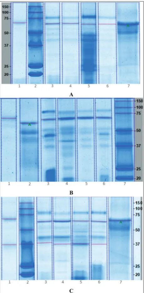
The mean molecular weights±standard error of the mean (SEM) of trypsins of C. othonopterus, C. parvi-pinnis and C. xanthulus were 24.4±0.2, 23.6±0.1 and 23.7±0.1 kDa (Fig. 1), respectively. Zymography of
the crude extract showed a clear band on a dark blue background (lane 7 in Fig. 1) produced by the activ-ity of this protease against casein, which matched ex-actly the band observed on the SDS-PAGE gel. Lipases of C. othonopterus and C. xanthulus had molecular weights of 65.8±0.3 (Fig. 2A) and 69.5±0.8 kDa (Fig. 2B), respectively. Two bands with lipase activity were detected for C. parvipinnis, one of 61.5±0.2 kDa and another one of 36.0±0.3 kDa (Fig. 2C). Lipase bands were evident from the reaction of the enzyme against its substrate and were observed as dark blue bands in the zymograms.
The concentrations of trypsins and lipases were analyzed by densitometry on the SDS-PAGE gels. Mean concentration values±SEM for trypsins and
li-Table 1. Biological parameters and indices of wild Cynoscion othonopterus, Cynoscion parvipinnis and Cynoscion xanthulus.
Biological
indices othonopterusC. parvipinnisC. xanthulusC.
Total weight (g) 1129.4±53.9 862.0±49.2 879.4±45.6
Total length (cm) 51.0±0.8 46.5±1.0 47.3±0.9
Liver weight (g) 14.2±1.3 10.0±0.6 12.7±1.3
Gonad weight (g) 0.9±0.3 4.0±0.9 2.0±0.5
Viscera weight (g) 82.1±3.9 65.8±4.6 70.7±4.5
HSI (%) 1.2±0.1 1.4±0.1 1.6±0.1
GSI (%) 0.08±0.02 0.53±0.11 0.24±0.05
VSI (%) 7.3±0.2 9.5±0.6 9.1±0.7
Total weight and length values are means±SEM of 33 C. parvipinnis, 15
C. othonopterus and 23 C. xanthulus. Organs, viscera and indices values are means±SEM of 15 fish from each species.
pases observed in the three sciaenids are presented in Table 2. For trypsin, the concentration ranged from 6.0-12.8 mg g-1 of tissue, and in C. othonopterus and C.
parvipinnis, the AI and PC appeared to have the largest concentrations of the enzyme. Lipase concentration estimated by densitometry ranged from 16.3-50.0 mg g-1 of tissue, with no noticeable differences between the analyzed sections of the GIT, except that in all sci-aenids the concentration in PI appeared to be lower.
Trypsin and lipase activities
Trypsin activity was significantly higher in AI of C. othonopterus (65.1 U mL-1, P=0.0352), in C.
parvipin-nis it was significantly higher in AI and PC (66.5 and 30.8 U mL-1, respectively; P=0.0286), whereas in C.
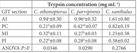
xanthulus, no significant differences (P=0.2757) in activity were evident between the four sections of the GIT, but numerically, more activity (108.8 U mL-1) was registered in the AI (Table 3). These observations were consistent with the concentrations of trypsin es-timated through the calibration curve with trypsin from bovine pancreas; the IA showed significantly higher activity in C. othonopterus (0.94 mg mL-1), in
C. parvipinnis it was significantly higher in the AI and PC (0.96 and 0.42 mg mL-1, respectively), and in C.
xanthulus, no significant differences in activity were observed, ranging from 1.61 mg mL-1 in the AI, to 0.38 mg mL-1 in the PI (Table 4). Optimum temperature for trypsin activity (Table 5) was 60°C (426.3 U mL -1) in C. othonopterus, though it was not significantly different from the activity observed at 55 or 65°C (307.1 or 318.5 U mL-1, respectively). In C.
parvipin-nis and C. xanthulus, trypsin activity was significantly higher (P<0.0001) at 65°C (699.4 and 160.7 U mL-1, respectively). Optimum pH for trypsin activity in C. othonopterus was 8.0 (797.9 U mL-1), but it was not significantly different from pH 7.0 or 9.0 (493.4 or 527.9 U mL-1, respectively). In C. parvipinnis activity was significantly higher (P<0.0001) at pH 8.0 (845.2 U mL-1), but not statistically different from pH 7.0 (653.3 U mL-1). Finally, in C. xanthulus the optimum pH for trypsin activity was 8.0 (782.8 U mL-1), which was significantly higher (P<0.0001) than the rest of the pH values tested (Table 6).
Lipase activity on the other hand, was significantly higher in the AI and PC of C. parvipinnis (133.3 and
Fig. 2. Lipases. A:C. othonopterus. Zymogram: Lane 1 – Ante-rior intestine; SDS-PAGE gel: 2 – Molecular weight standard, 3 – Anterior intestine, 4 – Middle intestine, 5 – Posterior intestine, 6 – Pyloric caeca, 7 – Bovine serum albumin (A: quantitative standard). B:C. xanthulus. Zymogram: Lane 1 – Anterior intes-tine; SDS-PAGE gel: 2 – Bovine serum albumin (A: quantitative standard), 3 – Anterior intestine, 4 – Middle intestine, 5 – Poste-rior intestine, 6 – Pyloric caeca, 7 – Molecular weight standard.
86.0 U mL-1, respectively; P=0.0039) and C. xanthulus (113.6 and 89.0 U mL-1, respectively; P=0.0007), as well as in C. othonopterus, but in this last sciaenid, the activity registered in MI (73.1 U mL-1) was not significantly different (P=0.0021) from that recorded in the AI or PC (101.0 and 81.3 U mL-1, respectively) (Table 3). The optimum temperature for lipase ac-tivity (Table 5) in C. othonopterus was 45°C (609.3 U mL-1; P=0.0428); in C. parvipinnis a significantly higher activity was registered at 40°C (1,225.7 U mL-1;
P<0.0001), and in C. xanthulus no significant differ-ences were observed between 40 (1,327.3 U mL-1) and
45°C (1,299.8 U mL-1). The optimum pH for activity of lipase in these sciaenids was 8.0 (1014.2-1185.6 U mL-1), and in all three species, this pH was signifi-cantly higher (P<0.0001) than the rest of the pH values evaluated (Table 6).
DISCUSSION
The weights and lengths of the experimental organ-isms in this study corresponded to large commercial-size sciaenids. The HSI (1.2-1.6%), GSI (0.08-0.53%) and VSI (7.3-9.5%) reported in this study are com-parable to values reported in the literature for these and other sciaenids; for C. parvipinnis, HSI and VSI values ranging from 0.9-2.6% and from 3.7-5.9%, re-spectively [35], and HSI values ranging from 2.2-2.9% for C. othonopterus [27] have been reported. For other sciaenids like Totoaba macdonaldi, also endemic to the Gulf of California, HSI values ranging from 1.6-2.2% and VSI of 3.6-4.5% [35] have been reported, as well as HSI ranging from 1.33-1.96% for Sciaenops ocellatus
[36]. Although comparable, differences between the observed and reported values may be explained by the size differences, since the values reported in the previous studies corresponded mostly to juvenile fish. Their origin is also different; in the previous studies
Table 2. Concentration (mg g-1 of tissue) of trypsin and lipase within the different sections of the GIT of C. othonopterus, C. parvipinnis and C. xanthulus determined by densitometry in SDS-PAGE gels.
Trypsin concentration (mg g-1 of tissue) Lipase concentration (mg g-1 of tissue)
GIT section C. othonopterus C. parvipinnis C. xanthulus C. othonopterus C. parvipinnis C. xanthulus
AI 12.8±0.9 8.1±2.7 7.0±1.4 35.8±2.2 35.8±3.8 45.4±3.2
MI 9.4±0.7 7.7±2.9 6.0±0.4 50.0±2.2 37.2±5.9 42.3±4.2
PI 9.2±1.1 7.3±2.4 7.0±0.2 28.3±0.2 16.3±6.2 25.1±2.8
PC 11.8±0.5 11.3±1.8 7.0±0.4 42.6±2.8 35.3±4.4 39.9±5.0
SDS-PAGE: sodium dodecyl sulfate polyacrylamide gel electrophoresis; GIT: gastrointestinal tract; AI: anterior intestine; MI: middle intestine; PI: posterior intestine; PC: pyloric caeca. Values are means±SEM of three fish per species.
Table 3. Activity (U mL-1) of trypsin and lipase within the different sections of the GIT of C. othonopterus, C. parvipinnis and C. xanthulus.
Trypsin activity (U mL-1) Lipase activity (U mL-1)
GIT section C. othonopterus C. parvipinnis C. xanthulus C. othonopterus C. parvipinnis C. xanthulus
AI 65.1a ±19.9 66.5a±21.3 108.8±52.3 101.0a±9.6 133.3a±19.6 113.6a±15.5
PC 17.3b±5.8 30.8ab±4.4 57.1±12.5 81.3ab±7.0 86.0ab±15.1 89.0ab±13.0
MI 24.6b±7.0 20.7b±1.8 85.2±25.3 73.1ab±8.7 65.3b±5.2 64.0bc±5.8
PI 17.8b±5.0 21.7b±5.1 27.9±1.4 50.7b±4.1 56.0b±6.5 44.0c±4.8
ANOVA P>F 0.0352 0.0286 0.2757 0.0021 0.0039 0.0007
GIT: gastrointestinal tract; AI: anterior intestine; MI: middle intestine; PI: posterior intestine; PC: pyloric caeca. Values are means±SEM of three replicates per species; each triplicate sample and its blank were run in duplicates. Means with different superscripts within the same column are significantly different (P≤0.05).
Table 4. Concentration (mg mL-1) of trypsin within the differ-ent sections of the GIT of C. othonopterus, C. parvipinnis and C. xanthulus.
Trypsin concentration (mg mL-1)
GIT section C. othonopterus C. parvipinnis C. xanthulus
AI 0.94a±0.30 0.96a±0.32 1.61±0.80
PC 0.21b±0.09 0.42ab±0.07 0.82±0.19
MI 0.32b±0.11 0.27b±0.03 1.25±0.38
PI 0.22b±0.08 0.28b±0.08 0.38±0.02
ANOVA P>F 0.0346 0.0290 0.2766
the reports are for cultured fish, whereas wild organ-isms were used in this study. Nevertheless, it is evident from these biological indices that the experimental fish in this study were in good physiological state.
Lipases are present in the GIT of carnivorous fish because, in spite of consuming large amounts of pro-tein in their natural diet, lipids are also a significant component. In C. othonopterus, the molecular weight of lipase was 65.8 kDa, fairly similar to the lipase of
Pagrus major of 64 kDa [14]. In C. parvipinnis, two bands of 61.5 and 36.0 kDa showed lipase activity, sim-ilarly to the observation in Oncorhynchus tshawytscha, where two bands of 79.6 and 54.9 kDa were reported and thus, the presence of two isozymes of lipase was
proposed [15]. Lipase of Atlantic cod Gadus morhua
has a molecular weight of 60 kDa [12], similar to the molecular weight of the first lipase observed in C. par-vipinnis. Finally, lipase of C. xanthulus had a molecular weight of 69.6 kDa, close in value to the pancreatic lipase of T. macdonaldi of 70.4 kDa [19], and to the 70 kDa purified lipase of Indian carp Catla catla [37]. Additional studies have reported molecular weights of 80.3 kDa in S. ocellatus [20], 74 kDa in common carp Cyprinus carpio [38] and 44.6 kDa for Macruronus novaezelandiae [15]. The molecular weight of lipase in marine finfish varies according to species; differ-ences in tridimensional structures may be attributed to differences in amino acid sequences constituting the primary protein structure as a result of genetic
di-Table 5. Activity (U mL-1) of trypsin and lipase within AI of C. othonopterus, C. parvipinnis and C. xanthulus at different temperatures.
Trypsin activity (U mL-1) Lipase activity (U mL-1)
Temperature C. othonopterus C. parvipinnis C. xanthulus C. othonopterus C. parvipinnis C. xanthulus
20°C 19.1d±0.8 15.8g±0.9 13.9d±1.9 62.1cd±24.9 219.1efg±28.8 57.2d±9.2
25°C 26.6d±3.6 18.8g±3.1 15.0d±1.4 148.9abcd±65.7 301.3def±17.5 135.5d±3.5
30°C 80.8cd±18.2 24.9fg±2.1 14.7d±0.6 271.7abcd±58.7 415.7cde±2.0 376.5c±10.8
35°C 100.5cd±18.5 27.8fg±4.8 20.7cd±1.6 339.0abcd±79.3 557.3c±48.1 628.6b±14.9
40°C 146.1cd±31.0 37.1fg±4.2 21.6cd±1.2 489.6abc ±78.4 1225.7a±5.5 1327.3a±8.0
45°C 184.4bc±34.4 82.7ef±12.7 24.2cd±0.7 609.3a±90.8 880.4b±82.3 1299.8a±4.5
50°C 229.9bc±41.8 132.3de±16.9 30.3cd±0.8 560.3ab±74.3 439.6cd±71.7 536.9b±58.7
55°C 307.1ab±57.7 200.2c±28.6 42.1bc±5.6 307.3abcd ±75.6 281.7defg±12.7 106.8d±14.7
60°C 426.3a±81.4 473.3b±8.7 62.6b±7.9 108.9bcd±29.7 172.4fg±50.3 38.8d±4.5
65°C 318.5ab±36.2 699.4a±27.6 160.7a±3.4 53.0cd±17.7 66.4g±23.5 33.7d±4.2
70°C 48.0d±2.9 146.2cd±11.5 64.9b±7.6 33.1d±17.5 38.5g±12.6 19.1d±5.0
75°C 36.2d±5.4 39.1fg±5.4 36.1cd±9.2 - -
-80°C 29.2d±5.3 38.7fg±5.9 35.3cd±5.5 - -
-85°C 28.9d±6.3 42.1fg±5.0 42.2bc±6.6 - -
-90°C 26.7d±5.8 40.6fg±5.0 27.2cd±6.6 - -
-ANOVA P>F < 0.0001 < 0.0001 < 0.0001 < 0.0428 < 0.0001 < 0.0001
AI: anterior intestine. Values are means±SEM of three replicates per species; each triplicate sample and its blank were run in duplicates. Means with different superscripts within the same column are significantly different (P≤0.05).
Table 6. Activity (U mL-1) of trypsin and lipase within AI of C. othonopterus, C. parvipinnis and C. xanthulus at different pH values.
Trypsin activity (U mL-1) Lipase activity (U mL-1)
pH C. othonopterus C. parvipinnis C. xanthulus C. othonopterus C. parvipinnis C. xanthulus
5 - - - 12.7cd±2.9 37.9e±4.5 30.2d±7.9
6 291.1b± 34.9 227.3cd±10.3 306.1cd±9.9 110.8cd±31.6 537.3d±17.9 179.4c± 12.7
7 493.4ab±20.2 653.3ab±75.8 499.8b±44.2 587.3b±69.8 987.9b±63.5 715.6b±27.6
8 797.9a±31.7 845.2a±48.2 782.8a±2.4 1014.2a±114.6 1185.6a±7.9 1158.4a±23.2
9 527.9a±72.3 462.5bc±61.7 325.9c±16.0 215.3c ±27.4 786.2c±8.3 730.4b±12.3
10 245.4b±6.3 211.5d±10.7 215.8d±11.3 6.9d±1.9 5.9e±1.6 11.1d±2.6
ANOVA P>F 0.0003 < 0.0001 < 0.0001 < 0.0001 < 0.0001 < 0.0001
versity, as well as the result of possible environmental pressures to which they have been subjected, resulting in evolutionary adaptations at a molecular level [39-41]; feeding habits most likely also play an important role in the structural differences observed in lipases and other digestive enzymes in finfish.
Lipase activity within the different sections of the GIT of C. othonopterus, C. parvipinnis and C. xan-thulus evaluated at 35°C confirmed that the AI is the section with the greatest enzymatic activity, though it is not significantly different from the activity in the PC. In general, activity gradually decreased towards the posterior end of the intestine in all three species. Higher lipolytic activities within the AI and PC in comparison with the distal sections of the intestine have also been reported in T. macdonaldi, Sparus au-rata and Thunnus orientalis [19,42,43]. These observa-tions coincide with earlier reports demonstrating that the digestion and absorption of lipids in marine finfish takes place within the AI [44,45]. The higher activ-ity of lipase within the anterior sections of the GIT is explained by the fact that pancreatic enzymes are secreted within the AI of finfish. Moreover, enzymes are proteins also exposed to hydrolysis by other prote-ases in the GIT; thus, when they reach the MI and PI they have been subjected to hydrolysis to some extent, explaining the lower enzymatic activity within these sections. It is interesting to note that the lipases of all three sciaenids had catalytic activity towards fatty acids esterified to triacylglycerol molecules present in olive oil, which leads us to assume that the catalytic mechanism in the lipases of the three species is quite similar to that observed in higher vertebrates, and that under culture conditions, balanced diets could include this type of vegetable oil as long as their nutritional requirements for essential fatty acids (mainly n-3 highly unsaturated fatty acids) are satisfied by other lipid sources, fish oil for instance.
The optimum temperature for lipase activity of
C. parvipinnis was 45°C; at 40 and 45°C C. xanthulus
showed no differences in activity, and for C. othonop-terus, 45°C was the temperature where the highest activity was recorded, quite similar to the observa-tion for T. macdonaldi with optimum lipase activity at 45°C [18,19]. Optimum lipase activities between 35 to 45°C in T. orientalis and Morone saxatilis [18] have been reported, and 35°C for O. tshawytscha and
M. novaezelandiae [15]. Differences in optimum tem-peratures for pancreatic enzymes could be explained by the differences in environmental temperatures ob-served in the habitats of the species, which clearly lead to adaptations [46]. Sciaenids in this study inhabit a geographical area with average temperatures ranging between 19.8±2.3 and up to 27.8±1.9°C [47]. The op-timum temperature for enzyme activity in finfish and higher vertebrates is usually higher than their opti-mum physiological temperature; this is explained by the increment observed in the molecular kinetic en-ergy at higher temperatures, up until the point where denaturation of the protein occurs [48]. Optimum pH for lipase activity on the other hand was 8.0 for all three sciaenids, coinciding with the value reported for
T. macdonaldi, M. saxatilis and T. orientalis [18,19]. For
O. tshawytscha and M. novaezelandiae, the optimum pH was reported to be 8.0-8.5 [15]. Thus, the optimum pH for enzymatic activity of lipase does not vary much among these sciaenids or other marine finfish.
Trypsin molecular weights for C. othonopterus,
C. parvipinnis and C. xanthulus were 24.4, 23.6 and 23.7 kDa, respectively, comparable to mammalian and other marine finfish trypsins, such as that of Sardinops sagax caerulea with 25 kDa [49], or that of Zosterises-sor ophiocephalus with 23.2 kDa [50]. The molecular weight of purified trypsin of Clarias macrocephalus ×
Clarias gariepinus is 24 kDa [51]. In Thunnus alalunga, trypsins A and B are 21 and 24 kDa, respectively [46]. In Colossoma macropomum,trypsin has a molecular weight of 23.9 kDa [52] and in Diapterus rhombeus
26.5 kDa [53]. As has been suggested, variability in molecular weights is expected even in closely related species because of genetic differences, differences in tridimensional structures and amino acid sequences in the proteins, as well as environmental adaptations [39-41], among other factors. Similar to lipase, the proteolytic activity of trypsin was higher in the AI of
to be more active and to be more concentrated within this section of the tract [56].
The optimum temperature for trypsin activity in
C. othonopterus was 60°C, but not significantly dif-ferent from that at 55 or 65°C, coinciding with the values for two trypsin isoforms of Katsuwonus pela-mis, 55 and 60°C [57] and the trypsins of C. mac-rocephalus × C. gariepinus [51], O. tshawytscha [15],
Sardina pilchardus [58] and Mugil cephalus [59], with an optimum temperature at 60°C. In C. parvipinnis
and C. xanthulus, the optimum temperature was 65°C, the same as for Centropomus undecimalis [60],
Atractosteus tropicus [61], Brycon orbignyanus [62] and T. thynnus [63]. Lower optimum temperatures for trypsin activity have been described in the litera-ture, such as those for S. sagax caerulea [49] and Ga-dus macrocephalus [64] at 50°C, or Balistes capriscus
[41], Pterygoplichthys disjunctivus [65] and Cirrhinus mrigala [66] with optimum temperatures as low as 30-40°C. Optimum temperature for activity for all enzymes could still be related to environmental tem-perature, in spite of being higher than their optimum physiological temperature [46,48]. All sciaenids in this study showed a drastic decline in trypsin activity after reaching 70°C, similarly to observations for trypsins of C. macrocephalus × C. gariepinus and T. alalunga
that also declined at 70°C [46,51] and of C. undeci-malis at 75°C [60], which is explained by the thermal denaturation of the protein. Heat causes the enzyme molecule to unfold, breaking down the secondary and tertiary structures of the protein and the enzyme loses its function. It has been suggested that alkaline proteases of aquatic organisms are usually stable and active under adverse conditions, such as temperatures between 50-60°C [67]; this is the reason why trypsins from C. othonopterus, C. parvipinnis and C. xanthu-lus are interesting candidates for the feed industry or for industrial applications such as the manufacture of detergents.
The optimum pH for trypsin activity in C. othonopterus was 8.0-9.0, in C. parvipinnis it was 7.0-8.0 and in C. xanthulus it was 8.0, all within the range proposed as the optimum pH for trypsins [49], 7.0-9.0. An optimum pH of 8.0 for trypsin activity of S. sagax caerulea has been reported [49], and the same value has been reported for C. macrocephalus × C. gariepinus [51] and O. tshawytscha [15]. An
op-timum pH value of 8.5 was reported for T. alalunga
[46] and D. rhombeus [53], and a range of 8.0-9.0 for the trypsins of S. pilchardus [58]. Thus, the optimum pH values of trypsin activity for several marine and freshwater fish closely resemble those observed in this study for our three studied sciaenids. The activity of trypsin decreased at either lower or higher pH values, presumably as a result of conformational modifica-tions resulting from changes in the electrical charge of side chains of amino acids, which in turn modify elec-trostatic interactions that stabilize the tertiary struc-ture of proteins [48] or cause the denaturation of the protein under antagonist acid or alkaline conditions of the aqueous solutions, resulting in lower enzyme activity [68]. In general, trypsins of aquatic organ-isms are active at pH values ranging from 7.0 to 10.0 and can hydrolyze a diversity of substrates [49,69]. Consequently, the trypsins from C. othonopterus, C. parvipinnis and C. xanthulus evaluated in this study may also be used as additives in detergent formula-tions, since they are all active at those alkaline pH values [70].
CONCLUSIONS
poten-tial candidates for some industrial applications, such as the food industry and the production of detergents.
Acknowledgments: We would like to thank the Consejo Nacional de Ciencia y Tecnologia (CONACYT-Mexico) for partly funding Ms. Carolina De La Reé-Rodríguez. The mention of trademarks or proprietary products does not constitute an endorsement of the product and does not imply its approval to the exclusion of other products that may also be suitable.
Author contributions: Mayra L. González-Félix and Martin Perez-Velazquez conceived and planned the study. Carolina De La Reé-Rodríguez and Martin Perez-Velazquez participated in fish collecting. Carolina De La Reé-Rodríguez and Mayra L. González-Félix carried out the laboratory analyses. Mayra L. González-González-Félix took the lead in writing the manuscript. All authors discussed the results and provided critical feedback to the manuscript.
Conflict of interest disclosure: The authors have no conflict of interest related to this work.
REFERENCES
1. Goodman B. Insights into digestion and absorption of major nutrients in humans. Adv Physiol Educ. 2010;34:44-53. 2. Nardini M, Dijkstra BW. α/β Hydrolase fold enzymes: the
family keeps growing. Curr Opin Struc Biol. 1999;9:732-7. 3. Smith LC, Faustinella F, Chan L. Lipases: three-dimensional
structure and mechanism of action. Curr Opin Struc Biol. 1992;2:473-651.
4. Khangembam BK, Kameshwar-Sharma YVR, Chakrabarti R. Purification and characterization of trypsin from the diges-tive system of carp Catla catla (Hamilton). Int Aquat Res. 2010;4:1-12.
5. Cao MJ, Osatomi K, Sujuki M, Hara K, Tachibana K, Ishihara T. Purification and characterization of two anionic trypsins from the hepatopancreas of carp. Fish Sci. 2000;66:1172-9. 6. Lowe ME. Structure and function of pancreatic lipase and
colipase. Annu Rev Nutr. 1997;17:141-58.
7. Lowe ME. The triglyceride lipases of the pancreas. J Lipid Res. 2007;43:2007-16.
8. Van Tilbeurgh H, Bezzine S, Cambillau C, Verger R, Carrière F. Colipase: structure and interaction with pancreatic lipase. Biochim Biophys Acta. 1999;1441:173-84.
9. Terzyan S, Wang CS, Downs D, Hunter B, Zhang X. Crystal structure of the catalytic domain of human bile salt activated lipase. Protein Sci. 2000;9:1783-90.
10. Kurtovic I, Marshall SN, Zhao X, Simpson BK. Lipases from mammals and fishes. Rev Fish Sci. 2009;17:18-40.
11. Léger C. Digestion, absorption and transport of lipids. In: Cowey CB, Mackie AM, Bell JG, editors. Nutrition and feed-ing of fish. London: Academic Press; 1985. p. 299-331. 12. Gjellesvik DR, Lombardo D, Walther BT. Pancreatic bile salt
dependent lipase from cod (Gadus morhua): purification and properties. Biochim Biophys Acta. 1992;1124:123-34. 13. Gjellesvik DR, Lorens JB,Male R. Pancreatic carboxylester
lipase from Atlantic Salmon (Salmo salar) cDNA sequence
and computer-assisted modelling of tertiary structure. Eur J Biochem. 1994;226:603-12.
14. Iijima N, Tanaka S, Ota Y. Purification and characterization of bile salt-activated lipase from the hepatopancreas of red sea bream (Pagrus major). Fish Physiol Biochem. 1998;18:59-69. 15. Kurtovic I, Marshall SN, Zhao X. Purification and proper-ties of digestive lipases from Chinook salmon (Oncorhynchus tshawytscha) and zeland hoki (Macruronus novaezelandiae). Fish Physiol Biochem. 2010;36:1041-60.
16. Nolasco H, Moyano-López F, Vega-Villasante F. Partial char-acterization of pyloric-duodenal lipase of gilthead seabream (Sparus aurata). Fish Physiol Biochem. 2011;37:43-52. 17. Holmes RS, Cox LA. Bioinformatics and evolution of
verte-brate pancreatic lipase and related proteins and genes. J Data Min Genom Proteomics. 2012;3:1-10.
18. Rueda-López S, Martínez-Montaño E, Viana MT. Biochemi-cal characterization and comparison of pancreatic lipases from the Pacific bluefin tuna, Thunnus orientalis; totoaba,
Totoaba macdonaldi; and striped bass, Morone saxatilis. J World Aquacult Soc. 2017;48:156-65.
19. González-Félix ML, Santana-Bejarano EB, Perez-Velazquez M, Villalba-Villalba AG. Partial characterization, quantifica-tion and activity of pancreatic lipase in the gastrointestinal tract of Totoaba macdonaldi. Arch Biol Sci. 2018a;70:489-96. 20. González-Félix ML, Gatlin III DM, Perez-Velazquez M, Webb
K, García-Ortega A, Hume M. Red drum Sciaenops
ocella-tus growth and expression of bile salt-dependent lipase in response to increasing dietary lipid supplementation. Fish Physiol Biochem. 2018b;44:1319-31.
21. Shahidi F, Vidanarachchi J. Enzymes from fish and aquatic invertebrates and their application in the food industry. Trends Food Sci Technol. 2001;12:435-64.
22. Gupta R, Beg QK, Lorenz P. Bacterial alkaline proteases: molecular approaches and industrial applications. Appl Microbiol Biotechnol. 2002;59:15-32.
23. Klomklao S. Digestive proteinases from marine organ-isms and their applications. Songklanakarin J Sci Technol. 2008;30:37-46.
24. Bougatef A. Trypsins from fish processing waste: characteris-tics and biotechnological applications: Comprehensive review. J Clean Prod. 2013;57:257-65.
25. Mendivil-Mendoza JE, Aragón-Noriega EA, Arreola-Lizár-raga JA, Rodríguez-Domínguez G, Castillo-Vargasmachuca SG, Ortega-Lizárraga GG. Indicadores de sustentabilidad
para la pesquería de curvina golfina Cynoscion
othonop-terus en el Alto Golfo de California. Rev Biol Mar Oceanog. 2018;531:119-30.
26. Perez-Velazquez M, Urquidez-Bejarano P, González-Félix ML, Minjarez-Osorio C. Evidence of euryhalinity of the Gulf cor-vina (Cynoscion othonopterus). Physiol Res. 2014;63:659-66. 27. González-Félix ML, Minjarez-Osorio C, Perez-Velazquez M,
Urquidez-Bejarano P. Influence of dietary lipid on growth performance and body composition of the Gulf corvina,
Cynoscion othonopterus. Aquaculture. 2015;448:401-9. 28. Minjarez-Osorio C, Castillo-Alvarado S, Gatlin DM III,
29. Fischer W, Krupp F, Schneider W, Sommer C, Carpenter KE, Niem VH. FAO identification guide for fishery purposes. Pacífico Centro-Oriental. Vol 3, Vertebrates. In: Chao NL, editor. Sciaenidae, Rome, Italy: FAO; 1995. p. 1427-518. 30. Laemmli UK. Cleavage of structural proteins during
the assembly of the head of bacteriophage T4. Nature. 1970;227:680-5.
31. García-Carreño FL, Dimes LE, Haard NF. Substrate-gel elec-trophoresis for composition and molecular weight of protein-ases or proteinaceous proteinase inhibitors. Anal Biochem. 1993;214:65-9.
32. Yadav RP, Saxena RK, Gupta R, Davidson WS. Rapid zymo-gram for lipase. BioTechniques. 1998;24:754-6.
33. Versaw WK, Cupett SL, Winters DD, Williams LE. An improved colorimetric assay for bacterial lipase in nonfat dry milk. J Food Sci. 1989;54:1557-8.
34. Erlanger B, Kokowsky N, Cohen W. The preparation and properties of two new chromogenic substrates of trypsin. Arch Biochem Biophys. 1961;95:271-8.
35. González-Félix ML, Pérez-Velázquez M, Cañedo-Orihuela H. The effects of environmental salinity on the growth and physiology of totoaba Totoaba macdonaldi and shortfin cor-vina Cynoscion parvipinnis. J Fish Biol. 2017;91:510-27. 36. Castillo S, Halligan S, Gatlin DM III. Growth responses of
juvenile red drum Sciaenops ocellatus to dietary phenylalanine and tyrosine can be used to calculate the total aromatic amino acid requirement. J Nutr. 2015;145:2341-6.
37. Kameshwar-Sharma YVR, Boora N, Tyagi P. Isolation, purifcation and characterization of secondary structure and kinetic study of lipase from Indian major carp, Catla catla
(Catla). Enz Eng. 2014;3:1-8.
38. Görgün S, Akpinar MA. Purification and characterization of lipase from the liver of carp, Cyprinus carpio L. (1758), liv-ing in Lake Tödürge (Sivas, Türkiye). Turk J Fish Aquat Sci. 2012;12:207-15.
39 Klomklao S, Benjakul S, Visessanguan W, Kishimura H, Simpson BK, Saeki H. Trypsins from yellowfin tuna (Thunnus albacores) spleen: purification and characterization. Comp Biochem Physiol. 2006;144:47-56.
40. Klomklao S, Benjakul S, Visessanguan W, Kishimura H, Simpson BK. Purification and characterization of trypsins from the spleen of skipjack tuna (Katsuwonus pelamis). Food Chem. 2007;100:1580-9.
41. Jellouli K, Bougatef A, Daassi D, Balti R, Barkia A, Nasri M. New alkaline trypsin from the intestine of grey triggerfish
(Balistes capriscus) with high activity at low temperature: Puri-fication and characterization. Food Chem. 2009;116:644-50. 42. Deguara S, Jauncey K, Agius C. Enzyme activities and pH
variations in the digestive tract of gilthead sea bream. J Fish Biol. 2003;62:1033-43.
43. Matus de la Parra A, Rosas A, Lazo JP, Viana MT. Partial char-acterization of the digestive enzymes of Pacific bluefin tuna
Thunnus orientalis under culture conditions. Fish Physiol Biochem. 2007;33:223-31.
44. Borlongan IG. Studies on the digestive lipases of milkfish,
Chanos chanos. Aquaculture. 1990;89:315-325.
45. Rust MB. Nutritional physiology. In: Halver JE and Hardy R, editors. Fish nutrition. 3rd ed. San Diego, CA, USA: Academic Press; 2002. p. 367-452.
46. Klomklao S, Benjakul S. Two trypsin isoforms from albacore tuna (Thunnus alalunga) liver: Purification and physicochem-ical and biochemphysicochem-ical. Int J Biol Macromol. 2018;107:1864-70. 47. National Institute of Fisheries and Aquaculture (INAPESCA). Temperatura superficial marina del Pacífico Mexicano. [last updated 2018 Feb 02; cited 2018 Aug 04]. Available from: https://www.gob.mx/cms/uploads/attachment/file/325216/ Temperatura_superficial_marina_del_Pac_fico_Mexi-cano10nov17_02_feb_18.pdf
48. Rodwell VW, Kenelly PJ. Enzyme kinetics. In: Murray RK, Granner DK, Mayes PA, Rodwell VW, editors. Harper’s Bio-chemistry. 26th ed. New York, USA: McGraw-Hill; 2003. p. 60-71.
49. Castillo-Yáñez FJ, Pacheco-Aguilar R, García-Carreño FL, Navarrete-Del Toro MA. Isolation and characterization of trypsin from pyloric caeca of Monterey sardine Sardinops sagax caerulea. Comp Biochem Physiol B. 2005;140:91-8. 50. Nasri R, Sila A, Ktari N, Lassoued I, Bougatef A,
Karra-Chaabouni M, Nasri M. Calcium dependent, alkaline deter-gent-stable trypsin from the viscera of goby (Zosterisessor ophiocephalus): purification and characterization. Process Biochem. 2012;47:1957-64.
51. Klomklao S, Benjakul S, Kishimura H, Chaijan M. 24 kDa Trypsin: a predominant protease purified from the viscera of hybrid catfish (Clarias macrocephalus × Clarias gariepinus). Food Chem. 2011;129:739-46.
52. Marcuschi M., Esposito TS, Machado MFM, Hirata I, Silva MV. Purification, characterization and substrate specific-ity of a trypsin from the Amazonian fish tambaqui ( Colos-soma macropomum). Biochem Biophys Res Commun. 2010;396:667-73.
53. Silva JF, Espósito TS, Marcuschi M, Ribeiro K, Cavalli RO, Oliveira V, Bezerra RS. Purification and partial character-ization of a trypsin from the processing waste of the silver mojarra (Diapterus rhombeus). Food Chem. 2011;129:777-82. 54. Langeland M, Lindberg JE, Lundh T. Digestive enzyme
activity in Eurasian perch (Perca fluviatilis) and Arctic charr (Salvelinus alpinus). J Aquac Res Development. 2013;5:1-8. 55. Torrissen KR. Characterization of proteases in the digestive
tract of Atlantic salmon (Salmo salar) in comparison with rainbow trout (Salmo gairdneri). Comp Biochem Physiol B: Comp Biochem. 1984;77:669-74.
56. Einarsson S, Davies PS, Talbot C. The effect of feeding on the secretion of pepsin, trypsin and chymotrypsin in the Atlantic salmon, Salmo Salar L. Fish Physiol Biochem. 1996;15:439-46. 57. Klomklao S, Kishimura H, Nonami Y, Benjakul S. Biochemi-cal properties of two isoforms of trypsin purified from the intestine of skipjack tuna (Katsuwonus pelamis). Food Chem. 2009;115:155-62.
58. Bougatef A, Souissi N, Fakhfakh N, Ellouz-Triki Y, Nasri M. Purification and characterization of trypsin from the viscera of sardine (Sardina pilchardus). Food Chem. 2007;102:343-50. 59. Guizani N, Rolle RS, Marshall MR, Wei CI. Isolation, puri-fication and characterization of a trypsin from the pyloric
ceca of mullet (Mugil cephalus). Comp Biochem Physiol.
1991;98:517-21.
Partial characterization of digestive proteases in the common snook Centropomus undecimalis. Int J Biol. 2016;8:1-11. 61. Guerrero-Zárate R, Álvarez-González CA, Olvera-Novoa
MA, Perales-García N, Frías-Quintana CA, Martínez-García R, Contreras-Sánchez WM. Partial characterization of diges-tive proteases in tropical gar Atractosteus tropicus juveniles. Fish Physiol Biochem. 2014;40:1021-9.
62. García-Carreño FL, Albuquerque-Cavalcanti C, Navarrete del Toro MA, Zaniboni-Filho E. Digestive proteinases of
Brycon orbignyanus (Characidae, Teleostei): Characteris-tics and effects of protein quality. Comp Biochem Physiol. 2002;132:343-52.
63. Essed Z, Fernández I, Alarcón FJ, Moyano FJ. Caracterización de la actividad de proteasa digestiva de atún rojo Thunnus thynnus (Linnaeus, 1758). Boletín. Instituto Español de Oceanografía. 2002;18:99-107.
64. Fuchise T, Kishimura H, Sekisaki H, Nonami Y, Kanno G, Klomklao S, Benjakul S, Chun B. Purification and character-istics of cold-zone fish trypsin. Food Chem. 2009;116:611-6. 65. Villalba-Villalba AG, Ramírez-Suárez JC, Valenzuela-Soto
EM, Sánchez GG, Ruiz GC, Pacheco-Aguilar R. Trypsin from
viscera of vermiculated sailfin catfish, Pterygoplichthys dis-junctivus, Weber, 1991: its purification and characterization. Food Chem. 2013;141:940-5.
66. Khangembam BK, Chakrabarti R. Trypsin from the digestive system of carp Cirrhinus mrigala: purification, characterization and its potential application. Food Chem. 2015;175:386-94. 67. Klomklao S, Benjakul S, Visessanguan W, Simpson BK,
Kishimura H. Partitioning and recovery of proteinase from tuna spleen by aqueous two-phase systems. Process Biochem. 2005;40:3061-7.
68. Klomklao S, Benjakul S, Kishimura H. Proteinases in hybrid catfish viscera: Characterization and effect of extraction media. J Food Biochem. 2010;34:711-29.
69. De Vecchi S., Coppes Z. Marine fish digestive proteases - rel-evance to food industry and the south-west Atlantic region- a review. J Food Biochem. 1996;20:193-214.