Role of Optical Coherence Tomography in the
Diagnosis of Spontaneous Coronary Artery
Dissection
Kosei Terada, Atsushi Tanaka, Yosuke Katayama, Manabu Kashiwagi, Tsuyoshi Nishiguchi,
Akira Taruya, Takashi Kubo, Takashi Akasaka.
1. Department of Cardiovascular Medicine, Wakayama Medical University, Wakayama, Japan
Corresponding author:
Atsushi Tanaka, Department of Cardiovascular Medicine, Wakayama Medical University, 811-1 Kimiidera, Wakayama, 641-8509, Japan.
E-mail: [email protected]
Introduction
Epidemiology of SCAD
Spontaneous coronary artery dissection (SCAD) is an infrequent and often missed diagnosis, especially in young females presenting with acute coronary syndrome (ACS). SCAD is defined as a spontaneous separation of the coronary artery wall that is not iatrogenic or related to trauma. Until recently, SCAD has been incorrectly believed to be a rare and challenging clinical condition, with an estimated prevalence ranging from 0.2% to 1.1% in angiographic series [1]. Vanzetto G et al reported that 23 (0.2 %) cases of SCAD were confirmed out of a cath-lab database of 11605 files [2]. The reasons why the incidence of SCAD has been largely underestimated are as follows: 1) initial presentation as sudden death; 2) underuse of coronary angiography (CAG) in young healthy females with chest pain; and 3) the well-known inherent limitations of CAG to detect disease of the coronary artery wall [1]. Currently, our understanding of this rare condition has been enriched by data from a large number of prospective and systematic studies [2-7]. The prevalence of SCAD is high as a cause of ACS in younger females. Nakashima T et al reported that SCAD may be up to 35 % of ACS in females ≤ 50 years of age [7]. Elkayam U et al reported that SCAD is the most common cause of pregnancy-associated myocardial infarction (MI) (43 %)
[5]. The average age of female with SCAD ranges from 45 to 53 years [8]. One study reported that men presented with SCAD at a slightly younger age than women (mean age 48.6±9.8 vs 52.3±9.2 years, p = 0.05) [9]. Although SCAD has been reported in all major racial and ethnic groups, the majority of patients are white. [4, 6, 8] In addition, the widespread use of new intracoronary imaging modalities, especially optical coherence tomography (OCT), in patients with a suspicion of SCAD with ambiguous angiographic features, has significantly enhanced our diagnostic accuracy. OCT can make a diagnosis of SCAD using similar criteria of pathology [10-13]. In a systemic series to assess the value of OCT in SCAD by Alfonso F et al, OCT provides unique insights in patients with a suspicion of SCAD where angiography alone has limited diagnostic value [11]. An OCT study that systematically use OCT for all ACS patients (n = 326) reported that SCAD was observed in about 4.0% of ACS subjects [14]. We should pay attention to study cohorts and diagnostic tools when we discuss the prevalence of SCAD. (Table 1) SACD might not be a rare disease.
Abstract
Spontaneous coronary artery dissection (SCAD) is an infrequent and often missed diagnosis among patients with acute coronary syndrome (ACS), especially young healthy females. Unfortunately, SCAD can result in significant morbidity, such as ischemia and infarction. Currently, there has been a surge in the diagnosis of SCAD due to the widespread use of new intracoronary imaging modalities, especially optical coherence tomography (OCT). However, no specific guidelines exist concerning appropriate treatment for SCAD. Moreover, the role of intracoronary imaging with OCT has yet to be fully established. The aim of our review is to provide a comprehensive contemporary update of SCAD; the epidemiology, etiology, diagnosis, management and cardiovascular outcomes are reviewed from the viewpoint of OCT.
Keywords: Spontaneous coronary artery dissection; Acute coronary syndrome: Optical coherence tomography
Citation: Terada K, Tanaka A, Katayama Y, Mashiwagi M, Nishiguchi T, Taruya A, Kubo T, Akasaka T. Role of Optical Coherence Tomography in the Diagnosis of Spontaneous Coronary Artery Dissection: International Cardiovascular Forum Journal. 2018;14:3-7. DOI: 10.17987/icfj.v14i0.531
ISSN: 2410-2636 © Barcaray Publishing
© 2018 Author(s). This is an Open Access article distributed under the terms of the Creative Commons Attribution CC-BY-4.0 license CC-BY-4.0 (http://creativecommons.org/ licenses/by/4.0/), which permits use, distribution and reproduction, provided the original work is properly cited. Published by Barcaray (International) Publishing.
Etiology of SCAD
Classically, SCAD has been believed to be associated with several specific clinical conditions, including fibromuscular dysplasia (FMD); multiple pregnancy; peripartum and perimenopausal periods; use of oral contraceptives; heavy isometric exercise; systemic inflammation, including systemic lupus erythematosus, Crohn’s disease, polyarteritis nodosa and sarcoidosis; and systemic connective tissue disorders, such as Ehlers-Danlos syndrome, Marfan syndrome and cystic medial necrosis [15, 16]. Recently, an intravascular ultrasound (IVUS) study proposed two mechanisms for the initiation of arterial dissection with SCAD [17]. One proposed mechanism is intimal tear and propagation of medial dissection. However, this mechanism might be clinically unimportant, and coronary angiography is notoriously suboptimal for visualizing intimal tears. IVUS, particularly OCT, has increased the detection of intimal rupture substantially without associated findings of SCAD [1]. Another possible mechanism is rupture of the vasa vasorum. The vasa vasorum is a network of small arterioles within the walls of arteries supplying blood to the walls and is closely associated with development of atherosclerosis [18]. When such rupture occurs, blood can pool within the intramural space, creating a false lumen filled with hematoma, namely, intramural hematoma (IMH). These morphologies seem to be consistent with previous pathological reports. Our OCT study also reported two types of SCAD, including dissection and intramural hematoma without intimal tear [14].
Diagnostic tools for SCAD
Coronary angiography is widely available and is the first-line examination for patients presenting with ACS. Early case reports indicate that clinical diagnosis of SCAD relies on visualization of a radiolucent intimal flap on coronary angiography. However, coronary angiography has significant limitations in diagnosing SCAD because an angiogram cannot image the arterial wall. Notably, the diagnosis of SCAD would be missed by angiography alone [7, 17, 19, 20]. It has been previously reported in a large database that IVUS improves the detection of silent SCAD
on angiography. However, it is not as widely available and is associated with additional risks and costs (Table 2). Moreover, precise detection of the entry site of SCAD is generally challenging on IVUS as well as on angiography. [17] On the other hand, OCT outweighs the disadvantages on IVUS. OCT is a recently developed intravascular imaging modality that uses near-infrared light to create images. The greatest advantage of OCT is its high resolution (10-20 μm), which is 10 times higher than that of intravascular ultrasound (IVUS). OCT can discriminate three layers of the coronary artery wall, demonstrating the intima as the rich layer nearest the lumen, the media as the signal-poor middle layer, and the adventitia as the signal-rich layer surrounding the signal-poor layer of the media. OCT allows a greater understanding of the pathophysiology of ACS and may have the potential to provide guidance for an appropriate patient-specific therapeutic approach [21]. In comparative studies of IVUS and OCT, OCT was shown to be more sensitive and accurate at detecting more characteristics of SCAD than IVUS, especially for identifying intimal tears and flaps [22]. OCT is able to readily visualize the double-lumen morphology characteristic of this entity and to identify the entry site, the circumferential and longitudinal extent of the disease, as well as the involvement of related side branches. The compromise of the true lumen and the distribution of the false lumen were also clearly visualized. OCT was especially of value in patients with suspicion of SCAD [11-13, 23]. (Figure 1)
Management of SCAD
Unlike in the case of atherosclerotic coronary artery disease, no specific guideline has been established concerning the appropriate treatment of SCAD, which may include medical therapy, percutaneous coronary intervention (PCI), coronary artery bypass grafting (CABG) or the optimal type of stents in otherwise atheroma-free vessels. There have never been any randomized trials that compared medical therapies or revascularization strategies. Current recommendations on management are largely based on expert opinions from observational series (Table 3).
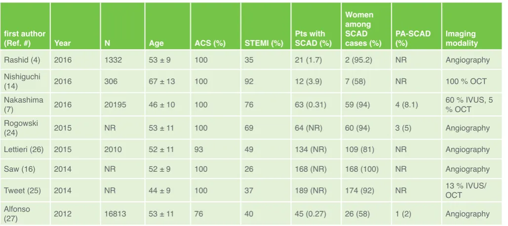
Table 1. Prevalence of SCAD
first author
(Ref. #) Year N Age ACS (%) STEMI (%) Pts with SCAD (%)
Women among SCAD
cases (%) PA-SCAD (%) Imaging modality
Rashid (4) 2016 1332 53 ± 9 100 35 21 (1.7) 2 (95.2) NR Angiography Nishiguchi
(14) 2016 306 67 ± 13 100 92 12 (3.9) 7 (58) NR 100 % OCT
Nakashima
(7) 2016 20195 46 ± 10 100 76 63 (0.31) 59 (94) 4 (8.1) 60 % IVUS, 5 % OCT Rogowski
(24) 2015 NR 53 ± 11 100 69 64 (NR) 60 (94) 3 (5) Angiography Lettieri (26) 2015 2010 52 ± 11 93 49 134 (NR) 109 (81) NR Angiography
Saw (16) 2014 NR 52 ± 9 100 26 168 (NR) 168 (100) NR Angiography
Tweet (25) 2014 NR 44 ± 9 100 37 189 (NR) 174 (92) NR 13 % IVUS/OCT
Alfonso
(27) 2012 16813 53 ± 11 76 40 45 (0.27) 26 (58) 1 (2) Angiography
Generally, conservative treatment is preferred for stable patients without ongoing pain on the basis of expert opinions derived from observational data [1, 24-27]. In the updated Vancouver cohort by Saw et al, 83% (232 of 280 patients) were treated conservatively, and subsequent revascularization in-hospital in this group was only 3.5% (2.9% PCI and 0.6% CABG) [23]. In contrast, patients with ongoing chest pain, ischemia, ST elevation, or hemodynamic instability should be considered for PCI or CABG, especially when the dissection affects major arteries with sizable myocardial jeopardy.
Emergent CABG may be considered if the dissection involves the left main trunk [28]. PCI of dissected coronary arteries can be notoriously challenging. Several series revealed poor technical success with PCI for SCAD. In the Vancouver cohort of 168 patients, PCI was successful or partial successful in only 64% (57% had extension of dissections during PCI, 12% required urgent CABG, and 6% had stent thrombosis), with long-term durable results in only 30% [29]. These results are a consequence of multiple challenges with PCI, as follows. It may be difficult to advance the coronary guidewire into the distal true lumen. The IMH of a dissected segment can also propagate with angioplasty, further decreasing arterial blood flow and extending the dissection. The dissection often involves a distal coronary artery, which is too small for stent implantation. Even if the dissected artery is large, the dissection is often extensive, requiring long stents and thus increasing the risk of restenosis. Furthermore, IMH resorbs and heals in the chronic phase, resulting in late stent malapposition.
Unfortunately, this may increase the risk of very late stent thrombosis [30]. Therefore, when PCI is attempted, patient cardiovascular outcomes rely on stenting strategy. The additional resolution from OCT imaging allows confirmation of the coronary guidewire position before placement of stents and shows the distal exit of the dissection and the intimal flap clearly [31, 32]. Thus, OCT is more helpful to determine PCI strategy in SCAD.
Because OCT provides detailed information about the exact location of the entry site, some experts report a single stent strategy that seals the entry site, thereby avoiding mid-term in-stent re-stenosis possibly induced by implantation of multiple long stents. The ability of OCT to visualize microstructures in real time enables the procedure to be carried out precisely. [33] On the other hand, some experts recommend a strategy of longer stents. Even though the lesion is relatively focal, they recommend longer stents, which provide adequate coverage for both edges of the lesion. This strategy has the advantage of accommodating extension of the IMH proximally and distally when compressed by the stent. As well as the single stent strategy, OCT can ensure adequate stent coverage and wall apposition.
Additionally, for longer lesions, a multistep approach of stenting the distal edge, followed by the proximal edge, and then stenting the middle of the dissection, may be useful in preventing IMH propagation [34]. Moreover, there have also been recent successful case reports of using cutting balloons under OCT guidance, which fenestrate the IMH to allow decompression of the false lumen into true lumen. In one of the cases, a 2.5 mm Table 2. Advantages and disadvantages of optical coher-ence tomography over angiography for SCAD imaging
Advantages Disadvantages
Definitive diagnosis of SCAD More invasive examination
Confirm true lumen entry Require anticoagulation, increasing a bleeding risk
Facilitate stent sizing Costly Confirm adequate stent
apposition Not available in all hospitals Confirm full coverage of
dissected segment A risk of extending dissection size by imaging itself
SCAD = spontaneous coronary artery dissection.
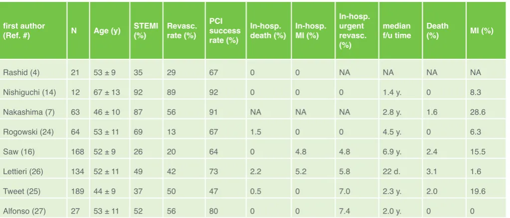
Table 3. Outcomes in Contemporary SCAD Series
first author
(Ref. #) N Age (y) STEMI (%) Revasc. rate (%) PCI success rate (%)
In-hosp.
death (%) In-hosp. MI (%)
In-hosp. urgent revasc. (%)
median
f/u time Death (%) MI (%)
Rashid (4) 21 53 ± 9 35 29 67 0 0 NA NA NA NA
Nishiguchi (14) 12 67 ± 13 92 89 92 0 0 0 1.4 y. 0 8.3
Nakashima (7) 63 46 ± 10 87 56 91 NA NA NA 2.8 y. 1.6 28.6
Rogowski (24) 64 53 ± 11 69 13 67 1.5 0 0 4.5 y. 0 6.3
Saw (16) 168 52 ± 9 26 20 64 0 4.8 4.8 6.9 y. 2.4 15.5
Lettieri (26) 134 52 ± 11 49 42 73 2.2 5.2 5.8 22 d. 3.1 1.6
Tweet (25) 189 44 ± 9 37 50 47 0.5 0 7.0 2.3 y. 2.0 19.6
Alfonso (27) 27 53 ± 11 52 56 80 0 0 7.4 2.0 y. 0 0
cutting balloon was dilated to 2 atm in the distal segment and 4 atm in the proximal segment. After ballooning, coronary flow was immediately restored to TIMI flow grade 3. OCT confirmed that the incisions on the dissected intima corresponded with the blade shape and that communications were successfully made between the false and true lumens [35]. In addition, considering frequent spontaneous healing of coronary dissection and a higher risk of complications with PCI in the setting of SCAD, bioresorbable vascular scaffolds (BVS) may offer more potential advantages over conventional stents. However, this state-of-the-art approach should be discussed [36].
Cardiovascular outcomes
The acute mortality rate in hospital was <5%, and in-hospital recurrent MI, need for urgent revascularization in conservatively managed patients, or other MACEs accounted for 5-10% [37]. Long-term recurrent SCAD rates after up to 5 years were
reported at ~27% [4, 7, 16, 25, 38, 39]. Some reports stated that long-term MACE rates were 15% to 37% at 5 to 7 years [7, 16, 24, 26]. Coronary imaging could alter the prognosis of SCAD. In our recent study, the long-term outcome of OCT-guided PCI for SCAD has been revealed. The prognosis of OCT-guided PCI SCAD is comparable with those of PCI for atherosclerosis lesions. Further study is needed to establish the efficacy of OCT-guided PCI for SCAD [38].
Conclusions
Coronary angiography is unable to establish an accurate diagnosis of SCAD, even in patients with strong clinical suspicion. OCT provides unique and specific insights on most relevant morphologic features of the condition, including entry tear, flap, double-lumen morphology, intramural hematoma, and associated thrombus. The ability of OCT to visualize microstructures in real time enables the procedure for SCAD to be carried out precisely, and that approach could improve the prognosis of SCAD.
Declarations of Interest
The authors declare no conflict of interest.
Acknowledgements
The authors state that they abide by the authors’ responsibilities and ethical publishing guidelines of the International Cardiovascular Forum Journal [40].
References
1. Saw J. Spontaneous coronary artery dissection. Can J Cardiol. 2013;29:1027-33. DOI:10.1016/j.cjca.2012.12.018.
2. Vanzetto G, Berger-Coz E, Barone-Rochette G, et al. Prevalence, therapeutic management and medium-term prognosis of spontaneous coronary artery dissection: results from a database of 11,605 patients. Eur J Cardiothorac Surg. 2009;35:250-4. DOI:10.1016/j.ejcts.2008.10.023. 3. Tweet MS, Gulati R, Aase LA, et al. Spontaneous coronary artery dissection:
a disease-specific, social networking community-initiated study. Mayo Clin Proc. 2011;86:845-50. DOI:10.4065/mcp.2011.0312.
4. Rashid HN, Wong DT, Wijesekera H, et al. Incidence and characterization of spontaneous coronary artery dissection as a cause of acute coronary syndrome - a single-center Australian experience. Int J Cardiol. 2016;202:336-8. DOI:10.1016/j.ijcard.2015.09.072.
5. Elkayam U, Jalnapurkar S, Barakkat MN, et al. Pregnancy-associated acute myocardial infarction: a review of contemporary experience in 150 cases between 2006 and 2011. Circulation. 2014;129:1695-702. DOI:10.1161/ CIRCULATIONAHA.113.002054.
6. Saw J, Aymong E, Mancini GBJ, et al. Non- atherosclerotic coronary artery disease in young women. Can J Cardiol. 2014;30:814-9. DOI:10.1016/j. cjca.2014.01.011.
7. Nakashima T, Noguchi T, Haruta S, et al. Prognostic impact of spontaneous coronary artery dissection in young female patients with acute myocardial infarction: a report from the Angina Pectoris-Myocardial Infarction Multicenter Investigators in Japan. Int J Cardiol. 2016;207:341-8. DOI:10.1016/j.ijcard.2016.01.188.
8. Tweet MS, Hayes SN, Gulati R, et al. Clinical features, management, and prognosis of spontaneous coronary artery dissection. Circulation. 2012;126:579-88. DOI:10.1161/CIRCULATIONAHA.112.105718.
9. Fahmy P, Prakash R, Saw J, et al. Pre-disposing and precipitating factors in men with spontaneous coronary artery dissection. JACC Cardiovasc Interv. 2016;9:866–8. DOI:10.1016/j.jcin. 2016.02.024.
10. Maehara A, Mintz GS, Castagna MT, et al. Intravascular ultrasound assessment of spontaneous coronary artery dissection. Am J Cardiol. 2002;89:466-8. DOI:10.1016/S0002-9149(01)02272-X.
11. Alfonso F, Paulo M, Gonzalo N, et al. Diagnosis of spontaneous coronary artery dissection by optical coherence tomography. J Am Coll Cardiol. 2012;59:1073-9. DOI:10.1016/j.jacc.2011.08.082.
12. Paulo M, Sandoval J, Lennie V, et al. Combined use of OCT and IVUS in spontaneous coronary artery dissection. JACC Cardiovasc Imaging. 2013;6:830-2. DOI:10.1016/j.jcmg.2013.02.010.
13. Saw J, Poulter R, Fung A. Intracoronary imaging of coronary fibromuscular dysplasia with OCT and IVUS. Catheter Cardiovasc Interv. 2013;82:e879-83. DOI:10.1002/ccd.24640.
c) b)
Figure 1. Combined imaging in a patient with spontaneous coronary artery dissection
a. Angiographic image of a SCAD lesion in the right coronary artery
b. Intravascular Ultrasound (IVUS) findings. IVUS image showing false lumen with intramural hematoma (IMH) (⁕).
c. Optical Coherence Tomography (OCT) images. OCT image showing false lumen with intramural hematoma (IMH) (⁕) and intimal rupture (arrow).
14. Nishiguchi T, Tanaka A, Ozaki Y, et al. Prevalence of spontaneous coronary artery dissection in patients with acute coronary syndrome. Eur Heart J Acute Cardiovasc Care. 2016;5(3):263-70. DOI:10.1177/2048872613504310. 15. Saw J, Ricci D, Starovoytov A, et al. Spontaneous coronary artery
dissection: prevalence of predisposing conditions including fibromuscular dysplasia in a tertiary center cohort. JACC Cardiovasc Interv. 2013;6:44-52. DOI:10.1016/j.jcin.2012.08.017.
16. Saw J, Aymong E, Sedlak T, et al. Spontaneous coronary artery dissection: association with predisposing arteriopathies and precipitating stressors and cardiovascular outcomes. Circ Cardiovasc Interv. 2014;7:645-55. DOI:10.1161/CIRCINTERVENTIONS.114.001760.
17. Maehara A, Mintz GS, Castagna MT, et al. Intravascular ultrasound assessment of spontaneous coronary artery dissection. Am J Cardiol. 2002;89:466-8. DOI:0.1016/S0002-9149(01)02272-X.
18. Rashid HN, Wong DT, Wijesekera H, et al. Incidence and characterization of spontaneous coronary artery dissection as a cause of acute coronary syndrome - a single-center Australian experience. Int J Cardiol. 2016;202:336-8. DOI:10.1016/j.ijcard.2015.09.072.
19. Mortensen KH, Thuesen L, Kristensen IB, et al. Spontaneous coronary artery dissection: a Western Denmark Heart Registry study. Catheter Cardiovasc Interv. 2009;74:710-7. DOI:10.1002/ccd.22115.
20. Vrints CJ. Spontaneous coronary artery dissection. Heart. 2010;96:801-8. DOI:10.1136/hrt.2008.
21. Low AF, Tearney GJ, Bouma BE, Jang IE. Technology insight: optical coherence tomography - current status and future development. Nat Clin Pract Cardiovasc Med. 2006; 3(3):154-62. DOI:10.1038/ncpcardio0482 22. Kume T, Akasaka T, Kawamoto T, et al. Assessment of coronary intimamedia
thickness by optical coherence tomography: comparison with intravascular ultrasound. Circ J. 2005;69(8):903-9. DOI:10.1253/circj.69.903
23. Saw J, Prakash R, Starovoytov A, et al. Cardiovascular outcomes in a large prospectively followed single-center cohort of spontaneous coronary artery dissection patients. J Am Coll Cardiol. 2016;67(13):457. DOI: 10.1016/ S0735-1097(16)30458-2
24. Rogowski S, Maeder MT, Weilenmann D, et al. Spontaneous coronary artery dissection: angiographic follow-up and long-term clinical outcome in a predominantly medically treated population. Catheter Cardiovasc Interv. 2017;89(1):59-68. DOI:10.1002/ccd.26383.
25. Tweet MS, Eleid MF, Best PJ, et al. Spontaneous coronary artery dissection: revascularization versus conservative therapy. Circ Cardiovasc Interv. 2014;7:777-86. DOI:10.1161/CIRCINTERVENTIONS.114.001659.
26. Lettieri C, Zavalloni D, Rossini R, et al. Management and long-term prognosis of spontaneous coronary artery dissection. Am J Cardiol. 2015;116:66-73. DOI:10.1016/j.amjcard.2015.03.039.
27. Alfonso F, Paulo M, Lennie V, et al. Spontaneous coronary artery dissection: long-term follow-up of a large series of patients prospectively managed with a “conservative” therapeutic strategy. JACC Cardiovasc Interv. 2012;5:1062-70. DOI:10.1016/j.jcin.2012.06.014.
28. Shamloo BK, Chintala RS, Singh SN, et al. Spontaneous coronary artery dissection: aggressive vs. conservative therapy. J Invasive Cardiol. 2010;22:222–8.
29. Saw J. Coronary angiogram classification of spontaneous coronary artery dissection. Catheter Cardiovasc Interv 2014;84:1115-22. DOI:10.1002/ ccd.25293.
30. Lempereur M, Fung A, Saw J. Stent malapposition with resorption of intramural hematoma with spontaneous coronary artery dissection. Cardiovasc Diagn Ther. 2015;5:323-9. DOI:0.3978/j.issn.2223-3652.2015.04.05.
31. Poon K, Bell B, Raffel OC, Walters DL, Jang IK, et al.Spontaneous coronary artery dissection: utility of intravascular ultrasound and optical coherence tomography during percutaneous coronary intervention. Circ Cardiovasc Interv. 2011;4(2):e5-7. DOI:10.1161/CIRCINTERVENTIONS.110.959593. 32. Satogami K, Ino Y, Akasaka T, et al. Successful stenting with optical
frequency domain imaging guidance for spontaneous coronary artery dissection. JACC Cardiovasc Interv. 2015;8(6):e83-5. DOI:10.1016/j. jcin.2014.12.247.
33. Nakagawa M, Shite J, Shinke T, Otake H, Okada K, Okita Y, Hirata K. Ability of optical coherence tomography to visualize the entry port of spontaneous coronary artery dissection. Circ J. 2011;75(10):2505-7. DOI:10.1253/circj. CJ-11-0329
34. Walsh S, Jokhi P, Saw J. Successful percutaneous management of coronary dissection and extensive intramural hematoma associated with ST elevation MI. Acute Cardiac Care. 2008;10:231-3. DOI:10.1080/17482940701802348. 35. Yumoto K, Sasaki H, Aoki H, et al. Successful treatment of spontaneous coronary artery dissection with cutting balloon angioplasty as evaluated with optical coherence tomography. J Am Coll Cardiol Intv. 2014;7:817-9. DOI:10.1016/j.jcin.2013.10.027.
36. Mahmood MM, Austin D. IVUS and OCT guided primary percutaneous coronary intervention for spontaneous coronary artery dissection with bioresorbable vascular scaffolds. Cardiovasc Revasc Med. 2017;18(1):53-7. DOI:10.1016/j.carrev.2016.09.005.
37. Thompson EA, Ferraris S, Gress T, Ferraris V. Gender differences and predictors of mortality in spontaneous coronary artery dissection: A review of reported cases. J Invasive Cardiol. 2005;17:59-61.
38. Nishiguchi T, Tanaka A, Akasaka T. Prognosis of spontaneous coronary artery dissection treated by percutaneous coronary intervention with optical coherence tomography. J Cardiol. 2017;70(6):524-9. DOI:10.1016/j. jjcc.2017.03.009.
39. Hill SF, Sheppard MN. Non-atherosclerotic coronary artery disease associated with sudden cardiac death. Heart. 2010 Jul;96(14):1119-25. DOI:10.1136/hrt.2009.185157.