Journal of Global Pharma Technology
Available Online at:
www.jgpt.co.in
RESEARCH ARTICLE
Targeted Systems in Cancer Treatment
Yuliya Tikhonova
1*,Shengnu Qiu
21. Sechenov First Moscow State Medical University (Sechenov University), Moscow, Russian Federation.
2. University College London, London, United Kingdom.
*Corresponding Author: Yuliya Tikhonova
Abstract
The World Health Organization (WHO) estimates that in 2015 cancer was the first or second leading death cause before the age of 70 in 91 out of 172 countries, and ranked third or fourth in the other 22. This study analyzes three main types of targeted cancer therapies: low molecular weight inhibitors; monoclonal antibodies; and immunotoxins. The low molecular weight inhibitors analyzed in the paper are Gleevec, Sprycel, VEGF targeting, Nexavar, Tykerb, Iressa, EGFR and HER2 Targeting, Torisel. The monoclonal antibodies include VEGF, EGFR and HER2, Herceptin, Avastin, CD20, Rituxan, Zevalin, Campath. The DAB389IL-2 (or Ontak - FDA Approved Medication for the Treatment of Acute T Cell Lymphoma) represents immunotoxins. It proves that depending on the location of the unregulated protein, different approaches are implemented. Monoclonal antibodies can block interactions and functions of unregulated proteins within the extracellular compartment, while low molecular weight inhibitors can reach proteins that are within the cell compartment. Currently, many of these drugs are used in combination with traditional cancer treatments to increase patient survival. As it was found, VEGF inhibitors, for example, Avastin, increased in low concentrations the survival time of patients with metastatic colorectal cancer from 13.8 to 21.5 months.
Keywords: Low molecular weight inhibitors, Gleevec, Pro-angiogenic molecules, Monoclonal antibodies.
Introduction
Cancer morbidity and mortality are rising rapidly around the world. The oncological situation remains unfavorable in both developed and developing countries. Every year, at least 12 million new cases are registered around the world. About 80% of them require chemotherapy (CT) either in an independent or in an adjuvant regimen [1, 2].At the beginning of our millennium, an active study of the CT optimization for malignant tumors treatment and the selection of the best modes and combinations continues.
However, the prognosis of CT sensitivity is very difficult and possible only for certain groups of patients (and for specific cases, it is practically unrealistic). Toxicity due to exposure to targets common with the tumor in the patient's body (bone marrow cells, mucous membranes, etc.) limits the use of many cytostatic [3, 5]. A serious CT limit is its focus on damage to the genome and/or the apparatus of dividing tumor cells.
Tumor cells are known for genome instability, which leads to increased resistance to anticancer drugs. The use of “new” cytostatic with the old mechanism of action rarely leads to significant success [6, 8]. The fundamental achievement of the last decade is the creation of a new class of “target molecular-oriented drugs” aimed to affect certain, predetermined intracellular molecular targets that are of key importance for the life of a tumor cell. All these targets are present in normal cells, but during tumor transformation, their overexpression or hyperactivation may occur, which is the basis for the use of certain targeted drugs.
modulate the same pathway, there are drugs that will be therapeutically effective for specific types of cancer. The following discussion will focus on both low molecular weight inhibitors and monoclonal antibodies for each target protein, whereas possible.
Targeted cancer therapy is the use of drugs or other substances that block cancer growth and spread. They affect certain molecules (“molecular targets”) that are involved in the growth, progression and spread of cancer. Targeted cancer treatments are sometimes referred to as “molecules with effects on molecules,” “therapy with effects on molecules,” “checkpoint drugs”, or similarly.
Targeted therapy differs from standard chemotherapy according to several criteria:
Targeted therapy affects specific molecular
targets associated with cancer, while most standard CT affects all rapidly dividing normal and cancer cells;
Targeted therapy is specifically selected or
designed to interact with its target, whereas many standard CT have been chosen for their ability to demolish cells;
Targeted therapy is often cytostatic (i.e.,
blocks tumor cells proliferation), while standard chemotherapeutic agents are cytotoxic (i.e., they kill tumor cells).
Targeted therapy is currently the focus of attention in the development of many new anticancer drugs. As a cornerstone of precision medicine, it uses information about human genes and proteins for the prevention, diagnosis, and treatment of diseases.
Most targeted cancer treatments are at various stages of preclinical and clinical trials for use alone or in combination with other protocols, although the US Food and Drug Administration (FDA) has approved over 15 targeted cancer therapies since 2000.
Scientists expected targeted cancer therapy
to be less toxic than traditional
chemotherapeutic drugs because cancer cells are more target-dependent than normal cells. However, targeted cancer therapy can have significant side effects.The most common observed side effects with targeted therapy are diarrhea and liver problems, such as hepatitis and elevated liver enzymes level. Other side effects include:
Skin problems (acne, dry skin, nail
changes, hair depigmentation);
Problems with blood coagulation and
wound healing;
High blood pressure;
Gastrointestinal perforation (a rare side
effect of some targeted treatments).
Some side effects of targeted therapies have been associated with the best patient outcomes. For example, patients who developed an acneiform rash (skin rashes resembling acne) during treatment with signal transduction inhibitors erlotinib (Tarceva) or gefitinib (Iressa), targeting the epidermal growth factor receptor, usually showed better response than those with no rash [11, 12].
Similarly, patients who developed high blood pressure during treatment with angiogenesis inhibitor bevacizumab generally had better results [13, 14]. A few targeted treatments approved for pediatric use may have various
side effects in adults, including
immunosuppression and impaired sperm production [15, 16].
Methods
The study analyzes three main types of targeted cancer therapies:
Low molecular weight inhibitors;
Monoclonal antibodies;
Immunotoxins.
Results
Low Molecular Weight Inhibitors
Inappropriate kinase activity is the main route through which cells become cancerous, avoiding the limitations of the normal cell cycle. Low molecular weight inhibitors bind competitively to an active or inactive ATP tyrosine kinase binding site.
They are used to target proteins that have become either unregulated or activated during cancer progression, such as BCR-ABL, Akt or mTOR (Table 1). Once these molecules bind to their specific target, they inactivate the tyrosine kinase domain and prevent the downstream signaling pathways. These drugs are very useful when administered to
patients in combination with
Besides, more than 20 different kinase targets are being developed in clinical trials [17, 18].
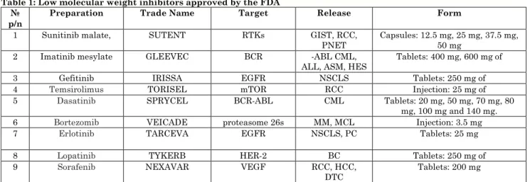
Table 1: Low molecular weight inhibitors approved by the FDA №
p/n
Preparation Trade Name Target Release Form
1 Sunitinib malate, SUTENT RTKs GIST, RCC,
PNET Capsules: 12.5 mg, 25 mg, 37.5 mg, 50 mg 2 Imatinib mesylate GLEEVEC BCR -ABL CML,
ALL, ASM, HES
Tablets: 400 mg, 600 mg of
3 Gefitinib IRISSA EGFR NSCLS Tablets: 250 mg of
4 Temsirolimus TORISEL mTOR RCC Injection: 25 mg of
5 Dasatinib SPRYCEL BCR-ABL CML Tablets: 20 mg, 50 mg, 70 mg, 80
mg, 100 mg and 140 mg.
6 Bortezomib VEICADE proteasome 26s MM, MCL Injection: 3.5 mg
7 Erlotinib TARCEVA EGFR NSCLS, PC Tablets: 25 mg
8 Lopatinib TYKERB HER-2 BC Tablets: 250 mg of
9 Sorafenib NEXAVAR VEGF RCC, HCC,
DTC Tablets: 200 mg
Note: RTKs - multiple tyrosine kinase receptors. GIST - gastrointestinal stromal tumors.RCC - renal cell carcinoma. pNET - pancreatic neuroendocrine tumors.CML - chronic myeloid leukemia. ALL - acute lymphoblastic leukemia. ASM - aggressive systemic mastocytosis. HES - gipereozinofilny syndrome. CEL - chronic eosinophilic leukemia. DFSP - dermatofibrosarcoma protuberans. EGFR - epidermal growth factor. NSCLS - non-small cell lung cancer.MM - Multiple Myeloma. MCL - mantle cell lymphoma. PC - pancreatic cancer. BC - breast cancer. HCC - hepatocellular carcinoma.DTC - differentiated thyroid carcinoma.
Targeting BCR-ABL
Drugs that target the chimeric BCR-ABL protein were one of the biggest success stories of target drug therapy for cancer. BCR-ABL is a clinically relevant drug target. This chimeric protein is usually found in almost all patients with chronic myeloid leukemia (СML). Currently, two new-generation Novartis and Sprycel low molecular mass inhibitors with a higher affinity for BCR-ABL than Gleevec have been created in the US market.
Gleevec
Gleevec (imatinib mesylate, Novartis) is a low molecular weight inhibitor used to treat several different types of cancer, including chronic myeloid leukemia with a positive response to the Philadelphia chromosome (Ph+ CML) and c-kit-positive gastrointestinal stromal tumors. Gleevec targets the BCR-ABL tyrosine kinase domain, although it has minimal effect on other tyrosine kinases. In CML, Gleevec inactivates the tyrosine kinase region of the chimeric BCR-ABL protein, which is a result of chromosome translocation [19, 16].
Translocation occurs when the q34 region of chromosome 9, containing ABL, merges with the q11 region of chromosome 22, containing BCR [11, 12]. This combination is called the Philadelphia (Ph) chromosome and results in an unregulated ABL tyrosine kinase domain during CML [9, 10]. Gleevec competitively binds to BCR-ABL at the ATP site, which leads to disruption of the tyrosine kinase
potential [17, 21]. Since this mutation occurs in almost all patients with CML, treatment of Gleevec results in a complete response in
approximately 98% of patients. In GIST
patients, the c-kit protein mutates, which leads to abnormal growth of GIST-cells. Gleevec is localized in the tyrosine phosphorylation site in the c-kit and competitively inhibits ATP binding to the site, effectively blocking the phosphorylation of the substrate for c-kit and leading to the termination of the signaling cascade [10, 8].
Patients with GIST usually have poor prognostic indicators due to the minimal response from radiotherapy [3, 8]. Although Gleevec is used to treat patients with GIST, there are currently no studies that would demonstrate that Gleevec improves patient symptoms or increases patient survival.
Sprycel
Sprycel (Dasatinib, Bristol-Myers Squibb) is a second-generation low molecular weight inhibitor that, like Gleevec, inhibits ATP binding in the BCR-ABL tyrosine kinase domain. However, Sprycel is recommended for use in Ph + CML patients who develop Gleevec resistance.
range of proteins than Gleevec, which leads to a marked reduction in the potential of drug resistance. In addition to using Ph+ CML as a treatment, Sprycel is also recommended for use for Ph+ acute lymphoblastic leukemia (ALL).
VEGF Targeting
VEGF is one of the most studied pro-angiogenic molecules, and it plays a role in several stages of the angiogenic process, both during development and in disease. The VEGF family consists of VEGF-A, VEGF-B, VEGF-C, VEGF-D, VEGF-E and placental growth factor (PlGF). VEGF-A consists of six isoforms (VEGF121, VEGF145, VEGF165, VEGF183, VEGF189, and VEGF206) due to alternative splicing from a single gene. There are also three separate receptors, including the VEGF-1 receptor (VEGFR-1; Flt-1), VEGFR-2 (KDR/Flk-1) and VEGFR-3 (Flt-4).
Receptors are transmembrane tyrosine kinases located on the surface of various cell types, including endothelial cells. Such receptors dimerize and become activated as a result of transphosphorylation after ligand binding. Activation of VEGFR-2 leads to an increase in vascular permeability and migration, as well as an increase in proliferation. These factors make it an ideal target for therapeutic intervention. Indeed, there are numerous low molecular weight inhibitors targeting VEGFR in various phases of preclinical and clinical trials [17, 18].
Nexavar
Nexavar (sorafenib, Bayer HealthCare) is a low molecular weight inhibitor that targets both VEGFR-2 and VEGFR-3, and also c-kit and PDGFR-P [15, 11].Nexavar is currently FDA approved for the treatment of inoperable hepatocellular carcinoma and progressive renal cell carcinoma (RCC). Nexavar can inhibit the RAF/MEK/ERK pathway by interacting with the kinase domain of VEGFR-2, VEGFR-3, c-kit, and PDGFR-P [11, 18].
These pathways are not regulated in cancer by inhibiting the pathway in the treatment of liver and kidney cancer. However, one of the common side effects of the VEGFR signaling pathway inhibitors is impaired wound healing, hypertension, skin rashes, and thrombosis.
EGFR and HER2 Targeting
The epidermal growth factor receptor (EGFR) is a transmembrane receptor involved in a wide range of cellular processes, including
cell growth, proliferation, survival,
migration, and invasion into tissues. The receptor consists of three separate domains, the extracellular ligand-binding region, the
transmembrane domain, and the
intracellular domain.
The binding of activating ligands induces receptor dimerization, leading to activation of
the tyrosine kinase domain,
autophosphorylation and subsequent
activation of downstream signaling
pathways. Expression and activity of EGFR are not regulated in most human epithelial tumors and, for this reason, is an attractive target for the development of new cancer treatments. Human epidermal growth factor-2 (HERfactor-2) is over-expressed in breast carcinomas and is associated with more aggressive forms of cancer.
Most likely, this is associated with increased
cell proliferation, angiogenesis, and
invasiveness of cancer cells. Overexpression of HER2 is also associated with anthracycline sensitivity and endocrine resistance. These data suggest that there are two different pathways in breast carcinomas: the pathway of tyrosine kinase receptor and the hormone receptor pathway. Because of this, breast carcinomas have one pathway sensitive to chemotherapeutic drugs, and a separate pathway sensitive to antiestrogens, which makes HER2 an ideal protein for targeted cancer therapy.
Iressa
These EGFR mutations result in higher sensitivity to treatment with either gefitinib or erlotinib (Tarceva), which also inhibits the activation and signaling of EGFR
Tykerb
Tykerb (lapatinib, GlaxoSmithKline) is a low molecular weight inhibitor that is specific for EGFR and HER2 and shows a higher affinity for both targets than Iressa [15, 16]. Instead of preventing the dimerization of EGFR and HER2, it inhibits ATP binding in the cleavage that binds HER2 and prevents the subsequent phosphorylation cascade [11, 16]. Thus, Tykerb causes PI3K to be blocked and Ras/MAPK directs and prevents the activation of downstream targets such as c-Myc, c-jun, and c-fos.
Tykerb is usually prescribed to patients who have stopped responding to treatment with Herceptin [22, 24]. Herceptin (trastuzumab; monoclonal antibody) is administered as the primary treatment since Herceptin has an increased risk of drug resistance. Tykerb is FDA approved for use in conjunction with
Xeloda (capecitabine), an oral
chemotherapeutic agent commonly used to treat metastatic breast tumors exhibiting over expression of HER2 [22, 23].
MTOR Targeting
mTOR is a kinase protein involved in the regulation of cell proliferation and growth. It was also shown that it plays a role in
catabolic processes and cytoskeleton
dynamics. Indeed, mTOR is a key convergence point for several signaling pathways, including phosphatase homolog (PTEN), which is a tumor suppressor.
The loss of PTEN function results in increased Akt/PKB and mTOR activation. Activation of these pathways leads to increased protein synthesis and penetration into the cell cycle. The other product of this pathway is the increased expression of hypoxia-induced factor-1a (HIF-1a) that blocks the mTOR pathway with angiogenesis [11, 8, 18].
PI3K/Akt/mTOR pathway is increased greatly with multiple cancers, including cancer of bladder, brain, breast, lungs, prostate, melanoma, renal cells and thyroid
gland [11, 18]. Normally inside the cells, the growth factors activate PI3K, but in the case of cancer cells, they can stimulate this pathway with no need for growth factors. As soon as mTOR (that lays below Akt) is
activated, this activates D-cyclin E
expression, the critical factor in cell cycle progression [11, 16, 12]. These data suppose that targeting mTOR can be used more widely in cancer treatment.
Torisel
Torisel (Temsirolimus, Wyeth Research) - is a low molecular inhibitor targeting mTOR, that was accepted for the treatment of progressing renal cell cancer (RCC) [24, 14]. During phase III of clinical trials administration of Torisel led to an increase in average survival rate (from 10.9 months to 7.3 months) comparing to patients taking interferon-alpha-2a (Roferon-A, Roche) [4, 7]. For patients taking both medications, the average survival rate decreased from 10.9 to 8.4 months. The reasons for this observed decrease in survival remain unresolved.
Torisel side effects are similar to other cancer treatments, such as skin rashes, nausea,
fatigue, and fluid retention. Certican
(everolimus, Novartis), a drug similar to Torisel, was first used in conjunction with organ transplantation (heart and kidney) to suppress the immune system and prevent rejection of the transplanted organ [11, 18].
Since these early studies, Certican has been shown to be another potent inhibitor of renal
cell carcinoma progression. Indeed,
everolimus (Afinitor; oral pills) has only recently received approval for the treatment of patients with RCC with no cancer response after treatment with alternative therapy.
Monoclonal Antibodies
One way to target specifically a protein that is deregulated during oncogenesis is to obtain a monoclonal antibody (Ab) against the
target protein. Researchers have created
monoclonal Ab that targets a wide range of proteins. They are involved in specific cancers (Table 2). There are three main mechanisms of cancer cell dysfunction; protein dysfunction and possible subsequent
signaling, antibody and
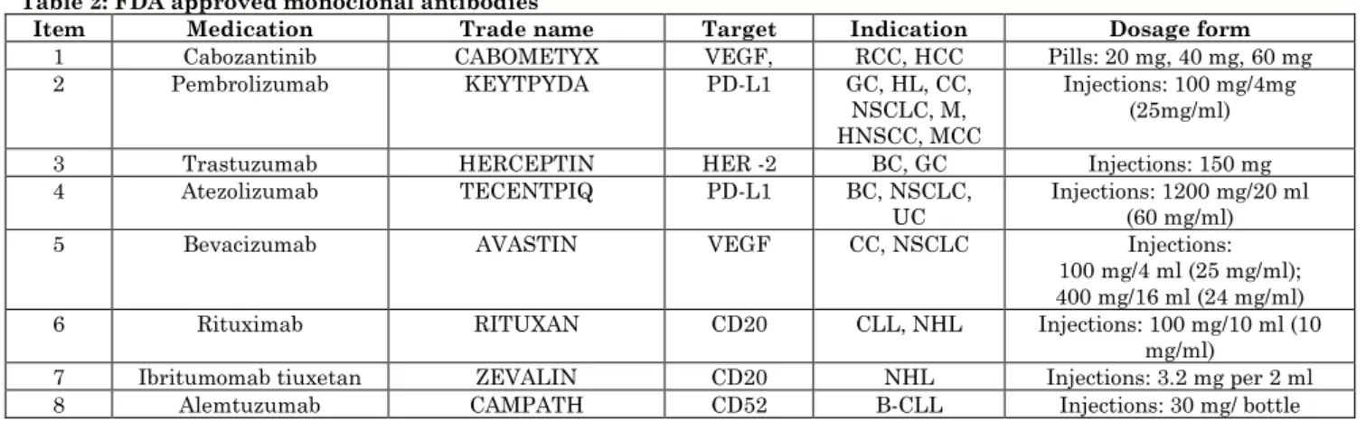
Table 2: FDA approved monoclonal antibodies
Item Medication Trade name Target Indication Dosage form
1 Cabozantinib CABOMETYX VEGF, RCC, HCC Pills: 20 mg, 40 mg, 60 mg
2 Pembrolizumab KEYTPYDA PD-L1 GC, HL, CC,
NSCLC, M, HNSCC, MCC
Injections: 100 mg/4mg (25mg/ml)
3 Trastuzumab HERCEPTIN HER -2 ВС, GC Injections: 150 mg
4 Atezolizumab TECENTPIQ PD-L1 BC, NSCLC,
UC Injections: 1200 mg/20 ml (60 mg/ml)
5 Bevacizumab AVASTIN VEGF CC, NSCLC Injections:
100 mg/4 ml (25 mg/ml); 400 mg/16 ml (24 mg/ml) 6 Rituximab RITUXAN CD20 CLL, NHL Injections: 100 mg/10 ml (10
mg/ml)
7 Ibritumomab tiuxetan ZEVALIN CD20 NHL Injections: 3.2 mg per 2 ml
8 Alemtuzumab CAMPATH CD52 В-CLL Injections: 30 mg/ bottle
Note: CC - cervical cancer, GC - gastric cancer, HL – Hodgkin’s lymphoma, NSCLC - non-small cell lung cancer, M - melanoma, HNSCC - head and neck squamous cell carcinoma, MCC - Merkel cell carcinoma, HCC - hepatocellular carcinoma, UC - urothelial carcinoma, BC - breast cancer, CLL - chronic lymphocytic leukemia, NHL - B-cell non-Hodgkin’s lymphoma, B-CLL - B-cell chronic lymphocytic leukemia
In addition to traditional cancer treatments, monoclonal Ab is currently the most widely used form of cancer treatment. This type of therapy uses a large amount of Ab produced outside the body instead of those produced by the patient’s own immune system. The first monoclonal Ab was obtained in the laboratory by fusing a mouse cancer cell (myeloma) with a mouse B-cell, creating a hybridoma.
All the original monoclonal Ab were obtained
using mouse cells, whereas modern
antibodies are either humanized or fully human, which makes them safer and more effective for treating people. Over the past decade, multiple monoclonal Ab has received FDA approval for the treatment of a wide range of cancer types, as described below. In addition, there are numerous preclinical and clinical trials involving monoclonal Ab for almost every type of cancer.
VEGF
Low doses of anti-VEGF monoclonal Ab were highly effective in treating cancer in combination with traditional CT [17, 18, 12]. Unexpectedly, high Ab doses were proved to reduce medication effectiveness and reduce the patient’s life expectancy. This harmful effect of high doses is explained as the drug inhibits the vascular network in the tumor and interferes with the chemotherapeutic drug delivery to the tumor. However, another possibility is that higher Ab doses cause an increase of VEGF expression, leading to ineffective treatment. At the same time, lower doses of antibodies to VEGF (Avastin) cause a decrease in intratumoral pressure
and provide better delivery of
chemotherapeutic agents.
Avastin
Avastin (bevacizumab, Genentech) is a monoclonal antibody targeted to VEGF. VEGF is a secreted glycoprotein that is involved in angiogenesis and is not highly regulated by cancer cells in the tumor microenvironment. VEGF levels correlate with poor survival outcomes and are among the causes of the potential anti-cancer benefits of a drug that inhibits VEGF signaling [9, 10]. Signal transmission through VEGF receptors (1, VEGFR-2, and VEGFR-3) activates the Raf-1-MEK-ERK pathway and the PKC-dependent pathway, which leads to targeted growth of endothelial cells [15].
These pathways are involved in numerous
biological processes, including cell
proliferation, differentiation, and apoptosis. Therefore, by blocking VEGF, the effect of ERK and PKC-dependent pathways on the cell is reduced. Avastin is used to treat metastatic breast cancer, colorectal cancer [10, 8]. After the removal of VEGF inhibitors from the patient’s system, tumor growth
increased. In phase II clinical trial (Avastin
with 5-fluorouracil (5-FU)) patients showed an increase in survival compared with a single treatment [11, 16].
EGFR and HER2
EGFR and HER2 are usually activated in metastatic breast cancer and NSCLC. A high expression of HER2 is detected in more aggressive cases of breast cancer.
Herceptin
Herceptin (Trastuzumab, Genentech) is a monoclonal Ab produced against the ectodomain HER2. HER2 is not regulated in approximately 30% of patients with breast cancer, and the levels of HER2 directly correlate with cancer progression [17, 10].
Herceptin binds to HER2 and interferes with receptor dimerization, which disrupts the intracellular signaling cascade that inhibits cell growth. Major downstream pathways blocked include C-y phospholipase (PLC-y), PI3K and Ras/MAPK pathways. In addition, Herceptin is able to increase the level of HER2 endocytosis, which leads to a decrease of HER2 present on the plasma membrane [11, 16].
Besides, Herceptin enhances the immune system's ability to detect cancer cells by binding to overexpressing HER2. Then the immune system, in particular, natural killer cells, detects the antibody and has a cytotoxic effect on cancer cells [19, 16]. Herceptin is useful for HER2-positive patients with breast cancer who have a relapse after an initial course of CT [19, 16]. In addition, clinical studies have shown that the use of Herceptin in combination with an anthracycline (doxorubicin) can increase survival by 20% compared with anthracycline alone [16, 18].
When using two drugs in combination, Herceptin is able to prevent the activation of anti-apoptotic signaling through the PLC-y/Akt pathway and allows doxorubicin to interfere with DNA replication and promote apoptosis. Herceptin side effects include heart dysfunction, skin rash, nausea and fatigue [19, 21].
CD20
Over 90% of B-cell lymphomas demonstrate the expression of cluster differentiation antigen 20 (CD20). By focusing on CD20 for non-Hodgkin B-cell lymphoma, treatment can better affect cancer cells expressing a distinct marker not expressed on normal cells.
Rituxan
In B-cell non-Hodgkin’s lymphoma and chronic lymphocytic leukemia, unregulated growth of B-cells is observed, which demonstrates the CD20 protein on their cell surface [19, 16]. Rituxan (rituximab, Genentech) is a monoclonal antibody targeting CD20 B-lymphocyte antigen and leading to cytotoxicity in both cancer and normal B-cells [16, 21]. Rituxan is FDA approved in combination with CT agents such as cyclophosphamide, doxorubicin, vincristine, and prednisone (CHOP) [16, 18].
In addition, Rituxan is used in combination with methotrexate to treat rheumatoid
arthritis. Rituxan binds its Fab domain to
CD20 on B-cells and induces apoptosis
through antibody-dependent-cellular
cytotoxicity [17, 12].
Once the anti-CD20 antibody binds, the Fc-region of Ab induces the apoptosis of effector cells. Natural killer (NK) cells, monocytes, and eosinophils are able to bind to the Fc-region, causing cytotoxins, granzyme B and perforin release in response to Fc binding, leading to cell apoptosis. Granzyme B cleaves the BH3 death agonist of the interacting domain (Bid), activates caspase-3, and leads to DNA fragmentation by activating carbamoyl phosphate synthetase 2, aspartate transcarbamylase and dihydroorotase (CAD).
Rituxan complement-dependent cytotoxic mechanism works by inducing apoptosis in
B-cells and activating immunological
mechanisms. Rituxan binds to CD20, causing complement activation and apoptosis [19, 16].
An important lesson that Rituxan
demonstrates, is that the unregulated protein involved in the formation of cancer should no longer be a therapeutic or primary target. This drug significantly changes the current paradigm of targeted drug development.
Zevalin
Zevalin (ibritumomab tiuxetan) binds
Hodgkin’s lymphoma due to higher survival rate [16, 20]. Monoclonal Ab labeling with radioisotopes targeting cancer cells allows these conjugates to serve as a kind of immunotoxin. Various toxins can have a long half-life in the body, increasing toxicity for both cancer and normal cells; however, different radioisotopes have a wide range of radiation decay properties to suit different uses for tumors.
CD52
B-cell chronic lymphocytic leukemia (B-CLL) is cancer in which too many lymphocytes accumulate in various organs of the body, including bone marrow, blood, and lymphatic tissues. Both mature T- and B-cells express the surface antigen CD52; however, this marker is not expressed on hematopoietic stem cells.
Campath
Campath (alemtuzumab, Genzyme) is a monoclonal antibody against CD52 antigen. After binding to CD52, Campath allows the immune system to act on cancer cells. Campath is approved for treating B-CLL as a single agent. Side effects include cytopenia, nausea, rashes, and infection [19, 24].
New Drugs Approved by FDA in 2019
In 2019 FDA approved the use of atezolizumab (TECENTRIQ®, Genentech Inc.) in combination with protein-related paclitaxel for adult patients with inoperable locally advanced or metastatic triple negative breast cancer (TNBC), whose tumors express PD-L1 (a membrane protein of the immunoglobulin superfamily, gene product CD274). The approval was based on
IMpassion130 (NCT02425891), a
multicenter, international, double-blind,
placebo-controlled, randomized study that included 902 patients with inoperable locally advanced or metastatic TNBC who had not
previously received chemotherapy for
metastatic disease.
Patients were randomized (1:1) to receive atezolizumab intravenous infusions (840 mg) or placebo on days 1 and 15 of each 28-day cycle, as well as protein-related paclitaxel
(100 mg/m2) administered by intravenous
infusion on days 1, 8 and 15 during the 28-day cycle. The most common side effects (reported in ≥20% of patients) with
atezolizumab with paclitaxel-associated
protein were: alopecia, peripheral
neuropathies, fatigue, nausea, diarrhea, anemia, constipation, cough, headache,
neutropenia, vomiting, and decreased
appetite. The recommended dose of
atezolizumab for patients with TNBC, whose tumors express PD-L1, is 840 mg when administered intravenously for 60 minutes
followed by the addition of 100 mg/m2 of
paclitaxel protein. For each 28-day cycle, atezolizumab is administered on days 1 and
15, and protein-bound paclitaxel is
administered on days 1, 8, and 15 until disease progression or unacceptable toxicity.
In addition, in 2019, injections of
trastuzumab and hyaluronidase for
subcutaneous administration were approved
(Herceptin Hylecta, Genentech Inc.).
Herceptin Hylecta is a combination of trastuzumab, HER2/neu receptor antagonist, and hyaluronidase, endoglycosidase, for the treatment of breast cancer with over expression of HER2. The approval was based
on two randomized trials, HannaH
(NCT00950300) and SafeHER
(NCT01566721). In HannaH, 596 patients with HER2-positive operative or locally
advanced breast cancer, including
inflammatory breast cancer, were
randomized to receive 8 cycles of either
Herceptin Hylecta or intravenous
trastuzumab concurrently with
chemotherapy, and followed by surgery and the continuation of Herceptin Hylecta therapy or intravenous trastuzumab, for an additional 10 cycles.
HannaH demonstrated comparability
between Herceptin Hylecta and IV
trastuzumab, based on the primary
endpoints of the full pathological response and pharmacokinetics. Pathological complete response (PCR) was observed in 118 patients (45.4%) of Herceptin Hylecta and in 107
patients (40.7%) who received IV
trastuzumab administration (95% CI for the difference in PCR: -4.0; 13).
SafeHER - A prospective, non-randomised, multidisciplinary, open-label study with two groups evaluating the overall safety and
tolerability of Herceptin Hylecta in
simultaneously with CT, either without adjuvant CT or in combination with neoadjuvant CT followed by trastuzumab. The most common side effects of Herceptin Hylecta observed in at least 10% of patients were fatigue, arthralgia, diarrhea, reaction at the injection site, upper respiratory tract infection, rash, myalgia, nausea, headache, edema, hyperemia, hyperthermia, cough and pain in limb. The recommended dose of Herceptin Hylecta is 600 mg/10.000 units (600 mg of trastuzumab and 10.000 units of hyaluronidase), administered subcutaneously for about 2-5 minutes once every three weeks.
In 2019, FDA approved trifluridine/tipiracil pills (LONSURF, Taiho Pharmaceutical Co., Ltd.). Which is a fixed combination of trifluridine, an inhibitor of the nucleosides metabolism, and tipiracil, a thymidine phosphorylase inhibitor, for adult patients with metastatic gastric or gastroesophageal junction (GEJ), adenocarcinoma, pretreated with at least two previous lines of chemotherapy, (including fluoropyrimidine, platinum, or taxane or irinotecan, and, if necessary, HER2/neu-targeted therapy).
The approval was based on TAGS
(NCT02500043), an international,
randomized, double-blind, placebo-controlled study on 507 patients with metastatic gastric or GEJ adenocarcinoma, who had previously received at least two chemotherapy lines. Patients were randomized 2:1 to take
Lonsurf (n=337) 35 mg/m2 orally twice a day
on days 1-5 and 8-12 of each 28-day cycle with the best supportive treatment (BSC) or appropriate placebo (n = 170) with BSC until disease progression or unacceptable toxicity.
In the TAGS study, the most frequent side effects or laboratory abnormalities (frequency ≥10%) happened in patients who received Lonsurf that occur with greater frequency than in patients who received placebo were: neutropenia, anemia, nausea, loss of appetite, thrombocytopenia, vomiting, and diarrhea.
The recommended dose and regimen of
Lonsurf is 35 mg/m2, the dose is administered
orally twice a day with meals on days 1 through 5 and days 8 through 12 of each 28-day cycle. In 2019, FDA approved pembrolizumab (KEYTRUDA, Merck) for the adjuvant treatment of melanoma patients
with impaired lymph nodes after complete resection.
The approval was based on
EORTC1325/KEYNOTE - 054
(NCT02362594), a randomized, double-blind, placebo-controlled study in 1019 patients with a fully resected stage IIIA, melanoma IIIB or IIIC. Patients were randomly assigned (1:1) to receive pembrolizumab 200 mg every three weeks or placebo for up to 1
year before disease recurrence or
unacceptable toxicity. 76% of patients received pembrolizumab for 6 months or longer. Pembrolizumab was canceled due to side effects in 14% of patients.
The most frequent side effects (for at least 10% of patients receiving pembrolizumab) were: diarrhea, pruritus, nausea, arthralgia, hypothyroidism, cough, rash, asthenia, influenza-like illness, weight loss, and hyperthyroidism. The recommended dose and
schedule for pembrolizumab for the
melanoma adjuvant treatment is 200 mg IV for 30 minutes every 3 weeks until disease recurrence or unacceptable toxicity for a maximum of 1 year.
In 2019 FDA approved cabozantinib (CABOMETYX, Exelixis, Inc.) for patients
with hepatocellular carcinoma (HCC)
previously treated with sorafenib. The approval was based on CELESTIAL (NCT01908426), a randomized (2:1), double-blind, placebo-controlled, multicenter study in patients with HCC who previously received sorafenib and are with hepatic
insufficiency class A. Patients were
randomized to receive cabozantinib 60 mg orally once a day (n=470) or placebo (n=237) until disease progression or unacceptable toxicity.
The most common side effects in ≥25% of patients who received cabozantinib in clinical trials were (in order of decreasing frequency): diarrhea, fatigue, loss of appetite, palmar and plantar erythrodysesthesia, nausea,
hypertension, and vomiting. The
recommended dose of cabozantinib is 60 mg orally, once a day, at least one hour before or 2 hours after a meal.
Immunotoxins
Initially, immunotoxins were obtained by chemically binding a toxin to a protein. Now they are produced by creating recombinant DNA that generates a fusion protein. This protein contains the toxin as an additional Ab domain or growth factor.
The immunotoxin binds to the cell surface protein and internalizes through clathrin-coated pits. After internalization, the toxin causes apoptosis of the target cell. Bound enzymes are highly toxic, and only one protein is required for the cytosol to cause an apoptotic effect. Plant and bacterial toxins for use in immunotoxins include diphtheria toxin (DT), antiviral protein (PAP), exotoxin A
(PE), ricin and saponin from Pseudomonas
[11, 18, 12].
Ontak
Ontak (denileukin diphitox, Ligand
Pharmaceuticals) is a chimeric protein of interleukin-2 (IL-2)/DT, used to treat patients with cutaneous T-cell lymphomas (CTCL) [11, 16]. Activated T-cells express high-affinity IL-2R in big amounts on their cell membrane, and thus IL-2R becomes a potential target in T-cell lymphomas. The chimeric protein IL-2/DT is obtained by generating recombinant DNA (rDNA). It fuses together the DT gene with a truncated binding region and the IL-2 gene.
Bacterial DT toxin leads to ADP-ribosylation of the elongation factor-2 (EF-2) and it
becomes inactive, preventing protein
synthesis [16, 12]. DT is extremely effective, and only one molecule is required to penetrate the cytosol of the cell to induce an
apoptotic response [16, 18]. In phase III of
clinical trials, Ontak had a 30% response in patients with CTCL. Some of the side effects of the drug included: hypotension, swelling, and nausea [11, 16]. In addition, Ontak was used in clinical trials with Targretin (bexarotene, Ligand Pharmaceuticals), which activates IL-2R [14, 18]. Targretin binds to the retinoic acid receptor (RAR), and these interactions activate p55 and p75 subunits of
IL-2R in T-cells. Ontak has a stronger effect on T-cells, which is shown by an increased amount of IL-2R because of Targretin administration [11, 12].
Conclusion
Many of the proteins with no regulation during the cancer staging are now beginning to be exposed to monoclonal Ab, low
molecular weight inhibitors or
immunotoxins. Depending on the location of the unregulated protein, different approaches might be implemented. Monoclonal Ab is able to block interactions and functions of unregulated proteins within the extracellular compartment, while low molecular weight inhibitors are able to reach proteins that are within the cell compartment.
However, one potential problem with low molecular weight inhibitors is that they can have a wide range of proteins with which they are associated. In addition, the amount of time and money required for low molecular weight inhibitors production is significantly greater than for monoclonal Ab, due to the significant amount of screening that is performed to detect a low molecular weight inhibitor affecting the target protein.
Immunotoxins are able to affect only the extracellular region of cell membrane proteins but also get into the cytosol. Once inside the cytosol, immunotoxins are incredibly effective in apoptosis induction. Currently, many of these drugs are used in
combination with traditional cancer
treatments to increase patient survival. VEGF inhibitors, for example, Avastin, increased in low concentrations the survival time of patients with metastatic colorectal cancer from 13.8 to 21.5 months. One of the
important lessons that monoclonal
antibodies, low molecular weight inhibitors, and immunotoxins demonstrate is the opportunity to heal.
References
1. Thongprasert S, Duffield E, Saijo N, Wu
YL, Yang JCH, Chu DT, Lam KC (2011)
Health-related quality-of-life in a
randomized phase III first-line study of gefitinib versus carboplatin/paclitaxel in clinically selected patients from Asia with
advanced NSCLC (IPASS). Journal of thoracic oncology, 6(11): 1872-1880.
2. Trent JC, Subramanian MP (2014)
3. Martin DE, Hall MN (2005) The
expanding TOR signaling network.
Current opinion in cell biology, 17(2): 158-166.
4. Wouters BG, Koritzinsky M (2008)
Hypoxia signalling through mTOR and the unfolded protein response in cancer. Nature Reviews Cancer, 8(11): 851.
5. Bedard PL, de Azambuja E, Cardoso F
(2009) Beyond trastuzumab: overcoming resistance to targeted HER-2 therapy in breast cancer. Curr. Cancer Drug Targets, 9: 148-162.
6. Hudes G, Carducci M, Tomczak P, Dutcher
J, Figlin R, Kapoor A, Kovacevic Z (2007) Temsirolimus, interferon alfa, or both for advanced renal-cell carcinoma. New England Journal of Medicine, 356(22): 2271-2281.
7. Kim NK, Park JK, Shin E, Kim YW (2014)
The combination of nuclear factor kappa
B, cyclo-oxygenase-2 and vascular
endothelial growth factor expression predicts poor prognosis in stage II and III colorectal cancer. Anticancer research, 34(11): 6451-6457.
8. Wang J, Taylor A, Showeil R, Trivedi P,
Horimoto Y, Bagwan I, El-Bahrawy MA
(2014) Expression profiling and
significance of VEGF-A, VEGFR2,
VEGFR3 and related proteins in
endometrial carcinoma. Cytokine, 68(2): 94-100.
9. Keating GM (2014) Bevacizumab: a review
of its use in advanced cancer. Drugs, 74(16): 1891-1925.
10.Mountzios G, Pentheroudakis G,
Carmeliet P (2014) Bevacizumab and micrometastases: revisiting the preclinical and clinical rollercoaster. Pharmacology & therapeutics, 141(2): 117-124.
11.Kreitman RJ (2006) Immunotoxins for
Targeted Cancer Therapy. AAPS J, 8: E532-51.
12.Farber S, Samimi S, Rosenbach M (2015)
Ulcerations within striae distensae
associated with bevacizumab therapy. Journal of the American Academy of Dermatology, 72: 1.
13.Pestalozzi BC, Holmes E, de Azambuja E,
Metzger-Filho O, Hogge L, Scullion M, Müller V (2013) CNS relapses in patients with HER2-positive early breast cancer
who have and have not received adjuvant trastuzumab: a retrospective substudy of the HERA trial (BIG 1-01). The lancet oncology, 14(3): 244-248.
14.Kodama T, Hasegawa M, Takanashi K,
Sakurai Y, Kondoh O, Sakamoto H (2014) Antitumor activity of the selective ALK
inhibitor alectinib in models of
intracranial metastases. Cancer
chemotherapy and pharmacology, 74(5): 1023-1028.
15.Slamon DJ, Leyland-Jones B, Shak S,
Fuchs H, Paton V, Bajamonde A, Baselga J (2001) Use of chemotherapy plus a monoclonal antibody against HER2 for metastatic breast cancer that over expresses HER2. New England Journal of Medicine, 344(11): 783-792.
16.Gopal AK, Press OW, Wilbur SM, Maloney
DG, Pagel JM (2008) Rituximab blocks binding of radio labeled anti-CD20 antibodies (Ab) but not radio labeled anti-CD45 Ab. Blood, 112(3): 830-835.
17.Beers SA, Chan CH, James S, French RR,
Attfield KE, Brennan CM, Glennie MJ (2008) Type II (tositumomab) anti-CD20 monoclonal antibody out performs type I
(rituximab-like) reagents in B-cell
depletion regardless of complement
activation. Blood, 112(10): 4170-4177.
18.Alewine C, Hassan R, Pastan I (2015)
Advances in anticancer immunotoxin therapy. Oncologist, 20: 176-185.
19.Kaminski MS, Tuck M, Estes J, Kolstad A,
Ross CW, Zasadny K, Wahl RL (2005) 131I-tositumomab therapy as initial treatment for follicular lymphoma. New England Journal of Medicine, 352(5): 441-449.
20.Sharkey RM, Press OW, Goldenberg D M
(2009) A re-examination of
radioimmunotherapy in the treatment of non-Hodgkin lymphoma: prospects for
dual-targeted antibody/radioantibody
therapy. Blood, 113(17): 3891-3895.
21.Jazirehi AR, Bonavida B (2005) Cellular
and molecular signal transduction
pathways modulated by rituximab
(rituxan, anti-CD20 mAb) in non-Hodgkin's lymphoma: implications in chemo sensitization and therapeutic intervention. Oncogene, 24(13): 21-21.
22.Olson EM, Abdel-Rasoul M, Maly J, Wu
and risk of central nervous system metastases as site of first recurrence in patients with HER2-positive breast cancer treated with adjuvant trastuzumab. Annals of oncology, 24(6): 1526-1533.
23.Camidge DR, Bazhenova L, Salgia R,
Langer CJ, Gold KA, Rosell R, Rivera VM (2015) Safety and efficacy of brigatinib (AP26113) in advanced malignancies,
including ALK+ non-small cell lung cancer (NSCLC). J. Clin Oncol., 33 (suppl; abstr 8062).
24.Sequist LV, Soria JC, Goldman JW,