Original Research Article
A study of the sociodemographic, clinical, pathological and radiological
profile of lung cancer in a tertiary care center
Silpa Kshetrimayum
1, Anand Srivastava
1, Surya Kant
1*, Ajay Kumar Verma
1,
Ved Prakash
1, Darshan Kumar Bajaj
1, Nuzhat Husain
2, M. L. B. Bhatt
3INTRODUCTION
Lung cancer now constitutes the majority of all cancer diagnosis in the world. It has the most unfavourable prognosis accounting for the maximum number of cancer related deaths worldwide.1,2 There are wide variations in the pattern of lung cancer according to ethnicity, geographical region and gender. Despite these variations,
the risk factors which include tobacco smoking and exposure to indoor air pollution via biomass fuel or environmental tobacco smoke (ETS) remain the same.3,4
Indian epidemiological data on lung cancer is scarce. The present study shows the sociodemographic, clinical, pathological and radiological profile of lung cancer in a north Indian setting.
ABSTRACT
Background: Lung cancer now constitutes the majority of all cancer diagnosis in the world. It has the most unfavourable prognosis accounting for the maximum number of cancer related deaths worldwide. The objective of this study was to study the complete profile of lung cancer. Prevalence of EGFR mutation in adenocarcinoma. Methods: 116 lung cancer patients were enrolled. They were subjected to diagnostic procedures like transthoracic FNAC/biopsy, bronchoscopy, thoracoscopy, closed pleural biopsy, lymph node FNAC/biopsy, besides routine blood and sputum examinations and CECT thorax. Data was analysed retrospectively after 1 year.
Results: Most patients presented in the sixth decade. Mean duration of symptoms was 7.24 months. 62% patients were smokers, 54.3% were exposed to non-smoke tobacco, 18.9% to environmental tobacco smoke (ETS) and biomass fuel. Mass as the single radiological lesion was the most common radiological finding. Pleural effusion was seen in 51.7% patients and was more common in females. Liver (3.4%) and brain (5.9%) were the most common sites of metastasis. EGFR mutation was positive in 34.2% of adenocarcinoma. Exon 19 deletion was more common. ALK was positive in 1 patient. Maximum number of patients (70.7%) presented in stage 4. Transthoracic biopsy could diagnose 61.2% of all lung cancers. Adenocarcinoma was the most common diagnosis (60.3%) and was more common in females and non-smokers.
Conclusions: Most patients present in an advanced stage. Adenocarcinoma now seems to be the most common histological subtype of lung cancer in India. EGFR mutation is common in the Indian population. Biomass fuel exposure is a significant risk factor in females. Bronchoscopy is the procedure of choice for diagnosing central tumours and transthoracic FNAC and Biopsy for peripheral tumours.
Keywords: Adenocarcinoma, EGFR, Lung cancer, NSCLC, Smokers
1Department of Respiratory Medicine, King George’s Medical University, Lucknow, Uttar Pradseh, India
2
Department of Pathology, Dr. Ram Manohar Lohia Institute of Medical Sciences, Lucknow, Uttar Pradesh, India
3Department of Radiotherapy, King George’s Medical University, Lucknow, Uttar Pradesh, India
Received: 27 June 2016 Accepted: 30 July 2016
*Correspondence: Dr. Surya Kant,
E-mail: skantpulmed@gmail.com
Copyright: © the author(s), publisher and licensee Medip Academy. This is an open-access article distributed under the terms of the Creative Commons Attribution Non-Commercial License, which permits unrestricted non-commercial use, distribution, and reproduction in any medium, provided the original work is properly cited.
METHODS
The study was conducted in the department of respiratory medicine, KGMU, Uttar Pradesh, Lucknow. All patients attending OPD or IPD from September 2014 to August 2015, irrespective of their age and sex, who presented with space occupying lesions in the thorax, large nodular shadows, consolidation and large effusions which were histologically or cytologically confirmed as primary bronchogenic carcinoma were included in the study. The patients were evaluated radiologically with chest X-ray posteroanterior view and contrast enhanced computed tomography scan (CECT) of the thorax and upper abdomen. CECT scan of the brain was done when indicated and for all patients with small cell histology. The diagnosis was established by various procedures including CT-guided transthoracic FNAC and biopsy for peripheral tumours, fibreoptic bronchoscopic wash, brush or biopsy and transbronchial nodal aspiration (TBNA) for central tumours and closed pleural or thoracoscopic guided biopsy for patients presenting with pleural effusions or a combination of various procedures. EGFR and ALK mutation studies were done for patients with histopathology of adenocarcinoma. Ultrasound of the whole abdomen was done for all patients as part of metastatic work up. Sputum smear examination for acid fast bacilli (AFB) and cytological evaluation for malignant cells was sent for patients wherever indicated. Blood was sent for complete hemogram and biochemical tests. For patients with pleural effusion, pleural fluid was sent for cell counts, biochemical tests, cytology and
adenosine deaminase (ADA) levels. The
sociodemographic history, family history, smoking history (type and number smoked, duration of smoking, exposure to ETS or biomass fuel) and past history were recorded. A total of 116 patients were enrolled and data was analysed retrospectively at the end of the 1 year study period.
RESULTS
Of the 116 patients, 87 (75%) were males and 29 (25%) were females. Male to female ratio was 3:1. Majority of the patients were Hindus (102/116, 87.9%). Muslims comprised the remaining 12.1% (14/116). No patients belonging to any other community reported during the study period. Most patients were farmers comprising 57.8% (67/116) of the study population. The mean age of presentation was 55.53±9.96 years for males and 54.52±9.96 years for females. Highest proportions of patients were in the age group 50-60 years (36.2%) followed by age group more than 60 years (30.2%). There were 3 patients in the age group 20-30, comprising 2.6% of the study population. (All three patients were found to have adenocarcinoma). The youngest patient was 20 years old.
Patients were classified into urban dwellers and rural dwellers according to the definition of urban and rural, published in Census of India, 2011. 17.2% (20/116)
patients were urban dwellers and 82.8% (96/116) were rural dwellers.
As per the modified Kuppuswammy classification 2015, most patients belonged to the lower class (77.6%, 90/116), followed by upper middle class (22.4%, 26/116). There were no patients belonging to upper class. The age, sex and sociodemographic distribution of patients is shown in Table 1.
Table 1: Sociodemographic profile of patients.
Socio-demographic characteristic
Male Female No.of
patients (N = 116) Age in years
30 3 0 3
31-40 6 3 9
41-50 21 6 27
51-60 28 14 42
>60 29 6 35
Religion
Hindu 78 24 102
Muslim 9 5 14
Occupation
Clerk 4 0 4
Farmer 64 3 67
Housewife 0 25 25
Labourer 8 0 8
Priest 4 0 4
Shopkeeper 2 0 2
Student 2 0 2
Teacher 3 1 4
Residence
Rural 76 20 96
Urban 11 9 20
Socioeconomic status
Low 71 19 90
Middle 16 10 26
High 0 0 0
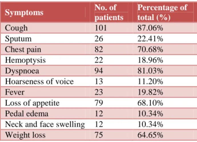
Table 2: Distribution of patients according to symptoms.
Symptoms No.of
patients
Percentageof total(%)
Cough 101 87.06%
Sputum 26 22.41%
Chest pain 82 70.68%
Hemoptysis 22 18.96%
Dyspnoea 94 81.03%
Hoarseness of voice 13 11.20%
Fever 23 19.82%
Loss of appetite 79 68.10%
Pedal edema 12 10.34%
Neck and face swelling 12 10.34%
The most common presenting symptom was cough, seen in 87.1% patients. Table 2 shows the number of patients with various symptoms. Haemoptysis was most commonly seen with small cell carcinoma (5 out of 9, 55.5%). Neck and face swelling (suggestive of superior vena cava obstruction) was also most common with small cell carcinoma (4 out of 9, 44.4%). Duration of symptoms ranged from less than 1 month to more than 12 months. Table 3 shows distribution of patients according to duration of symptoms. Overall mean duration of illness was 7.53±2.84 months for males and 6.38±3.14 months for females. Maximum proportion of patients (65.5%) presented between 6-12 months. 2 patients (1.7%) presented at more than 12 months (both were found to have squamous cell carcinoma). 1 patient (0.8%) presented with symptoms for less than one month. (He was diagnosed with adenocarcinoma).
Table 3: Distribution according to duration of symptoms.
Durationofsymptoms No.ofpatients
<1 month 1
1-<3 Months 1
3-<6 months 36
6-12 Months 76
>12 months 2
Total 116
Forty eight patients (41.4%) gave a history of taking treatment for tuberculosis in the past. It could not be documented how many had bacteriologically confirmed tuberculosis due to lack of past medical records for many patients. 6 patients (5.2%) had diabetes, 4 patients (3.4%) had hypertension and 1 patient (0.8%) who was diagnosed with squamous cell carcinoma had a history of prior malignancy in the tongue (he had undergone glossectomy for squamous cell carcinoma of tongue).
Table 4 shows distribution of patients according to addiction habit. 52 (59.7%) males and 11 (37.9%) females were tobacco chewers. 71 (81.6%) males and 1 (3.4%) female were current smokers or ex-smokers. 62 (71.3%) males and 1 (3.4%) female took bidi, 9 (10.3%) males took cigarette also. The mean pack years were 18.65±11.24 for males and 10.0±0.00 for females. The mean smoking index was 373.10±224.87 for males and 200.00±0.00 for females. 32.2% of males took alcohol and there were no females who took alcohol. History of any other addiction in the form of charas, ganja, bhang etc. could not be elicited from the patients. 22 (19%) patients were exposed to Environmental tobacco smoke (ETS). As per the National Cancer Institute, cancer progress report 2003, ETS is defined as secondhand smoke (also called passive smoke) which is the combination of “sidestream” smoke (the smoke given off by a burning tobacco product) and “mainstream” smoke (the smoke exhaled by a smoker). 22 (19%) patients, of which all were females, which is 75.9% of females were
exposed to biomass fuel. The mean duration of exposure was 18.36±9.32 years.
Table 4: Distribution according to addiction habit.
Addiction Male Female Total
Tobacco
chewing 52 11 63
Smoking 71 1 72
Alcohol 28 0 28
Type of smoking (N = 72)
Bidi 63 1 64
Cigarette 8 0 8
Pack years
(PY) 18.65±11.24 10.0±0.00
Smoking
index (SI) 373.10±224.87 200.00±0.00
Table 5 shows number of patients with various clinical examination finding. Peripheral lymphadenopathy was seen in 20 (17.2%) patients. The most frequently involved group was the axillary group (9 out of 20, 45%).
Table 5: Distribution according to clinical examination finding.
Examination finding
No. of
patients Percent (%)
Pallor 55 47.4%
Icterus 0 0%
Clubbing 34 29.3%
Cyanosis 3 2.6%
Peripheral
lymphadenopathy 20 17.2%
SVC obstruction 12 10.3%
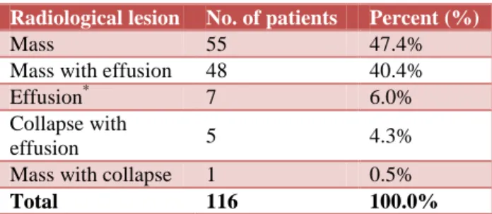
Mass as single lesion was the most common radiological lesion. It was seen in 55 (47.4%) patients. Mass with effusion was the second most common radiological finding, seen in 48 (41.4%) patients. Table 6 shows number of patients with various radiological lesions.
Table 6: Distribution according to radiological lesion.
Radiological lesion No. of patients Percent (%)
Mass 55 47.4%
Mass with effusion 48 40.4%
Effusion* 7 6.0%
Collapse with
effusion 5 4.3%
Mass with collapse 1 0.5%
Total 116 100.0%
*
No mass could be localized: Tx lesion.
centre of mass is within the hilar structures and peripheral tumours as those where the centre of the mass is within the parenchyma and with no or minimal contact with the hilar structures.5 In our study, 38 (32.8%) cases were central tumours, 71 (61.2%) were peripheral tumours and in the remaining 7 (6%) no mass could be localized (Tx lesion). Adenocarcinomas were mostly peripheral (48/70, 68.6%). SCC was mostly peripheral (18/31, 58.1%). All small cell carcinoma were found to be central (9/9,100%).
Pleural effusion was present in 60 (51.7%) patients. 46% males (40 of 87) had pleural effusion. Among all females, 69% (20 of 29) had pleural effusion. Adenocarcinoma was the most common diagnosis among those presenting with pleural effusion (46, 76.7%). Pericardial effusion was seen in 11 (9.5%) patients. All 60 (100%) pleural effusions were exudative in nature, 41 (68.3%) were haemorrhagic and the remaining 19 (31.7%) were straw coloured. Cytology was positive in only 7 (11.7%) cases. Sputum for malignant cells was positive in only 1 (0.9%) patient. He had a centrally located squamous cell carcinoma. AFB smear was positive in 1 (0.9%) patient Thirty eight patients underwent bronchoscopy. TBNA was positive in 13 (65%) out of 20 patients in which it was done, bronchial brush smear (BBS) in 4 out of 19 (21.1%), bronchioalveolar lavage in 12 out of 21 (57.1%)
and maximum yield was by endobronchial biopsy which was positive in 26 out of 32 (81.3%).
Table 7: Distribution according to location of tumour.
Type of cancer
Central location (no. and % of total)
Peripheral location (no. and % of total)
Total
Adenocarcinoma 15 (21.4%) 55 (78.6%) 70
SCC* 13 (41.9%) 18 (58.1%) 31
NSCLC (NOS)£ 0 (0%) 4 (100%) 4
SCLC+ 9 (100%) 0 (0%) 9
Adenosquamous 1 (50%) 1 (50%) 2
Total (N = 116) 38 (32.7%) 78 (67.2%) 116 *
Squamous cell carcinoma; £Non-small cell carcinoma, not otherwise specified, +Small cell carcinoma.
Table 8 shows the yield of various procedures. Multiple procedures were done in some patients. Transthoracic FNAC/biopsy was diagnostic in 71 (61.2%) cases, bronchoscopic procedures in 26 (22.4%), closed pleural biopsy in 18 (15.5%) and thoracoscopic guided biopsy in 4 (3.4%) cases. Other non-biopsy procedures like FNAC of peripheral lymph node or metastatic lesions were diagnostic in 4 (3.4%) cases.
Table 8: Table showing yield of various procedures (multiple procedures were done in some patients).
Transthoracic Biopsy
Endobronchial Biopsy
Closed pleural Biopsy
Thoracoscopic Biopsy
Non biopsy procedure*
Adenocarcinoma (n = 70) 45 9 16 4 0
SCC£ (n = 31) 20 11 1 0 2
SCLCµ (n = 9) 3 5 1 0 0
Adenosquamous (n = 2) 1 1 0 0 0
NSCLC (NOS)@ (n = 4) 2 0 0 0 2
Total 71 26 18 4 4
*non-biopsy procedures include FNAC of peripheral lymph node or metastatic lesions; £ Squamous cell carcinoma, µ Small cell lung cancer, @ Non-small cell lung cancer, not otherwise specified.
Table 9 shows the distribution of patients according to diagnosis. Adenocarcinoma constituted the maximum diagnosis (70, 60.3%) followed by squamous cell carcinoma (31, 26.7%). 9 patients had small cell carcinoma (7.7%), 2 had adenosquamous type (1.7%) and 4 patients had NSCLC, NOS (not otherwise specified) (2.6%). Among males the most common cancer was adenocarcinoma (48 out of 87, 55.2%), followed by squamous cell carcinoma (26 of 87, 29.9%), and small cell carcinoma (8 of 87, 9.2%). Females had the same sequence, adenocarcinoma (22 of 29, 75.9%), followed by squamous cell carcinoma (5 of 29, 17.2%) and small cell carcinoma (1 of 29, 3.4%). The most common cancer among non-smokers was adenocarcinoma (32/44, 72.7%).
Table 9: Distribution of patients according to diagnosis.
Diagnosis Male (no. and %)
Female (no.
and %) Total
Adenocarcinoma 48
(55.84%) 22 (75.86%) 70
SCC* 26
(29.88%) 5 (17.24%) 31
NSCLC (NOS)£ 3 (2.29%) 1 (3.44%) 4
SCLC+ 8 (9.19%) 1 (3.44%) 9
Adenosquamous 2 (2.29%) 0 (0.0%) 2
Total 87 29 116
*
EGFR and ALK mutation study was done in the 70 patients with adenocarcinoma. Table 10 shows sex wise distribution of EGFR mutation. Exon 19 deletion (E746-A750) was the most common subtype mutation. It was seen in 13 out of 16 patients (81.3%) in whom mutation subtype analysis could be done. Exon 21 mutation (L858R) was seen in the remaining 3 (18.7%). Exon 19 deletion was most common in both sexes. There was no female with Exon 21 mutation. 1 (1.4%) patient was positive for ALK mutation.
Table 10: Sex wise distribution of EGFR mutation.
Mutation Male (no. and %)
Female (no.
and %) Total
Exon 19 8 (72.72%) 5 (100.0%) 13
Exon 20 3 (27.27%) 0 (0.0%) 3
Not done 6 2 8
Total 17 7 24
The most common paraneoplastic syndrome was anorexia/fatigue. It was seen in 79 (68%) patients. It was followed by anemia seen in 55 (47.4%) patients. Clubbing was seen in 34 (29.3%) patients, hyponatremia in 1 (0.8%) and thrombophlebitis in 1 (0.8%) patient.
Other paraneoplastic syndromes- endocrine
(hypernatremia, hypercalcaemia, Cushing’s,
gynaecomastia, carcinoid), neurological (mononeuritis
multiplex), SLE, polymyositis/dermatomyositis,
vasculitis, haematological (leucocytosis, eosinophilia, thrombocytosis, leukaemoid reaction), DIC etc. were not reported in our study.
USG abdomen findings (metastasis not confirmed by invasive procedure): evidence of adrenal metastasis was seen in 1 (0.8%) patient. Hepatic metastasis was found in 4 (3.4%) patients, ascites in 2 (1.7%) patients and BPH in 6 (5.2%). Hepatomegaly without features suggestive of metastasis was found in 11 (9.4%) patients and splenomegaly in 3 (2.6%). Retroperitoneal mass which
was later confirmed to be metastasis from
adenocarcinoma was found in 1 patient (0.8%). Other findings were renal cyst in 2 (1.7%) patients and fibroid in 1 (0.8%) patient.
CECT head was done in all patients with small cell carcinoma and those patients presenting with symptoms of CNS involvement. It was done in a total of 34 patients (29.3%). 7 (6%) patients had evidence of CNS metastasis. One patient had multiple infarcts and the remaining 26 had normal findings. 3 out of 70 adenocarcinoma patients, that is 4.2% had brain metastasis, 1 out of 9 small cell carcinoma patients, that is 11.1% had brain metastasis and 3 out of 31 (9.6%) squamous cell carcinoma presented with brain metastasis. Brain metastasis was thus most common in small cell carcinoma.
Table 11 shows the distribution of patients according to TNM classification. No patient presented in stage IA, IB
or IIA. 1 (0.9%) patient presented in stage IIB (he had adenocarcinoma), 4 (3.4%) in stage IIIA and 29 (25%) in stage IIIB. Maximum number of patients presented in stage IV (82, 70.7%). No patient presented with ECOG 1. Most presented with ECOG 3 (68, 58.6%). 45 (38.3%) presented with ECOG 2 and 3 (2.6%) with ECOG 4.
Table 11: Distribution according to TNM staging.
TNM Male Female Total
T
Tx 9 4 13
T1 0 0 0
T2 5 3 8
T3 26 6 32
T4 47 16 63
N
N0 3 1 4
N1 4 0 4
N2 39 12 51
N3 41 16 57
M
M0 29 6 35
M1A 33 17 50
M1B 25 6 32
DISCUSSION
In our study the mean age of presentation was 55.53 years for males and 54.52 years for females. This is in conformity with the demographic data reported from various Indian studies as analysed by Behera where mean age was 52.16 years before 1985 and 54.6 years after 1985.6 Though most studies report females to be younger at presentation, there was no significant difference found in our study, maybe because of the lesser number of females enrolled in our study.7 There was a small percent of patients with age less than 30. All were found to have adenocarcinoma. This highlights the changing trends in lung cancer with growing incidence in the younger age group. As per data from the ICMR Cancer Registry, July 2002, males predominate with a M: F ratio of 4.5:1. In our study there was a slight difference with male to female ratio of 3:1. Pandhi et al in a recent article reported a male to female ratio of 2.7:1.8 These results show that the gap in incidence between male and female is gradually narrowing. This may be explained by the growing numbers of female smokers and the exposure of rural Indian women to biomass fuel. The growing awareness among females to seek medical advice may also have a contributory role.
Cough was the most common symptom (87.1%) in our study. The result was similar to that reported in various studies including that by Pandhi et al and Buccheri.8,9 Though many studies have reported hemoptysis to be most common in squamous cell carcinoma, we found it to be most common in small cell carcinoma.10 Hoarseness of voice was seen in 11.2%. It was much less than that observed by Pandhi et al where it was reported in 43.3% patients.8
In our study most patients presented between 6-12 months, mean duration was 7.53 months for males and 6.38 months for females. Ellis reported a median delay of 4.5 months until initiation of treatment in his study where patients presented to the health facility as early as 7 days after symptom onset.11 In our study, the earliest presentation was at 1 month of symptom onset. The patient was diagnosed to have adenocarcinoma. 2 of our patients presented with symptoms for more than one year’s duration. Both were found to have squamous cell carcinoma.
In this study, tuberculosis was an important entity in previous medical history of the patients. 41.1% patients had taken treatment for the same. (It is a limitation of our study that we could not confirm how many actually had bacteriologically confirmed tuberculosis due to non-availability of relevant documents with the patients). The increased incidence of lung cancer in such group of patients with prior history of tuberculosis was reported by Yu et al.12 We had one patient with prior cancer of the tongue. It has been reported earlier that metachronous lung cancer has been seen in patients with head and neck cancers by Erkal et al.13
The smoker to non-smoker ratio in our study was 1.6:1, similar to that reported by Pandhi et al (1.7:1). It has been estimated that 10-15% of lung cancer occurs in never smokers.8,14 17.4% of our patients were non-smokers. In our study, 75.9% of females were exposed to biomass fuel. No male was exposed to biomass fuel. Bruce et al15 also reported the higher risk for women in developing countries compared with developed countries for lung cancer due to exposure to biomass fuel.
The most common clinical examination finding was pallor, present in 47.4% of patients. Pandhi et al reported clubbing (50%) to be their most common clinical finding.8 Clubbing was seen in 29.3% in our study. Peripheral lymphadenopathy was seen in 17.2% in our study. It was reported in 30% by Pandhi et al.8 Superior vena cava obstruction was seen in 10.3% patients. Small cell carcinoma was found to be the most common cause of SVC obstruction in our study (44.4%). This is similar to that reported by Flounders.16 Some studies have reported adenocarcinoma as the most common cause of SVC.17,18
As in many other studies, mass as the single radiological lesion was the most common presentation in our study.8,19
Prior studies have reported 50% of adenocarcinomas to be peripheral and SCC to be mostly central.20 Both adenocarcinoma and SCC were found to be mostly peripheral in our study.
Most common paraneoplastic syndrome was anorexia and fatigue seen in 68% of patients. This conforms to the findings in most studies where it is reported to be 60%.21
Liver was the most common site of metastasis, seen in 3.4% patients. Kaqohashi et al reported that liver is a possible site of extra-thoracic spread of disease for some patients with lung cancer, especially with SCLC.22 In our study, all patients with liver metastasis were found to have adenocarcinoma, may be because of the overall increased incidence of adenocarcinoma . The incidence of brain metastasis in lung cancer at initial presentation has been described between 12 - 19% in various studies so far.23 Incidence of brain metastasis was 5.9% in our study. It was most common in small cell carcinoma (11.1%). Asymptomatic brain metastasis was not seen.
Pleural effusion was seen in 51.7% of patients similar to that reported by Pandhi et al.8 Dey et al reported it in 28% cases.24 100% of pleural effusion was exudative and majority (68%) were haemorrhagic as was found in various other studies.25 Pleural fluid cytology was positive for malignant cells in 11.7% in our study. This is
much less than 28% reported Kushwaha et al.26
Lung cancer was found to co-exist with pulmonary tuberculosis in one patient (0.8%). It is less than that reported by Varol et al where it was seen in 1.1%.27
Bronchoscopy was the procedure of choice for central tumours and transthoracic FNAB for peripheral tumours in our study. Overall the best diagnostic procedure was transthoracic FNAB (61.2% of all diagnosis). A similar finding was noted by Mehta et al.28
rearrangements were seen in 4-5% of all NSCLC patients.32
70.7% of patients presented in stage IV and 26.7% had distant metastasis. Only one patient was operable at presentation (0.8%). Various studies have reported
metastatic lung cancer to comprise around 50%.33
Majority of patients presented in ECOG 2 and 3 (38.8% and 58.6% respectively). This was consistent with that reported by Radzikowska et al.7
Adenocarcinoma was the most common lung cancer in our study (60.3%), followed by squamous cell carcinoma (26.7%). Houston et al. reported the same.34 Pandhi et al. and Behera D reported squamous cell carcinoma to be the most common type in India.6,8 Our findings are in conformity with studies in other parts of the world where adenocarcinoma has replaced squamous cell carcinoma as the most common type of lung cancer.35
CONCLUSION
Most patients present in an advanced stage.
Adenocarcinoma now seems to be the most common histological subtype of lung cancer in India. EGFR mutation is common in the Indian population. Biomass fuel exposure is a significant risk factor in females. Bronchoscopy is the procedure of choice for diagnosing central tumours and transthoracic FNAC and Biopsy for peripheral tumours.
Limitations of this study were study sample was not representative of the entire population. Upper section of the economic strata was almost not represented. A significant number of patients (48/116) gave history of taking treatment for tuberculosis. We could not confirm how many actually had tuberculosis due to non-availability of relevant documents with the patients. Evidence of metastasis as documented in ultrasound or CT head were not confirmed with invasive procedures. CT head was not done in all patients due to economic and social constraints.
Funding: No funding sources Conflict of interest: None declared
Ethical approval: The study was approved by the institutional ethics committee
REFERENCES
1. Bray F, Ren JS, Masuyer E, Ferlay J. Estimates of global cancer prevalence for 27 sites in the adult population in 2008. Int J Cancer. 2013;132(5):1133-45.
2. Cancer notification in India: an update. Aggarwal H,
Kumar P. South Asian J Cancer. 2014;3(4):236-7. 3. Behera D, Balmugesh T. Indoor air pollution as a
risk factor for lung cancer in women. J Assoc Physicians India. 2005;53:190-2.
4. Gray N. The consequences of the unregulated
cigarette. Tob Control. 2006;15(5):405-8.
5. Daniel R, John HM, Robert T. Influence of Type of
Cigarette on Peripheral versus Central Lung Cancer. Cancer Epidemiol Biomarkers Prev. 2005;14:576. 6. Behera D. Epidemiology of lung cancer – Global
and Indian perspective. JIACM. 2012;13(2):131-7.
7. Radzikowska E, Glaz P, Roszkowski K. Lung
cancer in women: age, smoking, histology, performance status, stage, initial treatment and survival. Population-based study of 20 651 cases. Ann Oncol 2002;13:1087-93.
8. Navin P, Balbir M, Nirmalchand K. Clinico
pathological Profile of Patients with Lung Cancer Visiting Chest and TB Hospital Amritsar. Sch. J. App. Med. Sci. 2015;3(2D):802-9.
9. Buccheri G, Ferrigno D. Lung cancer: clinical
presentation and specialist referral time. Eur Res J. 2004;24(6).
10. Ito M, Niho S, Nihei K, Yoh K, Ohmatsu H, Ohe Y.
Risk factors associated with fatal pulmonary hemorrhage in locally advanced non-small cell lung cancer. BMC Cancer. 2012;12:27.
11. Ellis PM, Vandermeer R. Delays in the diagnosis of
lung cancer. J Thorac Dis. 2011;3:183-8.
12. Yu YH, Liao CC. Increased lung cancer risk among
patients with pulmonary tuberculosis: a population cohort study. J of Thorac Oncol. 2011;6(1):32-7.
13. Erkal HS, Mendenhall WM, Amdur RJ, Villaret
DB, Stringer SP. J Clin Oncol. 2001;19(5):1358-62.
14. Jemal A, Siegel R, Ward E, Hao Y, Xu J, Murray T,
et al. Cancer statistics, 2008. CA Cancer J Clin. 2008;58(2):71-96.
15. Bruce N, Dherani M. Does household use of
biomass fuel cause lung cancer? A systematic review and evaluation of the evidence for the GBD 2010 study. Thorax. 2015;70(5):433-41.
16. Flounders J. Superior vena cava syndrome. Oncol Nurs Forum. 2003;30(4):E84-8.
17. Nunnelee JD. Superior vena cava syndrome. J Vasc
Nurs. 2007;25(1):2-5.
18. Rice TW, Rodriguez RM, Light RW. The superior vena cava syndrome: clinical characteristics and
evolving etiology.Medicine (Baltimore).
2006;85(1):37-42.
19. Behera D, Balamugesh T. Lung cancer in India. Indian J Chest Dis Allied Sci. 2004;46:269-81. 20. Mosmann MP. Solitary pulmonary nodule and (18)
F-FDG PET/CT. Part 1: epidemiology,
morphological evaluation and cancer probability. Radiol Bras. 2016;49(1):35-42.
21. Cella D, Davis K, Breitbart W, Curt G. Fatique Coalition. Cancer-related fatigue: prevalence of proposed diagnostic criteria in a United States sample of cancer survivors. J Clin Oncol. 2001;19(14):3385-91.
22. Kaqohashi K, Satoh H. Liver metastasis at the time
23. Srikanth SG, Jayakumar PN, Chandrashekar HS. CT features of intracranial metastases of unknown primaries.Neurol India. 2002;50:282-5.
24. Dey A, Biswas D, Saha SK, Kundu S, Sengupta A.
Comparison study of clinic radiological profile of primary lung cancer cases: An Eastern India experience. Indian J Cancer. 2012;49:89-95. 25. Light RW. Clinical practice. Pleural effusion. N
Engl J Med. 2002;346:1971-7.
26. Kushwaha R, Shashikala P, Hirmath S, Basavaraj H
G. Cells in pleural fluid and their value in differential diagnosis. J Cytol. 2008;25:138-43. 27. Varol Y, Varol U. Primary lung cancer coexisting
with active pulmonary tuberculosis. Int J Tuberc Lung Dis. 2014;18(9):1121-5.
28. Rivera MP, Mehta AC, Wahidi MM. Establishing
the diagnosis of lung cancer: Diagnosis and management of lung cancer, 3rd ed: American College of Chest Physicians evidence-based clinical practice guidelines. Chest. 2013;143(5):e142S-65S.
29. Kris MG, Johnson BE, Berry LD, Kwiatkowski DJ,
Iafrate AJ, Wistuba II. Using multiplexed assays of oncogenic drivers in lung cancers to select targeted drugs. JAMA. 2014;311(19):1998-2006.
30. Lynch TJ, Bell DW, Sordella R. Activating
mutations in the epidermal growth factor receptor
underlying responsiveness of non-small-cell lung
cancer to gefitinib. N Eng J Med
2004;350(21):2129-39.
31. Li M, Zhang Q, Liu L, Liu Z, Zhou L, Wang Z. The
different clinical significance of EGFR mutations in exon 19 and 21 in non-small cell lung cancer patients of China. Neoplasma. 2011;58(1):74-81. 32. Puey L, Paul M. Prevalence and natural history of
ALK positive non-small-cell lung cancer and the clinical impact of targeted therapy with ALK inhibitors. Clin Epidemiol. 2014;6:423-32.
33. Swensen SJ, Jett JR, Sloan JA, Midthun DE,
Hartman TE, Sykes AM. Screening for lung cancer with low-dose spiral computed tomography. Am J Respir Crit Care Med. 2002;165:508-13.
34. Houston KA, Henley SJ, Li J, White MC, Richards
TB. Patterns in lung cancer incidence rates and trends by histologic type in the United States, 2004-2009. Lung Cancer. 2014;86(1):22-8.
35. William T, Brambilla E. International Association for the Study of Lung Cancer/American Thoracic Society/European Respiratory Society International
Multidisciplinary Classification of Lung
Adenocarcinoma. J Thorac Oncol. 2011;6(2):244-85.
Cite this article as: Kshetrimayum S, Srivastava A,
Kant S, Verma AK, Prakash V, Bajaj DK, et al.A