A Multi-Centre Prospective Single Arm Intervention Trial Evaluating Focal Therapy using High Intensity Focused Ultrasound (Sonablate 500) for Localised Prostate Cancer
Study Protocol
Short Title: Focal Therapy for Prostate Cancer using HIFU INDEX Study Group
Protocol code Version 4
1.
Study Group
Chief Investigator Professor Mark Emberton Co-Principal Investigator Mr Hashim Uddin Ahmed Co-investigators (UCLH/UCL) Dr Clare Allen
Dr Julie Barber Mr Paul Cathcart Miss Louise Dickinson Dr Alex Freeman Dr Charles Jameson Dr Alex Kirkham Mrs Caroline Moore
Study Co-ordination Centre University College London Hospitals NHS Foundation Trust
Study Co-ordinator: Miss Louise Dickinson
Address: Department of Urology,
Ground Floor, 250 Euston Road, London, NW1 2PG
E-mail: [email protected]
Tel: 0207 380 9194
Fax: 0207 380 9303
Project Research Nurse Jane Coe and Helena Stone Department of Urology,
Ground Floor, 250 Euston Road, London, NW1 2PG
Project Manager TBC
Participating Centres (TBC) University College London Hospital NHS Foundation Trust
Basingstoke and North Hampshire NHS Foundation Trust
Imperial College Healthcare NHS Trust
Jewish General Hospital, McGill University, Montreal, Quebec, Canada
Oxford Radcliffe Hospitals NHS Trust Royal Marsden NHS Foundation Trust
University Hospitals Bristol NHS Foundation Trust
Sponsor University College London
Sponsor representative David Wilson
Joint UCLH/UCL Biomedical Research Unit, 1st Floor Maple House, 149 Tottenham Court Road, London W1T 7NF.
Postal address:
Joint UCLH/UCL Biomedical Research and Development (R&D) Unit, (1st Floor, Maple House), Ground Floor, Rosenheim Wing, 25 Grafton Way, London WC1E 6DB.
Signatures
The investigators and the sponsor have discussed this protocol. The investigators agree to perform the investigation and to abide by this protocol except in case of medical emergency or where departures from it are mutually agreed in writing.
Chief investigator
Mark Emberton, Professor of Interventional Oncology and Consultant Urological Surgeon UCL/UCLH
Sponsor
David Wilson, Research & Development, 1st Floor, Maple House, 149 Tottenham Court Road, London, W1T 7DN, UCL
SIGNATURE
DATE
SIGNATURE
Trial Steering Committee and Data Monitoring Committee
Independent Chair Dr John Fowler, Pelican Cancer Foundation Investigator members (TBC) Professor Mark Emberton
Mr Hashim U Ahmed Dr Julie Barber Dr Franck Bladou Mr Tom Leslie
Miss Louise Dickinson Mr Richard Hindley Mr Chris Ogden Mr Raj Persad Mr Matthias Winkler Service User Representatives Dr Michael Ford
Dr Morton Schatzman
2.
Details of healthcare professionals involved in the study from
the host centre and academic collaborators (alphabetical
order)
University College London Hospitals NHS Foundation Trust/ University College London (Sponsor)
Mr Hashim Uddin Ahmed - MRC Research Fellow/ SpR in Urology Dr Clare Allen - Consultant Radiologist
Dr Julie Barber – Lecturer in Statistics
Mr Paul Cathcart – NIHR Clinical Lecturer/ SpR in Urology
Miss Louise Dickinson – Clinical Research Fellow and NIHR Academic SpR Professor Mark Emberton – Reader and Consultant Urological Surgeon Dr Alex Freeman - Consultant Histopathologist
Dr Charles Jameson – Consultant Histopathologist Dr Alex Kirkham - Consultant Radiologist
Mrs Caroline Moore – Clinical Lecturer/ SpR in Urology
Jewish General Hospital, McGill University, Montreal, Quebec, Canada Dr Franck Bladou – Chief Urologist, Department of Urology
Department of Public Health, Erasmus MC, Rotterdam, Netherlands
Professor Ewout Steyerberg - Professor of Medical Decision Making and Research Methodology
Department of Urology, Memorial Sloan Kettering Cancer Centre, New York Professor John Mulhall – Professor of Urology and Director of Male Sexual Health
Table of Contents
1. Study Group...2
2. Details of all healthcare professionals involved in the study (alphabetical order) ....4
3. List of Abbreviations ...6
4. Study Protocol Summary ...7
5. Summary...9
6. Prostate Cancer: Background ...10
6.1 Standard care 6.2 Focal therapy 6.3 Multifocal versus unifocal/unilateral disease 6.4 Disease detection and localization 6.5 High intensity Focused Ultrasound 6.6 Focal therapy trials 6.7 Outcomes in Focal Therapy Trials 6.8 The rationale for a multicentre prospective single arm intervention study 6.9 Cost effectiveness modelling 7. Specific Aims of the study ...31
7.1 Research question 7.2 Objectives 7.3 Outcome measures 8. Study Design...36 8.1 Verification stage 8.2 Intervention stage 8.3 Training and quality control 9. Study Group...47
9.1 Eligibility 10. Trial Flow ...49
11. Individual Study Visits ...50
11.1 Visit schedule 11.2 Follow-up details 12. Evaluation of Safety and Tolerability...56
12.1 Adverse Event Monitoring 12.2 Adverse Event Definitions 12.3 Adverse Events Information Collection 12.4 Serious Adverse Events (SAE) Reporting 13. Data collection...58
14. Discontinuation of Study ...59
14.1 Study Discontinuation by the Sponsor 14.2 Study Discontinuation by the Chief Investigator 14.3 Discontinuation of Study for an Individual Centre 14.4 Discontinuation of Study for an Individual Patient 15. Statistical considerations...60
15.1 Sample size 15.2 Statistical analysis 16. Reporting and dissemination of results...64
17. Liabilities and Insurance ...64
18. Ethics ...65
19. References ...66
3.
List of Abbreviations
BNI BNI Bladder Neck Incision
CISC CISC Clean Intermittent Self Catheterisation
CRF CRF Case Report Form
CT CT Computed Tomography
CXR CXR Chest X Ray
DCE-MRI DCE-MRI Dynamic Contrast Enhanced Magnetic Resonance Imaging
DW-MRI DW-MRI Diffusion Weighted Magnetic Resonance Imaging
ECG ECG Electrocardiogram
ERSPC ERSPC European Randomised Study of Screening for Prostate
Cancer
FACT-P FOV Field of View
HIFU High Intensity Focused Ultrasound
ICS International Continence Score
IIEF IIEF International Index of Erectile Function
IMRT IMRT Intensity Modulated Radiotherapy
IPSS IPSS International Prostate Symptom Score
LREC LRES Local Research Ethics Committee
mpMRI mpMRI Multi-parametric Magnetic Resonance Imaging
MRSI MRSI Magnetic Resonance Spectroscopic Imaging
PCa PCa Prostate Cancer
PIN Prostate Intraepithelial Neoplasia
PSA Prostate-Specific Antigen
QoL QoL Quality of Life
RCT Randomised Controlled Trial
ROI Region of Interest
SAE Serious Adverse Event
SAR Serious Adverse Reaction
TPM Transperineal Template Mapping Prostate Biopsies
TRUS Transrectal Ultrasound
4.
Study Protocol Summary
Study title A Multi-Centre Prospective Single Arm Intervention Trial Evaluating Focal Therapy using High Intensity Focused Ultrasound (Sonablate® 500) for Localised Prostate Cancer Protocol code
Objective To evaluate medium term cancer control, genitourinary, rectal and overall health-related quality of life outcomes, and to model potential cost effectiveness of focal therapy for localised prostate cancer using HIFU
Study type Single arm (Phase II) therapeutic confirmatory
Study population Men with histologically proven localized low to intermediate prostate cancer
(PSA </=15ng/ml, Gleason ≤4+3, T1-T2cN0M0 disease). Sample size 140 patients
Duration of follow up 38 months follow-up post treatment
Test Product Sonablate 500 (Focus Surgery, Indianopolis, IN, USA) Sponsor (UK centres) University College London
Other participating centres (at
commencement of trial)
- Basingstoke and North Hampshire NHS Foundation Trust - Imperial College Healthcare NHS Trust
- Jewish General Hospital, McGill University, Montreal, Canada - Oxford Radcliffe Hospitals NHS Trust
- Royal Marsden NHS Foundation Trust
- University Hospitals Bristol NHS Foundation Trust Examination Dates • Screening visit
• Treatment (baseline)
• Follow up visits at 2 weeks, 6 weeks, 3, 6, 9, 12, 18, 24, 30, 36 and 38 months
• MRI and Trans-rectal biopsy at 12 months • MRI and Template biopsy at 36 months
Primary objectives • 1. To determine the proportion of men who are free of any prostate cancer in the treated area AND are free of clinically significant prostate cancer in the untreated area 36 months after focal therapy using HIFU
• 2. To determine the proportion of men who are free of clinically significant prostate cancer in the treated area AND are free of clinically significant prostate cancer in the untreated area 36 months after focal therapy using HIFU Secondary objectives 1. To determine the following after focal therapy using HIFU:
- rate of erectile dysfunction - time to return of erectile function - rate of loss of ejaculation
- rate of loss of orgasm
- rate of pain during intercourse
- number of men using phosphodiesterase-5 inhibitors to maintain erectile function
- rate of urinary incontinence (pad free, leak free and pad-free alone)
- time to return of continence (pad free, leak free and pad-free alone)
- rate of lower urinary tract symptoms - rate of bowel toxicity
- anxiety levels
- general health related quality of life
– to determine the histological outcomes in the treated and untreated area at 12 and 36 months
- proportion of men achieving trifecta status at 36 months - rate of secondary prostate cancer intervention (prostatectomy, radiotherapy, androgen ablation, whole-gland HIFU or cryosurgery)
- risk factors for failure defined as a) presence of any cancer and b) clinically significant cancer at study end
To analyse the following outcome parameters following focal therapy using HIFU:
- biochemical (PSA) kinetics including determining the optimal biochemical definition of failure
- describe composite outcomes of failure
2. To determine the costs of treatment and model potential cost effectiveness, by comparison to overall histological and functional outcomes at 36 months compared to other cohort trials involving the treatment of localized prostate cancer 3. To determine the clinical validity (sensitivity, specificity, negative and positive predictive values, inter-observer variability) of
- multi-parametric MR-imaging to predict presence of clinically significant prostate cancer on template transperineal prostate mapping biopsies prior to focal therapy
- MR-imaging changes to predict presence of residual/recurrent clinically significant prostate cancer on biopsy
- HistoScanning™ to predict presence of clinically significant prostate cancer on template transperineal prostate mapping biopsies prior to focal therapy
- HistoScanning™ to predict presence of residual/recurrent clinically significant prostate cancer on biopsy
4. To determine the following regarding MR-TRUS image registration
- Number of patients in whom the planned treatment volume was increased as a result of image registration
- Volume change between initial and registration-informed treatment plans
- Time required to plan the treatment manually versus registration-based planning alone or a combination of the two methods (as proposed in this protocol).
- Volume overlap between target volumes, as defined by the HIFU treatment plan, and the regions of necrosis visible in post-operative MR images
5.
Summary
IntroductionMen with localised prostate cancer currently face a difficult decision between radical therapies and active surveillance. The former provides a greater certainty of cancer control but with a significant risk of side effects (50% impotence, 5-10% incontinence, 5-20% bowel dysfunction); the latter entails living with the diagnosis of untreated cancer and the risk of progression. Focal therapy is a strategy that could complement the current choices available to men by offering potential functional preservation whilst ablating clinically significant cancer and monitoring untreated benign tissue or clinically insignificant cancer.
Aim and Objectives
Prospective trials using hemi-ablation with high intensity focused ultrasound (HIFU) (Sonablate 500) have demonstrated feasibility, safety, and encouraging functional outcomes and early cancer control with 90% of men achieving trifecta status (no erectile dysfunction, leak-free pad-free continence, cancer control). However, these trials have involved small numbers of patients with men selected for good baseline function. A multi-centre prospective trial within a larger cohort of men that better represents the patient population with prostate cancer (external validity) is required.
Design
A prospective multi-centre phase II, single arm, cohort study (therapeutic confirmatory) offering focal therapy using HIFU (Sonablate 500) to 140 men with histologically proven localised low to intermediate risk prostate cancer (PSA </=15ng/ml, </=Gleason 4+3, </=cT2cN0M0). Precise mapping and characterisation of the disease will be established using multi-parametric (mp)-MRI and transperineal template prostate mapping (TTPM) biopsies. Only the lobe with the dominant disease will be treated provided the contra-lateral side is free of clinically significant disease (Gleason >/=7 or >/=4mm maximum cancer core length on TTPM).
Outcomes
The primary outcome will be disease control. Disease control, as shown by histological outcomes, will be determined by TTPM biopsies at 36 months. Secondary functional outcomes will be assessed using validated patient questionnaires, including the evaluation of urinary, erectile, and bowel toxicity, and anxiety levels. In addition, we aim to determine the role of imaging and biochemical parameters in determining suitability for focal therapy and predicting histological outcomes of focal therapy, the rate of secondary prostate cancer therapy, and the potential cost-effectiveness of this new therapeutic pathway.
6.
Prostate Cancer: Background
Prostate cancer is the most common malignancy among elderly men and is the second leading malignancy in the Western world1. The rising number of men diagnosed with prostate cancer is a result of increasing life expectancy along with the current practice of screening by PSA blood tests2.
6.1
Standard Care
Men with localised prostate cancer (PCa) have to choose between active surveillance or radical therapy.
There are two problems with the choices available. First, the options sit at the extremes of care. At one extreme lies no treatment – in the refined form of active surveillance. This is an option that is not generally accepted and has shown only limited acceptance and use in Europe and the USA3,4. At the other extreme lies whole gland treatment, whether it is surgery (open, laparoscopic or robotic) or radiotherapy (external beam, high and low dose rate brachytherapy).
The difficult choices faced by men who have localised prostate cancer are further confounded by the findings from the recent publication of the third interim analysis from the European Screening study (ERSPC). This demonstrated a reduction in prostate cancer specific mortality from PSA screening and treatment5. The healthcare policy implications of screening need to be tempered. First, a randomised controlled study in the US has shown no difference between PSA screening and control6, although the control arm had a high degree of contamination since many men had already undergone a PSA test prior to enrolment. Second, there are considerable harms associated with a screening strategy. These include over-treatment and treatment-related harms.
The ERSPC showed that 1410 men need to be screened and 48 diagnosed and treated in order that one prostate cancer related death is avoided over a 9-year interval. Over-treatment becomes less of a problem if the treatment is cost-effective and associated with very low rates of harm, whilst eliminating potentially high-risk disease.
However, current treatments do not share these attributes. At present men can expect the following rates of toxicity: 30-90% erectile dysfunction, 20% incontinence and 5-20% rectal toxicity7,8.
6.1.1 Radical Therapy
The Scandinavian randomised controlled trial comparing surgery and watchful waiting showed an absolute risk reduction in preventing cancer mortality within 8 years of 5% (from 14% to 9%) 9. A recent update of this important RCT has shown that this absolute difference does not change with longer follow-up of 12 years 10.
This difference is probably smaller in the PSA−screened population, as lower risk disease is detected earlier11.
The advantage to radical therapy may also become smaller if watchful waiting is substituted with active surveillance (with selective delayed intervention); the comparison of active surveillance with radical therapy is the subject of an ongoing prospective randomised trial (ProStart trial, NCT00499174). This trial has closed in the UK due to poor recruitment.
It is widely accepted that whole-gland radical therapy causes significant side-effects as a result of damaging structures surrounding the prostate (bladder neck, external sphincter, neurovascular bundles, rectum). This occurs because the whole gland and capsule are treated irrespective of the volume or location of cancer. Radiotherapy causes moderate ano-rectal and urinary side-effects in 5−20% of men. Surgery causes chronic urinary symptoms in one third of men. Both modalities cause impotence in 30-90% of men depending on which modality is used (radiotherapy causes impotence in over 50%) and the particular series looked at (high volume centres of excellence get better results) 12.
The difference between newer forms of radical therapy, between say open surgery and laparoscopic/robotic, whilst declared to be significant, have not been borne out by retrospective studies13,14 and are in fact the subject of randomized controlled trials in themselves: (Lap vs. Open Prostatectomy NCT00695773; Robotic vs. Laparoscopic vs. Open NCT00578123; Laparoscopic vs. Open Europe Trial (LAPPRO) ISRCTN06393679; proposed UK Open vs. Robotic Radical Prostatectomy (OPERA).
Whilst recovery may be marginally quicker, rates of urinary incontinence, erectile dysfunction and cancer control are indistinguishable between the laparoscopic/robotic and open procedures. Whilst conformal and intensity modulated radiotherapy (IMRT) have lead to lower rates of toxicity, the incidence of such side-effects is still significantly high15.
In order to achieve the reduction in toxicities and enhanced recovery in radiation therapy and surgery, respectively, additional cost has been incurred due to an increase in specification of the technology platforms used to deliver these two types of care. Robotics has largely replaced open surgery in the USA and IMRT and proton therapy are increasingly being offered as standard or ‘best’ care16. If high quality prostate cancer care is to be offered to all that need it, irrespective of the ability to pay, then it will have to be both inexpensive and sustainable17.
Choice is only ever of value if it can be exercised and if the differences between the options are both important and meaningful.
Nonetheless, it should be noted that the European Association of Urology Guidelines 200818 as well as the American Urology Association guidelines19 regard curative standard whole gland treatments as radical prostatectomy and radiation therapy (external or brachytherapy). Other forms of treatment are experimental and should be evaluated within carefully conducted clinical trials.
6.1.2 Active Surveillance
Active surveilllance protocols usually involve monitoring the disease with clinical examination, prostate specific antigen (PSA) tests and biopsies at year 1 and then every
2-3 years or when PSA progression is detected. If these parameters demonstrate progression, men are offered radical treatment. A number of Phase II studies have shown that delayed intervention due to signs of progression occurs in approximately one-third of active surveilance groups within a 5 year follow-up from diagnosis 20, 21. However, there are a number of controversial aspects to active surveillance.
First, parameters indicating progression (clinical, biochemical and histological) have not been validated and indeed differ between groups in the USA/Canada and those in the UK.
Second, it is likely that a significant proportion of those men that ‘progress’ within 5 years do so not due to true cancer progression but due to the poor accuracy of diagnostic transrectal ultrasound guided biopsies in ascertaining baseline burden (cancer core length) and Gleason grade (undergrading occurs in one-third of men with Gleason 6)22, 23.
Third, there is some evidence that demonstrates men have increased levels of anxiety during this period24, 25, 26. Indeed, in some series about one–tenth of men decide to opt for radical therapy, despite having no objective measures of progression27. However, others have shown no increase in anxiety levels28, 29. These differences may be accounted for by the particular healthcare service in which these studies have carried out, with European centres demonstrating no change in anxiety levels.
Fourth, the latest series from Toronto has demonstrated that men who progress and have radical therapy have a higher than normal rate of biochemical failure30.
6.2
Focal Therapy
The attributes of a treatment that would appeal to patients faced with the therapeutic dilemma described above would have to share the well tolerated aspects of active surveillance and equal - or approach so closely as to be indiscernible - the oncological control afforded by surgery or radiotherapy.
One way of reducing the unwanted side-effects from radical treatment of PCa may be to direct treatment to only areas of cancer - this is deemed focal therapy.
Focal therapy is a new research area that has been subject to a number of key discussion papers by various groups and more recently, has been subject to discussions by the Food and Drug Agency in the USA 31, 32, 33, 34, 35,36, 37, 38, 39, 40, 41, 42. Focal therapy conferences have been held in 2008, 2009, and 2010 in Duke, Amsterdam and Washington DC. In addition, all the major urology and oncology conferences have held plenary sessions on the topic (AUA 2009; AUA 2010; ASCO-GU 2009; EAU 2009, EAU 2010).
Such a proposed change in treatment of prostate cancer reflects the management of all other solid organ cancers, in which organ preservation is fundamental to functional preservation (breast, kidney, liver, lung). With focal therapy of prostate cancer, it is proposed that by avoiding the bladder neck, rectum, external sphincter and at least one
neurovascular bundle, side-effects could be reduced. For this to be feasible, localisation of cancer within the prostate must be more precise to ensure that malignant areas are not left untreated and areas of clinically significant cancer and a margin of normal tissue are within the treatment zone. In addition, ablative technologies that can treat parts of the prostate are also required. Focal therapy can be delivered using a number of ablative modalities that can treat discrete areas of tissue. These include high intensity focused ultrasound (HIFU), cryosurgery, photodynamic therapy, brachytherapy and radiofrequency ablation, and thermal lasers 43, 44. All are at different stages of development in relation to their use as technologies capable of focal ablation. HIFU, cryosurgery and photodynamic therapy are at a stage of evaluation akin to phase II, non-comparative studies in drug development.
6.2.1 Focal therapy as an alternative to radical therapy or active surveillance
In order that as much information as possible is forthcoming from this evaluation it is desirable that the patient characteristics and the comparators are broad to reflect true clinical practice. There has been some debate recently on which group of men might serve as the ideal population when evaluating a novel intervention such as focal therapy
30, 31. It has been argued that many men with low risk disease probably do not need
treatment; if such men are comfortable pursuing a policy of active surveillance then this would appear to be the most appropriate therapy at the time of writing45. This is especially the case as, increasingly, men are being characterised with greater precision through the use of MR and intensified biopsy strategies46. The result is that the attribution of low risk status is much more secure than it has been historically.
In the USA, and in much of mainland Europe, there exists an imperative to treat all men who are diagnosed with prostate cancer. In these jurisdictions focal therapy tends to be seen as an alternative to active surveillance. Therefore, if active treatment is an inevitable consequence of diagnosis, the role of focal therapy might be to reduce both the harms and the costs associated with treating these men. Most commentators would agree that the oncological benefit that will result from this policy is marginal at best. In this context it is also likely that men with intermediate risk disease may be more likely to be offered radical whole gland therapy.
Defining who is and who is not a candidate for focal therapy is, in the absence of knowledge of the long-term outcomes of the intervention, possibly the most contentious and difficult issue in trial design. The arguments are polarized as follows. On the one hand, a novel intervention, that by definition has high levels of uncertainty associated with it, should only be offered to a group of men who have a low chance of disease progression and, as a result, a low chance of prostate cancer related death. This position is adopted in order to minimize the ‘loss to cure’ in the men that enroll into a non-randomised Phase I/II programme, or are randomized to the non-control arm of a randomized Phase II/III programme.
The alternative is to adopt the position that men with low risk characteristics are not destined to die of prostate cancer over a 15-20 year window47 and therefore any intervention has a very low chance of conferring benefit and therefore can do no more than incur cost and confer harm. This position would encourage the inclusion of patients that had characteristics that would increase their chances of disease progression if left untreated and might incorporate men with higher grade tumours, and at the same time
reduce the upper limit of tumour burden.
Our choice of a relatively broad spectrum of disease risk permits and encourages a range of views (that we feel reflects the equipoise of the investigators) on who should and who should not be offered focal therapy in the current UK climate.
6.2.2 Focal therapy as an alternative strategy to active surveillance in low risk disease The arguments for focal therapy to be carried out only within men suitable for active surveillance are to:
1) reduce the potential psychological morbidity associated with men not having treatment for a cancer – offering these men ‘some form of treatment is better than none’ 2) reduce the cancer progression and/or re-classification rate that currently occurs in about one-third of men who undergo active surveillance and require delayed intervention within 5 years.
Whilst up to 10% of men on active surveillance within 5 years choose to have intervention despite the absence of biochemical or histological progression48, questionnaire surveys have shown conflicting findings about the level of anxiety present in such cohorts. Furthermore, despite the progression rate the mortality rate has been very low, so that it could be argued that most men can avoid treatment and those that have delayed intervention have a period of time free of treatment-related side-effects. However, the period of lower side-effects could be extended if focal therapy were to be carried out at diagnosis or indeed, at the time of disease progression (instead of radical therapy).
The arguments against men who are suitable for active surveillance undergoing focal therapy are that any treatment within this group is liable to be over-treatment. Any treatment, regardless of the encouraging functional outcomes that it may demonstrate, will carry greater morbidity than a management strategy in which two-thirds of men with low-risk disease can avoid treatment whilst the others can delay such morbidity. Nonetheless, active surveillance is not without harm – clinical examination and PSA tests every 2-3 months and biopsies every 1-2 years. Sepsis rates are increasing and repeat biopsies carry a greater risk of sepsis requiring hospital admission49, and pose a significant and sustained healthcare burden for the individual and healthcare systems. In fact, the latest report from Toronto’s active surveillance cohort has demonstrated that of 450 on active surveillance, 117 were treated radically. In these 117 men, the PSA failure rate was 50%, a relatively high rate50.
6.2.3 Focal therapy as an alternative strategy to radical treatments in men with intermediate risk disease
It can be argued that the emergence of focal therapy as a strategy to reduce the side-effects of conventional whole-gland therapies requires us to evaluate its potential within men who, as a result of harbouring intermediate risk disease, would undergo radical therapy. Despite the higher risk disease status, evidence points towards the benefits of radical therapies with respect to cancer control and prevention of death is seen in very few men within 10 years, with this benefit probably seen after 10 years, and rare in men over the age of 6551. A strategy that treats all clinically significant cancer and carefully
monitors untreated tissue for de novo cancers and/or progression of clinically insignificant disease, may obviate the need for any further radical therapies in future or delay it for a number of years during which the man is free of treatment-related side-effects.
The theoretical problem posed by focal therapy is that selective treatment of a target volume of tissue deemed to contain a cancer may incur a miss due to poor targeting, poor staging, or both. The result would be that a cancer with metastatic potential may be given a time window to progress that would not have been available had radical whole gland therapy been employed.
Whilst possible, we have good cause to believe that this risk is low. The reasons are as follows:
a) An estimated lead time bias of approximately 5 years, perhaps more, conferred by screening versus non-screening in the recently published US screening study failed to demonstrate any difference in survival6.
b) In the very high risk disease seen in the Swedish study, progression rates are going to be greater than in the screened population. The difference in survival between men treated by radical prostatectomy versus those men who had delayed systemic therapy was small (5% absolute risk reduction)51. It therefore follows that the likely difference in survival between radical whole gland therapy and a targeted therapy to the dominant cancer is going to be considerably smaller than this.
c) The longer lead time seen in the European Screening study5 that resulted from the lower rates of ‘contamination’ from non-screened to screened groups did show a difference in survival. However, once again this difference was small, indicating that few men progress if left undiagnosed and untreated for a fairly prolonged period of time. Even if the Swedish arm of the European screening trial, which showed a number needed to treat of 12 over a longer period of time [15 years]52, were to be replicated across the whole European Screening study, this still represents a significant over-treatment burden. The risk to the man treated with focal therapy must be less than it was to the man randomized to non-screening.
d) The upfront diagnostic strategy and the treatment verification test after focal therapy that has been incorporated into this protocol has been planned with safety in mind. Template transperineal prostate mapping biopsies at entry reduce any staging error to a minimum. Exit biopsies will have a 95% probability of detecting any man with residual disease that may require either re-treatment or consideration of salvage therapy.
6.3
Multifocal versus unifocal/unilateral disease
6.3.1 Unilateral/Unifocal Prostate Cancer
A number of studies have shown that prostate cancer in the PSA screened era is increasingly unilateral or unifocal. Indeed, unilateral disease has been shown to exist in 20-40% of men, whilst unifocal disease in contemporary series may be present in 10-44% of men with newly diagnosed localised prostate cancer 53, 54, 55, 56, 57, 58, 59.
However, the data on multifocality arises from verification studies performed on men who have undergone radical prostatectomy. Such information is liable to be influenced by a significant degree of work-up bias. Men who are recommended to undergo (and agree to undergo) radical prostatectomy are subject to numerous selection events. They are likely to over-represent the proportion of men who have multifocal disease compared to those men with screen detected disease who opt for other management strategies (surveillance, radiotherapy/brachytherapy, minimally invasive treatments). Although this is more likely in European countries, and particularly in the UK in which active surveillance is well established, it is difficult to verify.
6.3.2 Multifocal Cancer
A strong argument against focal therapy is the fact that the majority of men with localised prostate cancer have multifocal disease. Indeed, at diagnosis most men have between 2 and 3 separate cancer foci. Amongst these foci there usually exists a dominant lesion that accounts for about 80% of the total tumour volume (mean tumour volume varies between 0.5 and 2.3cc)60, 61, 62, 63. The implication of this observation is that the ‘other’ non-dominant lesions account for 0.1 to 0.4cc of tumour on average. By far the majority of these small cancer foci will be of low grade and will conform therefore to most of the definitions of ‘indolence’ 64, 65.
Lesions above 0.5cc are the ones that tend to harbour Gleason scores of 7 or greater and are responsible for extra-capsular extension if present.
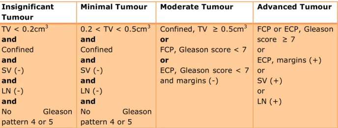
Epstein et al66 have classified foci into insignificant tumours, and minimal, moderate and advanced tumours using a radical prostatectomy series but drawing on the literature demonstrating pathological characteristics of tumours found in radical prostatectomy, autopsy studies and cystoprostatectomy.
Table 1. Categorization of Clinical Stage T1c Tumours 62 Insignificant
Tumour
Minimal Tumour Moderate Tumour Advanced Tumour TV < 0.2cm3 and Confined and SV (-) and LN (-) and No Gleason pattern 4 or 5 0.2 < TV < 0.5cm3 and Confined and SV (-) and LN (-) and No Gleason pattern 4 or 5 Confined, TV ≥ 0.5cm3 or FCP, Gleason score < 7 or
ECP, Gleason score < 7 and margins (-) FCP or ECP, Gleason score ≥ 7 or ECP, margins (+) or SV (+) or LN (+)
TV = Tumour Volume; FCP = Focal Capsular Penetration; ECP = Established Capsular Penetration; Plus sign, positive; Minus sign, negative; SV = Seminal Vesicles, LN = lymph nodes
Additional evidence pointing to the role of volume of cancer driving disease progression have emerged from retrospective cohorts evaluating rates of biochemical failure after
surgery and radiotherapy67, 68, 69. Other studies have shown total tumour volume predicts failure on univariate analysis but not on multivariate analysis likely due to the strong influence of Gleason score70, 71. Evaluating the predictive power of the index lesion seems to demonstrate a relationship72, 73. This may explain some of the discrepancy evident in the literature.
Evidence from molecular genetic studies, which point to a single clone being responsible for metastases demonstrate that there is usually only one clinically significant clone in the prostate and therefore presumably one clinically significant lesion. This study could not demonstrate whether the metastatic clone resided in the index lesion74. It may seem reasonable to propose that ablation of the dominant lesion(s) by volume and grade will give rise to disease control provided the remaining lesions can be well characterized in the pretreatment evaluation75. In fact, it could be argued that definitive knowledge of whether index lesions drive disease progression could only be answered within a clinical trial that involves careful selection and follow-up to ensure that progression of untreated areas of cancer is detected early.
6.4
Disease detection and localisation
6.4.1 Template Transperineal Prostate Mapping Biopsies in Localising Prostate Cancer It has been argued that template transperineal biopsies can serve this purpose to the highest available performance characteristics with sensitivity for 0.2cc and 0.5cc lesions above 90%76,77. We propose using a more accurate technique for disease characterization and localization, so that these errors are reduced significantly. This technique has been shown to be approximately 95% accurate in locating all significant tumor foci. Recently, the Colorado group demonstrated that prostate mapping biopsies detect all tumour subsequently found on whole-mount radical prostatectomy specimens 78, 79, 80. Further, it has been accepted as the standard to which trials in focal therapy should evaluate patients' eligibility81,82.
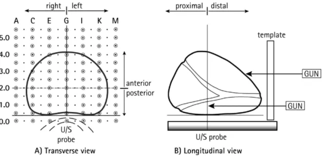
The graphs below represent template simulations on reconstructed radical prostatectomy specimens with each individual lesion also reconstructed. Although this represents the ideal setting for template biopsies, this work allows us to define what minimum amount of cancer within positive biopsy cores accurately represents a significant and insignificant lesion as defined by work drawn from the Stamey and Epstein groups.
A. Template Biopsy Simulations at Lesion Level
The ability of a threshold Cancer Core Length to Detect Lesions of >/=0.2cc. The point at which test performs optimally (Max. Youden index): Sen: 0.958, Spc: 0.861, ppv: 0.672, npv: 0.986. (Max. Youden: 0.818). Cut-off for cancer core length involvement is 4mm. In other words, if >/=4mm is used as the threshold we can be confident that 92% of all lesions >/=0.2cc will be detected (92% sensitivity). In addition, the level of specificity (also at 92%) demonstrates that 8% of lesions </=0.2cc will be falsely designated as a lesion >/=0.2cc.
B. Template Biopsy Simulations at Lesion level
The ability of a threshold Cancer Core Length to Detect Lesions of volume >/=0.5cc.
The point at which test performs optimally (Max. Youden index): Sen: 0.906, Spc: 0.927, ppv: 0.687, npv: 0.982. (Max. Youden: 0.833). Cut-off for cancer core length involvement is 6mm.
If we use the threshold of 4mm used for detection of >/=0.2cc lesions, then if a biopsy is >/=4mm then we can be confident that 98% of all lesions >/=0.5cc in volume will be detected. However, for this greater sensitivity we accept a greater degree of false-positive. In other words, using a 4mm threshold means that we only correctly designate 83% of lesions </=0.5cc as such.
In the focal therapy multicentre trial, we propose that any focus with more than one positive biopsy of greater than, or equal to, 4mm will be deemed above the threshold allowed for defining significant foci. This ensures that a cautious approach is adopted, one in which over-treatment of small lesions is built-in so as to ensure under-treatment of large lesions is less likely to occur.
These results allow us to derive two definitions of clinically significant cancer that approximate to the definitions used by Epstein in which green represents clinically insignificant disease, and yellow and red represent two thresholds for clinically significant disease. Importantly, these definitions can be applied prior to any therapy in contrast to Epstein’s definitions that require whole mount processing of the prostate.
6.4.2 Template Transperineal Prostate Mapping Biopsies in Determining Optimal Standard Care and Suitability for Focal Therapy
Our data also shows that if men with low to intermediate unilateral disease on TRUS are subjected to prostate mapping, the following change in management can occur with the potential for 90% of these men to be treated in a focal manner, as demonstrated by the following table.
Table 2: Template Biopsies and Change in Prostate Cancer Management Recommended Standard Management on TRUS Biopsy Recommended Standard Management on TPM
Focal Therapy strategy based on TPM
Radical Therapy Active Surveillance Radical Therapy Active Surveillance Hemiablation (unilateral any burden, Gleason </=4+3) Focal ablation of all foci (unilateral or bilateral, </=60% gland ablation, Gleason </=4+3) Index lesion ablation (dominant lesion(s) ablated with max 3mm Gleason 3+3 in untreated areas) 46 86 89 43 80 14 23
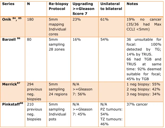
Depending on the intervention that is used, we have estimated that approximately 50-75% of men who currently undergo prostatectomy could have a form of focal therapy (based on an evaluation of their whole-mount specimens)83. Our template data demonstrates that 80-90% of men with low to intermediate risk cancer on TRUS biopsy could be suitable for focal therapy, if this is defined as hemiablation with absence of clinically significant prostate cancer in untreated areas. The data currently available suggest that the benefits of focal therapy are likely to be realized if at least 40-50% of the total prostate tissue is untreated and at least one neurovascular bundle is preserved. Table 3: Template Biopsy Series (other centres)
Series N Re-biopsy Protocol Upgrading >=Gleason Score 7 Unilateral to bilateral Notes Onik 84, 85 180 5mm mapping Individual cores 23% 61% 19% no cancer (35/36 had Max CCLI <5mm) Barzell 86 80 5mm sampling 28 zones 16% 54% 36 unsuitable for focal: 100% detected by TG; 14% by TRUS. 66 had TGB and TRUS at same time: 92% deemed suitable for focal; 45% by TGB Merrick87 294 previous neg. biopsies 5mm sampling 24 regions N/A >=Gleason 7: 56% 1 neg biopsy: 55% 2 neg biopsy: 42% 3 neg biopsy: 34% Pinkstaff88 210 previous neg. biopsies 5mm sampling Individual pots N/A >=Gleason 7: 45% N/A PZ tumours: 54% TZ tumours: 46% 37% cancer
6.4.3 Multi-parametric Magnetic Resonance Imaging
Studies suggest that multi-parametric MRI (mpMRI), which uses a combination of different functional sequences, has a high negative predictive value of 90-95% for lesions of greater than 0.2cc and 0.5cc in volume. In addition, mpMRI will allow the morphological characteristics of the tumours to be visualised so that margins are better incorporated within the treatment plan 89, 90, 91.
Imaging for prostate cancer with the use of mpMRI has progressed from its initial use to stage the disease to its present day capability to identify tumour burden and precise location of tumour foci within the gland. Traditional MRI uses T1 and T2-weighted
sequences, but newer sequences such as diffusion-weighted (DW), magnetic resonance spectroscopy imaging (MRSI), and dynamic contrast-enhancement (DCE) using intravenous gadolinium, have been used to improve the accuracy of this imaging modality 92.
A study by Kim et al 93, comparing DCE-MRI and T2-weighted sequences to locate tumour reported the sensitivity, specificity and accuracy for prostate cancer detection being 55%, 88% and 70% for T2-weighted imaging, and 73%, 77% and 75%, respectively, when DCE-MRI was added. They concluded that tumour contour detection was achieved in 67% of cases by T2-weighted imaging alone and in 90% by T2-weighted and DCE-MRI. Similar results were reported by Villers et al 94 who showed good accuracy for lesions 0.5cc or larger – the threshold for significant cancer lesions. It is therefore appropriate to use DCE-MRI to identify, localise, and aid in targeting the delivery of focal HIFU therapy to, the index lesion.
One inconsistency in studies using mpMRI is in the use of scoring systems to report on the probability of prostate cancer being present. Most studies apply a Likert-type scale, most frequently ranging between scores 1 and 5, to report on the imaging findings (e.g. 1=no cancer seen, 5=cancer definitely present)95, 96, 97. However, these scales are non-standardised and have not been validated. Furthermore, the prostate can be divided into sectors, or ‘Regions of Interest’ (ROI), whereby the prostate can be assessed for the presence of cancer for each individual region. This not only allows division of the prostate into different anatomical zones according to natural borders (such as between the peripheral and transition zones, which differ in how easily cancers can be diagnosed by imaging98), but also allows for direct comparison of imaging outcomes using radical prostatectomy specimens as the reference standard. The number of Regions of Interest used range from one (whole prostate)99, through six100, 101, fourteen102, and up to twenty103.
A European consensus meeting, initiated by the chief investigator (Emberton), and organised by the study co-ordinator (Dickinson) was held in December 2009 at The Royal College of Surgeons, London. Sixteen experts within radiology, urology and oncology attended, representing Bristol (Persad), Brussels (Tombal), Leeds (Carey), Lille (Puech, Villers), Nijmegen (Heijmink, Futterer, Barenstz), Mount Vernon (Hoskins, Padhani), Royal Marsden Hospital (Sohaib), and University College Hospital (Allen, Kirkham, Punwani, Ahmed, Emberton), and coordinated through the Royal College of Surgeons Clinical Effectiveness Unit (Dickinson, van der Meulen). Agreement was reached on the conduct of mpMRI for the detection and localisation of prostate cancer, and on the use of a scoring system for this purpose. A minimal and optimal number of Regions of Interest was also recommended as a result of this meeting, which were sixteen and twenty-seven ROIs, respectively. These recommendations will be implemented and integrated within this protocol to ensure reproducibility and standardisation of mpMRI sequences and reporting across all centres.
6.4.4 HistoScanning™
HistoScanning™ (Advanced Medical Diagnostics, Waterloo, Belgium) is a novel form of diagnostic imaging currently under evaluation for its ability to detect and localize clinically significant prostate cancers. The technique relies on the differing backscatter produced by tissue of altered morphology i.e. tumours, compared with normal tissue.
Algorithms are applied that convert backscatter signals into interpretable results indicating the presence or absence of disease. Retrospective analyses using radical prostatectomy specimens as the reference standard have demonstrated that Histoscan can reliably detect clinically significant lesions of at least 0.5cc in volume104 105. We propose a prospective assessment of this technique in the characterisation and localization of clinically significant prostate cancers, using template biopsies as the reference standard.
6.5
High Intensity Focused Ultrasound (HIFU)
6.5.1 Description of the Medical Device
HIFU works by focusing and depositing a large pulse of high-energy ultrasonic waves on a single area, thereby increasing the temperature to a point whereby, it causes coagulative necrosis. Focused ultrasound waves are emitted from a transducer and are absorbed in the target area of approximately 3x3x10mm of tissue. The result is a targeted thermal effect without damage to the tissue in the path of the ultrasound beam106.
The clinical applications of HIFU in organ-confined prostate cancer are continually being updated with the technique being used throughout the world. Two commercially available devices exist for HIFU therapy: Ablatherm (Edap- Technomed, Lion, France) and Sonablate (Focus Surgery, Indianopolis, IN, USA). This study will use the Sonablate device, which has a combined therapy-imaging transducer of different focal lengths, allowing precise control of energy delivery by each pulse.
International results107, 108 show no cancer detected in between 87-94% of men biopsied after whole gland ablation. Our own results have shown that we can ablate prostate cancer effectively in a whole-gland and focal manner109, 110
6.5.2 Known and Potential Risks of Whole-gland High Intensity Focused Ultrasound The HIFU procedure does not breach the skin or mucosal surfaces and is therefore considered safer than other minimally invasive techniques such as cryotherapy and photodynamic therapy. Morbidity associated with the latest generation Sonablate HIFU is as follows:
Urinary symptoms- reported by most during the first 2 months after treatment Symptomatic urinary tract infection (UTI): 5%
Urethral Stricture: 10%
Retrograde ejaculation: 3%
Epididymitis: 3%
Urinary retention requiring surgery: 2%
Impotence: 25-30%
Incontinence (transient): 0-2%
6.6
Focal Therapy Trials
6.6.1 Focal therapy case seriesFocal therapy involves locating and destroying the areas of cancer only whilst leaving the majority of prostate tissue untreated. To date, focal therapy case series have evaluated hemiablation of unilateral disease (Gleason ≤7, PSA≤15, ≤T2bNoMo) using cryosurgery and one using HIFU111. These have demonstrated impotence rates of approximately 15% with little to no incontinence112, 113, 114, 115, 116. These have used a variety of methods to identify unilateral disease from Doppler TRUS biopsies, TRUS alone and template biopsies and have generally shown poor reporting standards due to their retrospective nature with short follow-up and generally small numbers of patients (tables below). Table 4: Focal Therapy HIFU Retrospective Series
Muto et al (2008)
(Sonablate500)
Barret (2009)
(Ablatherm)
No. 29 12
Therapy Hemiablation Hemiablation
Biopsy TRUS Biopsy TRUS Biopsy
Mean PSA
(ng/ml)
5 (range 2-25) <10
Gleason Score </=8 </=7
Potency Not reported Not reported
Incontinence Not reported 0%
Disease Control 76.5% (biopsy) 58% (10 years) Table 5: Focal Therapy Cryosurgery Series
Onik 2009 Endocare Ellis 2007 Endocare Lambert 2007 Oncura Bahn 2006 Endocare Crawford 2009 Endocare COLD Registry 2009 Endocare No. 112 60 25 31 100 795
Therapy Hemi Hemi Hemi Hemi Focal ‘Focal/Partial’
Biopsy Template TRUS TRUS TRUS+Doppler Template TRUS
Mean PSA (ng/ml) 8.3 7.2 +/- 4.7 6 (range 1-13) 4.95 5.2 +/- 4.1 Gleason Score ≤6 </=8 </=7 </=7 </=7 </=8 Potency 85% 70.6% 70.8% 89% 83% 65% Incontinence 0% 3.6% 0% 0% - 2.8% F/U (mean, months) 43.2 15.2 28 70 - 12 Disease control 93% NED 76.7% (biopsy) 88% (>50% nadir reduction) 96% (biopsy) 92% (ASTRO) 97% (biopsy at 12/12) 4.5% (36/295) 25% (36/199) 83% (ASTRO)
6.6.2 Prospective Studies in Focal Therapy
Three UK single-arm, phase II, prospective IRB-approved and independently audited studies are close to completion at University College London (NCT00988130, NCT00561314, NCT00561262). Two of these are National Cancer Research Network approved. These trials have evaluated the side-effect profile of various forms of focal therapy using high intensity focused ultrasound (HIFU) in men with low-intermediate risk prostate cancer (Gleason ≤4+3, PSA≤15, T2cNoMo):
1. Hemiablation of unilateral PCa (20 patients treated and follow-up completed, accepted for publication in Journal of Urology) (Hemi-HIFU)
2. Focal ablation of only the cancer areas, whether unilateral or bilateral (43/43 recruited, in follow-up stage) (Focal-HIFU)
3. Index lesion(s) ablation of multifocal/bilateral prostate cancer (54/56 recruited) (Lesion Control-HIFU)
The first two trials have localised disease using 5mm-spaced transperineal template prostate mapping biopsies whilst the lesion control trial localises disease either with template biopsies or a combination of TRUS and multi-functional MRI. Such trials suggest that focal therapy can be delivered safely as a day-case procedure with most men discharged 3-5 hours after treatment.
Quality of life
Outcomes measured using validated questionnaires show (tables below):
1) urinary incontinence </=5%
2) erectile dysfunction (erections insufficient for intercourse) 5-10% (with return to baseline scores by 6-9 months)
3) preservation of wet ejaculation 50%
4) bowel dysfunction </=5%
Early Cancer Control
Biopsy of treated areas at 6 months
- absence of any cancer: 82-89%
- absence of clinically important cancer: 88-95% (Gleason </=3+3 and/or Max CCLI </-2mm)
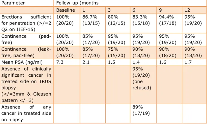
Table 6: Hemi-HIFU Trial Outcomes (UK National Cancer Research Network approved; Cancer Research UK endorsed; MRC funded)
Parameter Follow-up (months
Baseline 1 3 6 9 12 Erections sufficient for penetration (>/=2 Q2 on IIEF-15) 100% (20/20) 86.7% (13/15) 80% (12/15) 83.3% (15/18) 94.4% (17/18) 95% (19/20) Continence (pad-free) 100% (20/20) 85% (17/20) 95% (19/20) 95% (19/20) 95% (19/20) 95% (19/20) Continence (leak-free, pad-free) 100% (20/20) 85% (17/20) 75% (15/20) 90% (18/20) 90% (18/20) 90% (18/20) Mean PSA (ng/ml) 7.3 2.1 1.5 1.4 1.6 1.7 Absence of clinically significant cancer in treated side on TRUS biopsy (</=3mm & Gleason pattern </=3) 95% (19/20) (one refused) Absence of any cancer in treated side on biopsy
89% (17/19)
Table 7: Focal-HIFU Trial Outcomes (Interim) (UK National Cancer Research Network approved; MRC funded)
Parameter Follow-up (months)
1 3 6 9
Erections (hard for penetration) >/=2Q2 IIEF-15 70% (9/13) 73% (11/15) 85% (11/13) 100% (7/7) Urinary Continence (Pad-Free) 86%
(18/21) 94% (17/18) 100% (15/15) 100% (7/7) Urinary Continence (Leak-Free,
Pad-free 76% (16/21) 89% (16/18) 93% (14/15) 100% (7/7) Absence of clinically significant
cancer in treated side on biopsy (</=3mm & Gleason pattern </=3)
88% (15/17)
Absence of any cancer in treated side on biopsy
82% (14/17)
6.7
Outcomes in Focal Therapy Trials
There are no validated or agreed outcome measure other than prostate cancer related deaths or rate of metastatic disease that would serve as a meaningful clinical outcome measure across the therapies of focal therapy, active surveillance, radical prostatectomy and radical radiotherapy. However, in this focal therapy trial with a very low expected rate of death and metastatic progression in this sub-set of low-intermediate risk patients, some 10-15 years would have to pass after treatment before sufficient events were accrued in order to gain meaningful results on which to base the outcomes of this therapy. Furthermore, a trial would require 2000-3000 patients. Therefore, other outcomes will be required in the short to medium-term to serve as surrogate secondary outcome measures. These can be categorized into four groups.
• The first relates to treatment related side-effects and can be relatively well captured using validated questionnaires. These are principally directed at genito-urinary and bowel associated outcomes. In focal therapy studies reported to date, stability in terms of functional health status is achieved between 3 to 6 months following the intervention. This domain of outcome can therefore be derived relatively early.
• The second relates to a more global assessment of quality of life. Some are generic, and some designed specifically for the evaluation of patients with prostate cancer.
• The third, and most problematic area relates to the type and timing of the surrogate cancer related outcomes used (PSA, biopsy, imaging, additional therapy). This area needs some consideration in this proposal. The problem arises since radical therapies use very different outcome measures based on PSA kinetics with little consensus across different modalities of treatment. No such outcome measures have been validated for focal therapies. Indeed, focal therapy will be further problematic in this respect as tissue is left untreated – this tissue will inevitably give rise to PSA increases with time as the man ages. The table below illustrates which outcomes will be available on which group of patients at any given time point.
• The fourth relates to the costs of care and incorporates cost-effectiveness, cost utility and cost benefit. Apart from cost minimization exercises nested on the intervention versus known costs of alternative intervention, most economic analyses will require that cancer outcomes are derived as well as functional status and quality of life.
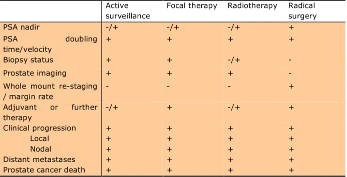
Table 8. Outcome measures that are applicable (+) or are not applicable (-) to the assessment of prostate cancer treatments
Active surveillance
Focal therapy Radiotherapy Radical surgery PSA nadir -/+ -/+ -/+ + PSA doubling time/velocity + + + + Biopsy status + + -/+ - Prostate imaging + + + -
Whole mount re-staging / margin rate - - - + Adjuvant or further therapy -/+ + -/+ + Clinical progression Local Nodal Distant metastases + + + + + + + + + + + + + + + +
Prostate cancer death + + + +
We propose the primary outcome measures detailed below (section 7.3.1). Prostate cancer has a long natural history with minimal expected impact on mortality rates over the time-scale of this trial. In addition, we need to establish the impact that this therapy may have on the patients’ daily quality of life. In respect to radical therapies, erectile dysfunction and urinary incontinence are the most common side effects encountered. These aspects will be assessed as part of the secondary outcome measures.
Erectile function, although multifactorial in aetiology, declines as part of the ‘normal’ aging process with a significantly greater prevalence of dysfunction in men over 70 years compared to those in their 50’s or 60’s117. Baseline erectile function is an important measure to establish in order to know the true impact of a therapy. Unfortunately, relatively few studies report on this. The range of reported erectile function rates is large. A review of the epidemiology of erectile function showed a prevalence of 5-20% of moderate to severe ED across all age groups. However, this prevalence ranged from 2-30% in the <50 year old age group to 38-57% in men of 70-79 years118. Salomon et al. reported dysfunction ranging from mild to severe on IIEF score in 48% of 1330 men with localized prostate cancer awaiting radical prostatectomy119. Whether there is a decline in continence function as part of normal aging is less certain but out of 3810 men within the ERSPC study, up to 9% of men had urinary leakage several times a week117.
The trifecta metric is a summary statistic that has been proposed as a useful measure of therapeutic success following radical surgery for men with early prostate cancer in which the rate varies between 45-62%. It incorporates the three domains that matter to patients – freedom from prostate cancer, erectile function and continence status120,121, 122. We will evaluate trifecta outcomes as a secondary outcome. However, we are aware that this measure is subject to population bias as it will select out those men with continence and normal erectile function at baseline.
6.8
The Rationale for a Multicentre Prospective Single Arm
Intervention Study
Verification of a new therapy as favourable, or equivalent, in outcome to ‘standard’ care is ideally sought through comparison with another matched control group. Randomised controlled trials (RCTs) offer the best method for minimising systematic bias and revealing the true effect of an intervention or drug. However, RCTs involving treatments of localised prostate cancer have had a historically poor patient uptake, as the reference ‘gold’ standard of care is not known. In addition, RCTs are expensive to run and involve huge infra-structural support. A number of trials in the USA have been forced to close due to lack of recruitment. The ProStart trial in the UK has also had to close for the same reason. It has been acknowledged by the Food and Drug Agency in the USA that comparative randomized trials will be problematic in this area due to lack of physician and patient equipoise. A randomized trial may be feasible if a pragmatic design is adopted but prior to acceptance of such a design, the number of centres with expertise in this complex intervention (mp-MRI, TTPM, focal HIFU) will need to be increased. Observational studies are a commonly used alternative to ascertain the effectiveness of a treatment. They are used to observe a treatment effect in a selected group of patients who are presumed to derive benefit from the treatment given. Although methodologically not as robust, and therefore prone to bias, they have some benefits over RCTs. The principal ones are those of enhanced external validity (many patients do not wished to be randomised and therefore refuse participation), and more rapid accrual compared to a randomised design.
For this reasons, a single arm medium term follow-up cohort intervention study has been designed.
At the time of writing the safety and tolerability aspects of focal therapy by HIFU are known as a result of the Phase I/II studies carried out at UCLH. The results have been presented and exist in the public domain in abstract form but have not yet been published (presented in tables above). These early studies were powered to detect a change in the proportion of men who could obtain an erection sufficient for penetration compared to their status prior to their treatment. The very low event rate for both erectile dysfunction and incontinence indicates that the ‘proof of concept’ has been demonstrated for focal therapy. Moreover, we can be relatively confident that, in expert hands, focal HIFU is safe.
Therefore, a multi-centre study is now required involving a larger group of patients for the following reasons:
1) To evaluate medium term cancer control using histological parameters.
2) To confirm that focal therapy can lead to low rates of genitourinary and rectal toxicity and minimal impact on quality of life within a large and more representative cohort of patients (greater precision around outcome measures).
3) To demonstrate that the skills (characterization through template prostate mapping and MRI as well as the treatment related skills) acquired by the team at UCLH are indeed
transferable to other providers.
4) To calculate costs of care and to model potential cost-effectiveness in comparison to alternative therapies.
If this single arm intervention study demonstrates acceptable outcomes to support the findings of the Phase I/II studies, it is anticipated that this preliminary study will lead onto a Phase III evaluation of focal therapy, prior to more widespread use of this technology.
6.9
Cost Effectiveness Modelling
Development of a cost effectiveness model for focal therapy outcomes could evaluate pre-treatment, treatment and post-treatment costs (blood tests, mpMRI, biopsies, management of side-effects, re-treatment of failures, recurrent costs), quality of life, and potential impact on survival. The benefits, costs and savings in using a quick treatment that utilises fewer resources (capital and recurrent), and achieving fewer side-effects through a reduction in the therapeutic burden using focal therapy, needs to be assessed against the greater costs incurred as a result of a requirement to characterise the disease accurately at the outset. A decision analytic disease model will become available to determine the place of focal therapy in relation to other treatment options for prostate cancer, including radiotherapy, surgery and active surveillance123.
This objective will utilise validated questionnaire data that is available from existing studies led by Erasmus MC, Rotterdam, the Netherlands. Such data will be used in the comparative modelling of the current diagnostic and therapeutic pathways. Details of the model to be used are specified in section 15.2.
In order to gain an understanding of how patient outcomes relate to other similar patient groups to derive incremental cost-effectiveness ratios, we are seeking to make direct outcome comparisons against other trials that have incorporated current standard care. Most single arm intervention observational studies of prostate cancer therapies use erectile function, urinary continence, and cancer control as outcome measures. We propose a retrospective analysis of data collected from other cohorts of men with similar disease risk profiles i.e. localised low-intermediate risk prostate cancer, and baseline characteristics e.g. age, who have undergone ‘standard’ prostate cancer treatments. A number of statistical methods can be used in order to reduce bias effects in observational studies. For example, propensity score matching uses a scoring system applied to study subjects, in order to ‘balance’ known confounding factors between exposed and non-exposed groups124. This method is thought to provide a more robust matching technique and can be applied where baseline characteristics differ between groups. This statistical analysis will enable us to model cost effectiveness.