The International Association for the Study of Lung Cancer
Lung Cancer Staging Project: Proposals Regarding the
Clinical Staging of Small Cell Lung Cancer in the
Forthcoming (Seventh) Edition of the Tumor, Node,
Metastasis Classification for Lung Cancer
Frances A. Shepherd, MD, FRCPC,* John Crowley, PhD,† Paul Van Houtte, MD,‡
Pieter E. Postmus, MD, PhD,
兩兩
Desmond Carney, MD, PhD,¶ Kari Chansky, MS,†
Zeba Shaikh, BSc,** and Peter Goldstraw, FRCS,
on behalf of the International Association for the Study of Lung Cancer International Staging
Committee and Participating Institutions
Background:Small cell lung cancer (SCLC) is usually classified using the limited and extensive definition. The tumor, node, metas-tasis (TNM) classification should also be applicable to SCLC, but it has only been reported in small surgical series. The current analysis looks to the impact of the TNM system on the clinical staging of SCLC and of the new International Association for the study of Lung Cancer (IASLC) proposals.
Methods:Using the IASLC database, survival analyses were per-formed for clinically staged patients. Prognostic groups were com-pared, and the new IASLC TNM proposals were applied to this population and to the Surveillance, Epidemiology, and End Results (SEER) database.
Results: The IASLC database contained 12,620 eligible cases of small cell histology. TNM staging was available for 8088 patients. Survival was directly correlated to both T and N category.
Differ-ences were more pronounced in patients without mediastinal or supraclavicular nodal involvement. Stage grouping using the sixth edition of TNM also differentiates survival except between IA and IB. Patients with pleural effusion regardless of the cytology have an intermediate prognosis between limited and extensive disease. The IASLC proposals for the seventh edition of the TNM classification also apply to this series of SCLC and to the SEER database. Conclusion:TNM staging is recommended for SCLC, and stratifi-cation by stage I-III should be incorporated in clinical trials of early-stage disease. Further studies are needed to clarify the impact of pleural effusion and the extent of N3 disease.
Key Words:Small cell lung cancer, Staging. (J Thorac Oncol.2007;2: 1067–1077)
L
ung cancer is the leading cause of cancer-related mortality in the western world, with recent statistics showing that it accounts for more than 1,000,000 deaths each year globally.1Small cell lung cancer (SCLC) represents only 15% to 20% of lung cancers overall, with incidence rates declining among men but continuing to increase among women in most coun-tries.2 It is distinguished by its rapid growth rate, early
dissemination to regional lymph nodes and distant sites, and its sensitivity to chemotherapy and radiotherapy.3The
intro-duction of chemotherapy to the treatment of SCLC led to significant improvements in median and 5-year survival rates in the 1970s through 1990s,4 but recently survival seems to
have reached a plateau for both localized and advanced SCLC.
Staging of lung cancer is second in importance only to the pathologic determination of cell type. Accurate staging provides prognostic information, and stage determines treat-ment strategies for all types of lung cancer. The first staging system for SCLC was introduced in the 1950s by the Veter-ans’ Administration Lung Study Group (VALSG) for use in
*Department of Medicine, Division of Medical Oncology and Hematology of the University Health Network, Princess Margaret Hospital and the University of Toronto; †Cancer Research and Biostatistics, Seattle, WA; ‡Department of Radiation Oncology, Institut Jules Bordet, Universite´ Libre de Bruxelles;兩兩Department of Pulmonary Diseases, Vrije Univer-siteit University Medical Center, Amsterdam, The Netherlands; ¶Depart-ment of Medical Oncology, Mater University Hospital, Dublin, Ireland; and **Royal Brompton Hospital, London, United Kingdom.6
The project was supported by the AJCC grant “Improving AJCC/UICC TNM Cancer Staging.”
Eli Lilly and Company provided funding to support the International Asso-ciation for the Study of Lung Cancer (IASLC) Staging Committee’s work to establish a database and to suggest revisions to the 6th edition of the TNM classification for Lung Cancer (staging) through a restricted grant. Lilly had no input into the committee’s analysis of the data or in their suggestions for revisions to the staging system.
Disclosure: The authors declare no conflict of interest.
Address for correspondence: Frances A. Shepherd, MD, FRCP(C), Princess Margaret Hospital, Suite 5-104, 610 University Avenue, Toronto, On-tario, Canada. E-mail: frances.shepherd@uhn.on.ca
Copyright © 2007 by the International Association for the Study of Lung Cancer
their randomized clinical trials.5This simple system divided
SCLC into two disease subgroups termed “limited” and “exten-sive” disease. Limited disease (LD) was characterized by tumors confined to one hemithorax, although local extension and ipsi-lateral, supraclavicular nodes could also be present if they could be encompassed in the same radiation portal as the primary tumor. No extrathoracic metastases could be present. All other disease was classified as extensive disease (ED).
In 1989, the International Association for the Study of Lung Cancer (IASLC) issued a consensus report6that, for the
first time in 30 years, introduced changes to the VALSG staging system. This report suggested that LD should be expanded to include tumors limited to one hemithorax with regional lymph node metastases, including hilar, ipsilateral
andcontralateral mediastinal and ipsilateralandcontralateral supraclavicular nodes. They also recommended that patients with ipsilateral pleural effusion, regardless of whether cytol-ogy is positive or negative, be considered to have LD if no extrathoracic metastases were detected.
The tumor, node, metastasis (TNM) staging systems of the American Joint Committee on Cancer Staging (AJCC)7
and the Union Internationale Contre le Cancer (UICC)8 are
also applicable to SCLC, but they are used less frequently in clinical practice because these two staging systems have historically relied on surgical confirmation for their accuracy, and patients with SCLC seldom present at a stage for which surgery is appropriate. However, in small surgical series of patients with SCLC, the TNM staging system has been shown to be prognostic of outcome.9 –21TNM staging in non-surgical
series is rarely reported.22,23
The IASLC has established a database containing stag-ing and a minimum of 5-year survival data for more than 100,000 patients with lung cancer from around the world, presenting between 1990 and the end of 2000. Among these are more than 12,000 patients with SCLC. This database was established to assess the current stage categories as defined by the AJCC7 and the UICC sixth edition,8 with the goal of
redefining and regrouping of T and N descriptors, to make recommendations for change if and where appropriate. These analyses were performed initially on the large 67,725 case non-small cell lung cancer (NSCLC) database,24 –27then
ap-plied to the smaller SCLC cohort. Proposed changes for the UICC seventh edition for NSCLC that are relevant for SCLC are mainly those that result in changes to T staging. It has been recommended that large sized tumors (⬎7 cm) be-come T3; additional nodules within the same lobe as the primary tumor also become T3; ipsilateral intrapulmonary nodules in other lobes become T4; and all pleural effusions regardless of cytology become M1. It is also recommended that pleural effusions in the absence of other metastatic sites be designated M1a, with M1b reserved for all other meta-static involvement.24,26
We herein report our analysis of clinical TNM staging for SCLC using the current sixth edition TNM staging system for lung cancer and compare this with the proposed new groupings as defined by the IASLC analyses in NSCLC. We focus on the clinical staging of SCLC because fewer than 5% of patients are eligible for pathologic surgical staging.
METHODS Statistical Analysis
The database on which these analyses are based was created by the IASLC to inform revisions for the seventh edition of the UICCTNM Classification of Malignant Tumors
staging manual in lung cancer due to be published early in 2009. To be eligible for inclusion in the IASLC database, patients were required to have a new diagnosis of either SCLC or NSCLC, an initial presentation from 1990 to the end of 2000, adequate staging information at baseline, and ade-quate follow-up for survival.
There were no minimal staging requirements for inclu-sion in the database, and details of staging were not recorded. TNM stage groupings for NSCLC were developed based on recommendations made by the individual T, N, and M subcommittees for changes to their respective components. Using these modified categories, a small number of candidate stage grouping schemes were developed, initially using a recursive partitioning and amalgamation algorithm.28 The
analysis grouped patients based on best stage (pathological if available, otherwise clinical) after determination of best-split points based on overall survival on indicator variables for the newly proposed T/M category and an ordered variable for N category. This analysis was performed on a randomly se-lected training set comprising two thirds of the available data that met the requirements for conversion to the newly pro-posed TNM categories (n⫽17,726), reserving 9133 cases for later validation. The random selection process was stratified by type of database submission and time of case entry (1990 –1995 vs 1995–2000).29 Selection of a final stage
grouping proposal from among the candidate schemes was based on its statistical properties in the training set and its relevance to clinical practice and was arrived at by consensus. Analyses focusing on sixth edition TNM were applied to the subset of SCLC cases for which complete clinical TNM stage was provided. The newly proposed TNM stage group-ings were applied to the set of SCLC cases that had sufficient descriptors to reclassify according to the new TNM stage scheme. Analyses focusing on pleural effusion required only a designation of limited or extensive stage disease by the VALSG system (or adequate TNM staging to derive this determination) and sufficient information to determine pres-ence/absence of pleural effusion.
Survival was measured from the date of entry (date of diagnosis for registries and the date of registration for proto-cols) and was estimated by the Kaplan-Meier method. Prog-nostic groups were compared formally by Cox proportional hazards regression analysis, using the SAS System for Win-dows Version 9.0 PHREG procedure.
RESULTS
Population
Of the 100,869 patients with information submitted to the IASLC database, 81,015 met the initial screening require-ments of having a new diagnosis rather than recurrent diag-nosis of either SCLC or NSCLC, presentation from 1990 to the end of 2000, adequate follow-up for survival, and ade-quate staging information at baseline. From the 13,290 SCLC
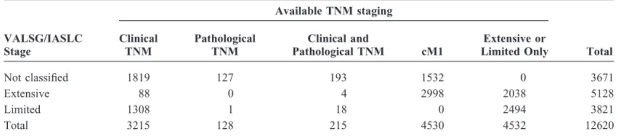
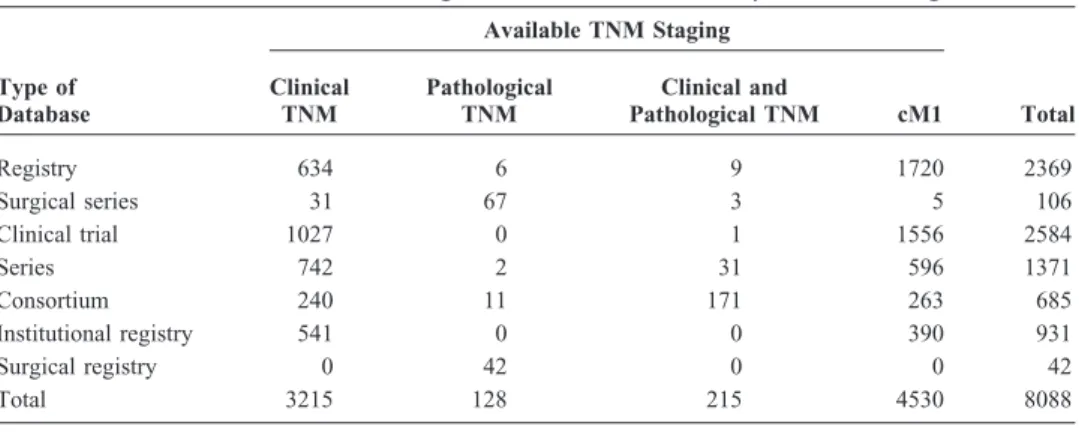
cases meeting the initial screen, 670 with mixed histology or with conflicts between TNM and extent of disease were excluded, to yield 12,620 eligible cases of small cell histol-ogy. Of the 12,620 eligible SCLC cases, 4532 had LD or ED classification only (Table 1). TNM staging was available for 8088 patients: 3430 cM0 cases had full clinical TNM data, 343 had full pathologic TNM staging, and 4530 were classi-fied as cM1 (clinical metastatic disease). The 12,620 cases originated from four global regions: Europe (56%), North America (35%), Asia (3%), and Australia (6%). Of the cases, 52% were from clinical trials, 43% from population-based registries or single-institution series or registries, and the rest from consortia. For cases with complete clinical TNM stag-ing, the proportions were: clinical trials 32%, population-based registries or single-institution series or registries 60%, and consortia 8% (Table 2). Because most patients with SCLC are staged only clinically, we focus on the clinical staging of this malignancy in this article; the surgical series will be reported separately. A complete listing of data con-tributors is given in Appendix A.
T Category
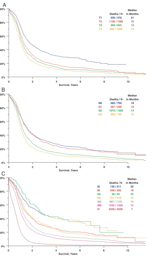
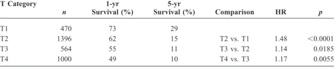
As shown in Figure 1A, increasing clinical T category, using the sixth edition TNM staging system was associated with progressively lower survival in patients who did not have evidence of metastatic (M1) involvement. Comparisons between T categories and 1- and 5-year survival rates are shown in Table 3. Patients with clinical T1 had significantly better survival than those with T categories 2– 4. A compar-ison of clinical category T1 versus T2 yields a hazard ratio (HR) of 1.48 (p ⬍ 0.0001), and the trend continues with significant differences between T2 versus T3 and T3 versus T4 (see Table 3). Thepvalue for the ordered log rank test (a test against ordered alternatives for survival data) is less than 0.0001.28,29
N Category
As shown in Figure 1Band Table 4, increasing clinical N category was associated with progressively lower survival. Patients with clinical N0 or N1 tumors had significantly better survival than did those with N2 or N3 tumors. The difference between cN0 and cN1 is not significant (HR 1.02,p⫽0.76). The cN2 and cN3, although significantly different, have similar median survivals (14 vs. 12 months, HR 1.18). The
difference between the N1 and N2 is the largest (median 19 vs. 14 months, HR 1.40, p ⫽ 0.0001) and represents the primary separation for clinical N category. The p value for the ordered log rank test is less than 0.0001.
Survival for clinical T categories within the four clin-ical N categories 0 –3 is shown in Figure 2. Although increas-ing T category seems to be associated with worse survival in each N category, the differences seem to be clinically mean-ingful only for patients without mediastinal or supraclavicular nodal involvement.
Survival According to TNM Stage
Survival by clinical TNM stage sixth edition is shown in Figure 1C. The near superimposition of stage IA together with IIA, and stage IB with IIB, are most likely a reflection of the separation between the T1 cases (comprising IA and IIA) and the T2 and T3 (IB and IIB). Pair-wise comparisons between adjacent stage groupings are shown in Table 5. All differences are significant, but for the reason just discussed, the hazard ratio for IIA versus IB is the reverse of what would be expected.
Pleural Effusion
In the proposals from the IASLC for the forthcoming (seventh) edition of the TNM classification for lung cancer, the presence of a pleural effusion in patients with NSCLC will now be considered as M1 disease. In the SCLC database, information concerning the presence or absence of pleural effusion or other distant sites was available for 1258 patients with otherwise limited disease (1113 of whom were without pleural effusion, 145 with pleural effusion) and 4500 patients with ED with other metastatic sites. Of the 145 cases of LD with pleural effusion, 81 were actually designated as ED without any other metastatic sites. These 81 were assumed to have LD with pleural effusion. Surgically managed cases were excluded. As can be seen from Figure 3A, the survival of patients with LD with effusion is intermediate between those of patients with LD without effusion and patients with ED (ordered log rankpvalue ⬍0.0001).
The result of cytologic examination of the pleural effusion was available for only 68 patients in the database. As shown in Figure 3B, the survival of patients with LD with effusion, whether cytologically negative or positive, re-mained intermediate between those of patients with LD
TABLE 1. Summary of 12,620 Small Cell Lung Cancer Cases from the IASLC International Staging Project Database VALSG/IASLC Stage Available TNM staging Clinical TNM Pathological TNM Clinical and Pathological TNM cM1 Extensive or
Limited Only Total
Not classified 1819 127 193 1532 0 3671
Extensive 88 0 4 2998 2038 5128
Limited 1308 1 18 0 2494 3821
Total 3215 128 215 4530 4532 12620
VALSG, Veterans’ Administration Lung Study Group; IASLC, International Association for the Study of Lung Cancer; TNM, tumor, node, metastasis.
without effusion and patients with ED, and the survival of patients with LD with positive effusions was superior to that of patients with ED (ordered log rank pvalue ⬍0.0001).
Proposals from the IASLC for Stage Groupings in the Seventh Edition of the TNM Classification for Lung Cancer
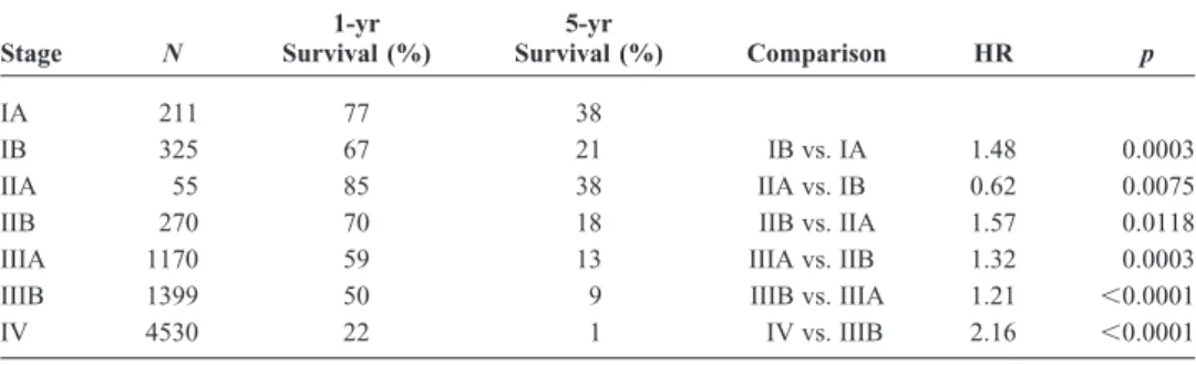
It was not possible to classify all SCLC cases according to the proposed IASLC stage groupings because details for the T descriptor frequently was inadequate, particularly with respect to size, presence of additional intrapulmonary nod-ules, and involvement of the pleura. Figure 4Ashows survival according to the TNM stage sixth edition for the smaller cohort of patients (n ⫽ 2464) with full T descriptor data. With the exception of the extremely small subgroup of eight patients with stage IIA, it can be seen that increasing stage is associated with progressively shorter survival. Figure 4B
shows the survival of the same cohort of patients using the IASLC stage groupings proposed for the UICC seventh edition, and Figure 4Cshows the proposed IASLC groupings applied to the much larger Surveillance, Epidemiology and End Results (SEER) data base of the National Cancer Insti-tute. In the SEER database, for patients registered with an initial diagnosis of SCLC during the years 1998 through 2000 inclusive, the proposed IASLC system works well with in-creasing stage being associated with dein-creasing survival.
DISCUSSION
At the time of initial diagnosis, approximately two thirds of patients with SCLC have clinical evidence of hematogenous metastases (M1); for the remaining one-third, most patients have clinical evidence of extensive nodal involvement in the hilar, mediastinal, and sometimes even the supraclavicular regions. For that reason, surgical resection is seldom offered; instead, patients are treated with systemic chemotherapy with or without radiation. Although TNM staging has been applied in small series of patients with SCLC treated either with surgery alone or with combined modality therapy including surgery,9 –21a
sim-pler staging system assigning patients to either LD or ED is used most frequently in clinical practice.5,6Patients with LD cancers
(confined to the thorax) are treated with chemotherapy and
thoracic radiation, whereas, for the most part, patients with ED (distant hematogenous metastases) receive only chemotherapy. In general, it has been believed that more precise staging of SCLC using the TNM system does not provide extra guidance with respect to selection of treatment modality because so few patients are eligible for surgery. However, a more precise definition of nodal involvement may be of particular relevance for radiation treatment. The simple des-ignation of LD does not provide adequate information for accurate radiotherapy planning because it does not differen-tiate between the presence or absence of ipsilateral and contralateral supraclavicular nodes, and ipsilateral and con-tralateral hilar or mediastinal nodes. According to the 1989 IASLC staging system for SCLC, all of these nodal stations are included in LD. In the modern era of conformal tech-niques and increasing radiation dose for SCLC, it is likely no longer appropriate to treat all patients with LD in the same way, and radiation fields must be determined using more precise nodal categories.
The SCLC subgroup of patients in the IASLC staging initiative represents the largest cohort of patients with this malignancy ever to be reported. In particular, it is the largest cohort of patients with SCLC to have been staged using the TNM system apart from small surgical series, most of which had fewer than 50 patients.9 –21These surgical series clearly
showed that TNM staging could identify patients with differ-ent prognoses.
The more important question, however, is whether the old VALSG and IASLC staging classifications should be abandoned and whether clinical TNM staging, as used for NSCLC, should be applied to SCLC as proposed by the UICC. In our large clinically staged series, patients with clinical T1 disease had significantly better survival than those with T2 disease (HR 1.48, p ⬍ 0.0001) and all other T categories, although they represented only 13.7% of all pa-tients. However, the test for trend among the four T groupings was highly significant. In a pattern of care study performed in Japan including only patients with limited disease, the T category was also a significant prognostic factor for survival in a multivariate analysis.18
TABLE 2. Sources of Small Cell Lung Cancer Cases with Complete TNM Stage
Type of Database Available TNM Staging Clinical TNM Pathological TNM Clinical and Pathological TNM cM1 Total Registry 634 6 9 1720 2369 Surgical series 31 67 3 5 106 Clinical trial 1027 0 1 1556 2584 Series 742 2 31 596 1371 Consortium 240 11 171 263 685 Institutional registry 541 0 0 390 931 Surgical registry 0 42 0 0 42 Total 3215 128 215 4530 8088
With respect to N category, patients with clinical N0 or N1 tumors had significantly better survival than those with N2 or N3 tumors, although the difference between N0 and N1 was not significant, and the greatest difference in survival was seen between N1 and N2. It is likely that the patients
with T1-2,N0-1,M0 tumors (i.e., stage IA, IB, IIA and IIB [N0 and N1]) are the same as those previously classified by the University of Toronto Lung Group as “very limited.”22
Unfortunately, the database did not contain enough information to determine whether the survival of patients
0% 20% 40% 60% 80% 100% 0 2 4 6 8 10 Survival, Years T1 T2 T3 T4 Deaths / N 335 / 470 1158 / 1396 484 / 564 883 / 1000 Median in Months 21 15 13 12 0% 20% 40% 60% 80% 100% 0 2 4 6 8 10 Survival, Years N0 N1 N2 N3 Deaths / N 562 / 750 267 / 348 1375 / 1592 656 / 740 Median in Months 18 19 14 12 0% 20% 40% 60% 80% 100% 0 2 4 6 8 10 Survival, Years IA IB IIA IIB IIIA IIIB IV Deaths / N 135 / 211 246 / 325 36 / 55 211 / 270 997 / 1170 1235 / 1399 4335 / 4530 Median in Months 30 18 33 18 14 12 7
A
B
C
FIGURE 1. Survival by (A) clinical T Category, (B) clinical N Category, and (C) clinical TNM category (sixth edi-tion of TNM).
with N3 contralateral mediastinal nodes differed from that of patients with N3 supraclavicular nodes, and it could not address differences in outcome for ipsilateral versus con-tralateral supraclavicular nodal involvement. In the IASLC staging system,6 both ipsilateral and contralateral
supracla-vicular nodes are included in LD. Therefore, more precise nodal categories should be recorded in future validation studies that will have prospectively collected data.
In the past, the traditional radiotherapy ports for chest irradiation for SCLC were large and sometimes included
TABLE 3. Overall Survival Comparisons for Clinical Category T1 to T4 (any N) M0 Small Cell Lung Cancer, IASLC Data
T Category n 1-yr Survival (%) 5-yr Survival (%) Comparison HR p T1 470 73 29 T2 1396 62 15 T2 vs. T1 1.48 ⬍0.0001 T3 564 55 11 T3 vs. T2 1.14 0.0185 T4 1000 49 10 T4 vs. T3 1.17 0.0055 HR, hazard ratio.
TABLE 4. Overall Survival Comparisons for Clinical Category N0 –N3 (any T) M0 Small Cell Lung Cancer, IASLC Database
N Category n 1-yr Survival (%) 5-yr Survival (%) Comparison HR p N0 750 68 24 N1 348 68 20 N1 vs. N0 1.02 0.7552 N2 1592 56 12 N2 vs. N1 1.40 ⬍0.0001 N3 740 50 9 N3 vs. N2 1.18 0.0006 HR, hazard ratio. cN0 M0 0% 20% 40% 60% 80% 100% 0 2 4 6 8 10 Survival, Years T1 T2 T3 T4 Deaths / N 135 / 211 246 / 325 79 / 95 102 / 119 Median in Months 30 18 17 13 cN1 M0 0% 20% 40% 60% 80% 100% 0 2 4 6 8 10 Survival, Years T1 T2 T3 T4 Deaths / N 36 / 55 132 / 175 61 / 72 38 / 46 Median in Months 33 20 12 14 cN2 M0 0% 20% 40% 60% 80% 100% 0 2 4 6 8 10 Survival, Years T1 T2 T3 T4 Deaths / N 112 / 148 590 / 684 234 / 266 439 / 494 Median in Months 16 15 12 12 cN3 M0 0% 20% 40% 60% 80% 100% 0 2 4 6 8 10 Survival, Years T1 T2 T3 T4 Deaths / N 52 / 56 190 / 212 110 / 131 304 / 341 Median in Months 15 12 13 11
A
B
C
D
“prophylactic” treatment of areas such as the contralateral hilum and both supraclavicular fossae. In essence, therefore, treatment frequently paralleled the IASLC definition of lim-ited disease. However, only moderate dose radiation could be delivered to such large volumes. The inability to deliver high doses to these large volumes may, in part, explain the high rates of failure at the primary site reported in most studies
despite the administration of thoracic radiotherapy. Several recent trends make more precise nodal staging critical for SCLC. As is the case in NSCLC, reduction of the radiother-apy field size to cover only the known area of macroscopic disease is being recommended for SCLC. Also, the high local failure rate despite the addition of radiation to chemotherapy is leading to the investigation of higher radiation doses or
TABLE 5. Overall Survival Comparisons for Clinical TNM Stage Category Small Cell Lung Cancer, IASLC Database
Stage N 1-yr Survival (%) 5-yr Survival (%) Comparison HR p IA 211 77 38 IB 325 67 21 IB vs. IA 1.48 0.0003 IIA 55 85 38 IIA vs. IB 0.62 0.0075 IIB 270 70 18 IIB vs. IIA 1.57 0.0118 IIIA 1170 59 13 IIIA vs. IIB 1.32 0.0003 IIIB 1399 50 9 IIIB vs. IIIA 1.21 ⬍0.0001 IV 4530 22 1 IV vs. IIIB 2.16 ⬍0.0001 HR, hazard ratio. 0% 20% 40% 60% 80% 100% 0 2 4 6 8 10 Survival, Years M0, PE Absent M0, PE Present M1 Deaths / N 966 / 1113 134 / 145 4313 / 4500 Median in Months 18 12 7 0% 20% 40% 60% 80% 100% 0 2 4 6 8 10 Survival, Years M0, PE Absent M0, PE Cyt (+) M0, PE Cyt (-) M1 Deaths / N 966 / 1113 35 / 40 24 / 28 4313 / 4500 Median in Months 18 12 13 7
A
B
FIGURE 3. Survival by pleural effu-sion status (surgical cases excluded).
A, All pleural effusions included.B, Only effusions with cytologic evalua-tion comparing cytology-positive with cytology-negative effusion.
more aggressive combined radio-chemotherapy regimens.30 –32
To avoid toxicity from large volume fields, it will be essential to identify the most appropriate patients for such therapy, and more precise staging will be key to the ability to do this. It will also be very important to examine the patient populations in reports of clinical trials of altered fractionation and high-dose radiation (particularly single-arm phase I or II studies)
because all patients with LD as defined by the IASLC may not be included in those studies, and this may have an effect on the survival outcomes. For example, in the landmark Turrisi trial, patients with pleural effusions, regardless of the cytology, and those with contralateral hilar or supraclavicular adenopathy were excluded.30In this analysis, the survival of
patients with IIIA and IIIB disease seems to be less than that
A
B
C
0% 20% 40% 60% 80% 100% 0 6 10 14 IA IB IIA IIB IIIA IIIB IV Deaths / N 17 / 25 20 / 27 2 / 8 83 / 100 326 / 377 473 / 535 1358 / 1392 Median in Months 31 26 NR 18 13 12 8 0% 20% 40% 60% 80% 100% IA IB IIA IIB IIIA IIIB IV Deaths / N 17 / 25 14 / 19 8 / 15 84 / 101 332 / 384 424 / 481 1400 / 1439 Median in Months 31 35 68 17 13 12 8 0% 20% 40% 60% 80% 100% 0 2 4 6 8 10 Survival, Years IA IB IIA IIB IIIA IIIB IV Deaths / N 83 / 127 80 / 114 71 / 100 45 / 52 537 / 637 545 / 611 3132 / 3243 Median in Months 26 21 15 12 13 11 6 Survival, Years Survival, Years 2 4 8 12 0 2 4 6 8 10 12 14FIGURE 4. Survival by (A) clinical sixth edition of TNM, including only those cases with sufficient information to derive IASLC proposed TNM stage; (B) IASLC proposed TNM stage; (C) survival by IASLC proposed TNM stage in the Surveillance, Epidemiol-ogy and End Results database.
reported in recent trials of high-dose radiation, hyperfraction-ated radiation, or concurrent chemotherapy and radiation therapy in LD SCLC. This may, in part, be because of less aggressive treatment practices in some parts of the globe where concurrent chemotherapy and radiation is seldom ad-ministered and where high-dose or hyperfractioned radiation is not considered standard. Alternatively, however, these survival rates may be a more accurate reflection of the true outcome of LD SCLC than those reported from clinical trials in which trial patients are a highly selected subgroup of LD overall.
If the proposals made by the IASLC for the forthcom-ing (seventh) edition of the TNM classification for lung cancer are accepted by the UICC and the AJCC, patients with pleural effusion will, in the future, be classified as having M1 (or extensive) disease. Our analysis shows that patients who have pleural effusion without extrathoracic metastases (M1a) have a survival that is intermediate between that of stage I–III without effusion and stage IV. A similar observation was reported by Perng et al. in a series of patients treated in Taiwan.23 Therefore, we recommend that pleural effusion
should be a stratification parameter in clinical trials for both LD and ED SCLC. Having even a small proportion of patients with pleural effusion in LD trials could affect out-come significantly if they are not balanced between the arms, as could patients with only a pleural effusion in ED trials. Unfortunately, we did not have enough patients in our anal-ysis to make a clear statement concerning cytology-positive or -negative effusions. Similarly, no information was avail-able with respect to the presence of a pericardial effusion. This information must be collected prospectively in future studies.
It seems that TNM staging can identify subgroups of patients with distinct prognoses from within the broad defi-nition of LD. However, the question still remains as to the value of applying the TNM system of staging to all patients with LD SCLC. Exact TNM stage is unlikely to alter treat-ment decisions for most patients with LD because combined modality therapy will be offered to patients with good per-formance status in most subsets of stage I, II, or III. The only potential exception to this might be the subgroup of patients with N3 supraclavicular nodal involvement, whether unilat-eral or bilatunilat-eral. Whether to include supraclavicular nodes in the radiation treatments field remains an unresolved issue and is one that will require careful documentation for future prospective database analyses.
Our analysis suggests that, in the context of clinical trials in LD, accurate TNM staging may be critical and suggests that stratification based on TNM stage may be important. Survivals for clinical stages I and II are signifi-cantly different from those of stage III with N2 or N3 involvement. Therefore, the SCLC staging subcommittee of the IASLC Lung Cancer Staging Project recommends that TNM staging should be applied to SCLC and that stratifica-tion for stage should be incorporated into all trials in LD SCLC, particularly those addressing thoracic and prophylac-tic cranial radiation questions and those that include a surgi-cal treatment arm. The small survival differences that are
expected in these trials could be affected significantly by imbalances in the two patient subgroups of stage I/II versus III. For tumor registries, the clinical and, where possible, pathologic TNM designations should be recorded. It will always be possible to derive the LD and ED subsets from these data.
In summary, the proposals made by the IASLC for the forthcoming (seventh) edition of the TNM staging system seem to have relevance to SCLC. The SCLC subcommittee of the IASLC Lung Cancer Staging Committee therefore rec-ommends that TNM staging be applied in SCLC and that stratification by TNM stage be incorporated into clinical trials in stage I-III SCLC. For prospective staging validation stud-ies, more information must be collected to define the N category more clearly and to determine whether there is a difference in prognosis for patients with cytology-negative or -positive pleural effusions and for patients with pericardial effusions. In addition, in future studies, data concerning other prognostic variables, such as gender and performance status, and more detailed staging data should also be collected for multivariate analysis to isolate the effect of stage in this malignancy.
REFERENCES
1. Parkin DM, Bray FJ, Ferlay L, Pisani P. Global cancer statistics, 2002.
CA Cancer J Clin2005;55:74–108.
2. Devesa SS, Bray F, Vizcaino AP, Parkin DM. International lung cancer trends by histologic type: male:female differences diminishing and adenocarcinoma rates rising.Int J Cancer2005;117:294–299. 3. Masters G. The clinical presentation of small cell lung cancer. In HI
Pass, DP Carbone, DH Johnson, JD Minna, AT Turrisi III (Eds.),Lung Cancer: Principles and Practice, Philadelphia: Lippincott Williams & Wilkins, 2005. pp. 304–314.
4. Janne PA, Freidlin B, Saxman S, et al Twenty-five years of clinical research for patients with limited small cell lung carcinoma in North America.Cancer2002;95:1528–1538.
5. Zelen M. Keynote address on biostatistics and data retrieval.Cancer Chemother Rep1973;4:31–42.
6. Stahel R, Ginsberg R, Havemann K, et al. Staging and prognostic factors in small cell lung cancer: a consensus report.Lung Cancer1989;5:119– 126.
7. FL Greene CC, Compton AG Fritz, JP Shah, DP Winchester (Eds). American Joint Committee on Cancer StagingAtlas. Chicago: Springer, 2006:167–176.
8. LH Sobin C Wittekind (Eds).UICC: TNM Classification of Malignant Tumours, 6th Ed. New York:Wiley, 2002.
9. Shepherd FA, Ginsberg RJ, Feld R, et al. Surgical treatment for limited small-cell lung cancer.J Thorac Cardiovasc Surg1991;101:385–393. 10. Meyer J, Comis RL, Ginsberg SJ, et al. The prospect of disease control
by surgery combined with chemotherapy in stage I and stage II small-cell carcinoma of the lung.Ann Thorac Surg1983;36:37–41. 11. Meyer JA, Gullo JJ, Ikins PM, et al. Adverse prognostic effect of N2
disease in treated small-cell carcinoma of the lung.J Thorac Cardiovasc Surg1984;88:495–501.
12. Shah SS, Thomson J, Goldstraw P. Results of operation without adjuvant therapy in the treatment of small cell lung cancer.Ann Thorac Surg
1992;54:498–501.
13. Lim E, Yap YK, De Stavola BL, Nicholson AG, Goldstraw P. The impact of stage and cell type on the prognosis of pulmonary neuroen-docrine tumors.J Thorac Cardiovasc Surg2005;130:969–972. 14. Shepherd FA. Surgical management of small cell lung cancer. In: KL
Franco, JB Putnam (Eds).Advanced Therapy in Thoracic Surgery,2nd Edition. Hamilton:BC Decker, 2005. pp. 101–112.
15. Inoue M, Miyoshi S, Yasumitsu T, et al. Surgical results for small cell lung cancer based on the new TNM staging system.Ann Thorac Surg
16. de Antonio DG, Alfageme F, Gamez P, et al. Results of surgery in small cell carcinoma of the lung.Lung Cancer2006;52:299–304.
17. Byhardt RW, Hartz A, Libnoch JA, Hansen R, Cox JD. Prognostic influence of the TNM staging and LDH levels in small cell carcinoma of the lung (SCCL)Int J Radiat Oncol Biol Phys1986;12:771–777. 18. Uno T, Sumi M, Sawa Y, et al. Process of care and preliminary outcome
in limited-stage small-cell lung cancer: results of the 1995–1997 patterns of care study in JapanInt J Radiat Oncol Biol Phys2003;55:626–632. 19. Darling G, Shepherd FA. Surgical management of small cell lung cancer. In: H Pass, J Mitchell, D Johnson, D Turrisi (Eds).Lung Cancer: Principles and Practice,3rd Edition. Philadelphia: JB Lippincott, 2005. pp. 475–490.
20. Waddell TK, Shepherd FA. Should aggressive surgery ever be part of the management of small cell lung cancer?Thorac Surg Clin2004;14: 271–281.
21. Shepherd FA, Feld R, Payne D. Small cell lung cancer. In: TW Shields, J Locicero III, RB Ponn, VW Rusch (Eds).General Thoracic Surgery.
Philadelphia: Lippincott Williams & Wilkins, 2005. pp. 1700–1737. 22. Shepherd FA, Ginsberg RJ, Haddad R, et al Importance of clinical
staging in limited small-cell lung cancer: a valuable system to separate prognostic subgroups.J Clin Oncol1993;11:1592–1597.
23. Perng RP, Chen CY, Chang GC, et al. Revisit of 1997 TNM staging system survival analysis of 1112 lung cancer patients in Taiwan.Jpn.J Clin Oncol2007;37:9–15.
24. Rami-Porta R, Ball D, Crowley JJ, et al. The IASLC Lung Cancer Staging Project: proposals for revision of the T descriptors in the forthcoming (7th) edition of the TNM classification for lung cancer.
J Thorac Oncol2007;2:593–602.
25. Rusch V, Crowley JJ, Goldstraw P, et al. The IASLC Lung Cancer Staging Project: proposals for revision of the N descriptors in the forthcoming (7th) edition of the TNM classification for lung cancer.
J Thorac Oncol2007;2:603–612.
26. Postmus PE, Brambilla E, Chansky K, et al. The IASLC lung cancer staging project: Proposals for revision of the M descriptors in the forthcoming (7th) edition of the TNM classification of lung cancer.
J Thorac Oncol2007;2:686–693.
27. Goldstraw P, Crowley JJ, Chansky K, et al. The IASLC Lung Cancer Staging Project: proposals for the revision of the TNM stage groupings in the forthcoming (7th) edition of the TNM classification for lung cancer.J Thorac Oncol2007;2:801–804.
28. Liu PY, Green S, Crowley JJ, et al. Testing against ordered alternatives for censored survival data.J Am Stat Assoc1993;88:153–160. 29. Liu PY, Tsai WY, Wolf M. Design and analysis for survival data under
order restrictions with a modified logrank test.Stat Med1998;17:1469– 1479.
30. Turrisi A, Kyungmann K, Blum R. et al. Twice-daily compared with once-daily thoracic radiotherapy in limited small-cell lung cancer treated concurrently with cisplatin and etoposide.N Engl J Med1999;340:265– 271.
31. Baas P, Belderbos JS, Senan S, et al. Concurrent chemotherapy (carbo-platin, paclitaxel, etoposide) and involved-field radiotherapy in limited stage small cell lung cancer: a Dutch multicenter phase II study.Br J Cancer2006;94:625–630.
32. DeRuysscher D, Bremer RH, Koppe F, et al. Omission of elective node irradiation on basis of CT-scans in patients with limited disease small cell lung cancer: a phase II trial.Radiother Oncol2006;80:307–312.
APPENDIX 1
IASLC International Staging Committee:
P. Goldstraw (Chairperson), Royal Brompton Hospital, London, UK; H. Asamura, National Cancer Centre Hospital, Tokyo, Japan; D. Ball, Peter MacCallum Cancer Centre, East Melbourne, Australia; V. Bolejack, Cancer Research and Biostatistics, Seattle, Washington, USA; E. Brambilla, Labo-ratoire de Pathologie Cellulaire, Grenoble Cedex, France; P. A. Bunn, University of Colorado Health Sciences, Denver, Colorado; D. Carney, Mater Misericordiae Hospital, Dublin, Ireland; K. Chansky, Cancer Research and Biostatistics,
Se-attle, Washington, USA; T. Le Chevalier, Institute Gustave Roussy, Villejuif, France; J. Crowley, Cancer Research and Biostatistics, Seattle, Washington, USA; R. Ginsberg (de-ceased), Memorial Sloan-Kettering Cancer Center, New York, USA; D. Giroux, Cancer Research And Biostatistics, Seattle, Washington, USA; P. Groome, Queen’s Cancer Re-search Institute, Kingston, Ontario, Canada; H. H. Hansen (retired), National University Hospital, Copenhagen, Den-mark; P. Van Houtte, Institute Jules Bordet, Bruxelles, Bel-gium; J.-G Im, Seoul National University Hospital, Seoul, South Korea; J. R. Jett, Mayo Clinic, Rochester, Minnesota, USA; H. Kato, (retired), Tokyo Medical University, Tokyo Japan; C. Kennedy, University of Sydney, Sydney, Australia; M. Krasnik, Gentofte Hospital, Copenhagen, Denmark; J. van Meerbeeck, University Hospital, Ghent, Belgium: T. Naruke (deceased), Saiseikai Central Hospital, Tokyo, Japan; E. F. Patz, Duke University Medical Center, Durham, North Caro-lina, USA; P. E. Postmus, Vrije Universiteit Medical Center, Amsterdam, the Netherlands; R. Rami-Porta, Hospital Mutua de Terrassa, Terrassa, Spain; V. Rusch, Memorial Sloan-Kettering Cancer Center, New York, USA; JP. Sculier, Insti-tute Jules Bordet, Bruxelles, Belgium; Z. Shaikh, Royal Brompton Hospital, London, UK; F. A. Shepherd, University of Toronto, Toronto, Ontario, Canada; Y. Shimosato (re-tired), National Cancer Centre, Tokyo, Japan; L. Sobin, Armed Forces Institute of Pathology, Washington, DC; W. Travis, Memorial Sloan-Kettering Cancer Center, New York; M. Tsuboi, Tokyo Medical University, Tokyo, Japan; R. Tsuchiya, National Cancer Centre, Tokyo, Japan; E. Vallieres, Swedish Cancer Institute, Seattle, Washington, USA; J. Vansteenkiste, Leuven Lung Cancer Group, Bel-gium; Yoh Watanabe, Kanazawa Medical University, Uchi-nada, Japan; and H. Yokomise, Kagawa University, Kagawa, Japan.
PARTICIPATING INSTITUTIONS:
O. Visser, Amsterdam Cancer Registry, Amsterdam, The Netherlands; R. Tsuchiya and T. Naruke, Japanese Joint Committee of Lung Cancer Registry; J. P. Van Meerbeeck, Flemish Lung Cancer Registry-VRGT, Brussels, Belgium; H.
〉u¨lzebruck, Thorax-klinik am Universitatsklinikum, Heidel-berg, Germany; R. Allison and L. Tripcony, Queensland Radium Institute, Herston, Australia; X. Wang, D. Watson and J. Herndon, Cancer and Leukemia Group B (CALGB), USA; R. J. Stevens, Medical Research Council Clinical Trials Unit, London, England; A. Depierre, E. Quoix and Q. Tran, Intergroupe Francophone de Cancerologie Thoracique (IFCT), France; J. R. Jett and S. Mandrekar, North Central Cancer Treatment Group (NCCTG), USA; J.H. Schiller and R. J. Gray, Eastern Cooperative Oncology Group (ECOG), USA; J.L Duque-Medina and A. Lopez- Encuentra, Broncho-genic Carcinoma Co-operative Group of the Spanish Society of Pneumology and Thoracic Surgery (GCCB-S), Spain; J. J. Crowley, Southwest Oncology Group (SWOG); J. J. Crowley and K. M. W. Pisters, Bimodality Lung Oncology Team (BLOT), USA; T. E. Strand, Cancer Registry of Norway; S. Swann and H. Choy, Radiation Therapy Oncology Group (RTOG), USA; R. Damhuis, Rotterdam Cancer Registry, The
Netherlands; R. Komaki and P. K. Allen, MD Anderson Cancer Center- Radiation Therapy (MDACC-RT), Houston, Texas, USA; J. P. Sculier and M. Paesmans, European Lung Cancer Working Party (ELCWP); Y. L. Wu, Guangdong Provincial People’s Hospital, Peoples Republic of China; M. Pesek and H. Krosnarova, Faculty Hospital Plzen, Czech Republic; T. Le Chevalier and A. Dunant, International Adjuvant Lung Cancer Trial (IALT), France; B. McCaughan and C. Kennedy, University of Sydney, Sydney, Australia; F. Shepherd and M. Whitehead, National Cancer Institute of Canada (NCIC); J. Jassem and W. Ryzman, Medical Univer-sity of Gdansk, Poland; G. V. Scagliotti and P. Borasio, Universita’ Degli Studi di Torino, S Luigi Hospital, Orbas-sano, Italy; K. M. Fong and L. Passmore, Prince Charles Hospital, Brisbane, Australia; V. W. Rusch and B. J. Park, Memorial Sloan-Kettering Cancer Center, USA; H. J. Baek, Korea Cancer Centre Hospital, Seoul, South Korea; R.P. Perng, Taiwan Lung Cancer Society, Taiwan; R. C. Yung, A. Gramatikova, John Hopkins University, USA; J. Vansteenk-iste, Leuven Lung Cancer Group (LLCG), Belgium; C. Brambilla and M. Colonna, Grenoble University
Hospital-Isere Cancer Registry, France; J. Hunt and A. Park, Western Hospital, Melbourne Australia; J. P. Sculier and T. Berghmans, Institute of Jules Bordet, Brussels, Belgium; A. K. Cangir, Ankara University School of Medicine, Ankara, Turkey; D. Subotic, Clinical Centre of Serbia, Belgrade, Serbia; R. Rosell and V. Aberola, Spanish Lung Cancer Group (SLCG), Spain; A. A. Vaporciyan and A. M. Correa, MD Anderson Cancer Center-Thoracic and Cardiovascular Surgery (MDACC-TCVS), Houston, Texas, USA; J. P. Pignon, T. Le Chevalier and R. Komaki, Institut Gustave Roussy (IGR), Paris, France; T. Orlowski, Institute of Lung Diseases, War-saw, Poland; D. Ball and J. Matthews, Peter MacCallum Cancer Institute, East Melbourne, Australia; M. Tsao, Prin-cess Margaret Hospital, Toronto, Ontario, Canada; S. Darwish, Policlinic of Perugia, Italy; H. I. Pass and T. Stevens, Karmanos Cancer Institute, Wayne State University, USA; G. Wright, St Vincent’s Hospital, Victoria, Australia; and C. Legrand and J. P. van Meerbeeck, European Organi-sation for Research and Treatment of Cancer (EORTC), Brussels, Belgium.