RECTAL CANCER
THESIS FOR DOCTORAL DEGREE (Ph.D.) AKADEMISK AVHANDLING
Avläggande av medicine doktorsexamen vid Karolinska Institutet försvaras offentligen i Lek-sellsalen, Eugeniahemmet, Karolinska Universitetssjukhuset, Solna, Stockholm
Fredagen den 29 januari 2016 kl 9.00
Av
Frida Lédel
Principal Supervisor: Associate Professor David Edler Department of Molecular Medicine and Surgery Karolinska Institutet Co-supervisor: Associate Professor Peter Ragnhammar Department of Oncology and Pathology Karolinska Institutet Opponent:Professor Henrik Grönberg Department of Medical Epidemiology and Biostatistics
Karolinska Institutet
Examination Board:
Professor Per Hall Department of Medical Epidemiology and Biostatistics
Karolinska Institutet
Professor Gudrun Lindmark Department of Clinical Sciences, Surgery Lunds Universitet Associate Professor Anders Höög Department of Oncology and Pathology Karolinska Institutet
INSTITUTIONEN FÖR MOLEKYLÄR MEDICIN OCH KIRURGI
Karolinska Institutet, Stockholm, Sweden
HER3 EXPRESSION AND PROGNOSIS IN
COLON AND RECTAL CANCER
Frida Lédel
All previously published papers were reproduced with permission from the publisher. Published by Karolinska Institutet
Layout Soraya Abdi
Picture on inside of cover; Jätten by painter Helmtrud Nyström © Frida Lédel, 2015
Printed by E-print AB 2015 ISBN 978-91-7676-203-5
My hand is weary with writing, My sharp quill is not steady, My slender-beaked pen jets forth A black draught of shining dark-blue ink. A stream of wisdom of blessèd God Springs from my fair-brown shapely hand: On the page it squirts its draught Of ink of the green-skinned holly. My little dripping pen travels Across the plain of shining books, Without ceasing for the wealth of the great― Whence my hand is weary with writing.
C
ONTENTS
________________________________________________________________________
ABSTRACT ... 9
LIST OF SCIENTIFIC PAPERS ... 11
THESIS AT A GLANCE ... 13
LIST OF ABBREVIATIONS ... 15
INTRODUCTION ... 17
Cancer in general ...17
Colon and rectal cancer ...18
Colon cancer ...18
Rectal cancer ...19
Symptoms ...20
Prevention ...20
Investigation ...20
Multidisciplinary team conference...21
Treatment ...21
Heredity ...26
Embryology and Tumor location ...28
Staging ...30
Pathology ...31
Inflammation and Immune response ...32
Metastases in CRC ...33
Genetic and epigenetic development in CRC ...35
Adenoma Carcinoma Sequence ...35
The MSI, CIMP and CIN phenotypes ...36
Biomarkers in CRC ...38
Stage II, III and biomarkers ...39
HER3 ...39
MMR ...41
HLA-A*02 genotype ...42
AIMS OF THESIS ... 45
PATIENTS AND METHODS ... 47
Patients ...47 Immunohistochemistry (IHC) ...48 Scoring ...50 HER3 ...50 MMR ...51 FISH ...51
RESULTS AND DISCUSSION ... 55
HER3 expression in primary tumor... 55
HER3 expression in colorectal metastases ... 57
HER3 expression and prognosis in colon and rectal cancer ... 58
HER3 phenotype, location and adjuvant therapy ... 59
Combining HER3, MMR and HLA-A*02 ... 61
HER3, MMR, HLA-A*02 ... 61
Prognosis and prediction ... 62
Discussion ... 63 CONCLUSIONS ... 67 FUTURE PERSPECTIVES ... 69 POPULÄRVETENSKAPLIG SAMMANFATTNING ... 73 ACKNOWLEDGEMENTS ... 75 REFERENCES ... 79 PAPER I-IV
A
BSTRACT
________________________________________________________________________
In Sweden, about 6000 patients get a colorectal cancer diagnosis annually. Advances in man-agement of both colon and rectal cancer have reduced mortality even though the incidences are increasing. To improve outcome further, it is important to identify prognostic and predic-tive factors for personalized, optimal surgery and to guide adjuvant treatment decisions. By selecting the right therapy combined with selection of the right patients, one can obtain indi-vidualization. Potential drug targets prevalent in primary tumors and metastases are of inter-est as well. HER3 is a transmembranous, epidermal growth factor receptor that is over ex-pressed in colon and rectal cancer. It affects cellular proliferation, differentiation and migra-tion in embryogenesis and in oncogenesis through activamigra-tion of intracellular signal pathways. This thesis investigates HER expression and prognosis in colon and rectal cancer, as well as in correlated lymph node and liver metastases. HER3´s association to a combination of bi-omarkers, MMR expression and HLA-A*02 genotype, is examined related to prognosis. Two cohorts of patients have been available for our investigation. Immunohistochemical (IHC) detection of HER3 and MMR expression was performed in a group of Swedish pa-tients with primary colon and rectal cancer of stage II and III. The papa-tients derived from a randomized Nordic trial aiming to evaluate additive effect of adjuvant chemotherapy to sur-gery. HER3 expression was also detected by IHC in a different group of patients, resected for both colorectal cancer (CRC) and corresponding liver metastases. FISH analysis was done to examine if gene amplification occurred associated to HER3 expression and HLA-A*02 geno-type was assessed by PCR.
In the initial study of patients with primary CRC of stage II and III, a high HER3 expression was seen in 70% of tumors and in 75% of lymph node metastases. Tumor and lymph node metastases correlated according to HER3 expression. HER3 did turn out to be an independent prognostic factor and high expression was associated to decreased survival. FISH analysis of CRC tumors did not show gene amplification with respect to HER3 expression.
A high expression of HER3 was seen in about 80% of primary CRC as well as in correspond-ing lymph node and liver metastases. There was a correlation between HER3 expression in primary tumor and metastases.
In the third study, high HER3 expression in colon cancer was associated to distal colon loca-tion and low-grade tumor. In distal colon cancer, high HER3 expression was of negative prognostic value according to disease free survival (DFS).
The results in our last study indicate that a combined analysis of HER3, MMR expression and HLA-A*02 genotype can have a prognostic value and can, to some extent, predict response to adjuvant 5-fluorouracil (5-FU) based chemotherapy in subgroups of primary colon cancer.
A single molecular biomarker or a combination of markers may play a relevant prognostic and predictive role though CRC is a complex and heterogeneous disease. Subtyping of CRC based on molecular, clinical and morphological features includes biomarkers and is difficult however, necessary when planning optimal treatment for each individual patient.
Key words: Colorectal cancer, colon cancer, HER3, IHC, lymph node metastases, liver me-tastases, prognosis, tumor location, biomarker, differentiation, MMR, HLA-A*02, adjuvant chemotherapy
L
IST OF
S
CIENTIFIC
P
APERS
________________________________________________________________________
The thesis is based on the following articles, referred to in the text by Roman numerals.
I. I HER3 expression in patients with primary colorectal cancer and corresponding lymph node metastases related to clinical outcome. Lédel F, Hallström M, Ragnhammar P, Öhrling K, Edler D.
European Journal of Cancer, 2014 February; 50(3):656-62
II. II HER3 expression in primary colorectal cancer including corresponding metastases in lymph node and liver.
Lédel F, Stenstedt K, Hallström M, Ragnhammar P, Edler D.
Acta Oncologica, 2015 April; 54(4):480-6
III.
III HER3 expression is correlated to distally located and low-grade colon cancer. Lédel F, Stenstedt K, Hallström M, Ragnhammar P, Edler D.
Accepted in Acta Oncologica, December, 2015
IV. IV A combined analysis of HER3, MMR and HLA-A*02 in colon cancer stage II and III.
Lédel F, Villabona L, Masucci G, Hallström M, Ragnhammar P, Edler D.
UNRELATED PAPER
The expression of CYP2W1 in colorectal primary tumors, corresponding lymph node metastases and liver metastases.
Stenstedt K, Hallstrom M, Lédel F, Ragnhammar P, Ingelman-Sundberg M, Jo-hansson I, Edler D. Acta Oncologica, 2014 July; 53(7):885-91
T
HESIS AT A
G
LANCE
________________________________________________________________________
Hypothesis Patients and methods Results Conclusion
I
The expression of HER3 in primary tumor of CRC is elevated and corre-lates to expression in lymph node metasta-ses. HER3 is associ-ated to survival and prognosis.
CRC, stage II and III, n=236,
IHC, FISH 70% of tumors had high HER3 expression and 75% of lymph node metastases. High HER3 expression was a negative prognostic factor. Correlation of HER3 ex-pression between tumor and metastases was observed. No gene amplification exist-ed.
High HER3 was de-tected in 70% of tu-mors, in 75% of lymph node metastases and correlated mutually. High HER3 expression was prognostic and predicted a worse outcome.
II
The expression of HER3 in CRC, in lymph node and liver metastases is elevated and expres-sion in tumor and metastases corre-lates.
Resected CRC + liver
metasta-ses, n=107, IHC 80% of primary tumors had high HER3 expression, 81% in lymph node and 82% in liver metastases. Correlation in HER3 expression be-tween tumor, lymph node and liver metastases was observed.
High HER3 expression was seen in about 80% of CRC and corre-sponding metastases. Correlation existed between HER3 expres-sion in tumor and metastases. III The expression of HER3 in primary colon cancer is elevated. HER3 is associated to prog-nosis and tumor location.
Colon cancer, stage II and III, n=521, IHC
High HER3 was expressed in 67% of colon tumors. High HER3 expression was asso-ciated with distal colon and low-grade tumor. In patients with distal tumor, high HER3 expression correlated to shorter DFS.
In this enlarged, refined study, high HER3 expression in colon cancer was associated to distal colon and low-grade tumor. High HER3 expression was of negative prognostic value in distal tumors.
IV A combined analysis of biomarkers HER3, MMR and HLA-A*02 sharpens prognostic value in colon cancer.
Colon cancer, stage II and III,
n=493, IHC, PCR Correlation between high HER3 expression and profi-cient MMR existed. When adjuvant therapy was given to high HER3/pMMR pat-ients, a tendency to prolong DFS was seen compared to only surgery. In females with stage III, receiving surgery alone, the combina-tions high HER3/HLA-A*02 and proficient MMR/HLA-A*02 had worse outcomes compared to not having HLA-A*02.
A combined analysis of HER3, MMR and HLA-A*02 held prog-nostic value and a tendency to prediction was observed regarding adjuvant chemotherapy in subgroups of pa-tients.
L
IST OF
A
BBREVIATIONS
______________________________________________________________________
5-FU 5-Fluorouracil
APC Adenomatous polyposis coli
BRAF Proto-oncogene B-Raf
CEA Carcinoembryonic antigen
CIMP CpG island methylator phenotype
CIN Chromosomal instability
CRC Colorectal cancer
CT Computed tomography
CTL Cytotoxic T-lymphocyte
d/pMMR Deficient/proficient mismatch repair
DFS Disease free survival
FAP Familial adenomatous polyposis
FFPE Formalin-fixed paraffin embedded
FISH Fluorescence in situ hybridization
HER3 Human epidermal growth factor receptor 3
HLA Human leucocyte antigen
HNPCC Hereditary non polyposis colorectal cancer
IHC Immunohistochemistry
KRAS Kirsten rat sarcoma viral oncogene
mCRC Metastasized colorectal cancer
MHC Major histocompatability complex
miRNA Micro ribo nucleic acid
MLH-1 MutL homolog 1
MRI Magnetic resonance imaging
MSH-2 MutS homolog 2
MSI Microsatellite instability
OS Overall survival
PET Positron emission tomography
PCR Polymerase chain reaction
TS Thymidylate synthase
I
NTRODUCTION
___________________________________________________________________
Cancer in general
The problem; suffering and dying people!
Cancer is a leading cause of death worldwide, accounting for 8,2 million deaths of which 694 000 constitutes of colon and rectal cancer (2012) [1].
Causes, why cancer?
Cancer arises from one single cell. It is a multistage process when a normal cell transforms into a tumor cell. These changes are the result of the interaction between one individual´s genetic factors and three categories of external agents;
Physical (e.g. radiation) Chemical (e.g. tobacco, food) Biological (e.g. intestinal flora) Ageing is also fundamental for the development of cancer. The incidence of a specific cancer
rises with age and that is due to a build-up of risks. Risk accumulation interacts with the cel-lular repair mechanisms that become less effective as a person grows older.
Figure 1.Ref; Hanahan and Weinberg 2011 HLA-A*02
HER3
Risk, “big five”
A large proportion, 30%, of cancer deaths could be prevented. Tobacco, alcohol, unhealthy diet, physical inactivity and some chronic viral infections (hepatitis B and C, HIV and HPV) are the “BIG FIVE” of the cancer safari.
Reduce cancer risk, HER3?
Through cancer prevention (the dream is to be able to use HER3 here), early detection (the dream is to also use HER3 here) and management of patients (we might have reasons to use HER3 here) the cancer risk can decrease. Maybe, I can contribute with this work to reduce the cancer burden.
Colon and rectal cancer
Colon and rectal cancer are diseases of welfare where the highest incidences are found in Europe, USA and Oceania. In Sweden, 6162 colorectal cancer (CRC) cases were diagnosed in 2013. CRC is the third most common cancer disease [2]. The age standardized mortality has decreased and overall survival has increased during the last 30 years [3]. The age specific incidence of CRC shows the same characteristic pattern for colon and rectal cancer, common in the elderly and unusual among the young [4]. Since both incidence and survival of CRC have increased during the last decades, the number of patients treated for the disease has grown. As for several other tumor forms, there is no single triggering factor known for CRC. Etiological factors can be carcinogens in feces and mutagens in Western food. The geograph-ical variation of CRC incidences is scattered and can change within a generation when mov-ing from a low risk to a high risk area. This might be explained by lifestyle or environmental factors like e.g. red meat, poor intake of fruit and vegetables, physical inactivity, obesity, tobacco and alcohol. Though the evidence for etiological factors are not so strong, a reduction of up to 30% of CRC have been estimated and could be prevented by change of food and lifestyle [5]. HLA genotypes differ in expression around the world in an evolutionary way and may also have an impact in CRC [6]. Other general features of CRC are that men have a slightly higher risk of getting adenomas as well as CRC and preferably distal colon and rectal tumors. Women tend to have an overweight of proximal tumors, especially at a higher age [3]. Diabetes, widespread, chronic ulcerative colitis and Crohn´s disease augment the risk of CRC [7, 5]. Age is the single most important risk factor followed by heredity for CRC in the clinic.
Colon cancer
The trend of incidence for colon cancer in Sweden is increasing over decades but in the last 5 years, it is actually declining. Another trend is that fewer deaths of colon cancer are seen in
the last 30 years (Figure 2). The 5-year survival rates for all stages of colon cancer were 65% for females and 62% for males in 2009 to 2013 [4].
Numbers/year/100 000 persons;
New cases Deaths
Figure 2. Colon cancer
Adapted from Nordcan
Rectal cancer
The trend of incidence for rectal cancer in Sweden is slightly increasing but flatter compared to colon cancer over decades. Like colon cancer, the trend is that fewer deaths of rectal cancer are seen in the last 30 years (Figure 3). The 5-year survival rate for all stages of rectal cancer is 66% for females and 62% for males from 2009 to 2013 [4].
Numbers/year/100 000 persons;
New cases Deaths
Figure 3. Rectal cancer
Symptoms
The majority of patients with CRC are asymptomatic. The symptoms can differ according to tumor location in proximal, distal or rectal cancer. Changed habits of defecation, blood or mucus in the stools, urgency, pain to defecate, abdominal pain (constant or intermittent), weight loss, anemia and abdominal swelling are all common symptoms. Large bowel ob-struction which constitutes of 20% of the onset of all CRC, bleeding or tumor associated perforation are late symptoms [8].
Prevention
Colorectal tumors are suitable for screening in a biological point of view but optimal meth-ods of investigation, which are easy to use, free of risk to patients, with a high sensitivity and specificity, are not fulfilled. As of today in literature, screening can probably result in increased survival in subgroups but might not affect the total CRC mortality [5, 8]. Screen-ing gains in colorectal cancer with fecal occult blood detection and colonoscopy for patients at the age of 60 are investigated in Sweden in the SCREESCO trial [9].
Investigation
To start the investigation with a colonoscopy and rectoscopy to get a biopsy and acquitting from synchronous tumors are gold standard in CRC. Computed tomography (CT) colography can be performed instead of colonoscopy but without the important biopsy op-portunity. Further, CT is done of the thorax and abdomen for a radiological, preoperative staging of the tumor and to detect metastases. A rectal digital exam should be done in the clinical examination. For rectal cancer, magnetic resonance imaging (MRI) is always done because of the clear anatomical vision of the pelvic space [10]. Positron emission tomogra-phy (PET-CT) is used to map a locally advanced, primary tumor or to evaluate if the meta-static situation is curative or palliative. MRI or ultrasound with contrast of the liver can be of complement in the preoperative investigation or in the follow up surveillance and radio-logical controls. Trans rectally performed ultrasound can be of complement to MRI. The tumor marker carcinoembryonic antigen (CEA) assessed from a blood sample, is used in clinical practice as a diagnostic tool as well as in the follow up of CRC to detect recurrenc-es [11].
Multidisciplinary team conference
During the last decade, multidisciplinary team (MDT) conferences have been established in Sweden. These conferences are structured meetings with colorectal surgeons, oncologists, radiologists, pathologists and specialized nurses and when needed, liver and thoracic sur-geons present. Each patient´s colorectal cancer case is individually discussed at least at two occasions, pre- and post-operative and again if a recurrence occurs. In a complicated case, the patient is discussed more frequent. Decisions concerning pre- and post-operative stag-ing, treatment and follow up are made at the MDT. The aim of the conference is to tailor the optimal surgical and oncological treatment for each individual patient. Improved sur-vival is a hard endpoint that is reached since implementation of MDT conferences [12]. Further systematic review of the MDT´s is needed. A panel of different biomarkers is be-coming standard in MDT settings of other solid tumors e.g. breast cancer and lung cancer, which are of help when making each patients very best decision [13, 14]. In CRC, mi-crosatellite instability (MSI) status and KRAS mutation of the tumor are sometimes com-plemented at the MDT to determine if adjuvant chemotherapy or epidermal growth factor receptor inhibitor e.g. cetuximab are going to have effect. HER3 expression or HLA-A*02 genotyping are not yet used in the clinic.
Treatment Surgery
Surgery is the primary treatment of CRC. Time between diagnosis and surgery should be minimized and no longer than 6 weeks. Curative resection of CRC is the strongest associat-ed factor of survival of the patient [8]. The surgical techniques in CRC have changassociat-ed step-wise with e.g. the total mesorectal excision (TME) in rectal cancer, “high thigh” ligation of colonic vessels as proximal to the aorta as possible in all bowel resections and taking the complementary mesentery en bloc, harvesting as many lymph nodes as possible [15, 16, 17, 18]. These improvements have all together prolonged survival. Morbidity and mortality related to surgery have decreased due to a more accurate pre-operative investigation, mod-ern anesthetic and surgical techniques. Open, laparoscopic or robotic approaches of CRC surgery are used. Principles of operation of CRC are resection of the tumor affected bowel segment with an adequate marginal. Bowel resection is done en bloc with corresponding mesentery, containing lymph vessels and regional lymph nodes. Resectable, distal metasta-ses (e.g. suspected malignant nodes of the para aorta, obturatorius foramen, groin or
liv-er/lung metastases) are taken away either at the operation of the primary tumor, before or after, regarding scenario. The entire abdominal cavity should always be examined for visi-ble or palpavisi-ble metastases [8]. Lymph node extirpation (regional and resectavisi-ble, metastatic distal) is necessary for the TNM-staging of the tumor but can also have a therapeutic rele-vance.
The different resections of the colon and rectum for tumors are; right and left hemicolectomy (extended or not), transeversectomy, total and subtotal colectomy (synchro-nous tumors, FAP or HNPCC), sigmoidectomy and rectal resection or amputation. Acute colon resections for colon cancer are done in 20% of all cases and should, if possible, be avoided [19]. An obstructing tumor is the most common cause but also perforation or bleeding can be reasons for acute surgery. Pre-operative, TNM-staging is strongly recom-mended even if the setting is acute or semi acute. Acute colon cancer surgery is more de-manding then elective and is often performed out of regular hours and without colorectal competence. For patients having acute resections of colon cancer, the prognosis is negative-ly affected [19]. The procedure can be more extensive due to the condition of bowels com-pared to elective surgery. Stoma of some kind is more common and often recommended in the acute setting [20]. Common complications of bowel surgery are leakage of the anasto-mosis, infections (wound, abdominal abscesses, pneumonia, urinary tract) and rarely occur-ring today, bleeding.
Radiation
Radiation has a direct tumor cell killing effect but can also sensitize cancer cells to be found by the immune response or recruit the immune response itself. Radiation is given to rectal cancer patients to reduce the risk of local recurrence [21]. If radiation has an effect, the tumor visibly shrinks and/or fibrosis transformation of the tumor is seen on MRI or in the pathology report. Preoperative given radiation according to standard (5x5 Gray) of rectal cancer has decreased the local recurrence rates [8]. The border between resectable and unresectable tu-mor is not razor sharp. The MRI can present three different tutu-mors; favourable “good” group, intermediate “bad” group and advanced “ugly” group [22]. Many studies have been conduct-ed on how and to whom to give radiation (optimal dose, administrative pattern, tumor type) and if it should be combined with chemotherapy [23, 24]. The category of “good” does not need radiation. The second category, “bad”, constitutes the largest group of patients and the risk of local recurrence is higher than the expected morbidity of radiation itself so standard radiation is recommended. In the category of “ugly”, radiation and neo adjuvant
chemothera-py is definitely needed to get a radical resection, to prevent local recurrence and in some cas-es to obtain shrinkage and convert an inoperable tumor to an operable case [25, 26]. Radia-tion can cause a complete response in some patients, which means that the tumor is no longer radiologically or pathologically detected [27]. MRI staging is the superior modality for choosing the right patients that need radiation but the tumor tissue biopsy also matters. Local recurrence risk has decreased because of better surgery and radiation but survival has not significantly improved yet. RAPIDO is an ongoing, randomized, multicenter study where patients of high risk of recurrence are included [28]. The hypothesis is that chemotherapy combined with radiation in rectal cancer can add in systemic effect and increase tumor con-trol locally, which might result in increased survival in advanced tumors. Radiation of colon cancer is not usually performed because of the non-fixed position of bowel tumor and simul-taneously risk of damage to the small bowels, which are sensitive to radiation. However, ra-diation of long term and high dose can be considered in locally advanced colon cancer cases without metastases combined with chemotherapy [29].
Adjuvant chemotherapy
The hard endpoint of systemic adjuvant chemotherapy in resected CRC patients is to eradi-cate the micro metastases that might exist [30]. While the pre-operative staging has improved, surgery and pathology have as well, thus resulting in stage migration. This means that the stage specific recurrence risk and stage survival have changed but not in total. This is known as the Will-Rogers phenomena [31]. 5-Fluorouracil (5-FU) based chemotherapy is standard adjuvant treatment for stage III colon cancer [32, 33]. Adjuvant 5-FU and e.g. calcium folinate (potentiates 5-FU) started within 8 weeks of surgery and given for 6 months to pa-tients <76, can reduce risk of recurrence with 30-40%. The 5-year survival increases about 10% [24, 32, 34]. Capecitabine (fluoropyrimidine), is an orally administrated prodrug of 5-FU and is an equivalent to intravenous 5-5-FU and calcium folinate [35]. The enzyme thymidylate synthase (TS) and DNA synthesis are inhibited by 5-FU. Leucovorin, calcium folinate and other similar agents stabilizes the binding of 5-FU metabolite, fluorodeoxyuridine monophosphate to nucleotide-binding site of TS, forming a ternary com-plex [33, 36].
Supplementary treatment to 5-FU is given to patients with high-risk tumors that can tolerate it. Oxaliplatin is an inhibitor of DNA replication, inhibiting topoisomerase that blocks DNA repair and is a down-regulator of TS [37]. Neuropathy is the main side effect of oxaliplatin
that can be permanent. Patients older than 70 years old probably have more side effects than gains of oxaliplatin but can tolerate 5-FU though biological age should be considered [38]. Both cetuximab and irinotecan are used in the metastatic CRC setting. For a simple overview see Table 1 below.
Table 1. Overview of CRC and oncological treatment.
More documented research exists in the colon cancer and adjuvant chemotherapy field com-pared to rectal cancer. Studies are now conducted for rectal cancer but the evidence base is still not completely solid. In general, colon and rectal cancer patients of stage II are not given adjuvant chemotherapy and colon cancer of stage III do get chemotherapy. Selected rectal cancer patients of stage III get adjuvant chemotherapy. The stage specific risk of recurrence or death, not regarding treatment, can be seen in Table 2.
Stage Risk of recurrence or death in 3-5 years
II (20-30%) 20-40%
III (30-40%) 40-60%
IV (20%) ~85%
Table 2. Risk and stage in CRC.
Stage II patients with colon cancer at risk with e.g. high-grade tumor, tumor vessel growth, T4 or few harvested lymph nodes, tumor perforation and relative low age are all parameters that often render in an adjuvant chemotherapy decision. If tumor perforation exists, the risk for recurrence is high which motivates adjuvant chemotherapy. Colon and rectal cancer pa-tients of stage III get adjuvant chemotherapy with a believed beneficial effect of 10-20% [32]. Oral capecitabine or intravenous 5-FU+calcium folinate with or without oxaliplatin do have a proven effect but is to a high extent dependent on risk factors, patient and tumor characteris-tics [39]. It is clinically relevant to even more specifically identify stage II and III patients that we know would or would not benefit from adjuvant chemotherapy. If guidelines of CRC
Adjuvant treatment Additional adjuvans Metastatic treatment
5-FU/calcium folinate or leucovorin
Oxaliplatin 5-FU/calcium folinate or leucovorin Capecitabine Capecitabine Oxaliplatin Irinotecan Cetuximab Bevacizumab Panitumumab
and adjuvant chemotherapy from the Swedish National Board of Health and Welfare are fol-lowed, recurrence or premature deaths are estimated to be prevented in 2-4/100 patients [3, 8]. The use of biological agents like cetuximab, panitumumab, bevacizumab and others have an effect in metastatic CRC (mCRC) but not yet a proven effect as neo adjuvant or adjuvant treatment. The ways to administrate chemotherapy are orally or intravenously.
Colon cancer and chemotherapy
5-FU has been given as adjuvant chemotherapy in colon cancer for many decades. The addi-tive effect of 5-FU to surgery is favorable in about 7% in colon cancer of stage II and III [32, 33]. A study from the nineties [36] and following studies still hold strong and patients with colon cancer stage III receive adjuvant chemotherapy today. The side effects are tolerable and only a few late side effects are seen of chemotherapy [33]. If oxaliplatin is given additionally to 5-FU/calcium folinate or capecitabine to patients with colon cancer stage III, the relative risk of recurrence decreases another 20%. In stage II it is still not completely clear if the gains of oxaliplatin are as large as in stage III [40, 41].
Rectal cancer and chemotherapy
Increased survival of the combination of radiation and neo adjuvant or adjuvant chemothera-py has been hard to prove in rectal cancer [24, 42]. Results from large scale colon cancer tri-als have been extrapolated to give adjuvant chemotherapy to rectal cancer patients of stage III and patients at risk in stage II. In non-radiated and radiated patients with tumors presenting a high grade of risk factors, oxaliplatin can be given additionally to adjuvant chemotherapy. Since TME surgery and radiation have been combined, the loco regional problem in rectal cancer is almost eliminated [43]. Neo adjuvant radiochemotherapy in rectal cancer patients is shown to improve local control in the advanced, “ugly”, group of tumors, but does not im-prove survival [21, 44]. The gain of adjuvant chemotherapy after neo adjuvant chemotherapy is unknown. If a tumor is down staged by radiation and chemotherapy, the gain of adjuvant chemotherapy is probably low [43, 45].
CRC, metastases and chemotherapy
CRC with limited liver and/or lung metastases can be operated with a curative intention more than once. If these patients are selected in a proper way, a 5-year survival of 45-50% is observed compared to all stage IV patients where the 5-year survival is much lower [46].
Neo adjuvant, adjuvant or peri-operative chemotherapy have been given in these cases after a MDT decision [47, 48]. The resection of CRC and liver metastases can be done synchro-nous or at two occasions depending on the level of difficulty. The EPOC study has shown that a sandwich model of peri-operative administration of chemotherapy, FOLFOX (5-FU+calcium folinate+oxaliplatin) and cetuximab prolongs DFS [47]. CRC and lung metas-tases are operated at two occasions.
Biological therapies
As an additional treatment to chemotherapy, four antibodies are used which are tyrosine kinase inhibitors (TKI); bevacizumab (VEGF inhibitor), cetuximab and panitumumab (EGFR inhibitors) and regorafenib (multitarget inhibitor). These new drugs have a well documented but restricted effect and are expensive. Cetuximab and panitumumab can be given without the combination of cytostatics in mCRC and give a remission of 10% in un-selected patients. RAS wildtype (KRAS and NRAS) in the tumor is a requirement for the EGFR inhibitors to work and raises remission with 20-30% [49, 50]. The two drugs are similar in the third line of mCRC in a randomized study [51]. Irinotecan can be combined with e.g. cetuximab and render patients an improved survival [52]. Other prognostic mark-ers like HER3 expression can interact with TKI´s.
Heredity
Familial syndromes are estimated to cause 25% of CRC but only 5% have an identified genetic defect [53]. Risk of hereditary cancer is when an individual has inherited a mutated gene that makes the person more prone to developing cancer. There are families where sev-eral members will develop colon or rectal cancer. In such families, cancer occurs more of-ten than would have been expected by chance yet is not detectable hereditary. Little is known about the causes of cancer in these families [53]. Epigenetic interactions might oc-cur between genes or gene clusters and the environment. This type of moderately increased cancer risk is named "familial colon and rectal cancer." Genetic testing for hereditary CRC can be done from a blood sample.
Hereditary nonpolyposis colorectal cancer (HNPCC)
HNPCC, also known as Lynch syndrome, is the most common form of hereditary colon cancer, accounting for ~3% of all colorectal cancer each year [54]. It should be suspected
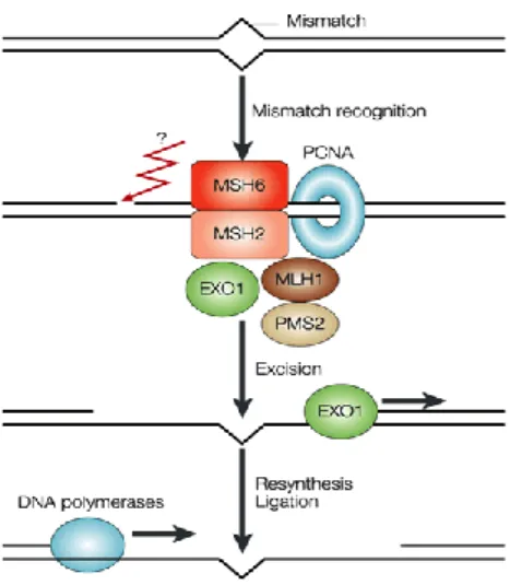
when CRC is diagnosed at a low age or heredity is present. Although not each and everyone with HNPCC will develop colorectal cancer, the risk is greatly increased compared with the general population. The CRC risk is approximately 80% over a lifetime. In HNPCC pa-tients, colonoscopy with polypectomi when needed, each 1-2 years should be performed, starting at 20-25 years of age. This procedure lowers the risk of CRC and extends expected survival [55]. The risk of developing other cancers in e.g. the endometrium, ovaries or pros-tate is increased in HNPCC. The genes that have been associated with HNPCC are inherited germline mutations in DNA mismatch repair (MMR) genes (MLH-1, MSH-2, MSH-6
and PMS-2) (Figure 10) [56]. The consecutive effect is widespread microsatellite instability (MSI).
Familial adenomatous polyposis (FAP)
FAP is a rare hereditary condition in which the patient has hundreds to thousands of polyps in the colon and rectum. FAP accounts for approximately ~1% of all colorectal cancer each year [57]. The polyps are adenomas and not cancer. However, they have the potential to develop into cancer. The polyps begin to occur in teenage years and malignification is complicated to monitor. Colonoscopy is recommended annually from 12 years of age until total colectomy [58]. The majority of tumors develop in the distal colon and rectum. If the colon of a person with FAP is not removed, eventually CRC will be developed. FAP pa-tients can also develop polyps in other gastrointestinal (GI) locations and there is an in-creased risk of GI cancers, desmoids, thyroid cancers and medulloblastoma. Non-cancerous features of FAP include soft tissue tumors like osteomas, skin cysts, dental abnormalities and congenital hypertrophia of the retinal pigment [59]. The gene associated with FAP is germline mutated APC that is an early and crucial step in the adenoma carcinoma se-quence. Patients with FAP have a mutation in the APC gene, which can be inherited (Figure 7) [60].
MYH-gene associated polyposis (MAP) is another inherited, rare form which develops later in life than FAP. People with MAP and FAP have very similar polyp development and the only way to distinguish between the syndromes is with genetic testing [60].
Other forms of hereditary colon cancers are; depending on chromosomal location or single nucleotide polymorphism (SNP´s). Juvenile Polyposis, Peutz-Jegher’s syndrome and PTEN hamartomas have a greater risk for CRC [53].
Embryology and Tumor location
Epidermal growth factors mediate their effect on epithelial cells in the colon and rectum through binding to naturally located epidermal growth factor receptors at the basolateral site of the cell membrane. The epidermal growth factor receptors play a major role in em-bryonic development, morphogenesis and maintenance of the colonic and rectal epithelium as well as in oncogenesis [61]. Survival, migration and mitosis of stem cells and pre-differentiated cells resulting in mature epithelial cells are regulated by the epidermal growth factor receptor (Figure 4).
Figure 4. Proliferation, migration and differen-tiation from stem cells to epithelial cells in the submucosal bowel crypts
Adapted from Nature Reviews Cancer
In CRC, this order of proliferation and differentiation of cells is changed and epidermal growth factor receptors are over expressed in an abnormal, distributional pattern in the complete cell membrane [62]. In all epithelial cell types, epidermal growth factor receptors exist which illustrate their modulatory functions on various differentiated cells of the colon crypt [62]. In a fetus, epidermal growth factor receptors increase in number during gestation and at the same time, ligand affinity decreases but in cancer this autonomal control is lost [63]. Epidermal growth factor receptors EGFR, HER2, HER3 and HER4 are important tar-gets for clinical applications in cancer detection and therapy because they regulate prolifer-ation, invasion, angiogenesis and metastases of cancer cells [63]. The location of tumors related to expression of epidermal growth factor receptors have been studied (Figure 5). EGFR, HER2 as well as HER3 are over expressed throughout the colon but at a higher ex-tent in distal colon and rectal tumors compared to proximal tumors [64, 65].
Figure 5. Tumor location related to phenotype and genetic background.
Adapted from Nature Reviews Cancer
One distinctive embryonic feature is the rudiment of midgut and hindgut resulting in prox-imal and distal colon. The caecum, ascending and 2/3 of the transverse colon derive from the midgut while the colonic segment including splenic flexure, left colon, sigmoid and rectum, derive from the hindgut. The dual blood supply of the colon reflects the different embryonic origins. Regarding innervation of the colon and rectum, it´s divided, where proximal colon is supplied by the vagus nerve, while the distal colon and rectum are main-tained from the S2, S3 and S4 [66]. The distinction between the colon and rectum is ana-tomical but surgical and radiotherapeutic management differs. Tumor location is potential with prognostic and predictive implications [67, 68].
Staging
Diagnostic information in the pathology report is based on the macro as well as the micro-scopic tissue analysis of a CRC tumor and is decisive for endomicro-scopic or surgical removal and for neo adjuvant or adjuvant treatment [69]. It additionally gives feedback to the MDT on the radiological tumor, node, metastases (TNM) staging, quality of surgery and results of onco-logical treatment. The pathologist is recommended to use the latest WHO Classification of Tumors of Digestive System 2010, as of date, the 7th edition of UICC, TNM Classification of Malignant Tumors [70].
T is how deep in the bowel wall the tumor is infiltrating. N gives information on the number of regional lymph node metastases. M is if distant metastases exist. Stages 0 to IV are used (Table 3).
Table 3. TNM and stage.
As an addition to TNM, certain validated risk factors are used to predict recurrence and me-tastases (Table 4) [70].
Risk factors of recurrence and metastases in CRC T4, N1-2, M
Number of analyzed lymph nodes <12 High grade tumor
Vascular invasion Lymphatic invasion Perineural tumor growth Tumor deposits
Tumor budding
Infiltrating tumor border Tumor perforation
Table 4. Pathological risk factors of CRC.
Pathology
According to the simplified paradigm of a CRC tumor, cancer arises from an adenoma esti-mated to take many years to malignify (Figure 6) [71].
Figure 6. Growth of tumor in the bowel wall and stages of CRC.
Adapted from National Cancer Institute
Surgical resection is the most effective treatment of CRC. Histopathological examination of the tumor specimen according to the TNM system can predict prognosis. The depth of tumor infiltration in T3 tumors is related to prognosis and is independent of stage. T4 tumors can break through the serous surface and have a worse prognosis compared to being spread to adjacent organs [72]. Dukes staging system (A-D) is today replaced by the 0-IV staging
sys-tem. The number of analyzed lymph nodes is of importance for correct staging and an indica-tor of surgical and pathology quality and the ruling ground for adjuvant chemotherapy deci-sion [72]. If more than 12 negative regional lymph nodes are found, this is predictive of stage I or II [73]. Low-grade cancer is defined to have ≥50% glandular formations and are well to moderately differentiated while high-grade cancer has ≤50% glandular formations and are low differentiated [72]. Tumor budding is a single cell up to a small cluster of undifferentiat-ed, stemlike cells adjacent to the tumor. It is suggested as the first event of migration of can-cer cells and is a negative prognostic factor [74]. Tumor deposits are larger clusters of tumor cells which are a kind of micro metastases. These are often seen close to the tumor as well [75]. Two types of tumor borders are identified, pushing or infiltrating where the latter is as-sociated to recurrence risk and metastases [74].
The surgeons are recommended to adequately mark important anatomical structures and send the specimen fresh or in formalin to the pathology department. A minimizing of autolysis should be pursued. A tumor is after resection, conserved through formalin fixation and paraf-fin embedded (FFPE). Sliced FFPE tumor sections (3-5µm) are fixed on glass slides and mul-tiple analyses can then be done, including IHC. From tumor tissue, one can extract DNA from fresh but also from fresh-frozen or formalin-fixed paraffin embedded tumor. Quality of DNA and DNA degradation are depending on the e.g. dilution of formalin solution, time to fixation and fixation time. DNA can be used for various types of molecular genetic analyses. Tissue micro array techniques are micro biopsies from the same or different paraffin embed-ded tumors that are analyzed at the same time with immunohistochemistry (IHC) or fluores-cence in situ hybridization (FISH). This is a time and tissue sparing method which enables multiple biomarker analyses in a number of tumors. For clinical practice in CRC, we are not there yet. Today only MSI-testing and KRAS (PCR) are reliable, validated biomarkers. Biobanks of CRC tumors are now established for clinical trials and for individual hospitals. This is going to be of great importance for future research possibilities regarding prognostic refinement and for identifying predictive factors of treatment. All biobanking need informed patient consent, ethical committee approval and a report to the National Board of Health and Welfare Registry of Biobanks.
Inflammation and Immune response
Long lasting inflammatory activity, e.g. ulcerative colitis, predisposes for CRC and in a life-time, 18% of malignancies occur [76]. A combination of colitis, having risk factors of CRC and heredity can have a large effect on incidence and prognosis. Aberrant activities of
JAK/STAT signaling pathways have been implicated in the development and spread of vari-ous cancer entities, among them CRC [77]. An equilibrium exists between immune response and CRC where the JAK/STAT pathway has an impact. The JAK/STAT complex is present in e.g. lymphocytes and is activated by interferons, interleukins, cytokines and growth fac-tors. STAT1 and 3 expression and activity constitute as independent favorable prognostic markers for CRC, where STAT protects the individual from cancer [77].
MSI tumors also present this balance between the immune system fighting the tumor while the tumor is trying to evade immune response [78]. Genetic MMR deficiency results in MSI tumors and represents a specific phenotype in CRC. Epigenetic silencing of DNA mismatch repair and hypoxia have been shown to affect inflammatory bowel disease-associated CRC [79]. Elevated microsatellite alterations at selected tetranucleotide repeats called EMAST is a commonly presented form of MSI that is initiated by inflammation and modulates CRC pro-gression [80].
The HLA-A*02 genotype might have an impact on prognosis since it is associated to ovarian tumors and colon tumors of advanced stages and to a worse prognosis [81, (Villabona et al. “Analysis of immune-related prognostic markers in colon cancer in patients randomized to surgery or surgery and adjuvant cytostatic treatment”, submitted 2015)]. One hypothesis be-hind this is that the HLA-A*02 genotype impairs immune response in CRC.
The HER complex is present in all epithelial cells in a normal state and in an aberrant pattern in CRC. Very little is known of HER3´s over expression in CRC and its relationship to im-mune response. What is evident though is that when inhibiting antibody treatment like cetuximab is given, acquired resistance occurs and HER3 might have an impact [82].
Information on HER3 expression in sporadic colon tumors and how it interacts with MMR and HLA-A*02 genotype is sparse. Different HER complex members can be targets for CTL´s in cancer cells. HER complex expression has been reported to associate to MHC class I. Over expression of HER complex might impair CTL mediated recognition of HLA-A*02 restricted tumor antigens [83, 84].
Metastases in CRC
The definition of metastases is the presence of tumor growth, away from the tumor, in distant lymph nodes or in other organs, documented by radiology, pathology or clinical examination. The survival is poor in CRC of stage IV (Table 2) [85]. The most common locals of CRC metastases are in the liver, the lung, para aortal lymph nodes and other distal lymph nodes
[86]. Among CRC patients, 15-20% have liver metastases at diagnosis, another 20% metasta-size metachronously [87]. Even if CRC is spread to the liver, it is potentially curable with surgery in 20% of all patients and it has markedly increased survival [87]. When investigating tumor biology of synchronous and metachronous colorectal liver metastases and molecular marker expression, it is concluded that most genetic aberrations in the primary tumor are maintained in the CRC liver metastases [88]. Synchronicity might imply a more aggressive disease though biological differences between primaries of synchronous and metachronous groups have been difficult to prove [89]. KRAS mutational status, which is important when considering anti EGFR therapy, is maintained in primary CRC and in corresponding metasta-ses [90]. In metastatic CRC treated with EGFR inhibitors while having high HER3 expres-sion, patients had a worse outcome compared with a low HER3 expressing tumor [91]. This implicates again that HER3 might interact with the response and with acquired resistance to anti-EGFR therapy in cancer [92]. A gene expression signature of the CRC metastases has been suggested and a high expression of these genes in the primary tumor correlated with worse outcome [93].
Genetic and epigenetic development in CRC
Adenoma Carcinoma Sequence
Adenomas are the most important lesions for carcinoma development as well as the sub-group of hyperplastic polyps. It is a continuous progression from normal epithelium to ab-errant crypt foci, to adenoma, to cancer and finally, to metastases. Approximately 10% of adenomas progress to CRC and it takes about 10-15 years [94]. With rising age, about 50% of the population has one or more adenomas in the colon. The size of the adenoma is asso-ciated to high-grade dysplasia [95]. The adenoma carcinoma sequence proposes that specif-ic mutations appear chronologically when a normal cell transforms to a carcinoma with a potential of metastatic behavior (Figure 7).
Figure 7.Markers of oncogenetic events can be seen in purple boxes and phenotypes in red box-es.
Modified from Nature Reviews Cancer
These mutations can affect genes and pathways that regulate differentiation and growth [96]. Mutated proto-oncogenes cause “gain of function”, which means uninhibited proliferation even in the absence of growth signals. Tumor suppressor genes handle “loss of function” in
the cell cycle and when these genes are silenced or mutated, the normal regulation and bal-ance between proliferation and apoptosis is disturbed.
The APC gene mutation, found in ~80% of CRC, is considered the initial event when a cell transforms into an adenoma [94]. The APC mutation is called the “gate keeper” of CRC. When the WNT pathway is active it increases proliferation in a cell and this is an early event in the adenoma carcinoma sequence. When APC is mutated, the WNT pathway is on nonstop and the cell proliferates [97]. A similar mechanism as for WNT is observed for mutated oncogene KRAS, seen in ~40% of CRC. KRAS is active between early and late adenoma transformation. KRAS is located at chromosome 12 and associated with growth of the adenoma through regulation of multiple functions in different pathways [98]. Mutated
BRAF is another proto-oncogene that affects the sequence in early malignant evolution and interacts with the MAPK pathway. BRAF mutation is less frequent (<10%) than KRAS in CRC [99]. The cell proliferation rate will increase with KRAS and BRAF mutations.
SMAD4 and TGFβR2 mutations also contribute in the malignifying process in CRC.
TGFβR2 is considered a driver of MSI [98]. Late events in the sequence are mutated tumor suppressor gene, TP53, “the guardian of the genome” (>50% of CRC) as well as
impair-ment of the whole TGFβ pathway and loss of chromosome 18q (includes SMAD 2 and 4) [98].
An alternative pathway from Fearon and Vogelsteins in CRC is observed for hyperplastic polyps where mutated BRAF starts the process and epigenetic events like promotor methyl-ation and gene silencing keep it going [100]. MSI tumors arise through a methylated or mutated MLH-1 gene or others. Hyperplastic polyps leading to CpG island methylation phenotype (CIMP) and MSI tumors are more frequently seen in proximal colon [101, 102].
The MSI, CIMP and CIN phenotypes
Parallel to the adenoma carcinoma sequence, there are three groups that are both independ-ent and mutually overlapping (Figure 7).
For solid tumors, chromosomal aberrations like aneuploidy or polyploidy are of im-portance. In CRC, about 85% of the tumors follow the adenoma carcinoma sequence and present numerical chromosomal alterations called chromosomal instability (CIN) [98]. In CIN tumors, chromosomal composition drives recurrent gains and losses that affect chro-mosomes in a non-random manner. The cause of CIN is not known, but it is suggested that alterations in the mechanism apparatus when chromosomes segregate in mitosis might be a
cause. CIN tumors are distally located, of low-grade, more often have malignant nodes and metastases and do not have peri-tumoral lymphocytic infiltrate when compared to MSI tu-mors. Associated mutated genes in CIN tumors are APC, KRAS and PI3K (Figure 7) [98]. Tumors in which microsatellite mutations are demonstrated are called microsatellite instable (MSI) and exist in 15% in sporadic CRC but also in HNPCC [102]. The MSI phenotype (de-scribed in page 42) has defects in the mismatch machinery where errors that are introduced during replication, are left unrepaired [102]. At least four genes are involved in dMMR in humans MLH-1, MSH-2, PMS-2 and MSH-6 (Figure 10) [56]. Deficient MMR genes can cause MSI due to either mutations or hyper methylation of promotors that silence MMR genes. The order of succession is that dMMR in sporadic cancer occurs after mutations of
APC and KRAS followed by a cascade of oncogenetic mutations (Figure 7) [103].
The methylation described above is an epigenetic chemical modification of DNA folding that leads to gene expression changes called CIMP [103]. Sporadic biallelic silencing of e.g. the
MLH-1 gene is a form of CIMP. Many genes have promoters embedded in clusters of cyto-sine-guanosine residues, CpG islands. The CIMP phenotype resembles the MSI phenotype but is BRAF mutated, absent of MLH-1 and PMS-2 proteins through methylated silencing of the MLH-1 gene and is diploid. The cause of CIMP is also unknown [103]. Both MSI and CIMP are consequences of a defect MMR system but through different mechanisms. When using IHC, which measures protein expression, both causes are covered. The CIN and MSI phenotype initially were considered mutually exclusive but now are found to partly overlap. The CIMP and MSI phenotype are also overlapping to a large extent [102, 103]
Biomarkers in CRC
The definition of a biomarker is a biological factor that can be of various sorts; peptide, re-ceptor, gene, allele or combinations that are used for risk prediction, as a diagnostic tool, for prognosis or prediction of treatment outcome [104].
The growing knowledge about molecular mechanisms of cancer in general has augmented expectations that compounds and abberations associated to oncogenesis can be used as bi-omarkers. In a typical CRC, about 80 genes are mutated whence only a small fraction is found in the majority of patients and most mutated genes are present at low frequencies. Therefore, a wider perspective then genetics is needed [105, 106]. Below is a table of bi-omarkers in CRC;
Type of biomarker Use Biomarker
Risk Assess cancer development e.g APC, SMAD4, MSH, MLH1, MSH6, PMS2, PTEN and others
Screening Cancer detection in asymp-tomatic population
Blood test, stool test (fecal hemoglobin)
Diagnosis Detect presence of cancer miRNAs, coloscopic mucosal staining Classification Phenotype stratification MSI, CIN, CIMP
Prognosis Outcome not related to therapy
MSI,miRNA,Coloprint,Oncotype DX and others
Prediction Predict response to therapy EGFR, KRAS, BRAF, PTEN, TP53, miRNA, MMR, HER3? HLA-A*02? Panel?
Table 5. CRC biomarkers in different areas.
Many biomarkers have been suggested but so far only KRAS and MSI are used as predictive biomarkers for the decision of therapy.
In recent years, complete cancer genomes, trascriptomes and exomes have been sequenced [106]. Epigenetic mechanisms like DNA methylation profiles have been used for CRC sub-grouping and miRNA have been showed to play a role in CRC [106, 107, 108]. Development of the molecular techniques and target oriented research have increased understanding of the human genome and its complexity as well as cancer specific aberrations. Micro RNA´s are short RNA´s that bind to complementary mRNA molecules, hindering translation to protein.
The non-protein coding transcripts of DNA where miRNA (which modulates cellular pro-cesses) originate from have been conserved throughout evolution [107]. Micro RNA´s circu-late in the blood and certain types of miRNA and changes in levels have been associated with tumor burden and cancer progression [108]. Comparing tumor exomes in individual patients can now identify cancer-driving mutations and sequencing differences between tumor and metastases providing information about genes important for the metastatic process [106].
Stage II, III and biomarkers
Meta analyses have clearly shown a better prognosis of MSI colorectal cancers compared to CIN [109]. The outcome of patients with CRC of stage II and III disease is difficult to pre-dict. Over expression, amplification and mutated HER complex members that signals through the MAPK and PI3K pathways are common in colorectal cancer and therefore a good target of treatment. EGFR inhibiting drugs are therapeutic alternatives to chemotherapy in mCRC. However, when KRAS is mutated and constantly active as well as the MAPK pathway, down-stream of EGFR, this will result in failure of EGFR inhibition. Further, only 30% of the wild-type KRAS patients will respond to EGFR inhibition. This indicates that there are other up-stream or downup-stream effectors involved. A panel of predictive markers for stratifying treat-ment decision might be a way to proceed. Also individual tumor sequencing is interesting as well as an individual comparison of healthy genome to tumor genome [105]. In the future clinical setting, these parameters can be examined further as a step forward in personalized prognostics and prediction in CRC of stage II and III.
HER3
The human epidermal growth factor receptor type 3 (HER3 or human ErbB3) is a transmembranous tyrosine kinase receptor belonging to the HER complex. HER3 is em-bedded in the plasma membrane of the cell and neuregulins are its extracellular ligands. This receptor regulates cellular proliferation, differentiation, apoptosis and migration during embryogenesis and oncogenesis. The HER complex consists of four members, EGFR/HER1/ErbB1, HER2/ErbB2, HER3/ErbB3 and HER4/ErbB4 [110]. When a ligand binds, receptor dimerization takes place. While EGFR, HER3 and HER4 are activated by extracellular ligands, HER2 is an orphan receptor and is nonstop active [110]. The HER complex can be normally expressed, over expressed in an embryological manner and gene amplified or over expressed in cancer. A connection between the HER complex and the
WNT pathway might exist [111]. A challenge in imaging of HER3 is a quite low receptor expression in tumors, usually below 50,000 receptors per cell, together with significant HER3 expression in normal tissues[112]. Dysregulation of the HER complex is associated with a histological, malignant phenotype in CRC where the normal basolateral expression transforms to over expression in the complete cell membrane[113]. Receptors of the HER complex have an extracellular domain, an intracellular tyrosine kinase domain, which acti-vate downstream signaling pathways, and an intracellular C-terminal tail (Figure 8).
Figure 8.Protein structure of HER3.
Adapted from Wikiprofessional
However, HER3 has an inactive intracellular tyrosine kinase domain and its activation de-pends on the heterodimer formation with other HER complex members, preferably HER2 [114]. The HER2-HER3 unit activates downstream signaling pathways, such as P13K/AKT and RAS/MAPK and is considered as one of the most potent heterodimers in tumorigenesis(Figure 9) [115].
Figure 9.HER3 expression, dimerization and function.
Evidence exists on the involvement of HER3 in various cancers, such as breast, prostate and colorectal cancer and its role in resistance to tyrosine kinase inhibiting (TKI) therapies [116]. The HER3 receptor is over expressed in about 70% of colon tumors [117, 118, 119]. Signaling can be affected by interdependency in between the HER complex (cross talk) [110]. For example, in response to trastuzumab (HER2 inhibitor) in HER2 over expressing breast cancer, HER3 can become up regulated and increase the signaling ability of HER2 as a compensation for its inhibition, which causes resistance to therapy[120]. It is also demon-strated that autocrine activation of HER2 with heregulin occurs through dimerization with HER3 in colon cancer[121].
The importance of HER3 in several cancers points to its potential as a molecular target in anti-cancer therapy. This has stimulated the development of appropriate pharmaceuticals [122, 123]. Currently, several HER3-targeting monoclonal antibodies are evaluated in clini-cal trials. To make anti-HER3 therapy effective, patients with HER3 over expressing tu-mors have to be identified.
Regarding the prognostic value of HER3 expression in colon cancer, the significance is not completely evaluated. Several reports have shown that HER3 expression is a prognostic factor in colorectal cancer (CRC) and might be of predictive value when cetuximab is given [124]. HER3 is found to be over expressed in primary colon cancer, rectal cancer, lymph node, liver metastases and it holds a certain prognostic value [117, 119, 125]. Studies indi-cate that the HER complex is a promising target for immunotherapeutic interventions with antibodies (TKI´s) and T-cells based approaches [124, 126].
MMR
Microsatellite instability (MSI) is an epigenetic or genetic instability found in sporadic colon and rectal cancer. Deficient MMR is a type of MSI where DNA errors are not repaired which result in a non-functional protein and in genomic instability. The deficiency or proficiency of MMR reflects the mechanism while MSI is the effect. Microsatellites are repeated units, one to six nucleotides long, usually located in non-coding regions and scattered throughout the genome and these sequences more frequently get errors. Deficient MMR tumors gain or lose repeat units at a higher frequency than in normal epithelial cells in the colon and rectum. Widespread MSI usually indicates mismatch repair deficiency (dMMR) [109]. The MMR genes maintain the stability and integrity of the genome. Inactivation of these genes promotes
accumulation at high speed of tumoral mutations. MMR genes are MLH-1, MSH-2, MSH-6
and PMS-2 (Figure 10) [127].
Figure 10. MMR, genetic function and in-volved genes. Adapted from Nature Reviews Cancer
Deficient MMR and MSI tumors characterizes a highly immunological subtype in CRC. This phenotype presents with topical immune growth control (raised number of cytotoxic tumor infiltrating lymphocytes), proximal colon location of tumor, female gender, high-grade tu-mor, apoptotic behaviour, low number of distant metastases and is of importance to inflam-matory response and cancer control [127]. MSI is observed in several types of cancers; en-dometrial, ovarian, urothelial and prostate [128]. CRC and prostate cancer are linked by he-redity [129]. MSI is used as a biomarker for prognosis and can predict sensitivity to 5-Fluorouracil (5-FU) in colon cancer [130, 131]. Moreover, MSI reveals genetic heterogeneity and expresses frame shift mutated proteins, a unique pool of tumor antigens that can act as T cells mediated immunotherapy targets and might have an immunological connection to HLA-A⃰02 (Figure 11) [132, 133].
HLA-A*02 genotype
Figure 11. HLA and MHC presentation.
HLA´s are variant alleles, expressed in all cells of the organism. MHC is the product of a HLA gene. The HLA-A*02 genotype is one of the most frequent HLA-A alleles in all ethnic populations and the most common haplotype in Sweden (58%) [6]. Red blood cells (no nu-cleus) and neurons (low antigen presentation) have an explicably lower expression of HLA. Cells without HLA expression could be at risk for apoptosis via natural killer (NK) cells acti-vated by absence of MHC class I. Cancer cells develop mechanisms to escape immune recognition. Certain HLA haplotypes are known to correlate to both risk of getting a cancer and prognosis in surviving cancer [133]. The loss or down-regulation of MHC class I has been studied [81]. The different HLA haplotypes have been associated with patients’ progno-sis in several cancers e.g. melanoma and ovarian cancer [134]. The locus of HLA is situated at chromosome 6 and encodes for peptide-presenting proteins. This is the first step in adap-tive immunity and protection against foreign pathogens or abnormalities detected in the cell and in the organism. T-cells recognize tumor cells through antigen presentation on cell sur-face regulated by human leucocyte antigen (HLA). HLA class I molecules, HLA-A, -B, and -C, generally present intracellular peptides. The variation of HLA´s is restricted to the exons. This polymorphism defines a group of peptides that can potentially bind to a specific MHC and respond to infectious or cancer agents [132]. HLA alleles might correlate to other bi-omarkers or predict response to oncological treatments. HLA´s have been studied in different tumors and in colon cancer [133]. When giving immune based therapies to cancer patients, the HLA phenotype is correlated with diverse outcomes [132]. Though, immunotherapies should not solely rely on HLA class I restricted CD8+ T-cells [132, 135].
Two biological hypotheses can be mentioned to justify the selection of HLA-A*02. One hy-pothesis is that having HLA-A*02 genotype reflects an impairment of the immune response to CRC, resulting in a more aggressive disease or that the HLA-A*02 genotype is important in late disease where we see the results [81]. The other hypothesis is that inherited “linkage disequilibrium”, a cluster of HLA alleles can involve nearby located genes for carcinogenesis and immune response. HLA-A*02 are over-represented and is a strong negative prognostic factor in ovarian cancer [81]. Prognosticity of HLA-A*02 has also been reported in prostate cancer [136]. Its relationship to colon cancer is not known but HLA-A*02 genotype has through personal communication of unpublished work been suggested to associate to nega-tive prognosis in females (Villabona et al. “Analysis of immune-related prognostic markers in colon cancer in patients randomized to surgery or surgery and adjuvant cytostatic treatment”, submitted 2015).Patients with HLA-A*02 genotype and CRC might have a significant worse outcome, however the genotype is not a risk factor [81]. It is probably the combined effect of
having HLA-A*02 genotype and loss or down-regulation of HLA class I MHC, that explains the poor prognosis.
A
IMS OF
T
HESIS
________________________________________________________________________
The overall aim of this thesis is to explore the HER3 expression in colon and rectal cancer.
The more specific aims are:
To evaluate the extent of HER3 expression in primary CRC.
To evaluate the extent of HER3 expression in colorectal cancer metastases. To evaluate impact of HER3 on prognosis in colon and rectal cancer.
To investigate if there are associations between HER3 expression and tumor phenotype, tu-mor location or outcome of adjuvant chemotherapy in colon and rectal cancer.
To evaluate the prognostic and predictive effect of combining biomarkers HER3, MMR and HLA-A*02 in colon cancer.
P
ATIENTS AND
M
ETHODS
________________________________________________________________________
Patients
In paper I, III and IV the tumors derived from 236 respectively, 521 respectively, 493 Swe-dish patients with radically resected colon and rectal cancer of stage II and III from 29 hospi-tals in Sweden (Figure 12). Surgery was performed between 1991 and 1997. The follow up ended in November of 2004. Surgical tumor specimens (FFPE) originated from an adjuvant Nordic trial (n=2224) where patients (≤75 years) with CRC were randomized to either sur-gery or sursur-gery plus 5-FU based adjuvant chemotherapy [33].
Figure 12.Flow chart of patients in paper I-IV.
In paper II, 107 patients with CRC and liver metastases were included (Figure 12). These patients all had resected primary tumors and liver metastases. The patients in this study were derived from a population-based cohort (n=255) undergoing liver resection for CRC