A 70-year-old male has excruci-ating pain in the lower left part of his face. This began 1 month ago. He describes it as being like a jolt of lightning that radiates from his left ear, down to his jaw, and to the side of his mouth. These jolts of pain occur numerous times each day. Between attacks his face seems normal. He denies any numbness or tingling sensations. There is no hearing abnormality. The pain is triggered by talking, chewing, or touch of the lower left part of his face. He is unable to eat or brush his teeth, particularly on the left
side, since he fears triggering another painful attack. He can only drink his meals through a straw and cannot lie in bed on his left side. He had the same symptoms about 2 years ago. At that time he was treated with a medication which helped; symptoms subsided, but he stopped taking the medicine. The pain is so distressing that the patient admits to contemplating suicide.
The general and neurologic exam is normal, except that he withdraws and will not let anyone touch the left side of his face.
C L I N I C A L C A S E
There are 12 pairs of cranial nerves emerging from the brain and radiating from its surface (Fig. 15.1). They pass through skull foramina, fissures, or canals to exit the cranial vault and then distribute their innervation to their respective structures in the head and neck. One of the cranial nerves, the vagus (L.,
“wanderer”) continues into the trunk where it innervates various thoracic and abdominal organs.
In addition to being named, the cranial nerves are num-bered sequentially with Roman numerals in the order in which they arise from the brain, rostrally to caudally. The
C H A P T E R 1 5
Cranial Nerves
CLINICAL CASE
OLFACTORY NERVE (CN I) OPTIC NERVE (CN II) OCULOMOTOR NERVE (CN III) TROCHLEAR NERVE (CN IV) TRIGEMINAL NERVE (CN V) ABDUCENT NERVE (CN VI) FACIAL NERVE (CN VII)
VESTIBULOCOCHLEAR NERVE (CN VIII) GLOSSOPHARYNGEAL NERVE (CN IX) VAGUS NERVE (CN X)
SPINAL ACCESSORY NERVE (CN XI) HYPOGLOSSAL NERVE (CN XII) SYNONYMS AND EPONYMS FOLLOW-UP TO CLINICAL CASE QUESTIONS TO PONDER
following list includes their names and corresponding numbers.
I Olfactory nerve.
II Optic nerve.
III Oculomotor nerve.
IV Trochlear nerve.
V Trigeminal nerve.
VI Abducent nerve.
VII Facial nerve.
VIII Vestibulocochlear nerve.
IX Glossopharyngeal nerve.
X Vagus nerve.
XI Spinal accessory nerve. XII Hypoglossal nerve.
Although the cranial nerves and their sensory and parasympathetic ganglia (Tables 15.1, 15.2) form part of the peripheral nervous system, the optic nerve is really an out-growth of the brain that emerges from the prosencephalon (not the brainstem as other cranial nerves), and is therefore not a typical cranial nerve. Moreover, part of the spinal accessory nerve arises from the cervical spinal cord; thus
there are only nine pairs of cranial nerves that emerge from the brainstem.
The main sensory and motor nuclei of the cranial nerves are shown in Fig. 15.2.
In describing the various functional components (modal-ities) of the cranial nerves, the definition of the following terms should be kept in mind: afferentis sensory input; efferent is motor output that may be somatic to skeletal muscles or visceralto smooth muscle, cardiac muscle, and glands, and Figure 15.1 = Ventral view of the brainstem showing the cranial nerves.
Ganglion Cranial nerve association
Trigeminal (semilunar, Gasserian) Trigeminal (V)
Geniculate Facial (VII)
Cochlear (spiral) Cochlear (VIII) Vestibular (Scarpa’s) Vestibular (VIII) Superior glossopharyngeal Glossopharyngeal (IX) Inferior glossopharyngeal Glossopharyngeal (IX)
Superior vagal Vagus (X)
Inferior vagal (nodose) Vagus (X) Table 15.1 = Sensory ganglia of the cranial nerves
special visceral efferent to striated muscles derived from the branchial arches; generalrefers to those components that may be carried by cranial nerves as well as spinal nerves; special refers to functional components that are carried by cranial nerves only. The following categories describe the functional components carried by the various cranial nerves (Table 15.3).
1 General somatic afferent(GSA). These fibers carry gen-eral sensation (touch, pressure, pain, and temperature)
from cutaneous structures and mucous membranes of the head, and general proprioception (GP) from somatic structures such as muscles, tendons, and joints of the head and neck. The trigeminal, facial, glossopharyngeal, and vagus nerves transmit GSA input to the spinal nucleus of the trigeminal nerve.
2 General somatic efferent (GSE). These fibers provide general motor innervation to skeletal muscles derived from embryonic somites. The oculomotor, trochlear, and abducent nerves innervate the extraocular muscles that Mesencephalic tract and nucleus of CN V Edinger–Westphal nucleus Oculomotor nucleus Trochlear nucleus Motor nucleus of CN V Abducens nucleus Superior salivatory nucleus Facial nucleus
Inferior salivatory nucleus Hypoglossal nucleus
Dorsal motor nucleus of CN X Nucleus ambiguus
Spinal accessory nucleus Main sensory
nucleus of CN V
Spinal nucleus of CN V
Nucleus of the solitary tract
Figure 15.2 = The nuclei of the cranial nerves. The sensory nuclei are illustrated on the left, and the motor nuclei on the right.
Ganglion Cranial nerve
association association Trigeminal Function(s)
Ciliary Oculomotor (III) Ophthalmic division Constricts pupil; lens accommodation Pterygopalatine Facial (VII) Maxillary division Lacrimation; nasal gland,
and minor salivary gland secretion Submandibular Facial (VII) Mandibular division Salivation (parotid gland) Otic Glossopharyngeal (IX) Mandibular division Salivation (submandibular
and sublingual glands)
Intramural Vagus (X) Gland secretion;
peristalsis Table 15.2 = Parasympathetic ganglia of
control eye movements, whereas the hypoglossal nerve supplies motor innervation to the muscles of the tongue, mediating movement of the tongue.
3 General visceral afferent(GVA). General sensation from the viscera is transmitted by the facial, glossopharyngeal, and vagus nerves.
4 General visceral efferent(GVE). These fibers provide vis-ceral motor (parasympathetic) innervation to the viscera. The only cranial nerves that transmit parasympathetic fibers are the oculomotor, facial, glossopharyngeal, and vagus nerves.
5 Special somatic afferent(SSA). These fibers carry special sensory input from the eye (retina), for vision, and from the ear (vestibular apparatus for equilibrium, and cochlea for hearing). The only nerves transmitting this component are the optic and vestibulocochlear nerves.
6 Special visceral afferent(SVA). These are special sensory fibers from the viscera. These fibers convey the special sense of smell transmitted by the olfactory nerve and the special sense of taste transmitted by the facial, glosso-pharyngeal, and vagus nerves.
7 Special visceral efferent(SVE). These motor fibers are special because they supply motor innervation to skeletal muscles of branchial arch origin. These fibers are carried by the nerves of the branchial arches, which are the trigeminal, facial, glossopharyngeal, and vagus nerves. Table 15.4 summarizes the modalities, nuclei, ganglia, and functions of the cranial nerves.
OLFACTORY NERVE (CN I)
The bipolar olfactory re-ceptor cells (first order sensory neurons) of the olfactory apparatus reside not in a sensory ganglion,
but instead in the olfactory epithelium (neuroepithelium)of the modified nasal mucosa lining the roof and adjacent upper walls of the nasal cavities (see Fig. 19.1). The axons of these bipolar neurons are SVA fiberstransmitting olfactory sensa-tion. These axons assemble to form bundles, the olfactory fila (L., “threads”), which collectively form cranial nerve I. The olfactory fila traverse the fenestrations of the cribriform plate of the ethmoid bone to terminate in the olfactory bulb where they synapse with second order relay neuronsand inter-neurons(see Chapter 19).
OPTIC NERVE (CN II)
The optic nervemediates the special sense of visionvia its SSA fibers. Light entering the eye activates cells known as rods and cones, the photoreceptors of the retina. Electrical signals generated by the photoreceptors are transmitted to other cells of the retina that process and integrate sensory input. The first order sensory bipolar neuronsof the visual pathway reside in the retina and transmit electrical signals of visual sensory input to the multipolar second order ganglion cellsof the retina. The ganglion cells give rise to unmyelinated axons that converge at the optic disc and traverse the lamina cribrosa, a sieve-like perforated area of the sclera, to emerge from the back of the eyebulb. At this point, the ganglion cell axons acquire a myelin sheath and assemble to form the optic nerve. This nerve, an outgrowth of the diencephalon, leaves the orbit via the optic canal to enter the middle cranial fossa. There, the optic nerves of the right and left sides join each other to form the optic chiasma(G., “optic crossing”) where partial decussation of the optic nerve fibers of the two sides takes place. All ganglion cell axons arising from the nasal halfof the retina decussate(through the central region of the chiasma) to the opposite optic tract. All ganglion cell axons arising from the temporal half of the Modality
General somatic afferent (GSA) General somatic efferent (GSE) General visceral afferent (GVA) General visceral efferent (GVE) Special somatic afferent (SSA)
Special visceral afferent (SVA) Special visceral efferent (SVE)
Function(s) General sensation and general proprioception
Motor supply to extraocular muscles Motor supply to tongue
General sensation from viscera Parasympathetic fibers to viscera Special sensory input from retina Special sensory input from vestibulocochlear apparatus Special sense of smell Special sense of taste Motor innervation to muscles of branchiomeric origin: mandibular, hyoid, 3rd, 4th, and 6th branchial arches Cranial nerves
V, VII, IX, X III, IV, VI XII VII, IX, X III, VII, IX, X II VIII I VII, IX, X V, VII, IX, X
Table 15.3 = Cranial nerve functional components.
The olfactory receptor cells reside in the olfactory epithelium, and not in a sensory ganglion as is typical of other cranial nerves
The optic nerve consists of the myelinated axons of the retinal ganglion cells
Cranial nerve I Olfactory II Optic III Oculomotor IV Trochlear V Trigeminal VI Abducent VII Facial
Table 15.4 = Sensory receptors.
Functional component (modality) SVA SSA GSE GVE (parasympathetic) GP GSE GP SVE GSA GP GSE GP SVE GVE (parasympathetic) SVA GVA Nucleus – – Oculomotor Edinger–Westphal Mesencephalic nucleus of the trigeminal Trochlear Mesencephalic nucleus of the trigeminal Motor nucleus of the trigeminal Main (chief, principal) nucleus of the trigeminal Spinal nucleus of the trigeminal Mesencephalic nucleus of the trigeminal Abducens Mesencephalic nucleus of the trigeminal Facial Superior salivatory Solitarius Solitarius Location of cranial nerve nuclei Telencephalon Diencephalon Mesencephalon (tegmentum) Mesencephalon (tegmentum) Mesencephalon Mesencephalon (tegmentum) Mesencephalon Metencephalon Metencephalon (pons) Metencephalon (pons to C3) Mesencephalon Metencephalon (pons) Mesencephalon Metencephalon (pons) Myelencephalon Myelencephalon Myelencephalon Ganglion – – – Ciliary (parasympathetic) – – – – Trigeminal – – – – Pterygopalatine (parasympathetic) Submandibular (parasympathetic) Geniculate Geniculate Distribution Olfactory mucosa Ganglion cells of retina All extraocular muscles
except the lateral rectus and superior oblique Sphincter pupillae muscle Ciliary muscle All extraocular muscles
except the lateral rectus and superior oblique Superior oblique Superior oblique Muscles of mastication: temporalis masseter medial pterygoid lateral pterygoid Mylohyoid, anterior belly of
the digastric Tensor tympani Tens veli palatini Scalp, anterior two-thirds
of the dura, cornea, conjunctiva, face, paranasal sinuses, teeth, gingiva, and anterior two-thirds of the tongue Muscles of mastication Periodontal ligament Lateral rectus Superior oblique Muscles of facial expression, platysma, posterior belly of the digastric, and stylohyoid Stapedius
Lacrimal gland Glands of the nasal cavity
and palate Submandibular and
sublingual glands Anterior two-thirds of the
tongue
Middle ear, nasal cavity, and soft palate
Function(s) Smell Vision Eye movement Pupillary constriction Lens accommodation Kinesthetic sense Eye movement Kinesthetic sense Chewing Tenses tympanic membrane Tenses soft palate General sensation Muscle stretch sensation Pressure sensation Eye movement Kinesthetic sense Facial expression Tension on stapes Lacrimation Mucous secretion Salivation Taste Visceral sensation
Cranial nerve VIII Vestibulocochlear Cochlear Vestibular X Glossopharyngeal X Vagus XI Spinal accessory XII Hypoglossal Table 15.4 = Continued. Functional component (modality) GSA SSA SSA SVE GVE (parasympathetic) SVA GVA GSA GVE (parasympathetic) SVE SVA GVA GSA SVE GSE Nucleus Spinal nucleus of the trigeminal
Dorsal and ventral cochlear Vestibular complex Ambiguus Inferior salivatory Solitarius Ambiguus Solitarius Spinal nucleus of the trigeminal Dorsal motor nucleus of the vagus Ambiguus Solitarius Solitarius Spinal nucleus of the trigeminal Ambiguus Hypoglossal Location of cranial nerve nuclei Metencephalon (pons) Myelencephalon Myelencephalon Myelencephalon Myelencephalon Myelencephalon Myelencephalon Myelencephalon Myelencephalon Myelencephalon Myelencephalon Myelencephalon Myelencephalon Myelencephalon Myelencephalon Ganglion Geniculate Spiral Vestibular – Otic (parasympathetic) Inferior ganglion of the glossopharyngeal Inferior ganglion of the
glossopharyngeal Superior ganglion of the
glossopharyngeal
Thoracic and abdominal submucosal and myenteric autonomic plexuses – Inferior (nodose) Inferior (nodose) Superior ( jugular) – – Distribution External auditory meatus
and area posterior to ear
Organ of Corti (inner ear) Utricle
Saccule Semicircular canal
ampullae (inner ear) Stylopharyngeus and
pharyngeal constrictors Parotid gland
Posterior one-third of the tongue and adjacent pharyngeal wall Middle ear, pharynx,
tongue, carotid sinus Posterior one-third of the
tongue, soft palate, upper pharynx, and auditory tube Thoracic and abdominal
viscera
Muscles of the larynx and pharynx
Epiglottis
Thoracic and abdominal viscera
Carotid body
Area posterior to the ear, external acoustic meatus, and posterior part of meninges Laryngeal muscles To sternocleidomastoid
and trapezius Muscles of the tongue
Function(s) General sensation Hearing Equilibrium Swallowing Salivation Taste Visceral sensation General sensation Gland secretion, peristalsis Phonation Taste Visceral sensation General sensation Phonation Head and shoulder
movements Tongue movement
retina proceed (through the lateral aspect of the chiasma) without decussatingand join the optic tract of the same side. The ganglion cell axons coursing in each optic tract curve around the cerebral peduncle to terminate and relay visual input in one of the following four regions of the brain: the lat-eral geniculate nucleus, a thalamic relay station for vision; the superior colliculus, a mesencephalic relay station for vision associated with somatic reflexes; the pretectal area, a mesencephalic region associated with autonomic reflexes; and the hypothalamus(see Figs 16.5, 16.7, 16.9).
OCULOMOTOR NERVE (CN III)
The oculomotor nerve
supplies skeletal motor
(somatomotor) innervation to the superior rectus, medial rectus, inferior rectus, and inferior oblique muscles (which move the bulb of the eye) and the levator palpebrae superioris muscle (which elevates the upper eyelid). It also provides parasympathetic The oculomotor nerve provides
motor innervation to four of the six extraocular muscles and the levator palpebrae superioris, and parasympathetic innervation to the sphincter pupillae and ciliary muscles
(visceromotor)innervation to the ciliary and sphincter pup-illae muscles, two intrinsic smooth muscles of the eye.
The triangular-shaped oculomotor nuclear complex is located in the mesencephalon. It is situated ventral to the periaqueductal gray, adjacent to the midline at the level of the superior colliculus. The oculomotor nucleus consists of several subnuclei representing each of the extraocular muscles. These subnuclei are composed of groups of nerve cell bodies of the GSE neurons that innervate the listed extraocular muscles and the levator palpebrae superioris muscle. The cell group innervating the levator palpebrae superioris is located in the midline, sending motor fibers to this muscle bilaterally (both right and left upper eyelids). The cell group innervating the superior rectus sends projections to the opposite side; whereas the cell group innervating the medial rectus, inferior oblique, and inferior rectus sends pro-jections to the same side.
The Edinger–Westphal nucleus, a subnucleus of the oculomotor nuclear complex is located dorsally, medially, and rostral to the GSE nuclear complex. It contains the cell bodies of GVE preganglionic parasympathetic neuronswhose axons join the GSE fibers as they converge and pass ventrally in the midbrain to emerge from the ventral aspect of the brain-stem in the interpeduncular fossa as the oculomotor nerve.
The oculomotor nerve proceeds anteriorly within the cra-nial vault, travels within the cavernous sinus, and by passing through the superior orbital fissure, enters the ipsilateral orbit. Within the orbit, the oculomotor nerve gives rise to
branches carrying the GSE fibers that innervate the levator palpebrae superioris muscle and all but two of the extraocu-lar muscles. The preganglionic parasympathetic fibers of the oculomotor nerve terminate in the ciliary ganglionwhere they synapse with postganglionic parasympathetic nerve cell bodies. Postganglionic parasympathetic fibers exit the gan-glion and reach the sphincter pupillae and ciliary muscles via the short ciliary nerves to provide them with parasympa-thetic innervation. The parasympaparasympa-thetic fibers, when stimu-lated, cause contraction of the sphincter pupillae muscle, which results in constriction of the pupil. Pupillary constric-tion reduces the amount of light that impinges on the retina. Stimulation of the parasympathetic nervous system causes pupillary constriction (whereas stimulation of the sympa-thetic nervous system, which innervates the dilator pupillae muscle, causes pupillary dilation). Ciliary muscle contraction releases the tension on the suspensory ligaments of the lens, changing its thickness to become more convex. This accom-modates the lens for near vision.
GSA pseudounipolar neurons, whose cell bodies reside within the mesencephalic nucleusof the trigeminal nerve, send their peripheral processes to terminate in the muscle spindles of the extraocular muscles. These fibers travel via the branches of the ophthalmic division of the trigeminal nerve. GSA (GP) sensory input is transmitted from the muscle spindles via the spindle afferents centrally to the trigeminal nuclear complex, mediating coordinated and synchronized eye movements by reflex and voluntary control of muscles.
Unilateral damage to the oculomotor nerve results in deficits in the ipsilateral eye. The following ipsilateral muscles will be paralyzed: the levator palpebrae superioris, resulting in ptosis(G., “drooping”) of the upper eyelid; the superior and inferior recti, resulting in an inability to move the eye vertically; and the medial rectus, resulting in an inability to move the eye medially. The eye deviates laterally (due to the unopposed lateral rectus), resulting in lateral strabismus. This causes the eyes to become misaligned as one eye deviates from the midline, resulting in horizontal diplopia(double vision). The inferior oblique is also paralyzed. Since the innervation to the lateral rectus (CN VI) and superior oblique (CN IV) muscles is intact and these two muscles are functional, the eye ipsilateral to the lesion deviates inferiorly and laterally (Fig. 15.3).
The sphincter pupillae muscle becomes nonfunctional due to interruption of its parasympathetic innervation. The pupil ipsilateral to the lesion will remain dilated (mydriasis) and does not respond (constrict) to a flash of light. This may be the first clinical sign of intracranial pressure on the GVE fibers of the oculomotor nerve. The ciliary muscle is also nonfunctional due to inter-ruption of its parasympathetic innervation, and cannot accommodate the lens for near vision (that is, cannot focus on near objects).
C L I N I C A L C O N S I D E R AT I O N S
Wrinkled forehead Raised eyebrow Lid droop Dilated pupil Downward abducted eyeFigure 15.3 = A lesion involving the left oculomotor nerve results in the following symptoms ipsilateral to the side of the lesion: (i) lateral strabismus, (ii) ptosis (drooping of the upper eye lid), (iii) pupillary dilation, (iv) loss of accommodation of the lens, and (v) downward and outward deviation of the eye.
TROCHLEAR NERVE (CN IV)
The trochlear nerve pro-vides motor innervation to only one of the extraocular muscles of the eye, the superior oblique muscle (a common mnemonic is SO4).
The nerve cell bodies of GSE neurons reside in the trochlear nucleus, which lies adjacent to the midline in the tegmentum of the caudal midbrain. Fibers arising from this nucleus initially descend for a short distance in the brainstem and then course dorsally in the periaqueductal gray mat-ter. The fibers decussate posteriorly and emerge from the
brainstem at the junction of the pons and midbrain, just below the inferior colliculus.
The trochlear nerve is unique because it is the only cranial nerve whose fibers originate totally from the contralateral nucleus, it surfaces on the dorsalaspect of the brainstem, and it is the smallest (thinnest) of the cranial nerves. As the trochlear nerve emerges from the brainstem, it curves around the cerebral peduncle and proceeds anteriorly within the cavernous sinus to pass into the orbit via the superior orbital fissure. Consequently, this cranial nerve has the longest intracranial course and is highly susceptible to increased intracranial pressure.
The trochlear nerve is the smallest (thinnest) cranial nerve and the only one whose fibers originate totally from the contralateral nucleus
C L I N I C A L C O N S I D E R AT I O N S
Head tilt Eyes rotate Normal
ANormal BLeft superior oblique paralysis
Head tilt to unaffected side
Right eye intorts Extorted left eye causing double vision
Figure 15.4 = (A) Normal: When the head is tilted, the eyes rotate in the opposite direction. (B) Left superior oblique paralysis following a lesion to the trochlear nerve: the affected eye becomes extorted with consequent double vision. To minimize the double vision, the individual tilts her head toward the unaffected side which intorts the normal eye.
TRIGEMINAL NERVE (CN V)
The trigeminal system con-sists of the trigeminal nerve, ganglion, nuclei, tracts, and central pathways. The trigeminal sensory pathway, which transmits touch, noci-ception, and thermal sensation, consists of a three neuron sequence (first, second, and third order neurons) from the periphery to the cerebral cortex respectively (Figs 15.5, 15.6). The peripheral processes of the first order neurons radiating from the trigeminal ganglion gather to form three separate
nerves, the three divisions of the trigeminal nerve whose peripheral endings terminate in sensory receptors of the oro-facial region. Their cell bodies are housed in the trigeminal ganglion. The central processes of these neurons enter the pons, join the spinal tract of the trigeminal, and terminate in the trigeminal nuclei where they establish synaptic contacts with second order neurons housed in these nuclei. The trigeminal nuclei, with the exception of the mesencephalic nucleus, contain second order neurons as well as interneu-rons. The second order neurons give rise to fibers that may or may not decussate in the brainstem and join the ventral or dorsal trigeminal lemnisci. These lemnisci ascend to relay The trigeminal nerve, the largest of the
cranial nerves, provides the major general sensory innervation to part of the scalp, most of the dura mater, and the orofacial structures
Damage to the trochlear nucleus results in paralysisor paresisof the con-tralateralsuperior oblique muscle, whereas damage to the trochlear nerve
results in the same deficits but in the ipsilateral muscle.
Normally, contraction of the superior oblique muscle causes the eye to intort(rotate inward) accompanied by simultaneous depression(downward) and lateral(outward) movement of the bulb of the eye. This is sometimes referred to as the “Salvation Army muscle” (“down and out”). Intorsion of the eyeball is the turning of the eyeball around its axis, so that the superior pole of the eyeball turns inward. Imagine that extreme intorsion (which we really can-not do) will bring the superior pole of the eye facing the medial wall of the orbit. When the superior oblique muscle is paralyzed, the ipsilateral eye will extort(rotate outward) accompanied by simultaneous upwardand outward movement of the eye (Fig. 15.4B). This is caused by the unopposed inferior oblique muscle and results in external strabismus.
Since the eyes become misaligned following such a lesion, an individual with trochlear nerve palsyexperiences vertical diplopia(double vision), accompanied by weakness of downward movement of the eye, most notably in an effort to adduct the eye (turn medially). The diplopia is most apparent to the individual when descending stairs or while reading (looking down and inward). To counteract the diplopia and to restore proper eye alignment, the individual realizes that the diplopia is reduced as he tilts his head towards the side of the unaffected eye (Fig. 15.4B). Normally, tilting of the head to one side elicits a reflex rotation about the anteroposterior axis of the eyes in the oppo-site direction (Fig. 15.4A), so that the image of an object will remain fixed on the retina. Tilting of the head toward the unaffectedside causes the unaffected eye to rotate inward and become aligned with the affected eye which is rotated outward. Also, pointing the chin downward (“chin tuck”) rolls the normal eye upward.
C L I N I C A L C O N S I D E R AT I O N S (
continued
)
Crossed Crossed Crossed VPM VPM PCG Main sensory nucleus PCG Third order fibers Third order fibers Tactile sense Tactile sense So V1 V2 V3 Si Sc Anterior CN V lemniscus (ventral trigeminal lemniscus) (second order fiber, crossed)Posterior CN V lemniscus (dorsal trigeminal lemniscus) (second order fiber, uncrossed)
mechanoreceptor information: discriminatory, tactile, and pressure
First order fiber V ganglion Aβ or Spinal CN V tract Spinal nucleus Figure 15.5 = The trigeminal pathway for touch
and pressure. Touch and pressure sensation from the orofacial structures is transmitted to the brainstem trigeminal nuclei, the main sensory nucleus, and the spinal nucleus via the central processes of first order pseudounipolar neurons whose cell bodies are located in the trigeminal ganglion. Second order neurons in these nuclei form the posterior and anterior trigeminal lemnisci which terminate in the ventral posterior medial nucleus of the thalamus (VPM). Third order neurons in the thalamus project to the postcentral gyrus. PCG, postcentral gyrus; Sc, subnucleus caudalis;
Si, subnucleus interpolaris; So, subnucleus oralis; V1,
ophthalmic division of the trigeminal nerve; V2, maxillary division of the trigeminal nerve; V3,
trigeminal sensory input to the ventral posterior medial (VPM) nucleus of the thalamus, where they synapse with third order neurons. The third order neurons then relay sen-sory information to the postcentral gyrus (somesthetic cor-tex) of the cerebral cortex for further processing.
The trigeminal nerveis the largest cranial nerve. It pro-vides the major GSAinnervation (touch, pressure, nocicep-tion, and thermal sense) to part of the scalp, most of the dura mater, the conjuctiva and cornea of the eye, the face, nasal cavities, paranasal sinuses, palate, temporomandibular joint, lower jaw, oral cavity, and teeth. It also provides SVE (bran-chiomotor)innervation to the muscles of mastication (tem-poralis, masseter, medial pterygoid, lateral pterygoid), and the mylohyoid, anterior belly of the digastric, tensor tympani, and tensor veli palatini muscles.
The trigeminal nerve is the only cranial nerve whose sen-sory root enters and motor root exits at the ventrolateral aspect of the pons (see Fig. 15.1). The larger, sensory root con-sists of the central processes (axons) of the pseudounipolar sensory neurons of the trigeminal ganglion. These axons enter the pons to terminate in the trigeminal sensory nuclear complex of the brainstem. The motor rootis smaller and con-sists of the axons of motor (branchiomotor) neurons exiting
the pons (Fig. 15.7). The motor root joins the sensory portion of the mandibular division of the trigeminal nerve just out-side the skull, to form the mandibular trunk. Before the motor root joins it, the trigeminal nerve displays a swelling, the trigeminal ganglion, which lies in a bony depression of the petrous temporal bone on the floor of the middle cranial fossa. Since this is a sensory ganglion there are no synapses occurring here. As the peripheral processes of the pseu-dounipolar neurons exit the ganglion, they form three divi-sions (hence “trigeminal,” meaning the “three twins”). These divisions traverse the foramina of the skull to exit the cranial vault on their way to reach the structures they innervate. The ophthalmic divisionis purely sensory and innervates the upper part of the face; the maxillary divisionis also purely sensory (although there may be some exceptions) and inner-vates the middle part of the face. The mandibular divisionis mixed, that is it carries sensory innervation to the lower face and branchiomotor innervation to the muscles listed above.
Trigeminal nuclei
The trigeminal system includes four nuclei: one motornucleus, the motor nucleus of the trigeminal; and three sensory nuclei, the main (chief, principal) sensory nucleus of the trigeminal, the mesencephalic nucleus of the trigeminal, and the spinal nucleus of the trigeminal (see Fig. 15.2; Table 15.5).
Motor nucleus
The motor nucleus of the trigeminal nerve contains the cell bodies whose axons form the motor root of the trigeminal nerve, which provides motor innervation to the muscles of mastication
Crossed VPM PCG Main sensory nucleus Third order fibers Pain and temperature So V1 V2 V3 Si Sc Anterior CN V lemniscus (ventral trigeminal lemniscus) (axon of second order neuron)
First order fiber CN V ganglion Aδ or C
Spinal trigeminal tract
Second order fiber
Figure 15.6 = The trigeminal pathway for pain and temperature. Pain and temperature sensation from the orofacial structures is transmitted to the brainstem subnucleus caudalis (Sc) of the spinal trigeminal nucleus via the
central processes of first order pseudounipolar neurons whose cell bodies are located in the trigeminal ganglion. Second order neurons from the subnucleus caudalis join the anterior trigeminal lemniscus to terminate in the ventral posterior medial nucleus of the thalamus (VPM). Third order neurons from the VPM terminate in the postcentral gyrus (PCG). For other abbreviations, see Fig. 15.5. Main nucleus So V1 V2 V3 Si Sc CN V ganglion Motor root of CN V Motor nucleus of CN V Myelin • Muscles of mastication • Mylohyoid
• Anterior belly of digastric • Tensor tympani • Tensor veli palatini
Figure 15.7 = Branchiomotor innervation of the trigeminal nerve. The motor nucleus of the trigeminal nerve contains the motoneurons whose axons assemble to form the motor root of the trigeminal nerve. The motor root exits the pons and joins the mandibular division of the trigeminal nerve and distributes to the muscles of mastication, the mylohyoid, the anterior belly of the digastric, the tensor tympani, and the tensor veli palatini muscles to provide them with motor innervation. For abbreviations, see Fig. 15.5.
The motor nucleus of the trigeminal is located at the midpontine levels, medial to the main sensory nucleus. It contains interneurons and the cell bodies of multipolaralpha and gamma motor (branchiomotor) neurons whose axons form the motor rootof the trigeminal nerve as they exit the pons. The branchiomotor fibers join the mandibular division of the trigeminal nerve and are distributed to the muscles of mastication as well as to the mylohyoid, anterior belly of the digastric, tensor tympani, and tensor veli palatini muscles.
Sensory nuclei
The sensory nuclei of the trigeminal nerve transmit sensory information from the orofacial structures to the thalamus
The sensory nucleiconsist of a long cylinder of cells, which extends from the mesencephalon to the first few cervical spinal cord levels. Two of these nuclei—the main sensory nucleus and the spinal nucleus of the trigeminal—receive the first order afferent terminals of pseudounipolar neurons whose cell bodies are housed in the trigeminal ganglion. These nuclei serve as the first sensory relay station of the trigeminal system.
The main(chief, principal) sensory nucleus of the trigem-inal nerveis located in the midpons. Based on its anatomical and functional characteristics, it is homologous to the nucleus gracilis and nucleus cuneatus. It is associated with the trans-mission of mechanoreceptor information for discriminatory (fine) tactileand pressure sense.
The mesencephalic nucleus of the trigeminal is unique, since it is a true “sensory ganglion” (and not a nucleus), containing cells that are both structurally and functionally ganglion cells. During development, neural crest cells are believed to become embedded within the CNS, instead of becoming part of the peripheral nervous system, as other sensory ganglia. This nucleus houses the cell bodies of sensory(first order) pseudounipolar neurons, thus there are no synapses in the mesencephalic nucleus. The peripheral large-diameter myelinated processes of these neurons con-vey GPinput from the muscles innervated by the trigeminal nerve and the extraocular muscles, as well as from the peri-odontal ligament of the teeth.
The spinal nucleus of the trigeminalis the largest nucleus of the three nuclei. It extends from the midpontine region
to level C3 of the spinal cord, and is continuous inferiorly with the dorsal-most laminae (substantia gelatinosa) of the dorsal horn of the spinal cord. This nucleus consists of three subnuclei: the rostral-most subnucleus oralis (pars oralis), the caudal-most subnucleus caudalis (pars caudalis), and the intermediate subnucleus interpolaris (pars interpolaris).
The subnucleus oralis merges with the main sensory nucleus superiorly and extends to the pontomedullary junction inferiorly. It is associated with the transmission of discriminative (fine) tactile sense from the orofacial region.
The subnucleus interpolarisis also associated with the transmission of tactile sense, as well as dental pain, whereas the subnucleus caudalisis associated with the transmission of nociception and thermal sensations from the head. The subnucleus caudalis extends from the level of the obex (medulla) to the C3 level of the spinal cord. It is the homo-logue of the substantia gelatinosa since their neurons have similar cellular morphology, synaptic connections, and functions. Since the subnucleus caudalis lies immediately superior to the substantia gelatinosa of the cervical spinal cord levels, it is also referred to as the “medullary dorsal horn.”
The trigeminal nerve does not have any parasympathetic nuclei in the CNS, or parasympathetic ganglia in the periph-eral nervous system. However, it is anatomically associated with the parasympathetic ganglia of other cranial nerves (oculomotor, facial, and glossopharyngeal) and carries their autonomic “hitchhikers” to their destination.
Trigeminal tracts
The spinal tract of the trigeminal nerve consists of ipsilateral first order afferent fibers of sensory trigeminal ganglion neurons and medi-ates tactile, thermal, and nociceptive sensibility from the orofacial region to the spinal nucleus of the trigeminal. The spinal tract of the trigeminal also carries first order sensory axons of the facial, glossopharyngeal, and vagus nerves. These nerves terminate in the spinal trigeminal nucleus, con-veying GVA or GSA sensory input from their respective areas of innervation to be processed by the trigeminal system. The spinal tract descends lateral to the spinal nucleus of the trigeminal, its fibers synapsing with neurons at various levels along the extent of this nucleus. Inferiorly this tract overlaps the dorsolateral fasciculus of Lissauer at upper cervical spinal cord levels.
The ventral trigeminal lemniscus (ventral trigeminothal-amic tract)consists of mainly crossed nerve fibers from the main sensory and spinal nuclei of the trigeminal. This tract relays mechanoreceptor input for discriminatory tactile and pressure sense(from the main nucleus) as well as sharp, well-localized pain and temperature and nondiscriminatory (crude) touchsensation (from the spinal nucleus) to the contralateral ventral posterior medial(VPM) nucleusof the thalamus.
The dorsal trigeminal lemniscus (dorsal trigemino-thalamic tract)carries uncrossed nerve fibers from the main sensory nucleus of the trigeminal, relaying discriminatory Motor nucleus
Sensory nuclei:
• Main (chief, principal) nucleus of the trigeminal • Mesencephalic nucleus of the trigeminal • Spinal nucleus of the trigeminal:
Subnucleus oralis Subnucleus interpolaris Subnucleus caudalis
Table 15.5 = The trigeminal nuclei.
The trigeminal system includes three tracts: the spinal tract of the trigeminal, the ventral trigeminal lemniscus, and the dorsal trigeminal lemniscus
tactile andpressure sense information to the ipsilateral VPM nucleus of the thalamus.
The thalamus also receives indirect trigeminal nociceptive (dull, aching pain) input via the reticular formation (reticulo-thalamic projections).
Trigeminal pathways
Touch and pressure sense
Nearly half of the sensory fibers in the trigeminal nerve are Aβ myelinated discriminatory touch fibers. As the central pro-cesses of pseudounipolar (first order) neurons enter the pons, they bifurcate into short ascending fibers, which synapse in the main sensory nucleus, and long descending fibers, which terminate and synapse mainly in the subnucleus oralisand less frequently in the subnucleus interpolarisof the spinal nucleus of the trigeminal. These fibers descend in the spinal trigeminal tract to reach their target subnuclei. Some second order fibers from the main sensory nucleus cross the midline and join the ventral trigeminal lemniscus to ascend and ter-minate in the contralateral VPM nucleus of the thalamus. Other second order fibers from the main sensory nucleus do not cross. They form the dorsal trigeminal lemniscus, and then ascend and terminate in the ipsilateral VPM nucleus of the thalamus. Descending fibers terminating in the subnu-cleus oralis or interpolaris synapse with second order neurons whose fibers cross the midline and ascend in the ventral trigeminal lemniscus to the contralateral VPM nucleus of the thalamus. The VPM nucleus of the thalamus houses third order neurons that give rise to fibers relaying touch and pressure information to the postcentral gyrus of the cerebral cortex.
Pain and thermal sense
The subnucleus caudalis is involved in the transmission of pain and thermal sensation from orofacial structures
The remaining half of the sensory fibers in the trigeminal nerve are similar to the Aδand C nociceptive and temper-ature fibers of the spinal nerves. As the central processes of pseudounipolar neurons enter the pons, they descend in the spinal tractof the trigeminal and most of them synapse in the subnucleus caudalisof the spinal nucleus of the trigeminal. Nociceptive sensory input relayed in the subnucleus caudalis is modified, filtered, and integrated prior to its transmission to higher brain centers.
Interneurons located in the subnucleus caudalis project superiorly to the subnucleus oralis and interpolaris of the spinal nucleus and to the main sensory nucleus of the trigem-inal, where they modulate the synaptic activity and relay of sensory input from all of these nuclei to higher brain centers. Furthermore, interneurons residing in the subnucleus oralis and interpolaris project to the subnucleus caudalis where they may in turn modulate the neural activity there.
Most of the second order fibers from the subnucleus caudalis cross the midline and join the contralateral ventral
trigeminal lemniscus, whereas others join the ipsilateral ven-tral trigeminal lemniscus. All the fibers ascend to the VPM nucleus of the thalamus where they synapse with third order neurons in that nucleus. The fibers of third order neurons ascend in the posterior limb of the internal capsule to relay somatosensory information from the trigeminal system to the postcentral gyrus of the somatosensory cortex for further processing.
Electrophysiological observations have indicated that electrical stimulation of the midbrain periaqueductal gray matter, the medullary raphe nuclei, or the reticular nuclei, has an inhibitory effect on the nociceptive neurons of the subnucleus caudalis.
Substance P, a peptide in the axon terminals of small-diameter first order neurons, has been associated with the transmission of nociceptive impulses. A large number of substance P axon terminals have been located in the sub-nucleus caudalis. Opiate receptors have also been found in the subnucleus caudalis, which can be blocked by opiate antagonists. These findings indicate that there may be an endogenous opiate analgesic system that could modulate the transmission of nociceptive input from the subnucleus caudalis to higher brain centers.
Motor pathway
The motor root fibers of the trigeminal nerve innervate the muscles of mastication
Branchiomotor neurons housed in the motor nucleus of the trigeminalgive rise to fibers which, upon exiting the pons, form the motor rootof the trigeminal nerve (see Fig. 15.7). This short root joins the sensory fibers of the mandibular division of the trigeminal nerve outside the skull. Motor fibers are distributed peripherally via the motor branches of the mandibular division, providing motor innervation to the muscles of mastication (temporalis, masseter, medial ptery-goid, lateral pterygoid) and the mylohyoid, anterior belly of the digastric, tensor tympani, and tensor veli palatini muscles.
Mesencephalic neural connections
Pseudounipolar neurons of the mesencephalic nucleus transmit general proprioception input to the main sensory and motor nuclei of the trigeminal and reticular formation
The peripheral processes of the pseudounipolar neurons housed in the mesencephalic nucleus of the trigeminal accompany the motor root of the trigeminal as they both exit the pons. These peripheral processes follow: (i) the motor branches of the mandibular division to the muscle spindles of the muscles of mastication; (ii) the orbital branches of the ophthalmic division to the muscle spindles of the extraocu-lar muscles; and (iii) the dental branches of the maxilextraocu-lary and mandibular divisions to the sensory receptors of the periodontal ligament of the maxillary and mandibular teeth, respectively. The central processes of the neurons
transmitting general proprioceptive input from all the muscles and from the periodontal ligament synapse in the main sensory nucleusand in the motor nucleusof the trigeminal, as well as in the reticular formation to mediate reflex responses.
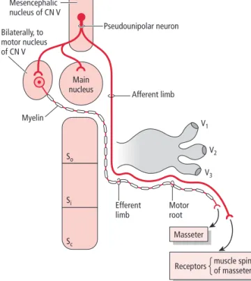
Jaw jerk (masseteric) reflex
The afferent and efferent limbs of the jaw jerk reflex are formed by the branches of the trigeminal nerve
The jaw jerk reflexis a monosynaptic, myotatic (G., “muscle stretch”) reflex for the masseter and temporalis muscles. A hammer gently tapped on the chin causes the intrafusal muscle fibers within the muscle spindles of the (relaxed) masseter and temporalis muscles to stretch, which stimulate the sensory nerve fibers innervating them. The cell bodies of these sensory pseudounipolar neurons are located in the mesencephalic nucleus of the trigeminal(Fig. 15.8). Their peripheral processes, which terminate in the muscle spindles (and are carried by branches of the trigeminal mandibular division), form the afferent limb of the reflex arc. The central processes of these neurons synapse in the motor nucleus of the trigeminal bilaterally, as well as in the main sensory nucleus and the reticular formation. The efferent limb of this reflex arc is formed by the motoneuron fibers traveling to the masseter and temporalis muscles (bilaterally, via motor branches of the trigeminal mandibular division) to cause them to contract and compensate for the stretch.
Main nucleus So V1 V2 V3 Si Sc Motor root Bilaterally, to motor nucleus of CN V Efferent limb Afferent limb Myelin Masseter
Receptors muscle spindlesof masseter Mesencephalic
nucleus of CN V
Pseudounipolar neuron
Figure 15.8 = The jaw jerk reflex. The mesencephalic nucleus of the trigeminal nerve contains the nerve cell bodies of pseudounipolar neurons whose peripheral processes terminate in the muscle spindles of the masseter muscle. Sensory information (about muscle stretch) is carried by the central processes of these neurons to the ipsilateral main sensory and bilaterally to the motor nucleus of the trigeminal nerve. The motor neurons innervating the masseter muscle cause its contraction. For abbreviations, see Fig. 15.5.
Skull fractures may cause a unilateral lesion of the branchiomotor fibers to the muscles of mastication, which will result in a flaccid paralysisor paresiswith subsequent muscle atrophyof the ipsilateral muscles of mastication. This becomes apparent upon muscle palpation when the patient is asked to clench his jaw. When depressing the lower jaw it deviatestowardsthe affected side (weak side) primarily due to the unopposed action of the lateral pterygoid muscle of the unaffected side. This impairs chewing on the lesion side due to muscle paralysis.
Damage to the fibers innervating the tensor tympani muscle results in
hyperacusis(acute sense of hearing) and impaired hearing on the ipsilateral side. Damage to the GSA fibers of the mandibular division will result in loss of sensation from the areas supplied by the branches of this division. Although the trigeminal nerve has an extensive distribution in the head, there is minimal overlapping of the areas innervated by its three divisions, especially in the cen-tral region of the face. Lesions in the peripheral branches of the trigeminal nerve can be located by testing for sensory deficits in the areas that are inner-vated by each of the three trigeminal divisions. If a lesion is located distal to the joining of the autonomic fibers that hitchhike with the trigeminal branches to the lacrimal gland or the salivary glands, then both sensory and autonomic innervation are interrupted.
Infection of the trigeminal ganglion by herpes zoster virus (known as shin-gles) causes a significant amount of pain as well as damage to the sensory
fibers of the three trigeminal divisions (the ophthalmic division is most com-monly infected). This results in loss of sensation on the affected side. Damage to the sensory fibers innervating the cornea (via the ophthalmic division) results in a loss of the corneal reflex when the ipsilateral eye is stimulated (afferent limb damage of the corneal reflex).
Trigeminal neuralgia (trigeminal nerve pain, tic douloureux) A common clinical concern regarding the trigeminal nerve is trigeminal neuralgia. This condition results from idiopathic etiology (unknown cause) and is manifested as intense, sudden onset, and recurrent unilateral pain in the distribution of one of the three divisions of the trigeminal nerve, most com-monly the maxillary division. There may be a trigger zone in the distribution of the affected trigeminal division, and if it is stimulated it may trigger an attack that usually lasts for less than a minute. This condition may be treated pharma-cologically or surgically. Surgical treatment includes sectioning of the affected trigeminal division as it emerges from the trigeminal ganglion or producing a lesion in the trigeminal ganglion. Although these procedures may alleviate the excruciating pain experienced by patients, they also abolish tactile sensation from the affected area. Sectioning of the descending spinal trigeminal tract proximal to its termination in the subnucleus caudalis selectively obliterates the afferents relaying nociception but spares the fibers relaying tactile sensa-tion from the orofacial region.
ABDUCENT NERVE (CN VI)
The abducent nervesupplies motor innervation to the lat-eral rectus muscle, which abducts the eye (a common mnemonic is LR6). The abducent nerve exits the brainstem at the pontomedullary junction, then courses anteriorly, traverses the cavernous sinus, and upon leaving the sinus it passes via the superior orbital fissure into the orbital fossa where it innervates the ipsilateral lateral rectus muscle.
Normally, both eyes move together regardless of the direction of gaze. This is achieved by precise coordinated action of all the extraocular muscles of both eyes. The oculo-motor, trochlear, and abducens nuclei are interconnected and are controlled by higher brain centers of the cerebral cortex as well as by the brainstem. During horizontal gaze, when looking to one side, the lateral rectus muscle of one side and the medial rectus muscle of the contralateral side contract simultaneously.
Abducens nucleus
The abducens nucleus and the internal genu of the facial nerve form an elevation, known as the facial colliculus (L., “little hill”) in the floor of the fourth ventricle. Axons emerging from the abducens nucleus belong to GSE nerve cell bodies. The axons course ventrally in the pontine tegmentum to exit in the ventral aspect of the brainstem at the pontomedullary junction.
The abducens nucleus contains two different populations of neurons (Fig. 15.9). One group (which makes up 70% of the nucleus neurons) consists of the GSE motoneurons, whose axons form the abducent nerve and project to the ipsilateral
lateral rectus muscle. The second group consists of internu-clear neurons. Their axons emerge from the nucleus, immedi-ately decussate and project via the contralateral medial longitudinal fasciculus (MLF) to the contralateral oculomotor nucleus. There the internuclear neuron terminals synapse with motoneurons that project to and innervate the medial rectus muscle. The MLF interconnects the abducens, tro-chlear, and oculomotor nuclei so that the two eyes move in unison. Thus the abducens nucleus mediates conjugate hor-izontal movement of the eyes.
When higher brain centers stimulate the abducens nucleus the following occur simultaneously:
1 Stimulation of the GSE motoneurons of the abducens nucleus that cause the ipsilateral lateral rectus muscle to contract, causing the eye to abduct.
2 Stimulation of the internuclear neurons of the same abducens nucleus that project, via the contralateral MLF, to the contralateral oculomotor nucleus. Here they form excitatory synapses with the motoneurons projecting to the contralateral medial rectus muscle causing it to contract so that the opposite eye adducts, resulting in coordinated lateral gaze.
GSA input from the lateral rectus muscle is transmitted centrally to the trigeminal nuclear complex via the processes of pseudounipolar neurons whose cell bodies are believed to reside in the mesencephalic nucleus of the trigeminal nerve.
Note that the clinical case at the beginning of the chapter refers to a patient suffering from intermittent excruciating unilateral pain in the lower half of the left side of his face.
1 Which cranial nerve provides sensory innervation to the lower half of the face?
2 Pain sensation from the lower half of the face is relayed to the brainstem by sensory neurons whose cell bodies are located in which ganglion?
3 In which brainstem nucleus is pain sensation from the lower half of the face relayed to?
4 Name the thalamic nucleus where pain sensation from the lower half of the face is relayed to.
The abducent nerve innervates only one extraocular muscle, the lateral rectus
The abducens nucleus mediates conjugate horizontal movement of the eyes Left eyeball LR MR MR Right eyeball LR Oculomotor nucleus Abducens nucleus
Center for conjugate horizontal eye movement Excitatory Right MLF CN III CN VI 30% interneuron 70% motoneuron
Figure 15.9 = The connections of the abducens nucleus with the oculomotor nucleus. Note that the abducens nucleus is the center for conjugate horizontal eye movement. It contains two populations of neurons: (i) lower motoneurons whose axons form the abducent nerve that innervates the lateral rectus muscle (LR); and (ii) interneurons whose axons cross the midline and join the contralateral medial longitudinal fasciculus (MLF) to synapse in the oculomotor nucleus with the motoneurons that innervate the medial rectus muscle (MR).
Abducent nerve lesion
A lesion in the abducent nerve causes paralysis of the lateral rectus muscle, resulting in medial strabismus and horizontal diplopia
A lesion in the abducent nerve (GSE, motor fibers) results in paralysis of the lateral rectus muscle that normally abductsthe eye. The eye will then deviate medially as a result of the unopposed action of the medial rectus (Fig. 15.10). The individual can turn the ipsilateral eye from its medial position to the center (looking straight ahead), but not beyond it. This paralysis results in medial strabismus (convergent, internal strabismus, esotropia). Since the eyes become misaligned, the individual experiences horizontal diplopia(double vision; i.e., a single object is perceived as two separate objects next to each other). The diplopia is greatest in an effort to look toward the side of the lesion and it is reduced by looking towards the unaffected side since the visual axes become parallel. The individual realizes that the diplopia is reduced by turning his head slightly so that his chin is pointing toward the side of the lesion. Bilateral abducent nerve lesion results in the individual becoming “cross-eyed.” Abducens nucleus lesion
A lesion involving the abducens nucleus results in medial strabismus, horizontal diplopia, and lateral gaze paralysis
A lesion involving the abducens nucleus (Fig. 15.11) results in the same deficiency as a lesion to the abducent nerve, with the addition of the inability to turn the opposite eye medially as the individual attempts to gaze toward the side of the lesion. This condition, referred to as lateral gaze paralysis, occurs because the damaged abducens nucleus no longer provides excitatory input to the opposite oculomotor nucleus neurons that innervate the medial rectus muscle.
Unilateral medial longitudinal fasciculus lesion: internuclear ophthalmoplegia
A lesion to one MLF results in internuclear ophthalmoplegia
If the oculomotor, trochlear, and abducent nerves and their nuclei are intact, but there is a unilateral MLF lesion, eye movements in all directions are pos-sible. However, since the connections between the nuclei of these nerves are interrupted, horizontal ocular movements will not occur in a conjugate fashion. When there is a lesion of the right MLF, and the individual attempts to gaze to the right, the lesion is not apparent, since both eyes can move simultane-ously to the right. However, when attempting to gaze to the left, the right eye cannot move inward (medially beyond the midline) but the left eye, which should move outward (laterally) in this lateral gaze, does since it is not affected. If you ask this same individual to look at a near object placed directly in front of him, which necessitates that both eyes adduct (converge), he is able to do so. This indicates that: (i) both oculomotor nerves (which innervate the medial recti) are intact; and (ii) the upper motoneurons arising from the motor cortex (which stimulate the motoneurons of the oculomotor nuclei) are also intact. Therefore, a unilateral lesion of the MLF becomes apparent only during conjugate horizontal eye movement, when gazing away from the side of the lesion.
“One-and-a-half”
A rare condition resulting from a lesion near the abducens nucleus, involving the ipsilateral abducens nucleus and decussating MLF fibers arising from the contralateral abducens nucleus
A rare condition referred to as “one-and-a-half” results following a lesion in the vicinity of the abducens nucleus, which involves the entire ipsilateral
C L I N I C A L C O N S I D E R AT I O N S
A B
Figure 15.10 = (A) Medial strabismus of the right eye due to paralysis of the lateral rectus muscle, resulting in diplopia (double vision). (B) To minimize the diplopia, the individual turns her head toward the side of the lesion, which abducts the normal eye.
abducens nucleus as well as the decussating MLF fibers arising from the con-tralateral abducens nucleus. If a lesion is present in the vicinity of the left abducens nucleus the following things happen:
1 The GSE motoneurons, whose axons form the left abducent nerve inner-vating the left lateral rectus, are damaged. Therefore, the left lateral rectus muscle is paralyzed.
2 The internuclear neuronshoused in the left abducens nucleus are also damaged. Their crossing fibers (coursing in the right MLF) do not, there-fore, form excitatory synapses with the motoneurons of the contralateral oculomotor nucleus that innervate the right medial rectus muscle.
3 The crossing fibers of the internuclear neurons arising from the contra-lateral (right) abducens nucleus are also damaged; thus they do not
form excitatory synapses with the motoneurons of the left oculomotor nucleus that innervate the left medial rectus.
Therefore, when attempting to gaze to the left, the left eye will not abduct and the right eye will not adduct during conjugate horizontal gaze to the left. When attempting to gaze to the right, the right eye responds normally, that is it is able to abduct, whereas the left eye will not be able to adduct during con-jugate horizontal gaze to the right. It is important to note that the innervation to all the extraocular muscles of both eyes is intact, except one—the left lateral rectus. If you ask this individual to look at a near object placed directly in front of him, both eyes will converge, since both medial recti and their innervation (branches of the oculomotor nerve) are intact. Thus this type of lesion becomes apparent only during conjugate horizontal eye movement.
C L I N I C A L C O N S I D E R AT I O N S (
continued
)
FACIAL NERVE (CN VII)
The facial nerve(Fig. 15.12)
provides branchiomotor
innervation to the muscles of facial expression, the platysma, the posterior belly of the digastric muscle, the stylohyoid muscle, and the stapedius muscle. It also trans-mits taste sensationfrom the anterior two-thirds of the tongue, as well as parasympathetic (secretomotor) innervation to the lacrimal, submandibular, and sublingual glands. Addi-tionally, it provides general sensationto the back of the ear, pinna, and external auditory meatus, as well as visceral sensa-tionfrom the nasal cavity and the soft palate.
The facial nerve consists of two parts: the facial nerve properand the nervus intermedius. The facial nerve proper is the motor root of the facial nerve consisting of the axons of SVE (branchiomotor) neuronswhose cell bodies reside in the facial nucleus. This nucleus contains subnuclei, each supplying specific muscles or groups of muscles. The nervus
intermedius is sometimes referred to as the “sensory root,” which is a misnomer since in addition to sensory fibers it also carries parasympathetic fibers. The nervus intermedius consists of the axons of the GVE (secretomotor) parasym-pathetic neurons, whose cell bodies reside in the superior salivatory nucleus. It also contains the central processes of first order, sensory pseudounipolar neurons whose cell bodies are housed in the geniculate (L., “bent like a knee”) ganglion, the only sensory ganglion of the facial nerve. Some of these pseudounipolar neurons transmit SVA (taste) sensa-tion from the anterior two-thirds of the tongue, others convey GSA sensation from the area posterior to the ear, whereas others carry GVA sensation from the nasal cavity and soft palate.
Both nerve roots (motor root and nervus intermedius) emerge from the brainstem at the cerebellopontine angle. Near their exit from the brainstem, the two roots of the facial nerve accompany one another to the internal acoustic meatus of the petrous portion of the temporal bone and Left eyeball LR MR MR Right eyeball LR Left oculomotor nucleus Right oculomotor nucleus Right MLF Left MLF Right abducent nerve Abducens nucleus Left abducent nerve 'Damage' 1 2
Figure 15.11 = A lesion of the left abducens nucleus will damage: (i) the lower motoneurons of the abducent nerve, paralyzing the left lateral rectus muscle (LR); and (ii) the interneurons that synapse with the lower motoneurons of the oculomotor nucleus that innervate the right medial rectus muscle (MR). The affected individual is unable to gaze to the side of the lesion (left) during conjugate horizontal eye movement. MLF, medial longitudinal fasciculus.
The facial nerve provides motor innervation to the muscles of facial expression
proceed to the facial canal where the nervus intermedius presents a swelling—the geniculate ganglion.
The facial nerve gives rise to three of its branches in the facial canal: the greater petrosal nerve, the nerve to the stapedius muscle (which innervates the stapedius muscle in the middle ear), and the chorda tympani nerve. The facial nerve exits the facial canal via the stylomastoid foramen and courses to the parotid bed where its main trunk gives rise to numerous muscular branches, which radiate from within the substance of the gland to innervate their respective muscles (muscles of facial expression, platysma, posterior belly of the digastric, and stylohyoid muscles).
The superior salivatory nucleus contains GVE pregan-glionic parasympathetic nerve cell bodies (Figs 15.12, 15.13)
whose axons leave the brainstem via the nervus intermedius. These preganglionic fibers are distributed by the greater pet-rosal and chorda tympani nerves. The fibers in the greater petrosal nerve subsequently join the nerve of the pterygoid canal to enter the pterygopalatine fossa where they terminate and synapse in the pterygopalatine ganglion, one of the two parasympathetic ganglia of the facial nerve. Postganglionic parasympathetic fibers from this ganglion are distributed to the lacrimal gland and the glands of the nasal and oral cavity to provide them with secretomotor innervation. The chorda tympani nerve joins the lingual nerve, a branch of the mandibular division of the trigeminal nerve. The chorda tympani carries preganglionic parasympathetic fibers to the submandibular ganglion (the second parasympathetic Abducens nucleus
Superior salivatory nucleus Solitary nucleus
Facial (motor) nucleus Nervus intermedius Geniculate ganglion
Greater petrosal nerve
Pterygopalatine ganglion
Chorda tympani nerve
Submandibular ganglion Supplies taste to anterior 2/3 of tongue Soft palate, nasal cavity Spinal nucleus of CN V Motor root
Posterior auricular nerve Temporal branch
Zygomatic branch
Buccal branch Mandibular branch Nerve to stylohyoid Nerve to posterior belly of the digastric muscle
Cervical branch
ganglion of the facial nerve), where the fibers synapse with its postganglionic parasympathetic neurons. The postgan-glionic parasympathetic fibers from this ganglion course to the submandibular and sublingual glands providing them with secretomotor innervation.
The geniculate ganglion houses the cell bodies of the SVA neurons, which are responsible for transmission of taste sensation from the anterior two-thirds of the tongue (Fig. 15.14). The peripheral processes of these neurons run in the chorda tympani, and reach the tongue via the lingual nerve of the mandibular division of the trigeminal nerve. The central processes of the SVA neurons enter the brainstem via the nervus intermedius to join the ipsilateral solitary tract and terminate in the solitary nucleus.
Other pseudounipolar neurons of the geniculate ganglion mediate GVA sensation. Their peripheral processes run in the greater petrosal nerve and terminate in the nasal cavity and the soft palate. Their central processes course in the nervus intermedius, join the ipsilateral solitary tract, and terminate in the solitary nucleus.
Still other pseudounipolar neurons of the geniculate gan-glion are responsible for pain, temperature, and touch sensa-tion from the pinna and the external auditory meatus (GSA fibers). The peripheral processes of these neurons terminate in the pinna and the external auditory meatus. Their central processes course in the nervus intermedius and join the spinal tract of the trigeminal, and terminate to synapse in the spinal nucleus of the trigeminal.
Superior salivatory nucleus (GVE)
Preganglionic parasympathetic Nervus intermedius (sensory root of facial nerve)
Geniculate ganglion Greater petrosal nerve Nerve of pterygoid canal
Pterygopalatine ganglion Submandibular ganglion Postganglionic parasympathetic Maxillary nerve Zygomatic nerve Zygomaticotemporal nerve Lacrimal nerve Chorda tympani Synapse Lingual nerve Greater and lesser palatine nerves Palatine minor salivary glands Nasal glands
Salivation Nasal secretions
Lacrimal gland Tears Secretomotor Sublingual branch Submandibular branch Sublingual gland Submandibular gland Salivation (secretomotor) Synapse Posterior inferior nasal nerves
Figure 15.13 = Parasympathetic innervation of the facial nerve. GVE, general visceral efferent.
Telencephalon (neocortex) Diencephalon Pons Medulla Solitary tract
Internal capsule (posterior limb)
Central tegmental tract
Geniculate ganglion Petrosal ganglion Nodose ganglion Chorda tympani CN VII CN IX CN X Parietal operculum and
parainsular cortex Taste buds (anterior 2/3 of tongue) Taste buds (posterior 1/3 of tongue) Taste buds (epiglottis) Parabrachial nucleus VPM of thalamus Solitary nucleus Hypothalamus Amygdala
Figure 15.14 = The gustatory pathway. Taste sensation is transmitted by cranial nerves VII (from the anterior two-thirds of the tongue), IX (from the posterior one-third of the tongue), and X (from the epiglottis). Taste sensation is relayed via the solitary tract to the solitary nucleus. The central tegmental tract arising from the solitary nucleus projects to the parabrachial nucleus and to the ventral posterior medial (VPM) nucleus of the thalamus, hypothalamus, and amygdala. The VPM nucleus of the thalamus projects to the gustatory cortex residing in the parietal operculum and the parainsular cortex. (Modified from Fix, JD (1995) Neuroanatomy. Williams & Wilkins, Media; fig. 20.2.)
A lesion to the facial nerve within the facial canal or near its exit from the stylomastoid foramen causes Bell’s palsy
A unilateral lesion of the facial nerve near its root or in the facial canal prior to giving off any of its branches (thus damaging all of its fibers), results in the fol-lowing conditions ipsilateralto the lesion: damage to the SVE (branchiomotor fibers), results in a flaccid paralysisor paresis(impairment) of the muscles of facial expression, the platysma, stylohyoid, and posterior belly of the digastric muscles with subsequent muscle atrophy. The stapedius muscle will also be
paralyzedand the individual will experience hyperacusis(an acute sense of hearing). Usually the stapedius muscle dampens vibrations of the ossicles, but when it is paralyzed, vibrations from the tympanic membrane are transmitted to the ossicles and subsequently to the inner ear receptors for hearing. Furthermore, damage of the SVA fibers relaying taste results in a loss of taste
from the anterior two-thirds of the tongue. Damage of the GVE parasym-pathetic fibers causes decreased salivary secretionfrom the submandibular and sublingual glands. Since both parotid glands (innervated by a different cranial nerve) and the contralateral sublingual and submandibular glands remain functional, it is difficult to determine from salivary action alone
whether there is an interruption of the parasympathetic innervation to the ipsilateral submandibular and sublingual glands. In addition, the efferent limb of the corneal blink reflex will be damaged.
Bell’s palsymay be idiopathic, or result following trauma or viral infection of the facial nerve within the facial canal or near its exit from the stylomastoid foramen. This condition is characterized by a paresis or paralysis of the muscles of facial expression ipsilateral to the lesion. Bell’s phenomenonis exhibited by individuals with a Bell’s palsy. As the individual attempts to close the eyes, the eye on the affected side deviates up and out.
A unilateral lesion of the facial nerve proximal to the geniculate ganglion causes loss of tear formation by the ipsilateral lacrimal gland. A condition referred to as “crocodile tear syndrome” (lacrimation while eating) may result as follows. As the preganglionic parasympathetic (“salivation”) fibers originating from the superior salivatory nucleus are regenerating, they may be unsuccessful at finding their way to their intended destination, the sub-mandibular ganglion, and instead take a wrong route to terminate in the pterygopalatine ganglion. The fibers then establish inappropriate synaptic contacts with postganglionic (“lacrimation”) neurons whose fibers project to the lacrimal gland.