Summary
The transradial approach increases patient comfort and reduces vascular complications and major bleed-ing. Although modern equipment has been improved and catheter sizes reduced, there remain specific tech-nical challenges in the practice of the transradial approach. Indeed, the transradial approach requires a longer learning curve than transfemoral access, but the transradial challenges are usually overcome with experience. Nowadays, in view of its benefits, there is no longer any justification for ignoring the transradial approach, the patient’s preferred access route for percutaneous coronary interventions. In this paper we highlight five rules which will transform you into a radialist and provide valuable tips and tricks.
Key words: transradial approach; coronary angiog-raphy; percutaneous coronary angioplasty; radial artery spasm; anatomical variations
As the radial artery (RA) is superficial and readily compressed, the transradial approach (TRA) reduces vascular complications and bleeding, major causes of morbidity and mortality, especially for patients under-going percutaneous coronary interventions (PCI). How-ever, TRA requires a longer learning curve than the transfemoral approach (TFA) due to specific technical challenges that are usually overcome with experience. After more than 1000 TRA procedures each, we felt it necessary to share some tips and tricks which will transform you into a TRA adept.
Five rules to transform you into a radialist
Blood supply to the hand is pro-vided by two palmar arches. The superficial palmar arch and the deeper palmar arch, which are important arterial connections
be-tween their major sources, the ulnar artery and the ra-dial artery. The RA is superficial and easily palpable. No major nerves or veins are located near the RA, thus minimising the risk of injury to these structures. Conversely, the ulnar artery is deeper and close to the ulnar nerve.
Rule N°1: be comfortable for radial artery puncture
Patient selection is described elsewhere [1]. RA punc-ture requires a palpable artery and a delicate touch. During the early phase of TRA experience, RA punc-ture is a frequent cause of failure. The groin should al-ways be prepared in case of emergency crossover to TFA.
Arm setup
The arm can be placed on a board anchored under the shoulder at an 80° angle from the body (fig. 1) or, according to the operator’s preference, directly in a support positioned along the body. A folded sheet or roll of gauze is usually placed under the wrist to achieve hyperextension. In our opinion the former setup provides a better wrist position for RA puncture, especially in overweight patients. In this setup the arm is positioned in a support along the body once the sheath has been inserted. The operator elevates the table to its full height to perform the puncture in a com-fortable standing position. Some operators prefer to be seated.
Funding / potential competing interests: No financial support and no other potential conflict of interest relevant to this article were reported.
Correspondence: Dr. Stephane Noble Département des Spécialités Unité de cardiologie interventionnelle Hôpitaux Universitaires de Genève CH-1211 Genève 4
stephane.noble[at]hcuge.ch
How to transform you into a radialist:
tips and tricks
Part II
Caroline Frangosa, Stéphane Nobleb
a Hôpital de La Tour, Meyrin, Genève b Hôpitaux Universitaires de Genève
is important to obtain a good backflow before introduc-ing the guide wire. Once the wire is inserted, a small skin incision at the base of the needle may facilitate sheath insertion, though, depending on the sheath used, it is not always necessary.
Rule N°2: make the patient comfortable
It is essential that the patient is comfortable and relaxed to prevent RA spasm (stress induces catecholamine re-lease and increases the risk of spasm). The procedural steps are explained to the patient by the operator to minimise patient anxi-ety.
Premedication
Anxiety relievers are useful to re-lax the patient, especially with in-experienced operators. In experi-enced hands, premedication need be administered only in anxious patients.
Arm support
The arm and wrist must be placed in a support (fig. 1), along the body, allowing the operator to work in similar conditions to TFA (comfort and distance to the X-ray genera-tor).
Local anaesthesia
Adequate local anaesthesia with subcutaneous xylocaine 1–2% (0.5 to 1.5 ml) with a 25-gauge short needle (2 ml syringe) minimises pain and helps prevent RA spasm. Infiltration of the RA should be avoided since it will increase RA spasm. Delicate massage of the wrist facilitates distribution of the xylocaine.
In a series of patients with ra-dial spasm due to failed attempts at RA puncture [3], the investiga-tors compared three different op-tions: the wait and see attitude, administration of 400 mcg of sub-lingual nitroglycerin or injection of 1 ml (200 mcg/ml) of nitroglycerin solution on the medial and lateral aspect of the RA location. The mean time for the pulse to reappear was 18 ± 5 min, 8 ± 1 min and 3 ± 1 min respectively and subsequently the rate of successful radial
cannula-needle selection and techniques for puncture
Two puncture techniques exist: the double-wall punc-ture technique with a sheath-covered needle, and the single-wall puncture technique with a bare needle, in line with the technique used by 60% of operators world-wide [2]. The choice of needle is a matter of operator’s preference. The RA should be punctured at a 30–45° angle, 1–2 cm proximal to the radial styloid process. It Figure 1
Arm setup for puncture.
A. Arm placed on a board anchored under the shoulder at an 80 degree angle from the body. A folded sheet is placed under the wrist. No venous catheter should be inserted in the area of the radial artery or on the hand. Ideally the venous catheter should be inserted on the contra-lateral arm. Arrow showing the support used to place the arm along the body.
B. Ready for anaesthesia and puncture.
C. Puncture in a standing position with the table elevated to its full height.
Rule N°3: use the right wire
Standard 175-cm long J-shaped 0.035’’ wire is the workhorse, as it progresses towards the arm easily and without resistance in the majority of cases. Neverthe-less, navigation through the brachial and subclavian arteries may sometimes be challenging, due to vessel loops, tortuosities or anatomical variations. The rate of upper limb anatomical variations ranges between 10 and 23% in different series [10] (table 1). Below we de-scribe commonly encountered situations and how to overcome difficulties.
Resistance advancing the wire or catheter.
Spasm or tortuosity, mostly at the level of the elbow, or the presence of a remnant artery (fig. 2) are the most common reason for resistance. The wire should never be advanced aggressively or against resistance. In-stead, radiobrachial injection of diluted contrast should be performed to assess anatomy. The table can be ro-tated for easier visualisation of the arm.
tion was 72%, 90% and 100%. Bertrand et al. refined the technique by routinely mixing xylocaine 2% and ni-troglycerin in the same syringe as the local anaesthesia [4].
sheath size
In cases where the RA is small, a 5 French (F) sheath is preferable, especially when the procedure is ex-pected to be a diagnostic coronary angiogram or the probability of angioplasty is low. If necessary, the angioplasty can be performed via a 5F guide catheter or sheathless guide catheter (Sheathless Eaucath, Asahi Intecc, Japan), or the sheath can be exchanged for a 6F.
Prevention of radial artery spasm
With respect to spasm prevention, success on the first attempt always achieves the best results. In addition to premedication and local anaesthesia, intraarterial ad-ministration of vasodilatators is essential. Various spasmolytic cocktails have been tried [5]. Verapamil is the most commonly used vasodilatator, being adminis-tered directly after sheath insertion in a dose of 2.5 up to 5.0 mg (diluted up to 10 ml with saline). The patient should be warned of a transient burning sensation in the forearm during injection. Mixing verapamil with blood (blood buffers reduce verapamil acidity) and slow injection reduce this unpleasant sensation.
Use of hydrophilic sheaths has been shown to re-duce pain and subsequently spasm. Moreover, use of smaller catheters (5F vs 6F) in the presence of a small RA further reduces the incidence of spasm. Thanks to automatic injectors which overcome the previous limi-tations of 5F catheters from the days of manual injec-tion, the vast majority of radialists routinely perform diagnostic coronary angiograms with 5F catheters re-gardless of the RA calibre and without impairment of image quality. With experience the procedure takes less time and fewer catheter manipulations are neces-sary, resulting in a marked decrease in RA spasm inci-dence.
Prevention of radial artery occlusion (RAo)
RA occlusion is an infrequent (3% to 10%) and clini-cally silent complication of TR catheterisation (in prop-erly selected cases). Unfortunately its occurrence pre-cludes any future TRA. There is evidence that up to half of RA occlusions are recanalised at 30-day follow-up [6]. Heparin has been shown to significantly reduce the incidence of RAO and a clear relationship exists be-tween the heparin dose and the rate of RAO [7]. 5000 IU heparin (or 70 IU/kg) is recommended, and most op-erators use an intraarterial route. Other potential pre-dictive factors of RAO are procedural time, operators in their learning curve, arterial diameter ratio to the sheath and compression time and pressure [8, 9].
Table 1
Most frequent upper limb artery anatomical variations in an angiographic series of 2211 consecutive patients.
– High bifurcation of the RA (8.3%) – Hypoplasia (7.7%)
– Tortuosity (3.8%) – Stenosis (1.7%) – Loop (0.8%)
– Retrooesophageal origin of the right subclavian artery or arteria lusoria (0.45%).
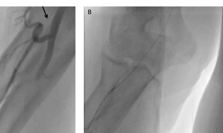
Figure 2
Anatomical variations.
Anatomical variations: a wire can be introduced into a remnant artery with a small arterial diameter (type 3) but failure to advance any catheter is the rule and the incidence of spasm is very high in such anatomy.
change wire (220 cm or 260 cm) or a 175 cm regular wire can be used. With the latter, if the wire is on the aortic cusp at the beginning of the exchange, loss of control of approximately 10 cm of the wire is not an is-sue. When the ascending aorta is difficult to reach, a long exchange wire should be used.
use of respiration
A long history of high blood pressure, especially in oc-togenarians, may be associated with considerable tor-tuosities of the brachiocephalic trunk and subclavian arteries, which may make the procedure more complex. Deep inspiration helps to reach the ascending aorta by modifying the angulation of the brachiocephalic trunk. Left anterior 30º projection is the optimal view for as-sessment of the ascending aorta as the different por-tions of the aorta are not superposed. However, when the wire repeatedly enters the descending aorta de-spite deep inspiration and the use of a JR4 oriented anteriorly, angiographic assessment is the next step. A retrooesophageal origin of the right subclavian artery (arteria lusoria), a rare anatomical variation, may become apparent, rendering the procedure by right TRA highly complex, whereas left TRA is eas-ier.
Usually when catheters reach the ascending aorta they fall into the right cusp, and engagement of the left cusp requires careful withdrawal until the tip retracts
Radial artery tortuosity or loop
A hydrophilic 0.035” J-shaped wire (Glidewire, Teru- mo, Japan) can cross any tortuosity or loop in the vast majority of cases, but must always be introduced under fluoroscopic guidance to ensure proper progress into the arm and ascending aorta without engaging small branches (risk of perforation) and avoid major head and neck vessels (vertebral and carotid arteries). The loop is usually straightened once crossed by the hydro-philic wire or the catheter (fig. 3). In severe radial ar-tery loop, when the 0.035” hydrophilic wire fails, a reg-ular 0.014” hydrophilic coronary angioplasty wire can be used under fluoroscopic guidance.
subclavian artery tortuosity or loop
A hydrophilic 0.035” J-shaped wire is the best choice in this situation. To overcome the difficulties of catheter manipulations once the subclavian level loop is crossed, a stiff wire (eg. Supracore, Abbott vascular) may help to introduce the catheter into the ascending aorta and rotate it for ostium cannulation. In extreme cases right coronary artery (RCA) cannulation may require a 5F guide catheter in association with a stiff wire; this does not impair image quality.
exchanging catheters
Catheter exchange should be performed over a 0.035” wire to avoid losing access (risk of spasm). A long
ex-Figure 3 Radial loop.
A. Large loop on the radial artery. Arrow showing a very small remnant artery.
Routine diagnostic catheter selection
For diagnostic angiography, 5F Judkins left (JL) 3.5 and Judkins right (JR) 4.0 remain the most commonly used catheter types according to the First Interna-tional Transradial Practice Survey [2], allowing a can-nulation success rate of over 90%. Dedicated single catheters to cannulate left and right systems have been developed (eg. Tiger [Terumo] and Kimny [Boston Sci-entific Corp, USA] catheters). Few operators use spe-over the right aortic leaflet and moves into the left
cor-onary cusp. Gentle inspiration may also facilitate movement into the left coronary cusp and help selec-tive left system cannulation.
Rule N°4: choose the right catheter
Selection of the catheter is important if good quality images are to be obtained and to ensure good support if PCI is necessary.
Figure 4
Barbeau guide catheter in a case of complex RCA PCI.
A. Antero-posterior and cranial projection showing a tortuous and diffusely diseased RCA (diagnostic JR 4 catheter).
B. RCA PCI performed with a 6F Barbeau guide catheter: after predilatation, positioning of a 3.0 × 38 mm Xience Prime drug-eluting stent (arrows) over a BMW wire. The Barbeau guide catheter provides a good backup support since it is well-seated against the posterior wall of the aorta (arrow head). A second wire (Buddy wire technique) is also used to improve stent delivery.
C. Insertion of a second Xience Prime stent (3.5 × 38 mm) proximal to the first one (arrows). D. Final result.
(in 1% of our RCA PCI), because we believe the benefits of this catheter are outweighed by a higher risk of os-tium dissection. Indeed, the Barbeau (Cordis Corpora-tion, Johnson and Johnson, USA) is our guide catheter of choice, especially for vertical RCA takeoff and when more backup support is required (fig. 4).
Rule N° 5: limit compression time
Since the artery is superficial, haemostasis is simple and easy to control. After removing the catheter over a 0.035”, the sheath is gently pulled back. Forceful pull-ing back of the sheath may result in avulsion of the RA [11]. Although a compressive bandage (simple gauze cial curves such as Amplatz left and right or
multipur-pose, and TRA pioneers have developed their own ded-icated catheters (eg. Barbeau and Fajadet).
Routine guide catheter selection
For left coronary interventions, the majority of opera-tors use extra backup guide catheters, the most popu-lar being the EBU 3.5 (Medtronic Inc) or XB 3.5 (Cordis Corporation), since the JL guide catheter does not offer adequate support by TRA. For RCA PCI, JR is the most commonly used catheter. When anatomical difficulties with the RCA require better support, some operators use the Amplatz left. We rarely use this guide catheter Figure 5
Sheathless catheter characteristics.
The sheathless 7.5F guide catheter (Asahi Intecc) has an inner diameter equivalent to a 7F conventional guide catheter and an outer diameter (2.49 mm) inferior to a 6F sheath (2.62 mm). Similarly, the 6.5F guide catheter has an inner diameter equivalent to a 6F conventional guide catheter and an outer diameter (2.16 mm) smaller than a 5F sheath (OD 2.29 mm).
and elastic bandage) is an option used in developing countries, the insertion of a compression device (TR-Band, Hemoband, Radistop, Easy radial, Radstat) is recommended. The “airbag”-based bracelet (TR-Band, Terumo) with progressive deflation is the most com-monly used dedicated radial compression device. To re-duce RA occlusion, compression time should ideally be limited to 3 hours (with progressive release). The con-cept of patent RA haemostasis has recently been
pro-moted (RA pulse assessment by plethysmography) to reduce the incidence of RAO, Pancholy et al. having shown a 75% RAO rate reduction at 30 days when ap-plying patent haemostasis under guided RA compres-sion [12]. The amount of air inflated in the TR-band usually varies between 13 to 18 ml. A Spanish study [9] showed that when the amount of air inflated was guided by mean arterial pressure (mean air volume of 8.8 ml), significantly fewer RAO were observed than Figure 6
Telescoping technique.
A. Picture showing the wide gap between an 0.035’’ wire and the 6F guide catheter.
B. Picture illustrating the telescoping technique with a long (125 cm) 5F Judkins right diagnostic catheter introduced through a 6F guide catheter.
C. Zoom on the distal end of the 5F diagnostic catheter. The gap from the 0.035’’ wire is undoubtedly smaller.
Table 2
Preferred route for each pattern of CABG.
Type of CABG Preferred route Catheter suggested
LIMA LRA Internal mammary curve
SVG to LCA (originating from the left side of ascending aorta)
LRA easier JR4
AL1, AL2 LCB SVG to RCA (originating from the right side
of ascending aorta)
LRA easier Multipurpose
JR4 RCB AL1, AL2
LIMA and RIMA Femoral
Double radial approach RRA
LCA = left coronary artery; LIMA = left internal mammary artery; LRA = left radial artery; RCA = right coronary artery; RIMA = right internal mammary artery; RRA = right radial artery; SVG = saphenous vein graft.
when the device was routinely inflated with 15 ml air. In our experience, 13 ml air is sufficient in the vast ma-jority of cases.
How to manage specific situations
Radial artery spasm
The loss of RA pulse is not only frustrating, but may render TRA nearly impossible. Spasm may occur either during puncture (increased risk with repeated punc-tures) or diagnostic and therapeutic procedures (usu-ally related to excessive catheter manipulation or mul-tiple catheter exchange). RA spasm is generally associ-ated with severe pain in the arm, justifying the administration of anxiety relievers and/or sedation.
RA spasm during the procedure
If spasm occurs during coronary angiography, admin-istration of anxiety relievers and a calcium-channel blocker (verapamil 2.5 mg) and/or intraarterial nitro-glycerin (100 to 250 ug) is helpful. Smaller size diag-nostic catheters (i.e, 4F instead of 5 or 6F) are a valid option. When a 6F or 7F guide catheter is needed, two options are available if catheter insertion is difficult or painful:
Sheathless catheter
Recently, sheathless hydrophilic guide catheters have emerged as an alternative when the RA is too small or in cases of spasm preventing the use of a 6F guide cath-eter. Sheathless guide catheters have an outer diame-ter (OD) more than 1F smaller than traditional guide catheters making 6F PCI in frail women, or complex Figure 7
Haematoma classification (published with permission of Dr O. Bertrand, Laval Hospital, Quebec; Modified from Bertrand et al. Circulation 2006; 114(24):2646–53. Table realised for standardisation of post-procedural haematoma assessement in the EASY study.
Grade I II III IV V
incidence ≤6% <3% <2% ≤0.1% <0.01%
Definition Local hematoma Hematoma with Forearm hematoma Hematoma and Ischemic
superficial moderate and muscular muscular threat
muscular infiltration, below infiltration (compartment infiltration the elbow extending above syndrome)
the elbow
Treatment Analgesia Analgesia Analgesia Analgesia Consider
Additional Additional Additional Bracelet Additional Bracelet sugery
Bracelet Bracelet Local ice Local ice
Local ice Local ice Inflated BP cuff Inflated BP cuff
Notes Inform physician Inform physician Inform physician STAT call to physician Remarks:
– Control blood pressure (BP): importance of pain management
– Consider interruption of any anticoagulation and/or antiplatelet infusion
– Follow forearm and arm diameters to evaluate requirement for additionnal bracelet and/or BP cuff inflation – Additional bracelet can be placed alongside artery anatomy
– Ice cubes in a plastic bag or washclosh are placed on the hematoma – Finger O2 saturation can be monitored during inflated blood pressure cuff
– To infalte blood pressure cuff, select a pressure of 20 mmHg < systolic pressure and deflate every 15 minutes – After bracelet removal, use «Velpeau bandage» around forearm/arm for a few hours to maintain mild positive pressure
PCI [13–14] requiring a 7F guide catheter possible (fig. 5). When using these sheathless catheters, be aware that the dilator is radiotransparent and hence the dilator should be held in place once the radio-opaque sheathless guide reaches the aortic cross, to prevent any potential damage to the aortic cusps.
Telescopic technique (mother and child technique).
This involves advancing a 6 or 7F guide catheter over a long 5F diagnostic catheter (125 cm JR 4). The 5F long diagnostic catheter serves as a dilator and reduces the mismatch between the catheter and wire (fig. 6). It has the advantage of allowing the use of any guide catheters you are accustomed to. On the other hand, the sheathless system described above is hydrophilic and moves forward very easily.
extreme spasm
Extreme radial or brachial artery spasm may prevent the removal of the catheter or sheath. On very rare oc-casions when anxiety relievers are not enough, deep se-dation/general anaesthesia may be required to relieve spasm and allow catheter removal.
Small calibre radial artery
RA is often smaller in women but this does not pre-clude radial access. Spasm is more likely to occur in the presence of small RA and in women, and thus special attention must be paid to anxiety, pain relief and punc-ture accuracy. Operators may be restricted to smaller conventional guide catheters to perform PCI, or they can use sheathless guide catheters or the telescoping technique. Moreover, contralateral RA should be checked as asymmetry of RA diameter may be present. A left radial approach can be adopted if left RA diam-eter seems significantly larger and hand collaterals testing is favourable.
Patients with coronary artery bypass grafts (CABG)
TRA can be successfully used for patients with CABG. However, transradial cannulation of the saphenous vein grafts (SVG) may be technically demanding. The preferred route for each pattern of CABG is described in table 2. TRA catheterisation of left internal mam-mary grafts (IMA) is easier than TFA. In cases of dou-ble IMA, catheter advancement into the contralateral subclavian artery has to overcome multiple angula-tions and high friction in the absence of adequate sup-port. It is therefore reasonable to favour TFA in pa-tients with bilateral IMA grafts even though successful contralateral access (preferably by right TRA) to the IMA have been reported [15]. Double TRA may be con-sidered in cases of inappropriate TFA access.
Local bleeding and haematoma
Vascular complications with TRA are extremely rare. Nevertheless, haematoma of the arm proximal to the elbow (non-access site related but secondary to small branch perforation or arterial laceration) must be promptly detected to ensure rapid management. Ber-trand et al. [16] graded forearm haematomas according to size with a 5 grade classification and provided the appropriate strategy to manage every grade (fig. 7). In cases of large or growing haematoma, a blood pressure cuff should be rapidly inflated at the site of the haema-toma at 20 mm Hg below the systolic pressure for 15 minutes, with repeated inflation if needed. This manœuvre allows sealing of the perforation, softening of the haematoma, and prevention of further swelling and the subsequent risk of compartment syndrome. In cases of late management, extensive arm haematoma may cause compartment syndrome, a very serious but fortunately very rare complication of TRA occurring in less than 0.01%. Surgical decompression may be re-quired in this clinical situation. Interestingly, when perforation of the RA occurs during a procedure, the best treatment may be to continue the procedure. When a catheter can cross and then tamponade the area of concern, the perforation will often seal by the end of the intervention.
Left transradial approach
The vast majority of operators (90%) use the right TRA as the initial access site since it allows the procedure to be performed with similar comfort to TFA (the right arm is positioned along the patient’s body). Left TRA can be used as a routine access or as an alternative to right TRA when unsuccessful. Left TRA is similar to TFA with respect to catheter selection and manipula-tion, and enables easier cannulation of the ostia than right TRA. Furthermore, in patients with left IMA graft catheterisation of the IMA is easier.
Patient preparation is similar to that for right TRA with a board under the left shoulder and the wrist in hyperextension. After obtaining left RA access, the pa-tient’s left arm is brought across the chest onto the ab-domen (left wrist close to the right groin area). A cush-ion is placed under the left shoulder to ensure arm sta-bility and patient comfort.
For left TRA, JL (3.5 or 4.0) and JR (4.0) catheters are the first choice. Progression of the wire can be dif-ficult through the elbow due to the half-bent arm and temporarily straightening the arm can be helpful. Sometimes use of a hydrophilic wire under fluoroscopic guidance may be necessary. Once the catheter is intro-duced into the brachial artery, the arm is repositioned on the abdomen.
In obese patients and particularly for shorter oper-ators, left TRA might be technically more demanding since the operator has to lean over the patient to reach the left wrist. Notably, since the operator stands closer to the X-ray generator in left TRA, there were some concerns regarding higher operator radiation exposure after left TRA than after right TRA. However this was not observed in the TALENT Dosimetric Substudy [17] which compared both TRA in a randomised trial in-cluding 770 patients in each group.
Conclusion
Specific technical challenges related to TRA are most often overcome with experience. When difficulties are encountered at any stage of wire progression, perfor-mance of an angiographic assessment is highly remended. Understanding the problem will prevent com-plications (dissection and perforation) and allow suc-cessful management. Knowing the anatomic variations and the technical recommendations to overcome such situations will undoubtedly help new TRA converts in achieving success.
References
1 Frangos C, Noble S. How to transform you into a radialist: literature review. Part I. Cardiovascular Medicine. 2011;14(10):277–82. 2 Bertrand OF, Rao SV, Pancholy S, et al. Transradial approach for
coro-nary angiography and interventions. Results of the first international transradial practice survey. J Am Coll Cardiol Intv. 2010;3:1022–31. 3 Pancholy SB, Coppola J, Patel T. Subcutaneous administration of
ni-troglycerin to facilitate radial artery cannulation. Catheter Cardiovasc Interv. 2006;68:389–91.
4 Bertrand OF, Larose E, Rodes-Cabau J. Sub-cutaneous nitroglycerin: good example of the “KISS” rule! Letter to the Editor Catheter Cardio-vasc Interv. 2006;70:161.
5 Varenne O, Jégou A, Cohen R, et al. Prevention of arterial spasm dur-ing percutaneous coronary interventions through radial artery: The SPASM study. Cathet Cardiovasc Interv. 2006;68:231–5.
6 Stella PR, Kiemeneij F, Laarman GJ, et al. Incidence and outcome of radial artery occlusion following transradial artery coronary angio-plasty. Catheter Cardiovasc Diagn. 1997;40:156–8.
7 Spaulding C, Lefèvre T, Funk F. Left radial approach for coronary an-giography: results from a prospective study. Cathet Cardiovasc Diagn. 1996;39:365–70.
8 Dahm JB, Vogelgesang D, Hummel A, et al. A randomized trial of 5 vs 6 French transradial percutaneous coronary interventions. Cath-eter Cardiovasc Interv. 2002;57:172–6.
9 Cubero JM, Lombardo J, Perdrosa C, et al. Radial compression guided by mean artery pressure versus standard compression with a pneu-matic device (RACOMAP). Catheter Cardiovasc Interv. 2009;73: 467–72.
10 Velsecchi O, Vassileva A, Musumeci G, et al. Failure of transradial ap-proach during coronary interventions : anatomic considerations. Catheter Cardiovasc Interv. 2006;67:870–8.
11 Arzamendi D, Romeo P, Gosselin G. Radial artery avulsion: a rare com-plication of percutaneous coronary intervention. Rev Esp Cardiol. 2011; 64:62.
12 Pancholy S, Coppola J, Patel T, et al. Prevention of radial artery occlu-sion-patent hemostasis evaluation trial (PROPHET study): a rand-omized comparison of traditional versus patency documented hemosta-sis after transradial catheterization. Catheter Cardiovasc Interv. 2008; 72:335–40.
13 Mamas MA, Fath-Ordoubadi F, Fraser DG. Atraumatic complex tran-sradial intervention using large bore sheathless guide catheter. Catheter Cardiovasc Interv. 2008;72:357–64.
14 Mamas M, D’Souza S, Hendry C, et al. Use of the sheathless guide cath-eter during transradial percutaneous coronary intervention: a feasibil-ity study. Catheter Cardiovasc Interv. 2010;75:596–602.
15 Burzotta F, Trani C, Hamon M, et al. Transradial approach for coronary angiography and interventions in patients with coronary bypass grafts : tips and tricks. Catheter Cardiovasc Interv. 2008;72:263–72. 16 Bertrand OF, De Larochellière R, Rodés-Cabau J, et al. A randomized
study comparing same-day home discharge and abciximab bolus only to overnight hospitalization and abciximab bolus and infusion after transradial coronary stent implantation. Circulation. 2006;114: 2636–43.
17 Sciahbasi A, Romagnoli E, Trani C, et al. Operator Radiation Exposure During Percutaneous Coronary Procedures Through the Left or Right Radial Approach: The TALENT Dosimetric Substudy. Circ Cardiovasc Interv. 2011;4(3):226–31.