Journal of Chemical and Pharmaceutical Research, 2015, 7(6):592-600
Research Article
CODEN(USA) : JCPRC5
ISSN : 0975-7384
Formation constants of some transition metal complexes of
2-hydroxybenzaldehyde [(1Z, 2E)-2-(hydroxyimino)-1-methyl propylidene]
hydrazone in mixed Solvent System
P. A. Shimpi
1,2*and R. G. Deshmukh
21
B. N. N. College, Bhiwandi, Dist: Thane, India
2Kokan Gyanpeeth Karjat College (ASC), Karjat, Dist: Raigad, India
_____________________________________________________________________________________________
ABSTRACT
A new ligand 2-hydroxybenzaldehyde[(1Z,2E)-2-(hydroxyimino)-1-methyl propylidine] hydrazone (HBHMPH) synthesised and reported for the first time in this work has been studied potentiometricaly in mixed solvent system (Dioxane + water ) at various ionic strengths to obtain the acidity constant of the ligand. Further, due to the perceived interests in the coordinating behaviour of this ligand with various transition metal ions, the potentiometric studies in solution similar to those for the ligand were carried out to obtain the metal-ligand formation constants. The studies revealed that the ligand behaves a monobasic acid in spite of two probable dissociable protons being present in it viz. the oximino proton and the phenolic proton. The nature of this monobasic character is studied. The metal complexes of Co (II), Ni (II), Cu (II), and Zn (II) show that the most stable species in
the ML2 complex.
Key words: Ionic strength, stability constant, metal ion complexes, potentiometric titration.
_____________________________________________________________________________________________
INTRODUCTION
Oximino ketones and their derivative have been widely reported with respect to their synthesis application such as analytical [1], bioinorganic system [2] catalyst [3,4] medicine[5,6]. This is primarily because of the inherent ambidentate character of the ligand so obtained as also due to the various positions of probable donor atoms in the ligand. As such, these ligands are known to report metal complexes of varied geometries, stabilities and applications.
With this interest in mind we are reporting in the present work a new ligand 2-hydroxybenzaldehyde[(1Z,2E)-2-(hydroxyimino)-1-methylpropylidine]hydrazones (HBHMPH) synthesised from a reaction between hydrazone of diacetyl monoxime and salicylaldehyde.
Formation studies in solution of the metal complexes Co (II), Ni (II), Cu (II), Zn (II) at various ionic strength in a dioxane: water (60 : 40 v/v) mixed solvent system have been studied in order to appreciate nature of complexation, stabilities of the complexes and effect of ionic strength on complexation in order to obtain the thermodynamic stability constant for the formation of metal complexes.
.
EXPERIMENTAL SECTION
SYNTHESIS OF LIGAND (HBHMPH)
O
H3C CH3
N OH
+
H2N NH2.H2O60
0C
- H2O
N
H3C CH3
N OH
NH2
DAMO HDAMO
N
H3C CH3
N OH
NH2
+
CHO
OH
H N
N C H3C N
H3C
OH
OH
HDAMO Structure of Ligand HBHMPH
6 g (0.05 M) Hydrazones of Diacetylmonoxime in 50 cm3 ethanol was mixed with 6.4 cm3 (0.05 M) Salicyaldehyde with constant stirring. After 5 minutes the yellow colour product was formed. The completion of reaction was checked by TLC. The yellow product formed was filtered, washed with distilled water and was recrystallized from ethanol to obtain shiny yellow crystals.
Structure of the ligand was confirmed by NMR, IR and UV-VS spectral studies.
Reagents and Materials
The stock solution of the ligand (0.05M) was prepared by dissolving the requisite quantity of the ligand in a minimum volume of dioxane subsequently diluted to final volume with dioxane. All chemicals used were A. R. grade KCl, HCl and KOH. Chlorides of metal ions were used to metal ion solution (0.01M) standardized by standard EDTA solution, volumetrically.[13] The ionic strength 0.05, 0.075, 0.1 mol dm-3 was maintained by using 1M KCl solution. The carbonate free potassium hydroxide solution (0.1M) was prepared in double distilled water and standardized against standard succinic acid solution.
Apparatus and Procedure
The pH meter (model EQ-610) equipped with combined glass electrodes and magnetic stirrer (accuracy ± 0.01). The pH meter was standardized before each titration with buffer solution of pH 4.00, 7.00 and 9.20 prepared from a Qualigens buffer tablets. All the pH metric titration were carried out 28°C in an inert atmosphere by bubbling oxygen free nitrogen gas through it and served the purpose of stirring the solution being studied. The pH-metric titrations of following solutions were performed against standard 0.1M KOH solution:-
a) Acid titration: 5cm3 of 0.100 mol dm-3 HCl + 30 ml dioxane + 13 cm3 distilled water.
b) Ligand titration: 5 cm3of 0.100 mole dm-3 HCl + 5cm3 of 0.05 mol dm-3 ligand +25cm3 dioxane + 13 cm3 distilled water.
c) Metal titration: 5cm3 of 0.100 mole dm-3 HCl + 5cm3 of 0.05 mole dm-3 ligand + 5cm3 of 0.010 mole dm-3 metal salt +25cm3 dioxane +8.05cm3 distilled water.
RESULTS AND DISCUSSION
Characterization of the ligand: (HBHMPH)
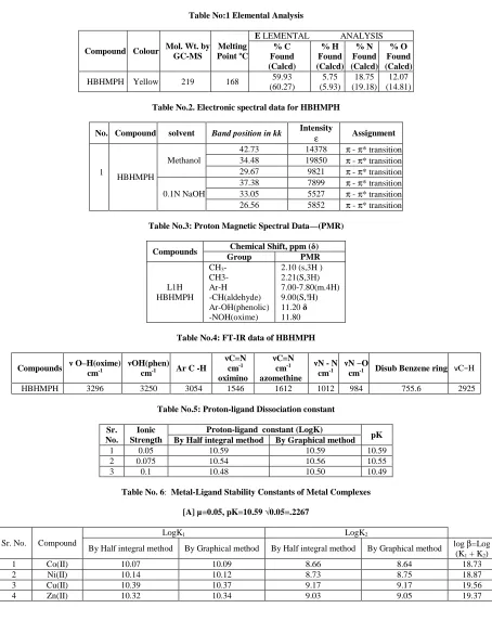
The analytical data of the ligand like elemental analysis, PMR, I.R, electronic spectra are given in Table No. 1, 2, 3 & 4. The molecular weight of the ligand was found by GC-MS spectra. Molecular formula corresponds to C11H13O2N3 . It was obtained in crystalline form, which melts at 168 0C. Solubility of ligand in dilute alkali indicates
the acidic nature of the oximino proton. The colour of ligand is yellow and soluble in chloroform, ethanol and dilute alkali. The molecular formula of ligand is C11H13O2N3.
Table No:1 Elemental Analysis
Compound Colour Mol. Wt. by GC-MS
Melting Point ºC
E LEMENTAL ANALYSIS
% C Found (Calcd) % H Found (Calcd) % N Found (Calcd) % O Found (Calcd)
HBHMPH Yellow 219 168 59.93
(60.27) 5.75 (5.93) 18.75 (19.18) 12.07 (14.81)
Table No.2. Electronic spectral data for HBHMPH
No. Compound solvent Band position in kk Intensity ε Assignment
1
HBHMPH
Methanol
42.73 14378 π - π* transition
34.48 19850 π - π* transition
29.67 9821 π - π* transition
0.1N NaOH
37.38 7899 π - π* transition
33.05 5527 π - π* transition
26.56 5852 π - π* transition
Table No.3: Proton Magnetic Spectral Data—(PMR)
Compounds Chemical Shift, ppm (δ) Group PMR
L1H HBHMPH
CH3-
CH3- Ar-H -CH(aldehyde) Ar-OH(phenolic) -NOH(oxime)
2.10 (s,3H ) 2.21(S,3H) 7.00-7.80(m.4H) 9.00(S,!H) 11.20 δ 11.80
Table No.4: FT-IR data of HBHMPH
Compounds ν O–H(oxime) cm-1
νOH(phen)
cm-1 Ar C -H
νC=N cm-1 oximino νC=N cm-1 azomethine
νN - N
cm-1
νN −O
cm-1 Disub Benzene ring νC−H
HBHMPH 3296 3250 3054 1546 1612 1012 984 755.6 2925
Table No.5: Proton-ligand Dissociation constant
Sr. No.
Ionic Strength
Proton-ligand constant (LogK)
pK By Half integral method By Graphical method
1 0.05 10.59 10.59 10.59
2 0.075 10.54 10.56 10.55
3 0.1 10.48 10.50 10.49
Table No. 6: Metal-Ligand Stability Constants of Metal Complexes
[A] µ=0.05, pK=10.59 √0.05=.2267
Sr. No. Compound
LogK1 LogK2
By Half integral method By Graphical method By Half integral method By Graphical method log β=Log (K1 + K2)
1 Co(II) 10.07 10.09 8.66 8.64 18.73
2 Ni(II) 10.14 10.12 8.73 8.75 18.87
3 Cu(II) 10.39 10.37 9.17 9.17 19.56
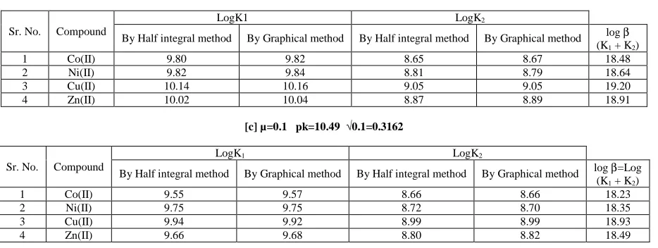
[image:3.595.79.533.166.739.2][B] µ=0.075 pk= 10.55 √0.075=0.2738
Sr. No. Compound
LogK1 LogK2
By Half integral method By Graphical method By Half integral method By Graphical method log β
(K1 + K2)
1 Co(II) 9.80 9.82 8.65 8.67 18.48
2 Ni(II) 9.82 9.84 8.81 8.79 18.64
3 Cu(II) 10.14 10.16 9.05 9.05 19.20
4 Zn(II) 10.02 10.04 8.87 8.89 18.91
[c] µ=0.1 pk=10.49 √0.1=0.3162
Sr. No. Compound
LogK1 LogK2
By Half integral method By Graphical method By Half integral method By Graphical method log β=Log (K1 + K2)
1 Co(II) 9.55 9.57 8.66 8.66 18.23
2 Ni(II) 9.75 9.75 8.72 8.70 18.35
3 Cu(II) 9.94 9.92 8.99 8.99 18.93
[image:4.595.76.543.83.259.2]4 Zn(II) 9.66 9.68 8.80 8.82 18.49
Table 7: Ionic strength and Metal stability constant
Ionic Strength µ
√µ Co Logβ Ni Logβ Cu Logβ Zn Logβ
0.05 0.227 18.60 18.78 19.42 19.21
0.075 0.2738 18.41 18.58 19.14 18.89
0.1 0.3162 18.28 18.41 18.92 18.61
UV-Visible spectral properties:[Table No.2] The electronic absorption spectrum of HBHMPH in methanol, in the
ultra-violet region reveals two high intensity bands at 42.73kK (ε=14378) and 34.48 kK (ε=19580) respectively. These may be due to π→π* transitions possible from the azomethine and oximino environment in the molecule. In several isonitrosoketone the band in the range 36.00- 40.00is reported to be due to π→π* transitions. In isonitrosopropiophenone, the π→π* transition band is observed at 39.68 kK. The band at 34.22 kK in the Ultra-Violet spectrum of HBHMPH is therefore believed to be due to the oxime group while the one at 42.73kK could be due to the hydrazonyl group. The ultra-violet spectrum of HBHMPH in dilute NaOH solution shows that the band at 34.48 kK in the methanolic solution spectrum has suffered a suppression along with a bathochromic shift to 33.11
kK (ε= 5527). This observation characteristically indicates the formation of the anion HBHMP- in the alkaline solution due to the deportation of the oximino group. This means that this band could have
its origin in the oximino linkage in the molecule as such, the band at 42.73 kK in the methanolic solution spectrum could be assigned to the azomethine linkage.
The PMR spectra data for ligand (HBHMPH) [Table No.3]
PMR spectral data of HBHMPH in D6DMSO reveals two singlet peaks at 11.8δ and 11.2δ. The intensity of the
peaks suggests that these are due to single deshielded protons. There are two likely such highly deshielded protons in the molecule: the oximino proton and phenolic proton. Many isonitrosoketones [2-4] reveal the oximino proton( =N-OH) in the region 8δ to 12δ in hydrozonyl derivative of isonitrosopropiophenol (HINPP) reported to be observed at 8.1δ [6] Of the two single proton, the oximino proton is likely to be more acidic than the phenolic proton in view of greater resonance stability of resultant anion.
HBHMPH = HBHMP - + H+
>C−C=N−OH→ >C−C=N−O- + H+↔ >C=C−N=O
Hence it is suggested that the peak at 11.8δ is due to oximino proton while the one at 11.3δ due to phenolic proton. In diacetylmonoxime it is reported at 11.70δ.while in HBHMPH it is appeared at11.8δ. In salicylaldehyde phenolic (-OH) proton reported at 11.00δ while in HBHMPH it occur at 11.2δ this indicate phenolic proton more shielded than oximino proton hence oximino proton easily releasable than phenolic proton. The singlet at 2.10δ and 2.21δ, 9.00δ assigned to the methyl group, methyne occur at the usual position in the HBHMPH. A broad multiplate between 7.00δ to 7.80δ has is origin in the phenyl ring protons of the HBHMPH.
IR Spectra of HBHMPH – [Table No.4]
the salicylaldehyde moiety and oximino OH group with both groups merged together since the OH vibrations are also expected to lower down from their usual range ~3500 cm-1 on account of H- bonding. The sharp band at 1612 cm-1 may be due to the perturbed > C=N stretching vibration of the azomethine (>C=N-N) group in ligand. In general, the >C=N vibration in isonitrosoketone[2-5] are known to occur at 1600 cm-1. Similar band occur at 1603, 1617cm-1 in benzyl-α-monoxime semicarbazone, benzyl-α-monoxime thiosemicarbazone [19]. These observations confirm the position of azomethine group at 1612 cm-1. The sharp band low intensity at 1546 cm-1 shows νC=N
stretching vibration of the oximino ( >C=NOH ) group in ligand. In general, the >C=N vibration in isonitrosoketone are shown to occur around 1600cm-1 In isonitrosopropiophenone and its hydrozonyl derivative this band is observed at 1597cm-1 and 1595 cm-1. While in diacetylmoxime it occurs at 1630 cm-1. This suggests unaffected nature of the oximino group in ligand. In ligand N-O stretching vibration band seen at 984cm-1. This band was seen at 997cm-1 in hydrozonyl derivative of isonitrosopropiophenone while in diacetylmonoxime this band occurs at 970 cm-1. The sharp band was seen at 755 cm-1 indicates HBHMPH contain disubstituted benzene ring.
From the above information from electronic spectra, PMR, IR spectra the suggested structure of the ligand is-
HBHMPH
FORMATION OF COMPLEXES IN SOLUTION
The ligand is colourless in 60: 40 % (V/v) dioxane water mixture. During the course of acid-ligand titration against 0 .1M KOH, light yellow coloured appeared at low pH. AS the titration goes on, the yellow colour become deepens at high pH ~ 8.80. This is because of the dissociation of proton from the oxime –OH group.
Similarly during metal ion titration, different colours are observed depending upon metal ions. In case of cobalt- reddish colour occur while in nickel- brown colour and in copper- green colour, zinc- yellow coloured appeared at different pH. Thus the colour changes significantly indicate formation of complexes.
The proton-ligand dissociation constant-
The ligand used in present investigation may be considered as monobasic acid containing only one dissociable proton from oxime (-NOH) group supported by PMR IR, electronic spectra and it therefore represented as HL. The dissociating equilibrium can be shown as
HL→ H+ +L-
The proton-ligand formation number nA, calculated by Irving-Rossetti expression. The pKa value of ligand
calculated by half integral method and graphical method. This is reported in Table no. – 5.
It can be seen that the pK values decreases with increase in ionic strength of the medium according to Debye Huckel theory. The proton-ligand formation number nA value less than 1.5 indicate that ligand containing only one
dissociable H+ ion from oxime.
Metal-ligand stability constant –[Table N0. 6]
Titration curves of the metal-ligand solution reveal that the n values range from 0 to 2.This indicate that at least two formation constants of the metal-ligand complex may be shown as a two step reaction’
M2+ + HL= ML− + H + and ML− + HL= ML2+ H+. Overall reaction is
Figure:1 Proton and metal ligand titration curves
The first step giving logK1 while the second step yielding logK2 value for the M-L bond stability. The curves also
reveal in all cases that the metal-ligand titration curves lie below those of the proton-ligand titration curves [Fig.No.1] indicating that the metal ligand complexes are more stable than the proton-ligand complexes.
The formation curves for the metal complexes were obtained by plotting the average number of ligand attached per metal ion(n) Vs. pL the free ligand exponent according to the Irving and Rossotti[14].The LogK1 and LogK2
calculated by n= 0.5 and n=1.5 respectively at different ionic strength.[Fig.No.2]. Higher value of LogK1 and
LogK2 showed that ligand are strong chelating agent and vice versa. . LogK1 and LogK2 also calculated by point
wise method-
log (n /1-n) Vs pL , log(2-n /n-1) Vs pL [Fig.No.3].The LogK1 and LogK2 given in Table- 6 From the data it is
found that-
1) The maximum value of n was about two indicating the formation of ML2 (metal: ligand) complexes only.
2) The order of the stability constant of metal complexes with respect in schiff bases was found to be Co(II) ˂ Ni (II)˂Cu(II) ˃ Zn(II) which is also in agreement with Irving and Williams order[18].
3) The order of stability constant decreases with increasing ionic strength according to Debye Huckel theory. 4) The difference between LogK1 and Log K2 complexes less than 2.5 indicating the simultaneous formation of 1:1
Figure:2 Formation of Cobalt complex
Calvin and Wilson [16] showed that resonance might affect the formation and stability of a chelate. They considered the formation of Cu2+ complexes of acetylacetone and some other β-diketones, of a group of substituted salicylaldehyde and 2-hydroxy-1-naphthaldehyde and 3-hydroxy-2-naphthaldehyde. All gave the same chelated stable rings and thus confirmed this view. Mellor et al [17] studied the stability of salicylaldehyde complexes in 50:50 v/v dioxan-water medium. They found that chelates follow the order as Pd > Cu > Ni > Co > Zn > Cd > Fe > Mn > Mg
Figure:3 Graphical representation: A represents log(n /1-n) verses pL B represents log(2-n /n-1) verses Pl
CONCLUSION
HBHMPH ligand is insoluble in water, but soluble in dilute alkali. It indicate it is in acidic nature and soluble in common organic solvent.
The dissociation constant of ligand is pK ~10.5. The order of stability constant and dissociation constant pK decreases with increases with ionic strength. While the order of stability constant with respect to metal ion is Co (II)˂ Ni (II) ˂ Cu(II) ˃Zn (II) which is in according with Irving –Williams natural order .
Acknowledgements
The authors are grateful to the Heads of the Chemistry Department, B. N. N. College, Bhiwandi and Kokan Gyanpeeth Karjat College (ASC), Karjat for providing the necessary facilities.
REFERENCES
[2] N. J. Patel and B.C. Haldar, J.Inorg. Nucl. Chem., 1967, 42,843. [3]N. V. Thakkar and B. C. Haldar, J Inorg. Nucl. Chem., 1980, 42,843 [5] R.G. Deshmukh, and N.V. Thakkar, Ind. J Chem., 1985, 24A, 1066. [6] R.G.Deshmukh, Ph.D. Thesis, Univ. of Bombay, 1992.
[7] A. Chakrabarti and B. Sahoo, Ind.J. Chem., 1981, 20A, 431. [8] S.Das, M. C. Chattopadhyaya. J. Ind. Chem. Soc., 2006. 83, 922.
[9] Omar M. EI-Roudi, Samir A. Abdel-Latif, J. Chem. Eng. Data., 2004, 49, 1193. [10] A.B. Naik, M.L. Narwade. Russ.J. Inorg., Chem., 2009, 35, 932.
[11] Tuba Sismanogh, Chinese chemical letters, 2003,14,1207
[12] Santosh D.Deosarkar, Ashok B. Kalambe, Der Pharma Chemica, 2011, 3(6); 75-83.
[13] G. H. Jeffery, J. Bassett , Vogel Textbook of Quantitative chemical analysis- 1956, 5th Ed. Longan publication.
[14] H. Irving and H.S. Rossotti. J Chem. Soc., 1953, 3397.
[15] R. Shakru, Sathish Kumar, K, Vijay Kumar Chityala, shivraj, J. Adv. Scient. Res. 2011, 2(4) 58-62. [16] M. Calvin and K.W. Wilson, J. Am. Chem. Soc., 1945,67,2330.
[17] D.P. Mellor and L. Maley, Nature., 1947,159, 370 [18] H. Irving and R. Williams, Nature., 1948,162, 746 . [19] Satish pingle, M.Sc. Thesis, Univ. of Mumbai, 2006.