Journal of Chemical and Pharmaceutical Research, 2014, 6(12):499-504
Research Article
ISSN : 0975-7384
CODEN(USA) : JCPRC5
Coumarins from the stem bark of Feronia limonia
Tjitjik Srie Tjahjandarie
Natural Products Chemistry Research Group, Organic Chemistry Division, Department of
Chemistry, Faculty of Science and Technology, Universitas Airlangga, Surabaya, Indonesia
_____________________________________________________________________________________________
ABSTRACT
Three coumarins were isolated from the stem bark of Feronia limonia namely as auraptene (1), osthol (2), and xhantotoxin (3). Their structures were elucidated by spectroscopic methods including UV, IR, HRESIMS, 1D and 2D
NMR analysis. Compounds 1–3 were evaluated for their cytotoxic properties against HeLa cells, showing their IC50
were 65.09, 7.62, and 21.51 ppm, respectively.
Keywords: Auraptene, Osthol, Xhantotoxin, Coumarin, Feronia limonia, Cytotoxic
_____________________________________________________________________________________________
INTRODUCTION
Feronia limonia belongs to the family Rutaceae, commonly known as Kawista in Indonesia. The phytochemical
investigations on Feronia limonia from different parts of this plant, have isolated various compounds, including alkaloids [1,2], coumarins [3,4,5], flavonoids [6,7], and tyramine derivatives [8]. In continuation of our phytochemical work of Indonesian Feronia plants aiming to find coumarin compounds from Feronia limonia, we report the isolation of coumarin compounds, auraptene (1), osthol (2), and xhantotoxin (3) from the methanol extract of the stem bark of Feronia limonia. The cytotoxic activity of compounds 1– 3 against HeLa is also briefly described.
EXPERIMENTAL SECTION
The stem bark of Feronia limonia was collected in July 2014 from Panjunan Village, Tuban District, East Java, Indonesia. The plant was identified at Herbarium Bogoriense, Center of Biological Research and Development, National Institute of Science, Bogor, Indonesia, and the voucher specimen was deposited in the herbarium. The dried and powdered stem bark of Feronia limonia (4.0 kg) were macerated in methanol at room temperature two times, and the methanol extract was evaporated under reduced pressure to give a dark brown residue (170 g). Furthermore, the methanol extract was partitioned with n-hexane and ethyl acetate. The ethyl acetate extract (47 g) was separated by vacuum liquid chromatography on silica gel eluted with n-hexane–ethyl acetate mixture with gradient amount of ethyl acetate (90:10, 80:20; 50:50 and 30:70) to give four major fractions A-D.The separation of fraction C (1.2 g) by flash chromatography with n-hexane-ethyl acetate (from 8:1 and 7:3) to give three subfractions C1-C3. Further purification of subfraction C2 (150 mg) by radial chromatography with n-hexane-diisopropylether
(from 9:1; 8:2, and 7:3) to give compound 1 (18 mg) and 2 (12 mg). The purification of subfraction C3 (100 mg) by
______________________________________________________________________________
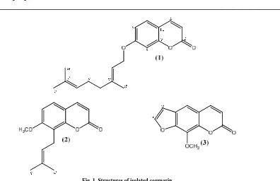
Fig. 1. Structures of isolated coumarin
Auraptene (1), pale yellow solid, UV (MeOH) λmaks nm (log ε): 224 (3.21), 289 (3.08), and 324 (3.43) nm.
HRESIMS m/z [M+H]+ 299.1650 (calcd for C19H23O3, 299.1647). 1H NMR (400 MHz, CDCl3): see Table 1. 13C
NMR (100 MHz, CDCl3): see Table 1.
Osthol (2), pale yellowsolid, UV (MeOH) λmaks nm (log ε): 235 (3.15), 255 (2.61), and 322 (3.01) nm. HRESIMS
m/z [M-H]- 229.0861 (calcd for C14H13O3, 229.0865). 1H NMR (400 MHz, CDCl3): see Table 2. 13C NMR (100
MHz, CDCl3): see Table 2.
Xhantotoxin (3), yellowsolid, UV (MeOH) λmaks nm (log ε): 248 (3.19), 264 (2.94), and 303 (2.90) nm. 1H-NMR
NMR (400 MHz, CDCl3): see Table 3. 13C NMR (100 MHz, CDCl3): see Table 3.
Cytotoxicity assay: Cytotoxic properties of the isolated compounds 1–3 on human cervical cancer (HeLa) were evaluated according to the method of MTT assay as described previously [9,10]. The cytotoxicity assay was performed against HeLa cells grown in RPMI 1640 medium containing 10% fetal bovine serum, 2 mg mL-1 sodium carbonate, 100 µg mL-1 penicillin sodium salt, and 100 µg mL-1 penicillin streptomycin sulfate. The cells were harvested at the log phase of growth, and then seeded into 96-well plates (1 × 10⁴ cells/well). After 24 h incubation at 37 ºC and 5% CO2 to allow cell attachment, the cultures were exposed to the test compounds 1-3 in DMSO at
various concentrations and incubated for 48 h followed by MTT [3-(4,dimethylthiazol-2-yl)-2, 5-diphenyltetrazolium bromide] assay at 570 nm
.
RESULTS AND DISCUSSION
Three coumarins, auraptene (1), osthol (2), and xhantotoxin (3) were isolated from the stem bark of Feronia limonia.
Their structures were elucidated with extensive by UV, IR, HRESIMS, 1D and 2D NMR spectra.
Auraptene (1) was obtained as pale yellowsolid, showed a quasimolecular ion [M+H]+ at m/z 299.1650 consistent to the molecular formula of C19H23O3. The UV spectrum of 1 exhibited absorption maximum for a coumarin structure
at λmax 224, 289, and 324 nm. The 1H-NMR spectrum of compound 1 showed three aromatic proton signals for ABX
system at δH 7.34 (1H, d, J = 8.6 Hz, 5); 6.83 (1H, dd, J = 8.6, 2.4 Hz, 6); and 6.80 ppm (1H, d, J = 2.4 Hz,
H-8). The 1H NMR spectrum of 1 also showed a pair doublets (J = 9.6 Hz) cis vinylic signal at δH 7.61, and 6.23
methine vinyl groups (δH 5.45, and 5.06 ppm). The 13C NMR spectrum of 1 (APT experiment, Table 1) showed the
presence of 19 carbon atom signals. Two carbon signals (δC 162.2 and 155.9 ppm) characteristic for the region
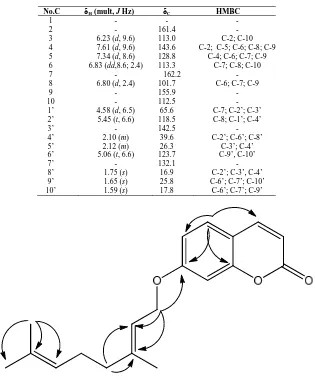
oxyaryl signals which indicate that the structure is a derivative of 7-hydroxycoumarin. The correlation of the one bond and the two/three bond 1H-13C compound 1 can be seen in the HMQC and HMBC spectra (Table-1). The presence of a geranyl group at C-7 showed in the HMBC spectrum, the long-range correlation between a proton signal methylene at δH 4.58 ppm with two quaternary atoms at δC (162.2 (C-7), 142.5 (C-3’) and one methine carbon
at δC 118.5 (C-2’). Based on data from 1D and 2D NMR of compound 1 is 7-O-geranylumbeliferon or known as
auraptene [3]. Other HMBC correlations consistent with the structure 1 are shown in Table 1.
Table 1. NMR spectroscopic data of auraptene (1)
No.C δH (mult, J Hz) δC HMBC
1 - - -
2 - 161.4 -
3 6.23 (d, 9.6) 113.0 C-2; C-10
4 7.61 (d, 9.6) 143.6 C-2; C-5; C-6; C-8; C-9 5 7.34 (d, 8.6) 128.8 C-4; C-6; C-7; C-9 6 6.83 (dd,8.6; 2.4) 113.3 C-7; C-8; C-10
7 - 162.2 -
8 6.80 (d, 2.4) 101.7 C-6; C-7; C-9
9 - 155.9 -
10 - 112.5 -
1’ 4.58 (d, 6.5) 65.6 C-7; C-2’; C-3’
2’ 5.45 (t, 6.6) 118.5 C-8; C-1’; C-4’
3’ - 142.5 -
4’ 2.10 (m) 39.6 C-2’; C-6’; C-8’
5’ 2.12 (m) 26.3 C-3’; C-4’
6’ 5.06 (t, 6.6) 123.7 C-9’, C-10’
7’ - 132.1 -
8’ 1.75 (s) 16.9 C-2’; C-3’, C-4’
9’ 1.65 (s) 25.8 C-6’; C-7’; C-10’
10’ 1.59 (s) 17.8 C-6’; C-7’; C-9’
Fig. 2. Significant HMBC correlation for 1
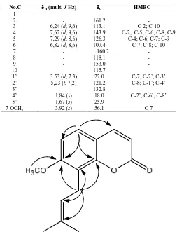
Osthol (2) was also obtained as pale yellowsolid. The ion peak at m/z 229.0861 [M-H]- in the HRESIMS spectrum gave the molecular C14H13O3. Based on UV, HRESIMS, 1H and 13C NMR spectrum suggesting that 2 is a coumarin
derivative containing one methoxyl group and one isoprenyl group. Analysis 1H-NMR spectrum of compound 2 showed a pair doublets (J = 8.6 Hz) signals for aromatic proton region at δH 7.29 and 6.82 ppm. The 1H NMR
spectrum of 2 also showed a pair doublets (J = 9.6 Hz) cis vinylic signal at δH 7.62, and 6.24 corresponding to the
coumarin with substitutent at C-7 and C-8. The existence of isoprenyl chain of compound 2 showed the presence of two methyl groups (δH 1.84, and 1.67 ppm), one methylene group (δH 3.53 ppm), and one vinyl group (δH 5.23
ppm). The methoxyl group showed singlet proton signal at δH 3.92 ppm. The placement of isoprenyl and methoxyl
[image:3.595.141.456.216.600.2]______________________________________________________________________________
proton of aromatic region at δH 7.29 with two oxyaryl carbon signals at δC [160.2 (C-7), and 153.0 (C-9)], and two
methine carbon signals at δC [143.9 (C-4), and 107.4 (C-6)] showed methoxyl at C-7. Thus the placement of
isoprenyl chain located at C-8. Based on data from 1D and 2D NMR, compound 2 is 8-isoprenyl-7-methoxycoumarin or known as osthol [3]. Other HMBC correlations consistent with the structure 2 are shown in Table 2.
Table 2. NMR spectroscopic data of osthol (2)
No.C δH (mult, J Hz) δC HMBC
1 - - -
2 - 161.2 -
3 6,24 (d, 9,6) 113.1 C-2; C-10
4 7,62 (d, 9,6) 143.9 C-2; C-5; C-6; C-8; C-9 5 7,29 (d, 8,6) 126.3 C-4; C-6; C-7; C-9
6 6,82 (d, 8,6) 107.4 C-7; C-8; C-10
7 - 160.2 -
8 - 118.1 -
9 - 153.0 -
10 - 115.7 -
1’ 3.53 (d, 7.3) 22.0 C-7; C-2’; C-3’
2’ 5,23 (t, 7,2) 121.2 C-8; C-1’; C-4’
3’ - 132.8 -
4’ 1,84 (s) 18.0 C-2’; C-6’; C-8’
5’ 1,67 (s) 25.9
[image:4.595.171.424.183.524.2]7-OCH3 3,92 (s) 56.1 C-7
Fig. 3. Significant HMBC correlation for 2
Xhantotoxin (3) was obtained as yellowsolid, and its UV spectrum exhibited absorption maximum of 248, 264, and 303 nm, typical for furanocoumarin derivative [3]. The 1H-NMR spectrum of compound 3 showed a pair of doublets (J = 9.6 Hz) at δH [7.75 (H-4); 6.36 (H-3)], a singlet aromatic region at δH 7.34 (H-5), and a pair of doublets (J = 2.2
Hz) at δH [7.67 (H-2’); 6.80 (H-3’)] suggested the signal of a furanocoumarin with substitutent at C-5 or C-8. The
methoxyl group showed singlet proton signal at δH 3.92 ppm suggested that the methoxyl group is either at 5 or
C-8 of the furanocoumarin structure. The one bond and two/three bonds 1H-13C correlations found in the HMQC and HMBC spectra of compound 3 (Table 3) unambiguously placed the methoxyl group at C-8 by the following observations. The presence of long-range correlations in the HMBC spectrum of 3 between the doublet proton signals (J = 2.2 Hz) of a furano group at δH 7.67 (H-2’), and 6.80 (H-3’) with one oxyaryl carbon signal at δC 147.8
(C-7), and the correlation between a proton aromatic signal at δH 7.34 with two oxyaryl carbon signals δC 147.8
(C-7), 143.1 (C-9) and one methine carbon signal δC 106.8 (C-3’) showed methoxyl at C-8. Based on data from 1D and
Table 3. NMR spectroscopic data of xhantotoxin (3)
No.C δH (mult, J Hz) δC HMBC
1 - - -
2 - 160.6 -
3 6.36 (d, 9.6) 113.0 C-2; C-10
4 7.75 (d, 9.6) 144.4 C-2; C-3; C-9; C-10
5 7.34 (s) 146.7 C-7; C-9; C-10; C-3’
6 - 105.4 -
7 - 147.8 -
8 - 132.9 -
9 - 143.1 -
10 - 116.6 -
1’ - - -
2’ 7.67 (d, 2.2) 114.9 C-7; C-3’
3’ 6.80 (d, 2.2) 106.8 C-7; C-2’
8-OCH3 4.29 (s) 61.4 C-8
Fig. 3. Significant HMBC correlation for 3
On cytotoxic evaluation against HeLa cells, compounds 1 - 3 exhibited IC50 values of 65.09 ± 0.017, 7.62 ± 0.004,
and 21,51 ± 0,003 ppm, respectively. The results of cytotoxic activity showed osthol (2), xhantotoxin (3) have moderate activity, and auraptene (1) was inactive [11]. These cytotoxic data suggested that the presence of methoxyl substituent at C-7 and isoprenyl at C-8 of the coumarin structure increases cytotoxic activity.
CONCLUSION
Three coumarins, auraptene (1), osthol (2), and xhantotoxin (3). have been isolated from the stem bark of of Feronia
limonia. The cytotoxic activity of compounds 1-3 against HeLa cells showed osthol>xhantotoxin>auraptene. The
structure-activity relationship of compounds 1–3 against HeLa cells suggested that the presence of isoprenyl group at C-8 and methoxyl group at C-7 on osthol (2) increases cytotoxic activity.
Acknowledgements
We would like to thank to Mr. Ismail Rachman from the Herbarium Bogoriense, Botanical Garden, Bogor, Indonesia for identifying the species.
REFERENCES
[1] P Pitchai; R Ulagi; PS Mohan; RM Gengan, Indian.J. Chem., 2012, 51B, 1771-1775. [2] MN Xue; HL Sheng; YP Li; XR Gao; DS Han, Chinese Chem.Lett., 2001, 12(3), 243-244. [3] S Ittipon; S; L. Surat, Chem. Nat. Compounds, 2012, 48(2), 308-309.
[4] HK Ki; KH Sang; YK Sun; HK Sung; RL Kang, Bull. Korean.Chem Soc., 2009, 30(9), 2135-2137. [5] A Amulaya; IR Shiddiqui, J Singh, Phytochem., 1989, 28(4), 1229-1231.
[6] I Javed, A Mohammad, J.Saudi.Chem.Soc., 2009, 13, 295-298. [7] R Mukhlesur; IG Alexander, Phytochem., 2002, 59, 73-77.
[8] G Parthasara; KG Mrinal; T Swapnadip; DD Jashodara; A Toshihiro; T Thositake; K Yumiko, Phytochem.,
1994, 37(3), 757-760.
______________________________________________________________________________
[10] YJ Hai; FW Chang; F Li; Y Kai; BF Jiang; FG Zhu; X Jing; WD Zhi; SD Shu; BY Hai, Molecules, 2013, 18, 10768-10775.