Direct and efficient cellular transformation of
primary rat mesenchymal precursor cells by
KSHV
Tiffany Jones, … , Mazen Arar, Shou-Jiang Gao
J Clin Invest. 2012;
122(3)
:1076-1081.
https://doi.org/10.1172/JCI58530
.
Infections by viruses are associated with approximately 12% of human cancer. Kaposi’s
sarcoma-associated herpesvirus (KSHV) is causally linked to several malignancies
commonly found in AIDS patients. The mechanism of KSHV-induced oncogenesis remains
elusive, due in part to the lack of an adequate experimental system for cellular
transformation of primary cells. Here, we report efficient infection and cellular transformation
of primary rat embryonic metanephric mesenchymal precursor cells (MM cells) by KSHV.
Cellular transformation occurred at as early as day 4 after infection and in nearly all infected
cells. Transformed cells expressed hallmark vascular endothelial, lymphatic endothelial,
and mesenchymal markers and efficiently induced tumors in nude mice. KSHV established
latent infection in MM cells, and lytic induction resulted in low levels of detectable infectious
virions despite robust expression of lytic genes. Most KSHV-induced tumor cells were in a
latent state, although a few showed heterogeneous expression of lytic genes. This efficient
system for KSHV cellular transformation of primary cells might facilitate the study of growth
deregulation mechanisms resulting from KSHV infections.
Brief Report
Virology
Find the latest version:
Direct and efficient cellular
transformation of primary rat
mesenchymal precursor cells by KSHV
Tiffany Jones,1,2,3 Fengchun Ye,1,4 Roble Bedolla,1,4 Yufei Huang,1,5Jia Meng,5 Liwu Qian,1,4 Hongyi Pan,1,4 Fuchun Zhou,1,4
Rosalie Moody,1,4 Brent Wagner,6 Mazen Arar,4 and Shou-Jiang Gao1,2,3,4,6
1Tumor Virology Program, Greehey Children’s Cancer Research Institute, 2Department of Molecular Medicine,
University of Texas Health Science Center at San Antonio, San Antonio, Texas, USA.
3Department of Molecular Microbioloby and Immunology, Keck School of Medicine, University of Southern California, Los Angeles, California, USA. 4Department of Pediatrics, University of Texas Health Science Center at San Antonio, San Antonio, Texas, USA.
5Department of Electrical and Computer Engineering, University of Texas at San Antonio, San Antonio, Texas, USA. 6Department of Medicine, University of Texas Health Science Center at San Antonio, San Antonio, Texas, USA.
Infections by viruses are associated with approximately 12% of human cancer. Kaposi’s sarcoma-associated
herpesvirus (KSHV) is causally linked to several malignancies commonly found in AIDS patients. The
mech-anism of KSHV-induced oncogenesis remains elusive, due in part to the lack of an adequate experimental
system for cellular transformation of primary cells. Here, we report efficient infection and cellular
transfor-mation of primary rat embryonic metanephric mesenchymal precursor cells (MM cells) by KSHV. Cellular
transformation occurred at as early as day 4 after infection and in nearly all infected cells. Transformed cells
expressed hallmark vascular endothelial, lymphatic endothelial, and mesenchymal markers and efficiently
induced tumors in nude mice. KSHV established latent infection in MM cells, and lytic induction resulted in
low levels of detectable infectious virions despite robust expression of lytic genes. Most KSHV-induced tumor
cells were in a latent state, although a few showed heterogeneous expression of lytic genes. This efficient system
for KSHV cellular transformation of primary cells might facilitate the study of growth deregulation
mecha-nisms resulting from KSHV infections.
Introduction
Oncogenic viruses are associated with close to 12% of human can-cer (1). Kaposi’s sarcoma–associated herpesvirus (KSHV) is caus-ally linked to Kaposi’s sarcoma (KS), primary effusion lymphoma, and a subset of multicentric Castleman’s disease, malignancies commonly found in AIDS patients (2). While KSHV encodes an array of genes targeting cellular oncogenic, tumor suppressor, and survival pathways, the mechanism of KSHV-induced oncogenesis remains unclear, in part because of the lack of an efficient system for cellular transformation of primary cells.
KSHV-infected tumor cells manifest the hallmark spindle-shape morphology, expressing vascular endothelial, lymphatic endothelial and mesenchymal markers (2). In culture, KSHV infects a variety of cell types. KSHV infection of human vascular and lymphatic endothelial cells converts them into spindle-shaped cells express-ing both lineage markers (3–11). Although the lifespan of primary human endothelial cells is prolonged by KSHV, their continuous growth relies on exogenous growth factors, and cellular transforma-tion remains elusive (3–5). These KSHV-infected cells do not induce any tumors when inoculated into immunocompromised mice.
The initial KSHV target cells could be precursor cells, and KS-like spindle endothelial precursor cells are present in the blood of KS patients (12, 13). Indeed, KSHV infects human mesenchymal
stem cells and hematopoietic precursor cells (14, 15). However, KSHV fails to immortalize and transform these cells. In contrast, transfection of mouse liver precursor cells with recombinant KSHV BAC36 genomes resulted in immortalization of a subset of cells, which induced tumors in nude mice despite the lack of cellular transformation phenotypes in culture (16). Nevertheless, KSHV infection as well as transfection and maintenance of KSHV genomes in mouse cells are inefficient. Here, we show that KSHV efficiently infects and transforms primary rat embryonic meta-nephric mesenchymal precursor (MM) cells. KSHV-transformed MM (KMM) cells efficiently induce tumors in nude mice. These results should facilitate study of the mechanisms of growth dereg-ulation by KSHV infection.
Results and Discussion
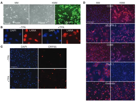
KSHV efficiently infects MM cells. To identify primary cell type(s) that can be transformed by KSHV, we infected cells of different origins (Table 1). MM cells were efficiently infected and transformed by KSHV. KSHV infection was monitored by examining the expres-sion of a GFP cassette in viral genome (ref. 17 and Figure 1A). KMM cells were positive for latent nuclear antigen (LANA, ORF73), with less than 1% positive for the late lytic protein ORF65 (Figure 1, B and C). Continuous passage of KMM cells changed neither the genome number per cell (Supplemental Figure 1; sup-plemental material available online with this article; doi:10.1172/ JCI58530DS1) nor the expression patterns of LANA and ORF65. Induction of viral lytic replication with 12-O-tetradecanoylphor-bol-13-acetate (TPA) did not affect LANA expression but increased
Authorship note: Tiffany Jones, Fengchun Ye, Roble Bedolla, and Shou-Jiang Gao contributed equally to this work.
Conflict of interest: The authors have declared that no conflict of interest exists.
brief report
ORF65-positive cells to 4%–5% (Figure 1, B and C). As controls, uninduced and induced MM cells were negative for LANA and ORF65 proteins (Supplemental Figure 2). The expression of lytic transcripts including transcripts of the immediate early gene RTA (ORF50), early genes ORF57, ORF59, kbZip (ORF-K8), vIL-6 (ORF-K2), and ORF49, and late genes ORF65 and ORF-K8.1 was increased,
[image:3.585.54.536.111.225.2]while that of the latent transcript vCyclin (ORF72) was not altered following lytic induction (Supplemental Figure 3). Interestingly, we detected only intermittent low levels of infectious virions in the supernatants of induced cells, suggesting an abortive viral lytic program. Similar results were observed when long-term-passage KMM cells were examined.
Table 1
KSHV infection of human, mouse, and rat cells
Cells Host Cell type Infection Persistence Immortalization Transformation efficiency efficiency
HUVECs Human Umbilical vein endothelial cells High High Senescence N/A
DMVECs Human Vascular endothelial cells Medium High Senescence N/A
MS1 cells Mouse Vascular endothelial cells Low Low N/A N/A
SVEC cells Mouse Lymphatic endothelial cells Low Low N/A N/A
MEFs Mouse Embryonic fibroblasts High Low Senescence N/A
JWEK cells Mouse Mesenchymal precursor cells Low Low N/A N/A
RAOECs Rat Aortic endothelial cells Low Low N/A N/A
MM cells Rat Mesenchymal precursor cells High High Yes Yes
NA, not applicable; MEF, mouse embryonic fibroblast; RAOEC, rat aortic endothelial cell.
Figure 1
Characterization of MM and KMM cells. (A) Morphology at day 2 after seeding. Cells were seeded at 5 × 10⁴ cells/well in 6-well plates. (B and C)
[image:3.585.54.532.341.699.2]MM and KMM cells expressed vascular endothelial markers β-catenin, vWF, and VEGFR-1; lymphatic endothelial mark-ers VEGFR-3 and LYVE-1; the mesenchymal marker vimentin; and the hematopoietic precursor marker CD34 (Figure 1D and Supplemental Figure 4). Vascular endothelial markers CD31, VCAM-1, and ICAM-1 and the lymphatic endothelial cell marker podoplanin were upregulated, while the hematopoietic precur-sor marker Thy1.1 was downregulated in KMM cells. Long-term passage of KMM cells did not change the patterns of cell surface markers. Thus, similar to vascular and lymphatic endothelial cells (7, 8, 10), KSHV reprograms MM cells to express a mixture of vas-cular endothelial, lymphatic endothelial, mesenchymal, and hema-topoietic precursor markers.
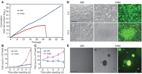
KMM cells are transformed. MM cells underwent crisis after 25–28 passages. In contrast, KMM cells were immortalized, as they could be maintained in continuous passage (Figure 2A). Com-pared with MM cells, KMM cells were smaller in size (Figure 1A) and had shorter doubling time as indicated by faster dilution of CFSE (13.1 ± 2.2 vs. 19.4 ± 2.4 hours at exponential phase; Supplemental Figure 5A), which was also demonstrated by an increase in cells per cluster at day 2 after seeding (7–10 vs. 2–3 cells/cluster; Figure 1A) and a faster growth rate (Figure 2B). Accordingly, KMM cells had more cells in S phase (50% vs. 40%) and fewer cells in G0/G1 phase (35% vs. 42%) than MM cells
(Sup-plemental Figure 5B), which was due to accelerated transition from G0/G1 to S phase, as shown by faster incorporation of BrdU
(Supplemental Figure 5C). In serum-free medium, MM cells were growth arrested starting at day 1 after seeding, with 85% cells
in G0/G1 phase and only 5% cells in S phase while KMM cells
continued to proliferate for up to day 4 after seeding, with only 68% cells in G0/G1 phase and 20% in S phase, and continued to
incorporate BrdU at a faster rate (Figure 2C and Supplemental Figure 5, D and E). MM cells were contact inhibited, while KMM cells lost contact inhibition, as shown by formation of foci and higher saturation density in culture (Figure 2D and Supplemen-tal Figure 5F) and formation of colonies in semisolid soft agar (Figure 2E). Growth in semisolid soft agar was observed as early as day 4 after infection, the earliest time point examined, and with close to 100% efficiencies. These results indicated that MM cells were efficiently immortalized and transformed by KSHV. We reproduced these results 8 times with MM cells isolated from 3 different batches. This transformation phenotype did not change following long-term passage of KMM cells. Furthermore, virions from BCP-1 cells also efficiently infected and transformed MM cells (Supplemental Figure 6), indicating that the transformation phenotype was not restricted to a specific viral isolate.
KMM cells induce tumors in nude mice. While MM cells failed to induce any tumors in nude mice, KMM cells efficiently induced tumors, with a mean incidence of 84.6% ± 11.2% and a mean latency of 12 ± 1.6 weeks (Figure 3, A and B). Tumors often mani-fested as reddish lesions and dissemination to visceral organs (Figure 3, C and D).
[image:4.585.54.534.81.333.2]H&E staining showed spindle-shaped tumor cells, many of which had mitotic figures and were positive for the proliferation marker Ki67 (Figure 3E and Supplemental Figure 7). Tumors contained many microvessels, slit-like spaces, and infiltrates of
Figure 2
Immortalization and transformation of MM cells by KSHV. (A) MM cells underwent crisis after 25–28 passages. KMM grew continuously without crisis. Cells were passaged every 3 days at 2 × 10⁴ cells/well in 24-well plates. (B) KMM cells grew faster than MM cells in regular medium
contain-ing serum. Cells seeded at 105 cells/well in 6-well plates were counted daily. (C) In serum-free medium, growth of MM cells stopped, while KMM
cells continued to grow for up to day 4 after seeding. Cells seeded at 105 cells/well in 6-well plates were counted daily. (D) KMM cells formed foci,
while MM cells were contact inhibited when they reached confluency. Cells seeded at 2 × 10⁵ cells/well in 6-well plates were cultured with daily
brief report
immune cells such as CD45R+ mouse B cells, which were
remi-niscent of CD8+ T cells and CD20+ B cells in KS tumors
(Sup-plemental Figure 8). These results indicated that KMM tumors consisted of proliferating spindle cells with vast inflammatory infiltrates and neoangiogenesis.
All tumor cells strongly expressed LANA protein, with 1%–3% of them expressing ORF65 protein (Figure 3E). Tumors had robust expression of latent LANA and vCyclin transcripts but heteroge-neous expression of lytic transcripts (Supplemental Figure 9). RTA was at levels close to those in uninduced KMM cells in 2 of 14 tumors; ORF57 and ORF59 were at levels higher than in uninduced cells in 12 of 14 tumors; and ORF65 was detected in 5 of 14 tumors, of which only 1 was at a high level while the other 4 were at levels similar to those in uninduced KMM cells. These results indicated a spontaneous default lytic program in tumors.
KMM tumor cells were positive for vascular endothelial markers β-catenin, CD31, VE-cadherin, ICAM-1, and VCAM-1; lymphatic endothelial markers LYVE-1, VEGFR-3, podoplanin, and PROX-1; hematopoietic precursor marker CD34; and mesenchymal mark-er vimentin, but wmark-ere negative for Thy1.1 (Figure 3E and Supple-mental Figure 7).
We have shown that KSHV can efficiently infect and trans-form MM cells. KMM cells were immortalized, proliferated at a faster rate than MM cells, grew in serum-free medium, lost con-tact inhibition in culture, and induced tumors in nude mice. Tumor cells acquired spindle morphology; formed slit-like spaces; and expressed vascular endothelial, lymphatic endothelial, and mesenchymal markers. Most tumor cells were in the latent state, but a few had heterogeneous expression of lytic genes. These path-ological and viral features resemble those in KS tumors
(Supple-Figure 3
[image:5.585.74.515.78.471.2]mental Figure 8). This system should be useful for dissecting fac-tors mediating KSHV-induced malignant transformation.
Because KSHV is a human virus, it is important to recognize the limitations of cross-species observations. While KSHV infects a variety of human cells in culture, cellular transformation of pri-mary human cells remains challenging (2). Furthermore, explant tumor cells from most KS tumors are neither immortalized nor transformed, despite their in vivo malignant proliferative nature (18–21). While the loss of viral genome could account for the lack of cellular transformation, it is clear that MM cells are distinct from human primary cells.
Besides the mesenchymal marker vimentin, MM cells expressed vascular endothelial markers β-catenin, ICAM-1, vWF, and VEGFR-1; lymphatic endothelial markers VEGFR-3 and LYVE-1; and hematopoietic precursor markers CD34 and Thy1.1. KSHV infection upregulated the vascular endothelial markers VCAM-1 and CD31 and the lymphatic endothelial marker podoplanin. Interestingly, Thy1.1 expression was almost completely shut down, while that of CD34 was maintained following KSHV infection. In differentiated cells, CD34 is often expressed in endothelial cells, while Thy1.1 is more confined to hematopoietic cells (22, 23). Human Thy1 expression is associated with tumor suppression of ovarian cancer (24). Thus, KSHV appeared to downregulate Thy1.1 expression to inhibit its tumor-suppressive effect, while at the same time converting the mesenchymal precursor cells into a mixed phenotype of vascular and lymphatic endothelial cells. This reprogramming process is not surprising, because MM cells are multipotent and can be differentiated into diverse cell types, including endothelial cells (25, 26). Nevertheless, whether mesenchymal precursor cells are the KSHV targets in vivo requires further investigation.
Methods
Cell culture and virus infection. Primary HUVECs and microvascular endothelial cells (DMVECs) from Lonza, MS1 and SVEC cells from ATCC, and primary rat aortic endothelial cells from Lifeline Cell Technology were maintained as described by the vendors. Mouse and rat MM cells as well as embryonic fibroblasts were isolated and maintained as previously described (27, 28). Primary cells of fewer than 5 passages were used for infection experiments. For subsequent characterizations, MM and KMM cells between 5 and 10 passages were used. KMM cells were cultured with 150 μg/ml hygromycin, which was removed 1 week before experiments. Growth of BAC36 and BCP-1 KSHV and infection were performed as previ-ously described (5, 17). Cells were split every 3 days, except for focus
forma-tion experiments, with daily change of medium. Cell numbers were deter-mined at passage. Cells were observed daily for crisis, including growth arrest and cell death. Semisolid soft agar assay was performed as previously described (29). Cells at specific days following KSHV infection were seeded, and pictures of colonies were taken at day 14 after seeding.
Immunodetection. Immunofluorescence assay, Western blotting, and immunohistochemistry were as previously described (5, 30). A rat monoclo-nal antibody to LANA (Abcam), mouse monoclomonoclo-nal antibodies to ORF65 (5), vimentin (BioCare Medical), and Thy1.1 (Santa Cruz Biotechnology Inc.), and rabbit polyclonal antibodies to vWF (Thermo Fisher Scientific), PROX-1 (Covance), Ki67 (Abcam), and CD31 (Abcam) were used. Rabbit polyclonal antibodies to VE-cadherin, β-catenin, ICAM-1, VCAM-1, LYVE-1, VEGFR-1, and VEGFR-3 and goat polyclonal antibodies to podoplanin and CD34 were from Santa Cruz Biotechnology Inc.
Cell cycle and CFSE analysis and BrdU incorporation. Cell cycle was ana-lyzed by propidium iodide staining. For CFSE assays, cells pulsed with 10 μM CFSE were analyzed daily for relative intensities (Invitrogen). BrdU incorporation was determined by pulsing cells with 10 μM BrdU for 4 hours, followed by staining with a mouse monoclonal antibody to BrdU (Invitrogen).
Growth of tumors. Subcutaneous growth of tumors was as previously described (29). Tumor volumes were measured twice a week using 0.2 cm3
as a threshold. Tumor analyses were performed at 20 weeks following inoculation or when the volume reached 1 cm3. Tumor images were taken
at the end of study. Fluorescence images were taken with an Image Station 2000R System (Kodak).
Statistics. Data are shown as mean ± SD. The 1-tailed Student’s test was used to compare data between experimental groups. Statistical significance was assumed at P values less than 0.05.
Study approval. The animal protocol was approved by the Institutional Animal Care and Use Committee of the University of Texas Health Science Center at San Antonio (Animal Welfare Assurance no. A3345-01).
Acknowledgments
This work was supported by grants from the NIH (CA096512, CA124332, and CA132637) to S.-J. Gao.
Received for publication April 15, 2011, and accepted in revised form December 14, 2011.
Address correspondence to: Shou-Jiang Gao, University of South-ern California, Keck School of Medicine, Los Angeles, California 90033, USA. Phone: 323.442.8028; Fax: 323.442.1721; E-mail: [email protected].
1. Parkin DM. The global health burden of infection-associated cancers in the year 2002. Int J Cancer. 2006; 118(12):3030–3044.
2. Ganem D. KSHV and the pathogenesis of Kaposi sarcoma: listening to human biology and medicine.
J Clin Invest. 2010;120(4):939–949.
3. Ciufo DM, et al. Spindle cell conversion by Kapo-si’s sarcoma-associated herpesvirus: formation of colonies and plaques with mixed lytic and latent gene expression in infected primary dermal micro-vascular endothelial cell cultures. J Virol. 2001; 75(12):5614–5626.
4. Flore O, Rafii S, Ely S, O’Leary JJ, Hyjek EM, Cesarman E. Transformation of primary human endothelial cells by Kaposi’s sarcoma-associated herpesvirus. Nature. 1998;394(6693):588–592. 5. Gao SJ, Deng JH, Zhou FC. Productive lytic
repli-cation of a recombinant Kaposi’s sarcoma-asso-ciated herpesvirus in efficient primary infection of primary human endothelial cells. J Virol. 2003;
77(18):9738–9749.
6. Moses AV, et al. Long-term infection and transfor-mation of dermal microvascular endothelial cells by human herpesvirus 8. J Virol. 1999;73(8):6892–6902. 7. Carroll PA, Brazeau E, Lagunoff M. Kaposi’s sar-coma-associated herpesvirus infection of blood endothelial cells induces lymphatic differentiation.
Virology. 2004;328(1):7–18.
8. Hong YK, et al. Lymphatic reprogramming of blood vascular endothelium by Kaposi sarcoma-associ-ated herpesvirus. Nat Genet. 2004;36(7):683–685. 9. Lagunoff M, et al. De novo infection and serial
transmission of Kaposi’s sarcoma-associated her-pesvirus in cultured endothelial cells. J Virol. 2002; 76(5):2440–2448.
10. Wang HW, et al. Kaposi sarcoma herpesvirus-induced cellular reprogramming contributes to the lymphatic endothelial gene expression in Kaposi sarcoma. Nat Genet. 2004;36(7):687–693. 11. An FQ, et al. Long-term-infected
telomerase-immortalized endothelial cells: a model for Kapo-si’s sarcoma-associated herpesvirus latency in vitro and in vivo. J Virol. 2006;80(10):4833–4846. 12. Massarelli G, Scott CA, Ibba M, Tanda F, Cossu A.
Immunocytochemical profile of Kaposi’s sarcoma cells: their reactivity to a panel of antibodies direct-ed against different tissue cell markers. Appl Pathol. 1989;7(1):34–41.
13. Browning PJ, et al. Identification and culture of Kaposi’s sarcoma-like spindle cells from the peripheral blood of human immunodeficiency virus-1-infected individuals and normal controls.
Blood. 1994;84(8):2711–2720.
14. Parsons CH, Szomju B, Kedes DH. Susceptibility of human fetal mesenchymal stem cells to Kapo-si sarcoma-associated herpesvirus. Blood. 2004; 104(9):2736–2738.
brief report
and in vivo. Blood. 2006;108(1):141–151. 16. Mutlu AD, et al. In vivo-restricted and reversible
malignancy induced by human herpesvirus-8 KSHV: a cell and animal model of virally induced Kaposi’s sarcoma. Cancer Cell. 2007;11(3):245–258. 17. Zhou FC, et al. Efficient infection by a recombinant Kaposi’s sarcoma-associated herpesvirus cloned in a bacterial artificial chromosome: application for genetic analysis. J Virol. 2002;76(12):6185–6196. 18. Aluigi MG, et al. KSHV sequences in biopsies and
cul-tured spindle cells of epidemic, iatrogenic and Medi-terranean forms of Kaposi’s sarcoma. Res Virol. 1996; 147(5):267–275.
19. Lebbe C, et al. Characterization of in vitro culture of HIV-negative Kaposi’s sarcoma-derived cells. In vitro responses to alfa interferon. Arch Dermatol Res. 1997;289(7):421–428.
20. Nakamura S, et al. Kaposi’s sarcoma cells: long-term culture with growth factor from
ret-rovirus-infected CD4+ T cells. Science. 1988; 242(4877):426–430.
21. Salahuddin SZ, et al. Angiogenic properties of Kaposi’s sarcoma-derived cells after long-term cul-ture in vitro. Science. 1988;242(4877):430–433. 22. Fina L, et al. Expression of the CD34 gene in
vascu-lar endothelial cells. Blood. 1990;75(12):2417–2426. 23. Pont S. Thy-1: a lymphoid cell subset marker capa-ble of delivering an activation signal to mouse T lymphocytes. Biochimie. 1987;69(4):315–320. 24. Abeysinghe HR, et al. THY1 expression is associated
with tumor suppression of human ovarian cancer.
Cancer Genet Cytogenet. 2003;143(2):125–132. 25. Nishinakamura R, Uchiyama Y, Sakaguchi M,
Fujimura S. Nephron progenitors in the meta-nephric mesenchyme. Pediatr Nephrol. 2011; 26(9):1463–1467.
26. Usui J, Yamada R, Kanemoto K, Koyama A, Nagata M. Murine metanephric mesenchyme possesses
characteristics of vascular endothelial cells in vitro.
Nephron Exp Nephrol. 2006;102(3–4):e93–e98. 27. Arar M, Xu YC, Elshihabi I, Barnes JL, Choudhury
GG, Abboud HE. Platelet-derived growth factor receptor beta regulates migration and DNA synthe-sis in metanephric mesenchymal cells. J Biol Chem. 2000;275(13):9527–9533.
28. Wagner B, et al. Mitogenic signaling via plate-let-derived growth factor beta in metaneph-ric mesenchymal cells. J Am Soc Nephrol. 2007; 18(11):2903–2911.
29. Gao SJ, Boshoff C, Jayachandra S, Weiss RA, Chang Y, Moore PS. KSHV ORF K9 (vIRF) is an oncogene which inhibits the interferon signaling pathway.
Oncogene. 1997;15(16):1979–1985.