Repairing skeletal muscle: regenerative
potential of skeletal muscle stem cells
Francesco Saverio Tedesco, … , Graziella Messina, Giulio
Cossu
J Clin Invest.
2010;
120(1)
:11-19.
https://doi.org/10.1172/JCI40373
.
Skeletal muscle damaged by injury or by degenerative diseases such as muscular
dystrophy is able to regenerate new muscle fibers. Regeneration mainly depends upon
satellite cells, myogenic progenitors localized between the basal lamina and the muscle
fiber membrane. However, other cell types outside the basal lamina, such as pericytes, also
have myogenic potency. Here, we discuss the main properties of satellite cells and other
myogenic progenitors as well as recent efforts to obtain myogenic cells from pluripotent
stem cells for patient-tailored cell therapy. Clinical trials utilizing these cells to treat
muscular dystrophies, heart failure, and stress urinary incontinence are also briefly outlined.
Review Series
Find the latest version:
Repairing skeletal muscle: regenerative
potential of skeletal muscle stem cells
Francesco Saverio Tedesco,1,2 Arianna Dellavalle,1 Jordi Diaz-Manera,1,3,4 Graziella Messina,1,5 and Giulio Cossu1,5
1Division of Regenerative Medicine, San Raffaele Scientific Institute, Milan, Italy. 2Vita-Salute San Raffaele and Open University, Milan, Italy. 3Hospital Santa Creu i Sant Pau, Neuromuscular Diseases Unit, Barcelona, Spain. 4Centro de Investigación Biomédica en Red
sobre Enfermedades Neurodegenerativas (CIBERNED), Madrid, Spain. 5Department of Biology, University of Milan, Milan, Italy.
Skeletal muscle damaged by injury or by degenerative diseases such as muscular dystrophy is able to regenerate
new muscle fibers. Regeneration mainly depends upon satellite cells, myogenic progenitors localized between
the basal lamina and the muscle fiber membrane. However, other cell types outside the basal lamina, such as
pericytes, also have myogenic potency. Here, we discuss the main properties of satellite cells and other myogenic
progenitors as well as recent efforts to obtain myogenic cells from pluripotent stem cells for patient-tailored cell
therapy. Clinical trials utilizing these cells to treat muscular dystrophies, heart failure, and stress urinary
incon-tinence are also briefly outlined.
Introduction
It has been known for more than a century that skeletal muscle, the most abundant tissue of the body, has the ability to regener-ate new muscle fibers after it has been damaged by injury or as a consequence of diseases such as muscular dystrophy (1). Mus-cle fibers are syncytial cells that contain several hundred nuclei within a continuous cytoplasm. Therefore, whether the process of regeneration depends upon the fusion of mononucleated precur-sor cells or upon the fragmentation of dying muscle fibers, which release new cells, remained controversial for a long time, even after the demonstration by Beatrice Mintz and Wilber Baker (2) that multinucleated fibers are formed by the fusion of single cells. In 1961, Alexander Mauro (3) observed mononuclear cells between the basal lamina that surrounds each muscle fiber and the plasma membrane of the muscle fiber and named them satellite cells (SCs) (Figure 1). SCs were later accepted to be, and are still considered today, the main players in skeletal muscle regeneration. SCs also contribute to the postnatal growth of muscle fibers, which in adults contain approximately 6–8 times more nuclei than in neo-nates, all of them being irreversibly postmitotic.
In addition to SCs, other progenitors located outside the basal lamina, including pericytes, endothelial cells, and interstitial cells, have been shown to have some myogenic potential in vitro or after transplantation. The developmental origin of these progenitors is unclear, as is their lineage relationship with SCs, even though they may feed, to some extent, into the SC compartment (4).
There is much interest in understanding the cellular and molec- ular mechanisms underlying skeletal muscle regeneration in dif- ferent contexts because such knowledge might help in the develop-ment of cell therapies for diseases characterized by skeletal muscle degeneration. These diseases include muscular dystrophy, the term for a group of inherited disorders characterized by progressive muscle wasting and weakness leading to a variable degree of mobil-ity limitation, including confinement to a wheelchair and, in the most severe forms, heart and/or respiratory failure (5). Many mus-cular dystrophies arise from loss-of-function mutations in genes encoding cytoskeletal and membrane proteins, the most common
and severe being Duchenne muscular dystrophy (DMD), which is caused by mutations in the gene encoding dystrophin, an integral part of a complex that links the intracellular cytoskeleton with the extracellular matrix in muscle. Muscular dystrophies are some of the most difficult diseases to treat, as skeletal muscle is composed of large multinucleated fibers whose nuclei cannot divide. Conse- quently, cell therapy has to restore proper gene expression in hun-dreds of millions of postmitotic nuclei (6).
In this Review, we discuss recent work indicating the possible exis-tence of a stem/progenitor cell compartment in adult muscle (see also ref. 7) as well as studies related to the derivation of myogenic cells from embryonic and induced pluripotent stem cells (PSCs) for the development of new cell therapy strategies for diseases of skeletal muscle. An overview of clinical trials based upon transplan-tation of skeletal muscle stem cells is also provided. Neither the role of SCs in aging skeletal muscle nor the SC niche are discussed here due to space constraints, and readers are directed to excellent recent reviews on these topics by Suchitra Gopinath and Thomas Rando (8) and Michael Rudnicki and colleagues (9), respectively.
SCs
Identification and characterization. The most stringent way to classify cells as SCs remains by determining their anatomical location: SCs are found underneath the basal lamina of muscle fibers, closely jux-taposed to the plasma membrane (3). SCs originate from somites (10, 11), spheres of paraxial mesoderm that generate skeletal muscle, dermis, and axial skeleton, but the exact progenitor that gives rise to SCs remains to be identified. SCs are present in healthy adult mammalian muscle as quiescent cells and represent 2.5%–6% of all nuclei of a given muscle fiber. However, when activated by muscle injury, they can generate large numbers of new myofibers within just a few days (12). Quiescent SCs (13) express characteristic (although not unique) markers. In the mouse, the most widely used of these markers is the transcription factor paired box 7 (Pax7) (14), which is essential for SC specification and survival (15). In contrast, Pax3 is expressed only in quiescent SCs in a few specific muscle groups such as the diaphragm (16). The basic helix-loop-helix (bHLH) gene myogenic regulatory factor 5 (Myf5 ) is expressed in the large major-ity of quiescent SCs, and for this reason, mice expressing nuclear
LacZ under the control of the Myf5 promoter (Myf5nlacZ/+ mice) have Conflict of interest: The authors have declared that no conflict of interest exists.
review series
been useful for identifying and characterizing SCs (17). Many other markers (18–29) have been identified and are listed in Table 1. Some of these surface markers are useful for isolating “purified” SC populations by cell sorting, but since each marker is not exclusively expressed on SCs, a combination of different markers must be used. Alternatively, transgenic mice such as those expressing GFP under the control of promoters that drive the expression of genes encod-ing SC markers — for example, the Pax3 promoter — can be used to isolate SCs (29–31). In humans, markers of both quiescent and activated SCs do not fully correspond to those in the mouse, and relatively little is known about them due to the difficulty of obtain-ing human tissue. For example, although CD34 is a marker of SCs in mice, it does not mark SCs in human muscle (32); and M-cad-herin is not as consistent a marker of SCs in humans as it is in mice. Among the more reliable markers of SCs in human muscle is CD56, although it also marks natural killer lymphocytes (33).
Activation. In response to a muscle injury, SCs are activated and start to proliferate; at this stage, they are often referred to as either myogenic precursor cells (mpc) or myoblasts (34, 35). Several signals, deriving both from damaged fibers and infiltrat-ing cells, are involved in SC activation, including HGF (36), FGF (37), IGF (38), and NO (39).
The progression of activated SCs toward myogenic differentia-tion is mainly controlled by Myf5 and myogenic differentiation 1 (MyoD) (17) and is followed by fusion into regenerating fibers. The whole process takes approximately 7 days in the mouse (40), during which time SCs undergo different fates, giving rise to a few Pax7+MyoD– cells, which return to quiescence (to maintain the progenitor pool), and many Pax7+MyoD+
cells, which are commit-ted to differentiation (41) (Figure 1). Notch signaling is thought to regulate this process through promotion of asymmetric divisions, although there is not agreement on the role of Numb (a Notch inhibitor and cell-fate determinant) in inducing differentiation (42) and sustaining self renewal (43). The occurrence of asymmetric cell division is also supported by the identification of a subpopulation of SCs able to retain BrdU after pulse-chase labeling, with some cells displaying selective template DNA strand segregation during mitosis (43, 44). In addition, Rudnicki and colleagues validated the label-retention model of SCs and demonstrated that approxi-mately 10% of Pax7+ mouse SCs had never expressed Myf5 and that these cells remain adherent to the basal lamina during asymmetric mitosis, generating one Pax7+Myf5– satellite “stem cell” and one Pax7+Myf5+ SC “progenitor,” eventually destined to differentiate (25) (Figure 1). The same group also elegantly described Wnt7a as regulating the symmetric expansion of Pax7+Myf5– SCs (45).
Transplantation. Because of their features, SCs were considered obvious candidates for a cell-therapy approach to treating muscular dystrophy. Pioneer studies demonstrated that intramuscular injec-tion of normal myoblasts (46) into mdx mice, which lack dystro-phin and are a model for DMD, resulted in fusion with host fibers and extensive dystrophin production. These studies led to several clinical trials in the early 1980s (see Muscular dystrophies section for details) that failed for a number of reasons, including poor survival and migration of donor cells after transplantation and rejection of the donor cells due to an immune response by the patients (47).
Many subsequent preclinical studies aimed to improve the sur- vival, proliferation, and differentiation of the SCs after engraft-
ment. For example, transplantation in dystrophic mouse mus-Figure 1
[image:3.585.47.538.75.345.2]cles of a single muscle fiber that contained as few as seven SCs led to an increasing number of new SCs that in turn generated more than 100 new muscle fibers and could also be activated after injury (48). This is a much more efficient way to gener-ate new muscle fibers than transplantation of cultured SCs, in which normally the number of donor-derived new fibers that are generated is several orders of magnitude less than the number of injected cells. Unfortunately, this method would be difficult to translate into clinical protocols.
In the past few years, several groups have succeeded in prospec-tively isolating “pure” populations of SCs by using a combination of different markers (Table 1), such as Pax3-GFP (30), CXCR4 and β1 integrin (49), α7 integrin and CD34 (50), or syndecan-3 and -4 (51). It is still unknown whether the different protocols allow isola-tion of the same cell population, enriched to different extents for a more primitive “stem-like” fraction. However, all these studies revealed that freshly isolated cells have a much greater capacity to generate dystrophin-expressing fibers in mdx mice than the same cells after in vitro expansion (30); the simplest explanation for this is that the “stem” fraction either dies in culture or generates differentiation-committed SCs. Importantly, a short culture peri-od of 3–4 days, without subculture, allowed lentiviral-mediated
transduction and thus genetic correction of SCs freshly isolated from mdx mice, without compromising myogenic potency in vivo (52). Despite these encouraging results, previous unsolved prob-lems still prevent the use of SCs to systemically treat patients with muscular dystrophy: in particular, the inability of these cells to cross the endothelial wall makes systemic delivery impossible and prevents possible healing of the diaphragm and cardiac muscles, both critical for patient survival (34).
Other myogenic progenitors
[image:4.585.55.530.103.455.2]The availability of cell-autonomous, tissue-specific transgenic mark- ers allowed the unequivocal demonstration of the existence of myo- genic progenitors originating from tissues other than skeletal mus-cle (53). Upon transplantation (either BM transplantation [BMT] or direct injection into skeletal muscle), these cells, identified by trans-gene expression, participate in muscle regeneration in wild-type and/or dystrophic mice (Figures 1 and 2; Table 2) and eventually enter the SC pool. The possibility that myogenic differentiation may depend upon fusion (and hence exposure to the dominant activity of MyoD) remains, but for skeletal muscle, this would be part of the physiological mechanism that creates the tissue. Below, we describe briefly some examples of these unorthodox myogenic cells.
Table 1
SC markers
Marker SC expression Localization Function Prospective Expression in Ref isolationA other tissues/cells
Pax7 100% of quiescent and Nucleus Transcription Pax7-GFP Absent 14 activated SCs factor
Pax3 Quiescent SCs Nucleus Transcription Pax3-GFP Melanocyte 16 (only in a subset of muscles) factor stem cells
Myf5 Most quiescent SCs and all Nucleus Transcription Myf5-nLacZ Absent 17 proliferating SCs and myoblasts factor
Syndecan-3 and -4 98% of quiescent Membrane Transmembrane Cell Brain, dermis, BM, 18 and activated SCs heparan sulfate sorting bone, smooth
proteoglycan muscle, tumors
VCAM-1 Quiescent and activated SCs Membrane Adhesion Cell Activated endothelial cells 19 molecule sorting
c-met Quiescent and activated SCs Membrane HGF receptor Not used Many tissues and tumors 20 Foxk1 Quiescent and activated SCs Nucleus Nuclear factor Not used Neurons 21 Cd34 Quiescent and activated SCs Membrane Membrane Cell Hematopoietic, endothelial, 13
protein sorting mast, and dendritic cells M-cadherin Quiescent and activated SCs; Membrane Adhesion Not used Absent 22
myoblasts protein
Caveolin-1 Quiescent and activated SCs; Membrane Membrane Not used Endothelial fibrous and 23
myoblasts protein adipose tissue
α7 Integrin Quiescent and activated SCs; Membrane Adhesion Cell Vessel-associated 24
myoblasts protein sorting cells
β1 Integrin Quiescent and activated SCs Membrane Adhesion Cell Many tissues 25
protein sorting
Cd56 Quiescent and activated SCs; Membrane Homophilic binding Cell Glia, neurons, and 26 myoblasts glycoprotein sorting natural killer cells
SM/C2.6B Quiescent and activated SCs; Unknown Unknown Cell Unknown 27
myoblasts sorting
Cxcr4 Subset of quiescent SCs Membrane SDF1 Cell HSCs, vascular endothelial 28 receptor sorting cells, and neuronal cells Nestin Around 98% of quiescent SCs Intermediate Intermediate Nestin Neuronal precursor cells 29
and myoblasts filament filament protein GFP
review series
Cells from ectoderm: neural stem cells. To date, neural stem cells (both murine and human) are the only ectoderm-derived stem cells that have been shown to differentiate into skeletal muscle when cocultured with skeletal myoblasts or transplanted into regenerat-ing skeletal muscle (54). Interestingly, cells expressing Myf5 exist in the brain and spinal cord, suggesting a cryptic potency that becomes apparent in vitro (55).
Hematopoietic cells. The first evidence of in vivo generation of skeletal muscle from BM cells was reported in 1998 (56) in a study that used transgenic mice expressing a nuclear LacZ under the control of the striated muscle promoter myosin light chain 1/3 fast (MLC3f ). After transplantation of BM from the trans-
genic mice and subsequent injury to the host muscle, unequivo-cal β -gal–positive nuclei were detected in regener-ated fibers, demonstrating that murine BM contains transplantable progenitors that can be recruited to an injured muscle through the circulation, where they participate in muscle repair (56). This opened the possibility of treating muscular dystrophy by BMT, but work in mice indicated that, unfortunately, the frequency of this event was too low, even in a chroni-cally regenerating dystrophic muscle and even if the side population (SP) progenitor–enriched fraction was transplanted (57, 58). To address this issue, subse-quent experiments were directed to identifying a rare, potentially highly myogenic progenitor, but those studies have so far had modest success. The hemato-poietic, CD45+ fraction of the BM has been identified as the cell population with myogenic potential (59), and retrospective analysis in a DMD patient that had undergone BMT confirmed the persistence of donor-derived skeletal muscle cells over a period of many years, again at very low frequency (60). Together, these data suggested that HSCs or a yet-to-be-identified cell that expresses several markers in common with true HSCs has myogenic potential. More recent approach-es confirmed that hematopoietic cells have myogenic potential but disagreed on the stage at which myo-genic differentiation would occur. One study reported that the progeny of a single mouse hematopoietic pro-genitor cell can both reconstitute the hematopoietic system and contribute, at low frequency, to muscle regeneration (61). However, a similar study showed that in response to injury, CD45+ hematopoietic pro-genitors contribute to regenerating mouse skeletal muscle through fusion of mature myeloid cells rather than fusion of the HSCs (62).
A subpopulation of circulating cells expressing CD133 (also known as Ac133), a well-characterized marker of HSCs, also expresses early myogenic markers (63). When injected into the circulation of dystrophic scid/mdx mice, CD133+ cells have been found to contribute to muscle repair, recovery of force, and replenishment of the SC pool. The group that discovered this also isolat-ed a population of muscle-derived stem cells (MDSCs) expressing CD133 (64). Furthermore, when CD133+ cells from DMD patients were genetically corrected by lentivirus-mediated exon skipping for dystrophin exon 51, these cells were able to mediate morphological and functional recovery in scid/mdx mice (64). Thus, differ-ent subpopulations of hematopoietic cells, whose characterization is still incomplete, seem to possess myogenic potency, but none of these exhibit this property at high frequency.
[image:5.585.47.323.82.517.2]Cells derived from mesoderm (other than hematopoietic cells) . Many dif-ferent types of mesoderm stem/progenitor cells have been shown to exhibit myogenic potential, usually after drug treatment, genet-ic modification, or coculture with SCs or myoblasts. In some cases, evidence of in vivo myogenesis has been documented. The list of such cells includes mesenchymal stem cells (MSCs), multipotent adult progenitor cells (MAPCs), MDSCs, CD133+ cells, mesoan- gioblasts (MABs), endothelial progenitor cells (EPCs), and adi-pose-derived stem cells, all of which are briefly described below or in Table 2. More details can be found in previous reviews (6, 32).
Figure 2
MSCs have been shown to be capable of skeletal myogenesis (65). However, recently, Perlingeiro and colleagues demonstrated that although Pax3 activation enabled the in vitro differentiation of murine and human MSCs into MyoD+ myogenic cells, these cells failed to cause functional muscle recovery in mdx mice, despite good engraftment (66). The reason for this failure remains unclear.
MABs are vessel-associated progenitors (67) that express early endothelial markers when isolated from the embryo and pericyte markers when isolated from postnatal tissues. Since MABs are able to cross the vessel wall and are easily transduced with lentiviral vectors, they have been used in preclinical models of cell therapy for muscular dystrophy. Intraarterial delivery of either wild-type or genetically corrected MABs morphologically and functionally ameliorated the dystrophic phenotype of mice lacking α -sarco-glycan (Sgca), which model limb-girdle muscular dystrophy 2D, a muscular dystrophy caused by mutations in the SGCA gene (68). In addition, intraarterial delivery of wild-type postnatal canine MABs resulted in extensive recovery of dystrophin expression and ame-liorated pathologic muscle morphology and function in golden retriever dogs that model DMD (69). Similar cells have been isolat-ed from human postnatal skeletal muscle and shown to represent a subset of pericytes and to give rise to dystrophin-positive muscle fibers when transplanted into scid/mdx mice (70). Based on these studies, a phase I clinical trial with MAB allotransplantation in DMD patients is planned for the near future.
Initially identified as circulating cells expressing CD34 and fetal liver kinase-1 (Flk-1; also known as VEGFR2), EPCs (71) were shown to be transplantable and to participate actively in angiogenesis in various physiologic and pathologic conditions (71). It was then shown that freshly isolated human cord blood CD34+ cells injected into ischemic adductor muscles gave rise not only to endothelial but also to skeletal muscle cells in mice (72). Consistent with this, Péault and colleagues have identified cells with high myogenic potential within the vascular endothe- lium of human adult skeletal muscle (73). These human myo-endothelial cells, which represent less than 0.5% of the cells in dissociated adult skeletal muscles, expressed both myogenic and endothelial cell markers (CD56+CD34+CD144+CD45–), exhibited long term proliferation, had a normal karyotype, and when transplanted into scid mice, were able to regenerate fibers in injured muscle (73).
Human multipotent adipose-derived stem (hMADS) cells, iso-
lated from adipose tissue, differentiate into adipocytes, osteo-blasts, and myoblasts (74). Recently, the myogenic and muscle repair capacities of hMADS cells have been enhanced by transient expression of MyoD (75). The easy availability of their tissue source, their strong capacity for expansion ex vivo, their multipo- tent differentiation, and their immune-privileged behavior sug- gest that hMADS cells could be an important tool for cell-medi-ated therapy for skeletal muscle disorders.
PSCs for muscle regeneration
PSCs can give rise to all cell types. Among the various PSCs, we limit our discussion to ES cells (76, 77) and induced pluripotent stem (iPS) cells (78), as they are, in practice, the two types of PSC most commonly used to direct differentiation toward a given cell type, skeletal muscle, for the purpose of this Review. PSCs hold tremendous hopes for the cell therapy of degenerative diseases; and iPS cells further offer the possibility of deriving patient-spe-cific PSCs (79) to study diseases in vitro (80) and the potential for genetic correction for autologous cell therapy.
Turning PSCs into skeletal muscle. A critical step in establishing the potential of PSCs as a therapeutic for skeletal muscle diseases is the development of techniques for the differentiation of these cells into tissue-specific progenitors suitable for transplantation. The most elegant way to obtain specific transplantable cell types is by exposing them in vitro to the same molecules that control their in vivo commitment during embryogenesis (reviewed in ref. 81), although the empirical testing of molecules and substrates could generate equally useful cells.
[image:6.585.51.533.112.235.2]Seminal studies from the mid-1990s described how ES cell– derived embryoid bodies (EBs), tridimensional structures formed when ES cells are grown in the absence of an embryonic fibroblast feeder layer, contained multinucleated muscle fibers that express skeletal muscle myosin heavy-chain genes (82, 83). Ten years later, it was documented for the first time in vivo that intramuscular injection of mouse EBs cocultured with mdx muscle–derived pro-genitors in mdx mice led to the production of a few clusters of donor-derived dystrophin-positive fibers (84). Recently, Studer and coworkers have derived transplantable myoblasts from human ES cells (85), while Chang and colleagues have generated transplantable satellite-like cells from mouse ES cells (86). Upon transplantation into mdx mice, the latter cells have been found to regenerate acutely and chronically injured muscle and could also be secondarily transplanted (86). MyoD-mediated myogenic con-version of ES cell–derived cells is another intriguing approach,
Table 2
Properties of myogenic progenitors other than SCs
Cell type Origin Proliferation In vitro myogenic Dystrophin Systemic Ref differentiation expression in vivo delivery
CD133+ Blood/skeletal muscle Low/high Induced by muscle cells /spontaneous Yes Yes 64
EPC Vessel wall Low Induced by muscle cells Not tested Yes 71
HSC BM Low Induced by muscle cells Yes Yes 56
MAB Vessel wall High Induced by muscle cells/spontaneous Yes Yes 68
MADS Adipose tissue High Spontaneous Yes Not done 74
MAPC Vessel wall High Induced by Aza-cytidine Not tested Not done 32 MDSC Skeletal muscle High Induced by muscle cells Yes Not done 32
MEC Vessel wall High Spontaneous Yes Not done 73
MSC Vessel wall High Induced by Aza-cytidine Yes Not done 65
review series
whose proof of principle dates to the early 1990s (87). On this front, Perlingeiro and colleagues recently achieved in vivo skele-tal muscle differentiation from purified PDGFRα+Flk1– progeni-tors isolated from EBs generated from mouse ES cells containing an inducible Pax3 gene (88). At the time of writing, there are no reports on the generation of myogenic cells from iPS cells, but assuming that the present protocols for ES cells can be adapted for iPS cells, we believe that in the next months, papers on this topic are likely to appear.
Muscle regeneration from ES/iPS cells via mesodermal progenitors. There is a large body of evidence indicating the existence of non-conventional muscle progenitors (see Other myogenic progenitors). Thus, the possibility of deriving mesodermal myogenic progeni- tors (89) from ES/iPS cells offers an alternative route for cell ther- apy for skeletal muscle regeneration. This approach has the advan-tage of producing myogenic progenitors that could be delivered systemically through the circulation.
In 2005, Studer and colleagues described the derivation from ES cells of mesenchymal precursors (90) that differentiated in vitro into different mesodermal lineages, including skeletal muscle. A recent article described the derivation from mouse ES cells of PDGFRα+ mesodermal progenitors that, after in vivo transplantation, expressed markers of SCs and contributed to muscle regeneration (91).
Issues to be solved. Despite the excitement for these novel strategies for treating degenerative muscular conditions, a number of safety and efficacy issues, some common to other cells (immunogenicity, survival, and differentiation), some prominent for ES/iPS cells, such as tumor formation, still need to be solved. For example, cyto- fluorimetric purification of committed progenitors (88) may dra-matically decrease the possibility of transplanting undifferentiated
tumorigenic cells. The use of standardized protocols for generat-ing iPS cells (92), together with stringent tumorigenic assays for the derived cell types, will certainly be a fundamental step toward their clinical application.
Skeletal muscle stem cells: past and ongoing clinical trials
Until now only SCs, and, to a very minor extent, CD133+ cells have been used in human clinical trials. The pathologies treated include forms of muscular dystrophy, heart failure associated with myo-cardial infarction (HFMI), and stress urinary incontinence (SUI).
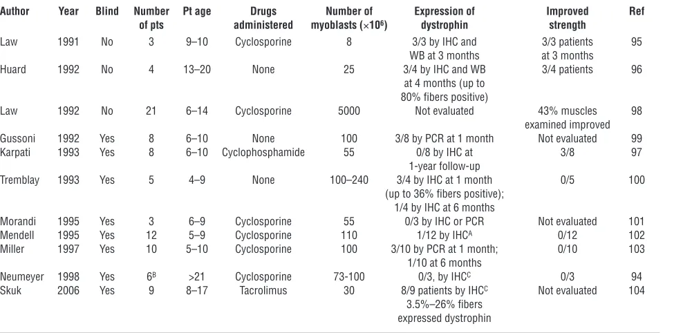
Muscular dystrophies . In 1990, Peter Law and collaborators report-ed the first SC transplant in a 9-year-old boy affected by DMD, showing safety and dystrophin production (93). Soon after, 11 clinical trials in DMD patients were conducted using intramus-cular injection of SCs (Table 3) (94–104). Although there were no adverse effects, new dystrophin production was demonstrated in many but not all cases and clinical benefit in none (6, 105). This is not surprising considering that intramuscular injection in sev-eral locations of a single muscle (or at most a few muscles) cannot elicit a general effect, although improved strength of the injected muscles was detected in 15% of the patients treated. Treating mus-cular dystrophies by intramuscular injection of myoblasts presents several problems that have not been solved yet. First, intramuscu-lar injected cells distribute locally, implying that a huge number of injections will have to be performed in order to treat a complete muscle (96). Second, immune responses toward the injected SCs have been described, even in the case of major histocompatibil-ity locus coincidence. Finally, the rapid death of most of the SCs in the first 72 hours after injection has been extensively report-ed (106, 107). Subsequent experimentation has been devoted to
solving these problems, and a phase I clinical trial has been com-Table 3
Clinical trials of myoblast transplantation in DMD patients
Author Year Blind Number Pt age Drugs Number of Expression of Improved Ref of pts administered myoblasts (×106) dystrophin strength
Law 1991 No 3 9–10 Cyclosporine 8 3/3 by IHC and 3/3 patients 95 WB at 3 months at 3 months
Huard 1992 No 4 13–20 None 25 3/4 by IHC and WB 3/4 patients 96 at 4 months (up to
80% fibers positive)
Law 1992 No 21 6–14 Cyclosporine 5000 Not evaluated 43% muscles 98 examined improved Gussoni 1992 Yes 8 6–10 None 100 3/8 by PCR at 1 month Not evaluated 99 Karpati 1993 Yes 8 6–10 Cyclophosphamide 55 0/8 by IHC at 3/8 97
1-year follow-up
Tremblay 1993 Yes 5 4–9 None 100–240 3/4 by IHC at 1 month 0/5 100 (up to 36% fibers positive);
1/4 by IHC at 6 months
Morandi 1995 Yes 3 6–9 Cyclosporine 55 0/3 by IHC or PCR Not evaluated 101 Mendell 1995 Yes 12 5–9 Cyclosporine 110 1/12 by IHCA 0/12 102
Miller 1997 Yes 10 5–10 Cyclosporine 100 3/10 by PCR at 1 month; 0/10 103 1/10 at 6 months
Neumeyer 1998 Yes 6B >21 Cyclosporine 73-100 0/3, by IHCC 0/3 94
Skuk 2006 Yes 9 8–17 Tacrolimus 30 8/9 patients by IHCC Not evaluated 104
3.5%–26% fibers expressed dystrophin
[image:7.585.54.548.114.355.2]pleted (104). Although encouraging results have been obtained, this method is still limited by the impossibility of delivering myo-blasts systemically through the circulation. Recently, Torrente and colleagues reported the first CD133+ cell transplant (108). They designed a phase I double-blind trial with an autologous trans-plant of unmodified, and thus still dystrophic, muscle-derived CD133+ cells in 8 boys affected by DMD exclusively to test safety; and indeed, no adverse events were reported.
Heart failure. Among different options to treat heart infarction, skeletal muscle–derived myoblasts were considered an optimal cell therapy, as they can be easily obtained from the same patient (avoiding the need for long-term immune suppression), rapidly expanded in vitro, and transplanted back in the patient heart. Pre-clinical experiments performed in animal models demonstrated their ability to engraft correctly, survive in postinfarction scars, differentiate into contractile skeletal muscle cells, and improve heart function (109–111), possibly also because they release angio-genic factors. Unfortunately, in these models, myoblasts were not able to differentiate into cardiomyocytes and did not integrate electrically with the host cardiomyocytes (112, 113). Despite this significant problem, several nonrandomized clinical trials using myoblasts to treat the infarcted heart demonstrated thickening of the LV, an increase in LV ejection fraction, and prevention of LV dilatation, with clinical improvement in some patients (114–120). Histological analysis showed the presence of new myofibers in the scar zone expressing skeletal muscle–specific myosin heavy chain (121). In general, the transplantation procedure was clinically well tolerated, but a high incidence of arrhythmias, some of which were fatal, was reported. In 2007, the results of the MAGIC study, an international phase II double-blind trial were published (122). Ninety-seven patients affected by HFMI were randomized to receive placebo, a low dose of myoblasts (400 × 106 cells), or a high dose of myoblasts (800 × 106 cells). LV end-diastolic volumes decreased substantially in patients receiving myoblasts, supporting a role for myoblasts in remodeling of the heart muscle. The incidence of secondary events, including arrhythmias, was not different between the groups, although all patients received the antiarrhyth-mic agent amiodarone and were implanted with a cardioverter defibrillator. The results of the CAuSMIC (123) and SEISMIC trials (124) have demonstrated safety and some clinical improve-ment. Ongoing trials include the MARVEL study, a phase II/III randomized, double-blind, placebo-controlled trial for which results are expected during the fall of 2009.
SUI. SC-derived myoblasts have also been used as cell therapy for individuals with SUI, which is characterized by the loss of small amounts of urine upon coughing, laughing, sneezing, exercising, or other movements that increase intraabdominal pressure. Myo-blasts may represent an interesting approach for the treatment of this disease (125), as the main cause of SUI is impaired tone of the urethral smooth and striated muscle, which is associated with atro-phy of the supporting structures of the urethra, the mucosa, and
vascular submucosa (126, 127). Treatment of SUI with SC-derived myoblasts has been performed using two different strategies: injec-tion of autologous myoblasts to improve the sphincter tone; and injection of myoblasts together with fibroblasts (isolated by dif-ferential adhesion from the same muscle biopsy) to both improve sphincter tone and treat mucosa atrophy. Until now, all published studies have been nonrandomized, open studies, demonstrating a remarkable clinical improvement in most of the patients treated (127, 128). Moreover, structural and functional techniques have demonstrated thickening of the urinary sphincter and an increase in maximum urethral closure pressure. The onset of improvement is not immediate and may be delayed up to six months after cell injection; however, the benefit, once obtained, lasts for a long peri-od of time, at least up to 12 months (126). Cystoscopic studies have not demonstrated overgrowth of myoblasts nor obstruction of the lower urinary tract (129). Unfortunately, these successful results have not been confirmed yet in a randomized study.
Conclusions
This is an exciting period for those studying the biology of skel-etal muscle stem cells and seeking to harness the information for clinical applications. By learning more about SCs and other meso-dermal skeletal muscle progenitors, we can learn how to better use them to repair muscle. The main limitations of SCs are loss of “stemness” upon culture and an inability to cross the vessel wall for systemic delivery. Limitations for other cell types are incom-plete characterization and their overall minor myogenic potency. Nevertheless, a phase I clinical trial with donor-derived MABs is planned for the end of 2010, given the fact that, because of exten-sive preclinical work, these cells appear at the moment as the best candidates for the cell therapy of muscular dystrophy. Moreover, the terrific prospective of deriving countless autologous, geneti-cally corrected iPS cells from patients certainly will set the stage for future cell therapies. Finally, we should remember that SCs have already been used for clinical trials for DMD, myocardial infarction, and SUI. The last seems to be a case of success for this approach and a situation from which we may also learn in order to redirect efforts toward therapies for myocardial infarction and muscular dystrophies, which have thus far been less successful.
Acknowledgments
Work in the authors’ laboratory is supported by grants from the European Community (OptiStem, Angioscaff, and MyoAmp), European Research Council, Téléthon, Association Française con-tre les Myopathies, Duchenne Parent Project, CureDuchenne, and the Italian Ministries of Research (FIRB) and Health.
Address correspondence to: Giulio Cossu, Division of Regenerative Medicine, San Raffaele Scientific Institute, 58 via Olgettina, 20132 Milan, Italy. Phone: 39-02-2643-4954; Fax: 39-02-2643-4621; E-mail: cossu.giulio@hsr.it.
1. Carlson BM. The regeneration of skeletal muscle. A review. Am J Anat. 1973;137(2):119–149. 2. Mintz B, Baker WW. Normal mammalian muscle
differentiation and gene control of isocitrate dehydrogenase synthesis. Proc Natl Acad Sci U S A. 1967;58(2):592–598.
3. Mauro A. Satellite cells of skeletal muscle fibers.
J Biophys Biochem Cytol. 1961;9:493–495.
4. Cossu G, Biressi S. Satellite cells, myoblasts and other occasional myogenic progenitors: possible
origin, phenotypic features and role in muscle regeneration. Semin Cell Dev Biol. 2005;16(4-5):623–631.
5. Emery AE. The muscular dystrophies. Lancet. 2002;359(9307):687–695.
6. Cossu G, Sampaolesi M. New therapies for Duch-enne muscular dystrophy: challenges, prospects and clinical trials. Trends Mol Med. 2007;13(12):520–526. 7. Buckingham M, Montarras D. Skeletal muscle stem
cells. Curr Opin Genet Dev. 2008;18(4):330–336.
8. Gopinath SD, Rando TA. Stem cell review series: aging of the skeletal muscle stem cell niche. Aging Cell. 2008;7(4):590–598.
9. Kuang S, Gillespie MA, Rudnicki MA. Niche regu- lation of muscle satellite cell self-renewal and dif-ferentiation. Cell Stem Cell. 2008;2(1):22–31. 10. Shi X, Garry DJ. Muscle stem cells in
develop-ment, regeneration, and disease. Genes Dev. 2006;20(13):1692–1708.
review series
stem cell birth and properties. Semin Cell Dev Biol. 2007;18(6):870–882. 12. Whalen RG, Harris JB, Butler-Browne GS, Sesodia S. Expression of myosin isoforms during notexin-induced regeneration of rat soleus muscles. Dev Biol. 1990;141(1):24–40. 13. Beauchamp JR, et al. Expression of CD34 and Myf5 defines the majority of quiescent adult skeletal mus-cle satellite cells. J Cell Biol. 2000;151(6):1221–1234.
14. Zammit PS, et al. Pax7 and myogenic progres-sion in skeletal muscle satellite cells. J Cell Sci. 2006;119(pt 9):1824–1832.
15. Kuang S, Charge SB, Seale P, Huh M, Rudnicki MA. Distinct roles for Pax7 and Pax3 in adult regenera-tive myogenesis. J Cell Biol. 2006;172(1):103–113. 16. Relaix F, et al. Pax3 and Pax7 have distinct and
overlapping functions in adult muscle progenitor cells. J Cell Biol. 2006;172(1):91–102.
17. Tajbakhsh S, et al. Gene targeting the myf-5 locus with nlacZ reveals expression of this myogenic fac-tor in mature skeletal muscle fibres as well as early embryonic muscle. Dev Dyn. 1996;206(3):291–300. 18. Cornelison DD, Filla MS, Stanley HM, Rapraeger AC, Olwin BB. Syndecan-3 and syndecan-4 spe-cifically mark skeletal muscle satellite cells and are implicated in satellite cell maintenance and muscle regeneration. Dev Biol. 2001;239(1):79–94. 19. Rosen GD, et al. Roles for the integrin VLA-4 and
its counter receptor VCAM-1 in myogenesis. Cell. 1992;69(7):1107–1119.
20. Cornelison DD, Wold BJ. Single-cell analysis of regulatory gene expression in quiescent and acti-vated mouse skeletal muscle satellite cells. Dev Biol. 1997;191(2):270–283.
21. Garry DJ, et al. Myogenic stem cell function is impaired in mice lacking the forkhead/winged helix protein MNF. Proc Natl Acad Sci U S A. 2000;97(10):5416–5421.
22. Irintchev A, Zeschnigk M, Starzinski-Powitz A, Wernig A. Expression pattern of M-cadherin in normal, denervated, and regenerating mouse mus-cles. Dev Dyn. 1994;199(4):326–337.
23. Gnocchi VF, White RB, Ono Y, Ellis JA, Zammit PS. Further characterisation of the molecular sig-nature of quiescent and activated mouse muscle satellite cells. PLoS One. 2009;4(4):e5205. 24. Blanco-Bose WE, Yao CC, Kramer RH, Blau HM.
Purification of mouse primary myoblasts based on alpha 7 integrin expression. Exp Cell Res. 2001;265(2):212–220.
25. Kuang S, Kuroda K, Le Grand F, Rudnicki MA. Asymmetric self-renewal and commitment of satel-lite stem cells in muscle. Cell. 2007;129(5):999–1010. 26. Betsholtz C. Insight into the physiological func-tions of PDGF through genetic studies in mice.
Cytokine Growth Factor Rev. 2004;15(4):215–228.
27. Fukada S, Higuchi S, Segawa M, Koda K, Yama-moto Y, et al. Purification and cell-surface marker characterization of quiescent satellite cells from murine skeletal muscle by a novel monoclonal antibody. Exp Cell Res. 2004;296(2):245–255.
28. Sherwood RI, et al. Isolation of adult mouse myo-genic progenitors: functional heterogeneity of cells within and engrafting skeletal muscle. Cell. 2004;119(4):543–554.
29. Day K, Shefer G, Richardson JB, Enikolopov G, Yablonka-Reuveni Z. Nestin-GFP reporter expres-sion defines the quiescent state of skeletal muscle satellite cells. Dev Biol. 2007;304(1):246–259. 30. Montarras D, et al. Direct isolation of satellite
cells for skeletal muscle regeneration. Science. 2005;309(5743):2064–2067. 31. Bosnakovski D, et al. Prospective isolation of skel-etal muscle stem cells with a Pax7 reporter. Stem Cells. 2008;26(12):3194–3204. 32. Peault B, et al. Stem and progenitor cells in skeletal muscle development, maintenance, and therapy.
Mol Ther. 2007;15(5):867–877.
33. Illa I, Leon-Monzon M, Dalakas MC. Regenerating and denervated human muscle fibers and satellite cells express neural cell adhesion molecule recog-nized by monoclonal antibodies to natural killer cells. Ann Neurol. 1992;31(1):46–52.
34. Price FD, Kuroda K, Rudnicki MA. Stem cell based therapies to treat muscular dystrophy. Biochim Bio-phys Acta. 2007;1772(2):272–283.
35. Dhawan J, Rando TA. Stem cells in postnatal myo- genesis: molecular mechanisms of satellite cell qui-escence, activation and replenishment. Trends Cell Biol. 2005;15(12):666–673.
36. Tatsumi R, Anderson JE, Nevoret CJ, Halevy O, Allen RE. HGF/SF is present in normal adult skel-etal muscle and is capable of activating satellite cells. Dev Biol. 1998;194(1):114–128.
37. Floss T, Arnold HH, Braun T. A role for FGF-6 in skeletal muscle regeneration. Genes Dev. 1997;11(16):2040–2051.
38. Musaro A. Growth factor enhancement of muscle regeneration: a central role of IGF-1. Arch Ital Biol. 2005;143(3-4):243–248.
39. Wozniak AC, Anderson JE. Nitric oxide-depen- dence of satellite stem cell activation and quies-cence on normal skeletal muscle fibers. Dev Dyn. 2007;236(1):240–250.
40. Zammit PS, et al. Kinetics of myoblast prolifera-tion show that resident satellite cells are competent to fully regenerate skeletal muscle fibers. Exp Cell Res. 2002;281(1):39–49.
41. Zammit PS, et al. Muscle satellite cells adopt diver-gent fates: a mechanism for self-renewal? J Cell Biol. 2004;166(3):347–357. 42. Conboy IM, Rando TA. The regulation of Notch signaling controls satellite cell activation and cell fate determination in postnatal myogenesis. Dev Cell. 2002;3(3):397–409. 43. Shinin V, Gayraud-Morel B, Gomes D, Tajbakhsh S. Asymmetric division and cosegregation of tem-plate DNA strands in adult muscle satellite cells.
Nat Cell Biol. 2006;8(7):677–687.
44. Conboy MJ, Karasov AO, Rando TA. High incidence of non-random template strand segregation and asymmetric fate determination in dividing stem cells and their progeny. PLoS Biol. 2007;5(5):e102. 45. Le Grand F, Jones AE, Seale V, Scime A, Rudnicki
MA. Wnt7a activates the planar cell polarity path-way to drive the symmetric expansion of satellite stem cells. Cell Stem Cell. 2009;4(6):535–547. 46. Partridge TA, Morgan JE, Coulton GR, Hoffman
EP, Kunkel LM. Conversion of mdx myofibres from dystrophin-negative to -positive by injection of nor-mal myoblasts. Nature. 1989;337(6203):176–179.
47. Cossu G, Sampaolesi M. New therapies for muscu-lar dystrophy: cautious optimism. Trends Mol Med. 2004;10(10):516–520.
48. Collins CA, et al. Stem cell function, self-renewal, and behavioral heterogeneity of cells from the adult mus-cle satellite cell niche. Cell. 2005;122(2):289–301. 49. Cerletti M, et al. Highly efficient, functional
engraftment of skeletal muscle stem cells in dys-trophic muscles. Cell. 2008;134(1):37–47. 50. Sacco A, Doyonnas R, Kraft P, Vitorovic S, Blau HM.
Self-renewal and expansion of single transplanted muscle stem cells. Nature. 2008;456(7221):502–506. 51. Tanaka KK, et al. Syndecan-4-expressing muscle
progenitor cells in the SP engraft as satellite cells during muscle regeneration. Cell Stem Cell. 2009;4(3):217–225.
52. Ikemoto M, et al. Autologous transplantation of SM/C-2.6(+) satellite cells transduced with micro-dystrophin CS1 cDNA by lentiviral vector into mdx mice. Mol Ther. 2007;15(12):2178–2185.
53. Cossu G. Unorthodox myogenesis: possible devel-opmental significance and implications for tissue histogenesis and regeneration. Histol Histopathol. 1997;12(3):755–760.
54. Galli R, et al. Skeletal myogenic potential of
human and mouse neural stem cells. Nat Neurosci. 2000;3(10):986–991.
55. Tajbakhsh S, et al. A population of myogenic cells derived from the mouse neural tube. Neuron. 1994;13(4):813–821.
56. Ferrari G, et al. Muscle regeneration by bone marrow-derived myogenic progenitors. Science. 1998;279(5356):1528–1530.
57. Gussoni E, et al. Dystrophin expression in the mdx mouse restored by stem cell transplantation.
Nature. 1999;401(6751):390–394.
58. Ferrari G, Stornaiuolo A, Mavilio F. Failure to correct murine muscular dystrophy. Nature. 2001;411(6841):1014–1015.
59. McKinney-Freeman SL, et al. Muscle-derived hema-topoietic stem cells are hematopoietic in origin.
Proc Natl Acad Sci U S A. 2002;99(3):1341–1346. 60. Gussoni E, et al. Long-term persistence of donor
nuclei in a Duchenne muscular dystrophy patient receiving bone marrow transplantation. J Clin Invest. 2002;110(6):807–814.
61. Corbel SY, et al. Contribution of hematopoi-etic stem cells to skeletal muscle. Nat Med. 2003;9(12):1528–1532.
62. Camargo FD, Green R, Capetanaki Y, Jackson KA, Goodell MA. Single hematopoietic stem cells gen- erate skeletal muscle through myeloid intermedi-ates. Nat Med. 2003;9(12):1520–1527.
63. Torrente Y, et al. Human circulating AC133(+) stem cells restore dystrophin expression and ameliorate function in dystrophic skeletal muscle. J Clin Invest. 2004;114(2):182–195.
64. Benchaouir R, et al. Restoration of human dystro- phin following transplantation of exon-skipping-engineered DMD patient stem cells into dystrophic mice. Cell Stem Cell. 2007;1(6):646–657.
65. Dezawa M, et al. Bone marrow stromal cells gen-erate muscle cells and repair muscle degeneration.
Science. 2005;309(5732):314–317.
66. Gang EJ, et al. Engraftment of mesenchymal stem cells into dystrophin-deficient mice is not accompanied by functional recovery. Exp Cell Res. 2009;315(15):2624–2636.
67. Minasi MG, et al. The meso-angioblast: a multipo- tent, self-renewing cell that originates from the dor-sal aorta and differentiates into most mesodermal tissues. Development. 2002;129(11):2773–2783. 68. Sampaolesi M, et al. Cell therapy of
alpha-sar-coglycan null dystrophic mice through intra-arterial delivery of mesoangioblasts. Science. 2003;301(5632):487–492. 69. Sampaolesi M, et al. Mesoangioblast stem cells ameliorate muscle function in dystrophic dogs. Nature. 2006;444(7119):574–579. 70. Dellavalle A, et al. Pericytes of human skeletal mus-cle are myogenic precursors distinct from satellite cells. Nat Cell Biol. 2007;9(3):255–267.
71. Asahara T, et al. Isolation of putative progeni-tor endothelial cells for angiogenesis. Science. 1997;275(5302):964–967.
72. Pesce M, et al. Myoendothelial differentiation of human umbilical cord blood-derived stem cells in ischemic limb tissues. Circ Res. 2003;93(5):e51–e62. 73. Zheng B, et al. Prospective identification of myo-genic endothelial cells in human skeletal muscle.
Nat Biotechnol. 2007;25(9):1025–1034.
74. Meliga E, Strem BM, Duckers HJ, Serruys PW. Adipose-derived cells. Cell Transplant. 2007;16(9):963–970. 75. Goudenege S, et al. Enhancement of myogenic and muscle repair capacities of human adipose-derived stem cells with forced expression of MyoD. Mol Ther. 2009;17(6):1064–1072. 76. Evans MJ, Kaufman MH. Establishment in cul-ture of pluripotential cells from mouse embryos. Nature. 1981;292(5819):154–156.
1998;282(5391):1145–1147.
78. Yamanaka S. A fresh look at iPS cells. Cell. 2009;137(1):13–17.
79. Nishikawa S, Goldstein RA, Nierras CR. The promise of human induced pluripotent stem cells for research and therapy. Nat Rev Mol Cell Biol. 2008;9(9):725–729.
80. Park IH, et al. Disease-specific induced pluripotent stem cells. Cell. 2008;134(5):877–886.
81. Murry CE, Keller G. Differentiation of embry-onic stem cells to clinically relevant popula-tions: lessons from embryonic development. Cell. 2008;132(4):661–680.
82. Robbins J, Gulick J, Sanchez A, Howles P, Doetschman T. Mouse embryonic stem cells express the cardiac myosin heavy chain genes during development in vitro. J Biol Chem. 1990;265(20):11905–11909. 83. Sanchez A, Jones WK, Gulick J, Doetschman T,
Robbins J. Myosin heavy chain gene expression in mouse embryoid bodies. An in vitro developmental study. J Biol Chem. 1991;266(33):22419–22426.
84. Bhagavati S, Xu W. Generation of skeletal mus-cle from transplanted embryonic stem cells in dystrophic mice. Biochem Biophys Res Commun. 2005;333(2):644–649. 85. Barberi T, et al. Derivation of engraftable skeletal myoblasts from human embryonic stem cells. Nat Med. 2007;13(5):642–648. 86. Chang H, et al. Generation of transplantable, func-tional satellite-like cells from mouse embryonic stem cells. Faseb J. 2009;23(6):1907–1919. 87. Dekel I, Magal Y, Pearson-White S, Emerson CP, Shani M. Conditional conversion of ES cells to skeletal muscle by an exogenous MyoD1 gene. New Biol. 1992;4(3):217–224. 88. Darabi R, et al. Functional skeletal muscle regen-eration from differentiating embryonic stem cells.
Nat Med. 2008;14(2):134–143.
89. Bianco P, Cossu G. Uno, nessuno e centomila: search-ing for the identity of mesodermal progenitors.
Exp Cell Res. 1999;251(2):257–263.
90. Barberi T, Willis LM, Socci ND, Studer L. Deri-vation of multipotent mesenchymal precursors from human embryonic stem cells. PLoS Med. 2005;2(6):e161.
91. Sakurai H, Okawa Y, Inami Y, Nishio N, Isobe K. Paraxial mesodermal progenitors derived from mouse embryonic stem cells contribute to muscle regeneration via differentiation into muscle satel-lite cells. Stem Cells. 2008;26(7):1865–1873.
92. Maherali N, Hochedlinger K. Guidelines and tech-niques for the generation of induced pluripotent stem cells. Cell Stem Cell. 2008;3(6):595–605. 93. Law PK, et al. Dystrophin production induced by
myoblast transfer therapy in Duchenne muscular dystrophy. Lancet. 1990;336(8707):114–115.
94. Neumeyer AM, et al. Pilot study of myoblast trans-fer in the treatment of Becker muscular dystrophy.
Neurology. 1998;51(2):589–592.
95. Law PK, et al. Myoblast transfer therapy for Duchenne muscular dystrophy. Acta Paediatr Jpn. 1991;33(2):206–215.
96. Huard J, et al. Human myoblast transplantation between immunohistocompatible donors and
recipients produces immune reactions. Transplant Proc. 1992;24(6):3049–3051.
97. Karpati G, et al. Myoblast transfer in Duchenne muscular dystrophy. Ann Neurol. 1993;34(1):8–17.
98. Law PK, et al. Feasibility, safety, and efficacy of myo- blast transfer therapy on Duchenne muscular dys-trophy boys. Cell Transplant. 1992;1(2-3):235–244. 99. Gussoni E, et al. Normal dystrophin transcripts
detected in Duchenne muscular dystrophy patients after myoblast transplantation. Nature. 1992;356(6368):435–438.
100. Tremblay JP, et al. Results of a triple blind clini-cal study of myoblast transplantations without immunosuppressive treatment in young boys with Duchenne muscular dystrophy. Cell Transplant. 1993;2(2):99–112.
101. Morandi L, et al. Lack of mRNA and dystro-phin expression in DMD patients three months after myoblast transfer. Neuromuscul Disord. 1995;5(4):291–295.
102. Mendell JR, et al. Myoblast transfer in the treat-ment of Duchenne’s muscular dystrophy. N Engl J Med. 1995;333(13):832–838.
103. Miller RG, et al. Myoblast implantation in Duch-enne muscular dystrophy: the San Francisco study.
Muscle Nerve. 1997;20(4):469–478.
104. Skuk D, Goulet M, Tremblay JP. Use of repeating dispensers to increase the efficiency of the intra-muscular myogenic cell injection procedure. Cell Transplant. 2006;15(7):659–663.
105. Partridge T. The current status of myoblast trans-fer. Neurol Sci. 2000;21(suppl 5):S939–S942. 106. Fan Y, Maley M, Beilharz M, Grounds M. Rapid
death of injected myoblasts in myoblast transfer therapy. Muscle Nerve. 1996;19(7):853–860. 107. Guerette B, et al. Prevention by anti-LFA-1 of acute
myoblast death following transplantation. J Immunol. 1997;159(5):2522–2531.
108. Torrente Y, et al. Autologous transplantation of mus-cle-derived CD133+ stem cells in Duchenne muscle patients. Cell Transplant. 2007;16(6):563–577. 109. Atkins BZ, et al. Intracardiac transplantation of
skeletal myoblasts yields two populations of striated cells in situ. Ann Thorac Surg. 1999;67(1):124–129.
110. Taylor DA, et al. Regenerating functional myocardi-um: improved performance after skeletal myoblast transplantation. Nat Med. 1998;4(8):929–933.
111. Pouzet B, et al. Factors affecting functional out- come after autologous skeletal myoblast trans-plantation. Ann Thorac Surg. 2001;71(3):844–850; discussion 850–851.
112. Reinecke H, Poppa V, Murry CE. Skeletal muscle stem cells do not transdifferentiate into cardio-myocytes after cardiac grafting. J Mol Cell Cardiol. 2002;34(2):241–249.
113. Leobon B, et al. Myoblasts transplanted into rat infarcted myocardium are functionally iso-lated from their host. Proc Natl Acad Sci U S A. 2003;100(13):7808–7811.
114. Smits PC, et al. Catheter-based intramyocardial injection of autologous skeletal myoblasts as a primary treatment of ischemic heart failure: clini-cal experience with six-month follow-up. J Am Coll Cardiol. 2003;42(12):2063–2069.
115. Menasche P, et al. Myoblast transplantation for heart failure. Lancet. 2001;357(9252):279–280. 116. Menasche P, et al. Autologous skeletal
myo-blast transplantation for severe postinfarction left ventricular dysfunction. J Am Coll Cardiol. 2003;41(7):1078–1083.
117. Dib N, et al. Safety and feasibility of autologous myoblast transplantation in patients with ischemic cardiomyopathy: four-year follow-up. Circulation. 2005;112(12):1748–1755.
118. Hagege AA, et al. Skeletal myoblast transplanta-tion in ischemic heart failure: long-term follow-up of the first phase I cohort of patients. Circulation. 2006;114(suppl 1):I108–113.
119. Herreros J, et al. Autologous intramyocardial injec-tion of cultured skeletal muscle-derived stem cells in patients with non-acute myocardial infarction.
Eur Heart J. 2003;24(22):2012–2020.
120. Steendijk P, et al. Intramyocardial injection of skeletal myoblasts: long-term follow-up with pres-sure-volume loops. Nat Clin Pract Cardiovasc Med. 2006;3(suppl 1):S94–S100.
121. Pagani FD, et al. Autologous skeletal myoblasts transplanted to ischemia-damaged myocar-dium in humans. Histological analysis of cell survival and differentiation. J Am Coll Cardiol. 2003;41(5):879–888.
122. Menasche P, et al. The Myoblast Autologous Grafting in Ischemic Cardiomyopathy (MAGIC) trial: first randomized placebo-controlled study of myoblast transplantation. Circulation. 2008;117(9):1189–1200.
123. Dib N, et al. One-year follow-up of feasibility and safety of the first U.S., randomized, controlled study using 3-dimensional guided catheter-based delivery of autologous skeletal myoblasts for isch-emic cardiomyopathy (CAuSMIC study). JACC Car-diovasc Interv. 2009;2(1):9–16.
124. Serruys PW. Final results of a phase I/II random-ized, open-label trial to evaluate intramyocardial autologous skeletal myoblast transplantation in congestive heart failure patients: The SEISMIC Trial. Paper presented at: 57th Annual Scientific Session of the American College of Cardiology; March 29-April 1 2008; Chicago, IL, USA. 125. Smaldone MC, Chancellor MB. Muscle derived stem cell therapy for stress urinary incontinence.
World J Urol. 2008;26(4):327–332.
126. Mitterberger M, et al. Adult stem cell therapy of female stress urinary incontinence. Eur Urol. 2008;53(1):169–175.
127. Carr LK, et al. 1-year follow-up of autologous mus-cle-derived stem cell injection pilot study to treat stress urinary incontinence. Int Urogynecol J Pelvic Floor Dysfunct. 2008;19(6):881–883.
128. Strasser H, et al. Transurethral ultrasonography- guided injection of adult autologous stem cells ver-sus transurethral endoscopic injection of collagen in treatment of urinary incontinence. World J Urol. 2007;25(4):385–392.