MicroRNA-486–dependent modulation of
DOCK3/PTEN/AKT signaling pathways
improves muscular dystrophy–associated
symptoms
Matthew S. Alexander, … , Jeffrey J. Widrick, Louis M.
Kunkel
J Clin Invest.
2014;
124(6)
:2651-2667.
https://doi.org/10.1172/JCI73579
.
Duchenne muscular dystrophy (DMD) is caused by mutations in the gene encoding
dystrophin, which results in dysfunctional signaling pathways within muscle. Previously, we
identified microRNA-486 (miR-486) as a muscle-enriched microRNA that is markedly
reduced in the muscles of dystrophin-deficient mice (
Dmd
mdx-5Cvmice) and in DMD patient
muscles. Here, we determined that muscle-specific transgenic overexpression of miR-486
in muscle of
Dmd
mdx-5Cvmice results in reduced serum creatine kinase levels, improved
sarcolemmal integrity, fewer centralized myonuclei, increased myofiber size, and improved
muscle physiology and performance. Additionally, we identified dedicator of cytokinesis 3
(DOCK3) as a miR-486 target in skeletal muscle and determined that DOCK3 expression is
induced in dystrophic muscles. DOCK3 overexpression in human myotubes modulated
PTEN/AKT signaling, which regulates muscle hypertrophy and growth, and induced
apoptosis. Furthermore, several components of the PTEN/AKT pathway were markedly
modulated by miR-486 in dystrophin-deficient muscle. Skeletal muscle–specific miR-486
overexpression in
Dmd
mdx-5Cvanimals decreased levels of DOCK3, reduced PTEN
expression, and subsequently increased levels of phosphorylated AKT, which resulted in
an overall beneficial effect. Together, these studies demonstrate that stable overexpression
of miR-486 ameliorates the disease progression of dystrophin-deficient skeletal muscle.
Research Article
Muscle biology
Find the latest version:
MicroRNA-486–dependent modulation
of DOCK3/PTEN/AKT signaling pathways
improves muscular dystrophy–
associated symptoms
Matthew S. Alexander,1,2 Juan Carlos Casar,3 Norio Motohashi,4 Natássia M. Vieira,1,2 Iris Eisenberg,5 Jamie L. Marshall,1,2 Molly J. Gasperini,1 Angela Lek,1,2 Jennifer A. Myers,1
Elicia A. Estrella,1,6 Peter B. Kang,1,6 Frederic Shapiro,7 Fedik Rahimov,1,2 Genri Kawahara,1,2 Jeffrey J. Widrick,1 and Louis M. Kunkel1,2,8,9
1Division of Genetics and Genomics, Boston Children’s Hospital, Boston, Massachusetts, USA. 2Department of Pediatrics and Genetics,
Harvard Medical School, Boston, Massachusetts, USA. 3Departamento de Neurología, Escuela de Medicina, Pontificia Universidad Católica de Chile,
Santiago, RM, Chile. 4Stem Cell Institute, Paul and Sheila Wellstone Muscular Dystrophy Center, Department of Neurology,
University of Minnesota Medical School, Minneapolis, Minnesota, USA. 5Center for Human Placenta Research, Department of Obstetrics and Gynecology,
Hadassah-Hebrew University Medical Center, Mt. Scopus Jerusalem, Israel. 6Department of Neurology and 7Department of Orthopedic Surgery,
Boston Children’s Hospital and Harvard Medical School, Boston, Massachusetts, USA. 8The Manton Center for Orphan Disease Research,
Boston Children’s Hospital, Boston, Massachusetts, USA. 9Harvard Stem Cell Institute, Cambridge, Massachusetts, USA.
Duchenne muscular dystrophy (DMD) is caused by mutations in the gene encoding dystrophin, which results
in dysfunctional signaling pathways within muscle. Previously, we identified microRNA-486 (miR-486) as a
muscle-enriched microRNA that is markedly reduced in the muscles of dystrophin-deficient mice (Dmd
mdx-5Cvmice) and in DMD patient muscles. Here, we determined that muscle-specific transgenic overexpression of
miR-486 in muscle of Dmd
mdx-5Cvmice results in reduced serum creatine kinase levels, improved sarcolemmal
integrity, fewer centralized myonuclei, increased myofiber size, and improved muscle physiology and
perfor-mance. Additionally, we identified dedicator of cytokinesis 3 (DOCK3) as a miR-486 target in skeletal muscle
and determined that DOCK3 expression is induced in dystrophic muscles. DOCK3 overexpression in human
myotubes modulated PTEN/AKT signaling, which regulates muscle hypertrophy and growth, and induced
apoptosis. Furthermore, several components of the PTEN/AKT pathway were markedly modulated by miR-486
in dystrophin-deficient muscle. Skeletal muscle–specific miR-486 overexpression in Dmd
mdx-5Cvanimals
decreased levels of DOCK3, reduced PTEN expression, and subsequently increased levels of phosphorylated
AKT, which resulted in an overall beneficial effect. Together, these studies demonstrate that stable
overexpres-sion of miR-486 ameliorates the disease progresoverexpres-sion of dystrophin-deficient skeletal muscle.
Introduction
Mammalian skeletal muscle is a dynamic organ capable of repair-ing itself followrepair-ing injury or atrophy arisrepair-ing from prolonged disuse. The skeletal muscle–regenerative process is a well-regulated process involving a variety of muscle progenitor cells and signaling mole-cules that work systematically to repair the damaged myofiber. The skeletal muscles of Duchenne muscular dystrophy (DMD) patients undergo waves or cycles of degeneration followed by regeneration, muscle tearing, and inflammation that lead to progressive muscle weakness and multi-organ complications, among which respira-tory failure and cardiomyopathy are life threatening (1). There have been several novel approaches to treating the disease, such as the use of exon-skipping morpholinos or 2′-O-methyl phosphorothio-ate (2′OmePS) antisense oligonucleotides (AONs), which restore a truncated dystrophin open reading frame (ORFs) (2–5). While these therapeutic strategies have shown promise in human clinical
trials, the long-term functional consequences of these treatments have yet to be fully elucidated (3, 6). Additional strategies are needed to treat the already damaged muscle and dysregulated secondary signaling pathways that exist due to chronic dystrophin deficiency (6–8). One strategy that has shown beneficial effects in dystrophin-deficient animal models is the modulation of components of the PTEN/AKT signaling pathway (9–11). PTEN, a tumor suppressor gene with diverse functions in multiple tissues, is induced in dys-trophin-deficient muscle (12–14). This increase in Pten expression levels is accompanied by a subsequent reduction in phosphorylated
Akt, both of which have been shown to regulate glucose and insulin signaling pathways in skeletal muscle (15–19). Transgenic overex-pression of a constitutively active version of Akt in skeletal muscle resulted in muscle hypertrophy and improved muscle histology and physiology in Dmdmdx mice (9, 10, 20). Direct suppression of
Pten in mouse myoblasts increased myogenic differentiation with a subsequent increase in phosphorylated Akt (21). Ablation of Pten
exclusively in mouse skeletal muscle resulted in leaner muscle and an improved ability to regenerate muscle on a high-fat diet (22).
To identify novel therapies and targets for treating dystrophin-deficient skeletal muscle, animal models have been proven to be useful (23). The Dmdmdx mouse contains a spontaneous nonsense Conflict of interest: Louis M. Kunkel is a consultant for Pfizer Inc., Summit
Corpo-ration PLC, and Tarix Pharmaceuticals for muscle disease drug therapies and for Syn-apDx Corporation for autism screening technologies. Peter B. Kang was previously a coinvestigator on a study funded by ISIS Pharmaceuticals, was a consultant for Third Rock Ventures, and serves on the DMD Advisory Board of Sarepta Therapeutics.
research article
mutation in exon 23 of the mouse Dystrophin (Dmd) gene and has been shown to have muscle deficits associated with dystrophin deficiency (24–26). An ENU-mutagenesis screen later identified several additional mouse strains containing mutations elsewhere in the mouse Dmd gene (27). One strain of dystrophic mice from that screen, Dmdmdx-5Cv, contains a nonsense mutation in exon 10, shows the fewest number of revertant fibers among all the Dmdmdx mouse strains, and has a severe diaphragm muscle-force deficit (28, 29). Given the severity of the disease phenotypes observed in the Dmdmdx-5Cv mice, this mouse strain is highly useful for testing novel drug compounds and factors that might ameliorate clinical pathological aspects of dystrophin deficiency in an animal model. MicroRNAs are 21–24 nucleotide small RNAs that have been shown to regulate gene expression by either degrading their target mRNA or interfering with the ribosome’s ability to translate the target mRNA (30). MicroRNAs can regulate several target genes in a single pathway or multiple signaling pathways at critical time points in development and disease progression (31–34). The abil-ity to quickly modulate the expression levels of disease-relevant microRNAs using viral overexpression or antisense inhibitors, makes microRNA-based therapies an attractive therapy for the treatment of complex diseases in which many signaling pathways are significantly affected. Previously, we identified the microRNA microRNA-486 (miR-486) as being significantly reduced in expres-sion levels in dystrophin-deficient DMD muscle, but not in the milder Becker muscular dystrophy (BMD) muscle (35, 36). miR-486 is highly enriched in muscle, and may promote myogenic dif-ferentiation by downregulating the expression of the transcription factor Pax7 in skeletal muscle satellite cells (37, 38). Here, we show that transgenic overexpression of miR-486 in the muscles of dys-trophic (Dmdmdx-5Cv) mice ameliorates many of the clinical signs associated with dystrophin deficiency. Dystrophic mice that over-express miR-486 have reduced serum creatine kinase (CK) levels, improved muscle physiology, and improved muscle performance. Additionally, we show that miR-486 overexpression directly mod-ulates the PTEN/AKT signaling pathway in normal and dystro-phic mouse muscle. Finally, we identify dedicator of cytokinesis 3 (DOCK3) (also called modifier of cell adhesion [MOCA]) as a direct target of miR-486 in normal and dystrophic muscles and show that it plays a functional role in myotube survival. Together, these studies identify miR-486 as a modulator of disease pathol-ogy in dystrophic skeletal muscle and open up the possibility of using miR-486 overexpression as a potential novel therapy for muscular dystrophy.
Results
miR-486 is enriched in myogenic tissues but reduced in dystrophic muscle. Previously, we identified miR-486 as being strongly reduced in expression levels in human dystrophic muscle (35, 36). miR-486 expression is highly enriched in developing mouse muscle and is induced during myogenic differentiation of muscle stem cells (36–38). We first sought to examine the expression of human miR-486 (hsa-miR-486), specifically the human miR-486-5p sequence, in a panel of normal adult tissues (Sanger miRBase: http://mirbase.org/). Human miR-486-5p was expressed at the highest levels in the skeletal muscle and heart tissues and at more moderate levels in lung, brain, and bladder (Supplemental Fig-ure 1A; supplemental material available online with this article; doi:10.1172/JCI73579DS1). To confirm these findings in another mammalian organism, we analyzed a similar array of adult mouse
tissues for miR-486-5p expression levels. Mouse miR-486-5p was similarly enriched in the adult heart and skeletal muscles while also showing moderate expression in the brain and bladder tis-sues (Supplemental Figure 1B). Given the similarities between mouse and human miR-486 expression, we next analyzed the pat-tern of miR-486 in both oxidative and glycolytic muscle groups consisting of the tibialis anterior (TA), extensor digitorum longus (EDL), gastrocnemius, soleus, triceps, paraspinal muscles, and the diaphragm in both normal and dystrophin-deficient (Dmdmdx-5Cv strain) muscles. miR-486 was expressed in all 7 of the muscle groups analyzed, but reduced in expression in all of the Dmdmdx-5Cv muscles except for the paraspinal muscles (Supplemental Figure 1C). These results in murine dystrophin-deficient muscle are in agreement with our previous finding that miR-486 transcript lev-els are strongly reduced in human DMD muscle biopsies (35, 36).
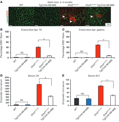
Muscle-specific overexpression of miR-486 improves skeletal muscle histology in dystrophic muscle. Suppression of miR-21, a fibroblast-proliferation–inducing microRNA that is induced in dystrophin-deficient muscle, resulted in improved pathology in Dmdmdx mouse muscles through modulation of the PTEN/AKT signal-ing pathway (39). Thus, we postulated that overexpression of a microRNA that is reduced in dystrophic muscle, miR-486, might have a similar beneficial effect in delaying or ameliorating some of the signs of muscular dystrophy in Dmdmdx-5Cv mice. To determine the functional consequences of miR-486 overexpression in muscle on a dystrophic background, we mated the Tg(Cmk-Mir486) (MCK promoter) mice with the Dmdmdx-5Cv mice. We performed histologi-cal analysis of the muscles of the Dmdmdx-5Cv Tg(Cmk-Mir486) adult (2 to 4 months old) mice to determine what effect or effects miR-486 overexpression might have on disease progression. Centralized myonuclei are indicative of dystrophin deficiency in mice and can be used as an indicator of myofiber regeneration (23, 25). The TA and diaphragm muscles of Dmdmdx-5Cv Tg(Cmk-Mir486) mice revealed reduced numbers of centralized myonuclei compared with their Dmdmdx-5Cv control littermates (Figure 1, A and B). Histological examination of the gastrocnemius muscles from adult Dmdmdx-5Cv Tg(Cmk-Mir486) mice also displayed improved overall histology and reduced numbers of centralized myonuclei, further supporting a functional role for miR-486 overexpression as a means of delaying the disease progression of muscular dys-trophy (Supplemental Figure 2, A and B). A previous study has shown that the diaphragm muscle of the Dmdmdx-5Cv strain is more severely affected by dystrophin deficiency than other muscles (29). We measured the muscle fiber cross-sectional area (CSA) in WT, Tg(Cmk-Mir486), Dmdmdx-5Cv, and Dmdmdx-5Cv Tg(Cmk-Mir486) mice to determine what effects miR-486 might have on overall myofiber size. The Tg(Cmk-Mir486) mice displayed larger overall myofiber sizes when compared with their WT littermates, sug-gesting that miR-486 might be inducing myofiber hypertrophy (Figure 1C). Similarly, the Dmdmdx-5Cv Tg(Cmk-Mir486) showed sig-nificantly larger myofibers compared with their Dmdmdx-5Cv litter-mates, comparable in size to WT myofibers (Figure 1C). Thus, the overall histology of Dmdmdx-5Cv dystrophic mice that overexpress the miR-486 transgene was greatly improved when compared with their Dmdmdx-5Cv littermates.
signifi-cantly less EBD infiltration in their TA muscles than their Dmdmdx-5Cv littermates, suggesting that miR-486 overexpression ameliorat-ed myofiber damage (Figure 2, A and B). These results were also observed in the gastrocnemius muscles, indicating that miR-486 muscle overexpression may have beneficial effects in preventing muscle membrane permeability and overall membrane instabil-ity that is often associated with loss of dystrophin (Figure 2C).
[image:4.585.87.494.81.557.2]Dysregulated serum biomarkers, such as CK and alanine transami-nase (ALT), are hallmarks of dystrophic muscle and are often used to diagnose the severity of muscle disease in both humans and mice (23, 42–44). We next measured the levels of serum CK in adult WT, Tg(Cmk-Mir486), Dmdmdx-5Cv, and Dmdmdx-5Cv Tg(Cmk-Mir486) mice. There were no significant differences in levels of serum CK between WT and Tg(Cmk-Mir486) mice; however, Dmdmdx-5Cv mice showed
Figure 1
Overexpression of miR-486 in dystrophic Dmdmdx-5Cv mouse muscles reduces dystrophic disease histopathology. (A) H&E staining of adult TA
and diaphragm muscles from WT, Tg(Cmk-Mir486), Dmdmdx-5Cv, and Dmdmdx-5Cv Tg(Cmk-Mir486) mice. Arrowheads demarcate centralized
nuclei, a classic finding in dystrophic disease and regenerating skeletal muscle. Scale bars: 50 μm. (B) Summary graphs of centralized nuclei
counted from the TA and diaphragm muscles of adult Dmdmdx-5Cv and Dmdmdx-5Cv Tg(Cmk-Mir486) mice. (C) Fiber type cross-section area
analy-sis of the TA muscles of WT, Tg(Cmk-Mir486), Dmdmdx-5Cv, and Dmdmdx-5Cv Tg(Cmk-Mir486) adult mice. Six-hundred fibers from 5 separate mice
research article
approximately 20-fold higher levels of serum CK as compared with WT mice (Figure 2D). Remarkably, the levels of serum CK were reduced by half in adult Dmdmdx-5Cv Tg(Cmk-Mir486) mice, again sug-gesting decreased membrane permeability (Figure 2D). Similarly, the levels of ALT were also reduced in Dmdmdx-5Cv Tg(Cmk-Mir486) mice when compared with Dmdmdx-5Cv littermates (Figure 2E). Together, these results demonstrate that miR-486 muscle overex-pression improves the serum biomarkers that are typically dysregu-lated in dystrophic mouse muscle toward near normal levels.
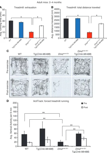
Overexpression of miR-486 improves Dmdmdx-5Cv muscle physiological
function. To determine whether miR-486 overexpression would improve muscle physiological function, we tested age-matched WT, Tg(Cmk-Mir486), Dmdmdx-5Cv, and Dmdmdx-5Cv Tg(Cmk-Mir486) mice using a series of treadmill, grip strength, activity, and
iso-lated muscle-force output assays. As expected, Dmdmdx-5Cv mice performed poorly on the treadmill assay compared with WT and Tg(Cmk-Mir486) mice in both the time to overall exhaus-tion and total distance tests (Figure 3, A and B). The Dmdmdx-5Cv Tg(Cmk-Mir486) mice showed marked improvements in both total running distances and time to exhaustion compared with Dmdmdx-5Cv littermates (Figure 3, A and B). Previous studies have shown that
Dmdmdx mice have reduced overall and vertical activity before and after forced exercise (45, 46). We measured the total activity of all 4 mouse genotype cohorts before and after forced downhill treadmill exercise. While Dmdmdx-5Cv mice showed severe decreases in overall and vertical activity before and after forced exercise, the
[image:5.585.93.491.82.488.2]Dmdmdx-5Cv Tg(Cmk-Mir486) mice displayed activity patterns similar to those of both WT and Tg(Cmk-Mir486) mice (Figure 3, C and D). Figure 2
Dmdmdx-5Cv Tg(Cmk-Mir486) mice have improved muscle membrane integrity and serum biochemistry. (A) EBD (red fluorescence)
immunohistochemistry of TA muscle biopsies taken from WT, Tg(Cmk-Mir486), Dmdmdx-5Cv, and Dmdmdx-5Cv Tg(Cmk-Mir486) mice. Sections were
costained with Laminin α-2 antisera (Lama2; green) to demarcate the myofiber basal lamina. Arrowheads demarcate EBD-positive myofibers. Scale bars: 50 μm. (B and C) Graph showing the quantification of EBD-positive myofibers from the TA (B) and gastrocnemius (C) muscles in WT (black
bars), Tg(Cmk-Mir486) (blue bars), Dmdmdx-5Cv (red bars), and Dmdmdx-5Cv Tg(Cmk-Mir486) (white bars) mice. Six-hundred fibers from 3 separate
miR-486 overexpression improves Dmdmdx-5Cv muscle strength and
tetanic force output. We tested cage-grip strength (the total force of a mouse’s front paws to grasp a wire cage) in the 4 mouse geno-type cohorts as an in vivo output of muscle strength. The Tg( Cmk-Mir486) mice generated similar overall cage-grip force output when compared with WT controls (Figure 4A). More impor-tantly, Dmdmdx-5Cv Tg (Cmk-Mir486) mice showed a significant increase in the overall force output and were able to hold onto their cages for longer periods of time compared with Dmdmdx-5Cv littermates (Figure 4, A and B). These findings demonstrate that loss of muscle-force output in dystrophin deficiency is
signifi-cantly improved when miR-486 is overexpressed. We next per-formed physiological assays on isolated groups of muscles (EDL, soleus, and diaphragm strips) biopsied from adult WT, Tg( Cmk-Mir486), Dmdmdx-5Cv, and Dmdmdx-5Cv Tg(Cmk-Mir486) age-matched cohorts. The specific force (tetanic force per muscle physiologi-cal CSA) of EDL, soleus, and diaphragm muscles is reduced in
Dmdmdx and Dmdmdx-5Cv mice (29, 47). We observed no significant differences in specific force in the muscles of WT versus Tg( Cmk-Mir486) mice, but a marked improvement in the muscles from
Dmdmdx-5Cv Tg(Cmk-Mir486) mice when directly compared with
[image:6.585.49.413.80.598.2]Dmdmdx-5Cv littermates (Figure 4C). These studies demonstrate
Figure 3
miR-486 overexpression in
Dmdmdx-5Cv mice improves
dys-trophic muscle physiology. (A)
Graph of forced downhill tread-mill running of WT (black bars), Tg(Cmk-Mir486) (blue bars),
Dmdmdx-5Cv (red bars), and Dmdmdx-5Cv Tg(Cmk-Mir486)
(white bars) mice measuring time of endurance (total time before the mice reached exhaus-tion) in minutes. (B) Graph of
forced downhill treadmill running measuring total distance trav-eled in centimeters (cm) of the 4 genotype cohorts. (C)
Repre-sentative ActiTrack activity plots of the 4 mouse genotype cohorts measured before (pre-exercise) and after (post-exercise) forced downhill treadmill running. Thin lines represent mouse distance traveled, while darker lines represent instances of rearing (vertical activity). (D) Summary
research article
that muscle-force output is improved in the murine muscles that overexpress miR-486 on the Dmdmdx-5Cv background.
Skeletal muscle fiber types are not significantly affected by miR-486 over-expression. In DMD muscle, the fast twitch type II fibers are the first group to break down, while the slow type I fibers remain are the last group to be affected (48). A previous study of skeletal mus-cle fiber types revealed a greater number of slow, type I fibers in the soleus muscles of Dmdmdx-5Cv mice (49). We tested whether any of the beneficial effects observed in the Dmdmdx-5Cv Tg(Cmk-Mir486) mice might be due to changes in muscle fiber type composition. Using immunofluorescence staining with monoclonal myosin heavy chain antibodies, we counted the total numbers of type I, type IIa, and type IIb in adult (2 to 4 months old) WT, Tg( Cmk-Mir486), Dmdmdx-5Cv, and Dmdmdx-5Cv Tg(Cmk-Mir486) age-matched cohorts (50). As expected, we observed a shift from fast to slow type myofibers in the soleus muscles of Dmdmdx-5Cv mice when com-pared with their age-matched WT cohort (Supplemental Figure 3, A and B). We did observe a slight overall increase in the number of type IIa oxidative fibers and a corresponding decrease in the num-ber of type IIb glycolytic finum-bers in the TA muscles of the Tg(
Cmk-Mir486) mice (Supplemental Figure 3A). Previous studies have shown that alterations of the PTEN/AKT signaling components in mouse muscles, while inducing muscle hypertrophy, had very modest effects on muscle fiber type specification (51–53). Thus, it is likely that the alteration of fiber type composition in miR-486– overexpressing mice might be due to the changes in expression lev-els of signaling components including the PTEN/AKT pathway. However, no change in the fiber type distribution was observed when Dmdmdx-5Cv and Dmdmdx-5Cv Tg(Cmk-Mir486) soleus, TA, or gastrocnemius muscles were compared (Supplemental Figure 3, A and B). These findings suggest that the beneficial effects seen in miR-486–overexpressing Dmdmdx-5Cv mice are most likely indepen-dent of any significant changes in muscle fiber type composition.
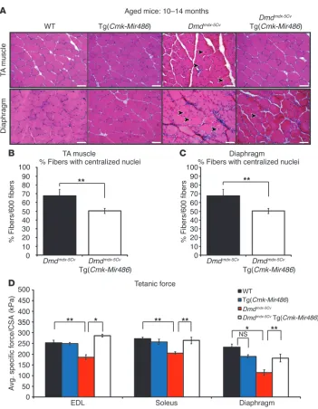
[image:7.585.65.521.81.434.2]Aged dystrophic mice show similar beneficial effects in their muscles resulting from miR-486 transgenic overexpression. It has been well doc-umented that dystrophic disease symptoms, such as muscle weak-ness, increased serum CK levels, and loss of muscle force, occur in aged Dmdmdx mice (54). In the more severe dystrophic Dmdmdx-5Cv mice, muscle fatigue and functional deficits have been reported as early as 8 weeks and progressively worsen in mice that have been Figure 4
miR-486–overexpressing dystrophic muscles have improved strength and tetanic force outputs. (A) Cage-grip strength measurements of mice
of the 4 genotype cohorts measured as kg × 10–5 and normalized to mouse weight (grams). (B) Two-limb (forelimbs) wire hang-time
measure-ments (seconds) of each of the 4 genotype cohorts. (C) Tetanic muscle-force measurements, normalized to CSA, of isolated EDL, soleus, and
diaphragm muscles. Eight (WT and Tg[Cmk-Mir486]) and 12 (Dmdmdx-5Cv and Dmdmdx-5Cv Tg[Cmk-Mir486]) adult (2 to 4 months old) male mice
aged to 1 year (29). To determine whether the beneficial effects observed in the adult 2- to 4-month-old mice were maintained in aged dystrophic muscles, we performed similar histological, biochemical, and physiological measurements on aged Dmdmdx-5Cv (10 to 14 months old) mice that overexpressed the miR-486 transgene. We observed a similar decrease in the amount of cen-tral myonuclei and overall improved histology of the TA muscles and diaphragm in aged Dmdmdx-5Cv Tg(Cmk-Mir486) mice when compared with their Dmdmdx-5Cv littermates (Figure 5, A–C). Addi-tionally, serum profiling of aged Dmdmdx-5Cv Tg(Cmk-Mir486) mice revealed a similar reduction in the overall serum CK levels when compared with the dystrophic Dmdmdx-5Cv littermates (Supplemen-tal Figure 4, A and B). Finally, we measured the specific force of the EDL, soleus, and diaphragm muscles from aged Dmdmdx-5Cv Tg(Cmk-Mir486) and Dmdmdx-5Cv mice along with aged WT and Tg(Cmk-Mir486) controls. We observed that the aged Dmdmdx-5Cv Tg(Cmk-Mir486) mice had maintained their specific muscle strength, similar to that of WT and Tg(Cmk-Mir486) mice, when
compared with their Dmdmdx-5Cv littermates (Figure 5D). Together, these studies demonstrate that the beneficial effects observed with the overexpression of miR-486 in the skeletal muscles of
Dmdmdx-5Cv mice are maintained in older dystrophic mice.
Overexpression of miR-486 reduces Pten expression while increasing phosphorylated Akt levels in skeletal muscle. miR-486 was previously shown to target PTEN and PTEN/AKT signaling components in both heart and skeletal muscles (36, 37, 55). We next measured the protein levels of PTEN and AKT directly in the TA muscles of both WT and Dmdmdx-5Cv mice, which overexpress miR-486. Overexpression of miR-486 strongly reduced the expression of
[image:8.585.45.396.77.529.2]Pten in the muscles of Tg(Cmk-Mir486) mice compared with their WT control littermates (Figure 6, A and B). Similarly, the over-all protein levels of Pten in the Dmdmdx-5Cv Tg(Cmk-Mir486) mice were also reduced compared with the levels in their Dmdmdx-5Cv littermates, implicating Pten as a direct in vivo target of miR-486 in skeletal muscle (Figure 6, C and D). The total levels of all 3 Akt isoforms (Akt1/2/3) remained unchanged, while the levels
Figure 5
Aged dystrophic mice show similar improved pathology resulting from miR-486 transgenic overexpression. (A) Representative H&E staining of
TA and diaphragm muscles taken from aged (10 to 14 months old) WT, Tg(Cmk-Mir486), Dmdmdx-5Cv, and Dmdmdx-5Cv Tg(Cmk-Mir486) mice.
Arrowheads represent centralized myonuclei. Scale bars: 50 μm. (B)
Graphs of centralized myonuclei counts of the TA and (C) diaphragm
muscles of aged Dmdmdx-5Cv and Dmdmdx-5Cv Tg(Cmk-Mir486) mice.
Three mice (n = 6 TA and n = 3 dia-phragm muscles) were used, with 600 TA and 200 diaphragm individual myofibers being counted. (D) Tetanic
muscle-force measurements, normal-ized to CSA, of isolated EDL, soleus, and diaphragm muscles. Five (WT and Tg[Cmk-Mir486]) to 8 (Dmdmdx-5Cv
and Dmdmdx-5Cv Tg[Cmk-Mir486])
research article
of phosphorylated AKT (S473 and T308) at 2 residues critical for its kinase function, significantly increased in the miR-486– overexpressing muscles (Figure 6, A–D). Thus, the overexpression of miR-486 in the skeletal muscles of Tg(Cmk-Mir486) mice sig-nificantly alters the expression levels of key components of the PTEN/AKT signaling pathway.
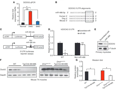
DOCK3 is a direct downstream target of miR-486 in skeletal muscle. To identify novel direct targets of miR-486 that might be modulating the pathology of dystrophic muscle, we utilized a bioinformatic approach using publically available software algorithms, which predict targets of microRNAs (TargetScan [www.targetscan.org], MicroCosm [http://www.ebi.ac.uk/enright-srv/microcosm/ htdocs/targets/v5/], miRDB [http://mirdb.org/miRDB/]). We focused on predicted miR-486 targets whose mRNA transcripts were conversely elevated in DMD muscle and cross-referenced those targets with previously published DMD biopsy microarray data sets for mRNA transcripts that were elevated in DMD muscles (12, 56). Several miR-486–validated targets, including PTEN and FOXO1, were confirmed as elevated in DMD muscle by Western blot analysis (data not shown). One predicted target, DOCK3 (pre-viously referred to as MOCA, modifier of cell adhesion), was found to be elevated in expression in DMD muscle biopsies (Figure 7A). The DOCK3 3′ UTR has a strongly evolutionarily conserved miR-486–binding seed site among mammals including mouse,
[image:9.585.78.510.80.386.2]dogs, and humans (Figure 7B). MicroRNAs can be functionally screened to determine whether they have repressive activity in cells by fusing the 3′ UTR of the miRNA target to luciferase, overex-pressing the microRNA (or scrambled miR control), and mea-suring the amount of luciferase reporter activity. We cloned the human DOCK3 3′ UTR containing the miR-486 seed site to a lucif-erase reporter and transfected the construct into the nonmyogenic HEK293T cell line along with either miR-486 or scrambled miR control constructs and assayed for luciferase activity (Figure 7C). miR-486 suppressed human DOCK3 3′ UTR luciferase reporter activ-ity when compared directly to scrambled miR controls (Figure 7D). Likewise, mutation of critical nucleotides in the miR-486 seed site of the DOCK3 3′ UTR resulted in the failure of miR-486 over-expression to suppress the luciferase levels of the reporter con-struct (Figure 7D). To test whether DOCK3 was a direct target of miR-486, we overexpressed miR-486 using either miR-486–overex-pressing or scrambled miR control lentiviruses in normal human primary myotubes and analyzed DOCK3 protein levels. Overex-pression of miR-486 inhibited endogenous DOCK3 protein levels in normal human primary myotubes (Figure 7E). Given the iden-tification of Dock3 mRNA transcript as a downstream target of miR-486 in skeletal muscle, we sought to measure DOCK3 expres-sion levels during critical time points in myogenic differentiation. Human primary myoblasts from unaffected individuals and from Figure 6
PTEN/AKT signaling is dysregulated in miR-486–overexpressing mouse muscle. (A) Western blot images of whole-tissue lysates from adult
WT and Tg(Cmk-Mir486) mouse TA muscles. Three mice were used per genotype cohort. (B) Western blot densitometry values of
phospho-AKT (S473), phospho-phospho-AKT (T308), phospho-AKT1/2/3, and PTEN protein images normalized to both the first lane and the β-actin–loading control. (C)
Western blot images of whole-tissue lysates from adult Dmdmdx-5Cv and Dmdmdx-5Cv Tg(Cmk-Mir486). Three mice were used per genotype cohort.
(D) Western blot densitometry values of phospho-AKT (S473), phospho-AKT (T308), AKT1/2/3, and PTEN protein images normalized to both
DMD patients were cultured in proliferative (high serum) con-ditions and induced to differentiate (low serum). DOCK3 tran-script was not detectable under proliferative conditions, but was strongly increased in both normal and DMD muscle cells during later differentiation time points (Supplemental Figure 5A). West-ern blot analyses further confirmed the mRNA data (Supplemen-tal Figure 5B). In comparison, DMD patient myoblasts showed significantly higher levels of DOCK3 protein expression at early myogenic differentiation time points and higher DOCK3 expres-sion overall when compared with normal controls (Supplemen-tal Figure 5B). Additionally, Dock3 mRNA transcript levels were strongly increased in both normal and Dmdmdx-5Cv muscles during days 1 through 7 after cardiotoxin-induced TA muscle injuries (Supplemental Figure 5C). As found in human muscle biopsies,
mouse Dock3 transcript levels were significantly higher in the
[image:10.585.47.539.79.456.2]Dmdmdx-5Cv injured muscles as compared with WT controls. The time points of days 3 to 7 are characterized by highly regulated stages of muscle regeneration and myoblast fusion (57). Thus, it is possible that the increase in Dock3 expression we observed sug-gests that Dock3 might be an essential regulator of these processes during normal muscle regeneration. We next determined whether DOCK3 protein levels were reduced by miR-486 overexpression in vivo by measuring DOCK3 protein expression in the TA muscles of the Tg(Cmk-Mir486) mice. DOCK3 protein levels were reduced in Tg(Cmk-Mir486) mice in both the WT and Dmdmdx-5Cv background, supporting the conclusion that DOCK3 is regulated by miR-486 (Figure 7, F and G). These results demonstrate that DOCK3 is expressed at later time points of myogenic differentiation and Figure 7
DOCK3 is a direct target of miR-486 in skeletal muscle. (A) Real-time qPCR of human DOCK3 expression levels in normal, DMD, and BMD
muscle biopsies. Expression levels are normalized to the 18sRb loading control. (B) Evolutionary conservation of the miR-486 seed site in
mam-malian DOCK3 3′ UTRs. Human, dog, and mouse are shown aligned with the seed region of miR-486-5p (boxed inset). (C) Schematic of 3′ UTR of the miR-486 target fused to a luciferase reporter construct. The miR-486 seed site is mutated in the mutant constructs. (D) Relative luciferase
fold expression of the human DOCK3 3′ UTR fused to luciferase and transfected into HEK293T cells with either miR-486 or scrambled miR control plasmids. (E) Western blot analysis of human DOCK3 protein expression in primary human myotubes overexpressing either miR-486 or
scrambled miR control lentivirus. β-Tubulin is shown as a loading control. (F) Western blot of DOCK3 protein in WT, Tg(Cmk-Mir486), Dmdmdx-5Cv,
and Dmdmdx-5Cv Tg(Cmk-Mir486) TA muscle lysates. GAPDH is shown as a loading control. (G) Densitometry graph of DOCK3 protein expression
research article
its expression is strongly induced in dystrophic muscle where miR-486 expression is significantly reduced.
DOCK3 overexpression induces myotube apoptosis and affects PTEN/AKT signaling. Previously, we demonstrated that inhibition of miR-486 expression resulted in increased apoptosis and decreased cellular migration following a scratch wound injury in human muscle cells (36). The increased levels of muscle cell apoptosis and poor cellu-lar migration mimic findings reported for DMD muscle cells, with reduced cell proliferation and migration capabilities, in addition to an induction of caspase-mediated cell death (58–61). While there have been several studies showing that DOCK3 regulates cancer cell shape and migration via the RAC1/CDC42/RHOA signaling
[image:11.585.46.542.77.433.2]path-way, little is known about DOCK3 function in skeletal muscle (62, 63). Previous studies have shown that DOCK3 overexpression can regulate neuronal cell death in either positive or negative fashions, depending on the cell type and signaling pathways activated (64). To test DOCK3 function in skeletal muscle, we generated stable primary human myotube cell lines that overexpressed either full-length human DOCK3 or a vector control. DOCK3-overexpressing myoblasts were more elongated when compared with vector con-trol myoblasts, but had no increased cell death (data not shown). Upon induction of myogenic differentiation via serum withdrawal, DOCK3-overexpressing human myotubes formed smaller, mal-formed mononuclear myotube aggregates when compared with Figure 8
DOCK3 overexpression induces apoptosis and affects PTEN/AKT and RAC1/RHOA signaling in primary human myotubes. (A) Phase microscopy
images of primary human myotubes expressing either a control vector (pCI-Neo) or HA-DOCK3. Stable-transfected (vector or HA-DOCK3) pri-mary myoblasts were differentiated into myotubes by adding differentiation medium for 72 hours prior to imaging. Note the large phase-bright cell aggregates in the DOCK3-overexpressing primary myotubes as compared with vector controls. Scale bars: 100 μm. (B) Activated caspase-3 is
induced in primary human myotubes that overexpress DOCK3. Three replicates in separate wells were used for both DOCK3 and vector controls. (C) Western blot images showing DOCK3 overexpression affects PTEN/AKT signaling levels. Western blot images of HA (DOCK3), PTEN, pAKT
(S473 and T308), AKT (pan/recognizes all 3 AKT isoforms), PROCASPASE3 (top arrowhead), cleaved CASPASE-3 (bottom arrowhead), and a GAPDH loading control taken from 30 μg of protein whole-cell lysates overexpressing either the control empty vector or HA-DOCK3 in primary human myotubes. (D) TUNEL assay on DOCK3-overexpressing and control vector–overexpressing primary human myotubes. The
TUNEL-positive nuclei (red) are costained with DAPI (blue) in the vector (pCI-HA) control– and HA-DOCK3–overexpressing samples. (E) The percentage
of TUNEL-positive myonuclei in the vector control– or HA-DOCK3–overexpressing normal primary myotubes is shown on the graph. Two-hundred myonuclei were counted from at least 10 different fields for each condition. Three separate 2-well chamber slides were used in this experiment (n = 6 wells per condition). *P < 0.005. (F) DOCK3 overexpression fails to induce active RAC1 (RAC1-GTP) in normal and DMD primary myotubes
vector control myotubes (Figure 8A). DOCK3-overexpressing myo-tubes had higher expression of activated caspases-3/7 compared with controls, suggesting increased cell death (Figure 8B). Several studies have shown that PTEN/AKT signaling levels are linked to myotube growth and survivability (15, 16, 65). We next tested whether DOCK3 overexpression was linked to the expression levels of PTEN/AKT signaling factors. Overexpression of DOCK3 in myo-tubes resulted in decreased phosphorylated AKT (S473 and T308), with a corresponding increase in PTEN protein levels (Figure 8C). Additionally, we observed a significant increase in the levels of cleaved caspase-3 in the DOCK3-overexpressing myotubes, indi-cating the induction of cell death pathways. We next performed a TUNEL assay and quantified the number of TUNEL-positive myo-nuclei on DOCK3-overexpressing and control vector overexpress-ing primary myotubes. We observed a significant increase in the amount of TUNEL-positive nuclei in the DOCK3-overexpressing myotubes, further indicating that DOCK3 overexpression induces apoptosis (Figure 8, D and E). These results are consistent with our findings demonstrating that miR-486 overexpression has a cytoprotective effect in muscle via modulation of the PTEN/AKT signaling pathway and that DOCK3 levels also have an important function in the regulation of this pathway.
Overexpression of DOCK3 fails to activate RAC1 in dystrophin-deficient myotubes. DOCK3 has been shown to be an essential activator of the Rho-GTPase protein RAC1 by directly binding to RAC1 at the plasma membrane, thereby subsequently regulating cellular membrane spreading in an actin-dependent manner (66). RAC1 is a small GTPase protein and its active form (RAC1-GTP) has been shown to activate phosphorylated-AKT whilst modulating the several PTEN/AKT signaling components (67). The dystro-phin-glycoprotein complex (DGC) is an essential docking point for RAC1 binding to dystroglycan and an essential interaction for RAC1 activation (68, 69). Previous studies have demonstrated that RAC1 activation of phosphorylated AKT is dependent on cellular membrane stability in that atrophied laminin-depleted and β-dystroglycan–depleted muscle RAC1 failed to activate AKT (70). We hypothesized that DOCK3 might similarly fail to activate RAC1 and subsequently affect the PTEN/AKT signaling pathway due to the lack of cellular membrane stability found in dystrophin-deficient muscle. We tested whether overexpression of DOCK3 was capable of increasing the levels of the active form of RAC1 in normal and DMD myotubes. As expected, overexpression of DOCK3 in normal human primary myotubes resulted in higher protein levels of activated RAC1 (RAC1-GTP) (Figure 8F). Con-Figure 9
RAC1 activation is induced in miR-486–overexpressing muscles. (A–D) The overexpression of miR-486 in dystrophic Dmdmdx-5Cv muscles
results in increased levels of active Rac1 (Rac1-GTP). (A) Western blot images of whole-tissue lysates from adult (2 to 4 months old) WT and
Tg(Cmk-Mir486) mouse TA muscles. Three mice were used per genotype cohort. (B) Densitometry graph of the Western blot images of
whole-tissue lysates from adult WT and Tg(Cmk-Mir486) mouse TA muscles. (C) Western blot images of whole-tissue lysates from adult (2 to 4 months
old) Dmdmdx-5Cv, and Dmdmdx-5Cv Tg(Cmk-Mir486) mouse TA muscles. (D) Densitometry graph of the Western blot images of whole-tissue lysates
[image:12.585.69.515.83.396.2]research article
versely, overexpression of DOCK3 in DMD human primary myo-tubes resulted in no change in the levels of RAC1-GTP (Figure 8F). These findings suggest that DOCK3 cannot activate RAC1, pos-sibly due to the cellular membrane instability caused by dystro-phin deficiency. Recently, it has been shown that RAC1 activation is essential for normal myoblast fusion (71, 72). We next tested whether or not overexpression of miR-486 affected the amount of active RAC1 via RAC1-GTP immunoprecipitation assays in nor-mal and DMD primary human myotubes. The overexpression of miR-486 resulted in no significant change in the amounts of active RAC1 in normal myotubes (Supplemental Figure 6). However, we observed significantly increased levels of RAC1 in miR-486– overexpressing DMD myotubes (Supplemental Figure 6). These findings suggest that RAC1 activation is induced by miR-486 overexpression, which had failed to occur when DOCK3 was overexpressed in DMD myotubes. Given that miR-486 overexpres-sion seems to delay the myofiber membrane instability resulting from the lack of dystrophin, we measured the levels of active Rac1 via immunoprecipitation assays in Tg(Cmk-Mir486) mice on nor-mal and Dmdmdx-5Cv backgrounds. In Tg(Cmk-Mir486) mice under normal background, we observed no change in the levels of active Rac1 compared with WT littermate controls (Figure 9, A and B). This finding implies that, despite the reduction of DOCK3 pro-tein, activated Rac1 can still function normally in skeletal muscle. Other Rac1 downstream signaling factors were also not significant-ly altered by miR-486 overexpression, with the exception of Cdc42 (Figure 9, A and B). In contrast, we detected significantly higher levels of active Rac1 in the Dmdmdx-5Cv Tg(Cmk-Mir486) mice when compared directly to Dmdmdx-5Cv littermates (Figure 9, C and D). This suggests that the increase in sarcolemma stability resulting from miR-486 muscle overexpression facilitates the remaining Dock3 (or other GTPases) to activate Rac1 and, subsequently, other downstream signaling factors. Our overall findings demon-strate that miR-486 overexpression in skeletal muscle can delay or prevent many of the histological, biochemical, and physiological deficits associated with dystrophin deficiency in mice.
Discussion
The overexpression of miR-486 transcript appears to have pro-found beneficial effects in ameliorating the disease progression in dystrophic skeletal muscle. miR-486 overexpression in dystrophic mouse skeletal muscle prevents the degeneration of the muscle myofiber membranes that is often associated with the disease. We have demonstrated that DOCK3 is a target of miR-486 and that overexpression of miR-486 suppresses DOCK3 expression while increasing the levels of phosphorylated AKT. Furthermore, we demonstrate that overexpressed DOCK3 cannot activate its downstream target RAC1 (an activator of PTEN/AKT signaling in normal muscle) in dystrophin-deficient muscle. Additionally, we demonstrate that the overexpression of miR-486 in dystrophin-deficient human myotubes and Dmdmdx-5Cv mice is able to restore RAC1 activity and potentially block the cell death that occurs in DMD disease pathology.
miR-486 as a modulator of PTEN/AKT signaling in skeletal muscle. In our present study, we identified miR-486 as a potent regulator of PTEN/AKT signaling in skeletal muscle in a dystrophic mouse model. These findings complement our previous findings in which we overexpressed miR-486 in primary human myoblasts and dem-onstrated that miR-486 directly inhibits PTEN protein levels while inversely inducing the levels of phosphorylated AKT (36). These
findings are consistent with 2 other studies that demonstrated that miR-486 overexpression could directly inhibit PTEN protein levels in rat cardiomyocytes or when injected as an oligo mimic directly into the skeletal muscle (37, 55). The function of PTEN in skeletal muscle is independent and distinct of its well-described role as a tumor-suppressor gene (73). Deletion of Pten in skeletal myofibers resulted in improved muscle regeneration in mice fed a high-fat diet (22). Genetic deletion of Pten in the Myf5 lineage resulted in mice having increased body weights due to a shift in the tissue distribution of white fat adipocyte progenitors (74). These results are in agreement with our previously described studies in Tg(Cmk-Mir486) mice, which showed significant increases in fat pad, but not muscle weight, due to a reduction of Pten levels in the myofiber (36). Indeed, it has been recently demonstrated that human patients with a PTEN haploinsufficiency have increased insulin sensitivity, obesity, and elevated phosphorylated-AKT lev-els in their adipose tissue (75). Additionally, by reducing Pten lev-els in dystrophic mouse muscle due to miR-486 overexpression, phosphorylated Akt levels significantly increased. Previous studies have shown that overexpression of the constitutively active myris-toylated form of Akt (myr-Akt) induces muscle hypertrophy both in vitro and in vivo and offers protective effects against muscle degeneration and damage in the Dmdmdx strain (10, 17, 20).
DOCK3 is an essential regulator of PTEN/AKT and RAC1 signaling in normal and dystrophic muscle. In the present study, we demonstrate that DOCK3 is a direct target of miR-486 in both normal and dys-trophic muscle. DOCK3 has been shown to be an essential regula-tor of cell movement and apoptosis via the modulation of RAC1 signaling by converting RAC1-GDP into RAC1-GTP via DOCK3’s GTPase activity (66, 76). Normal cellular migration and viability are significantly affected if DOCK3 is overexpressed or silenced, depending on the cell type (62, 64). Dock3-deficient mice have axonal degeneration, and a family carrying mutations resulting in nonfunctional DOCK3 protein have been linked to attention defi-cit hyperactivity disorder–like phenotype (77, 78). DOCK3 protein expression levels are thought to be regulated by let-7i in a process that has been shown to be essential for RAC1 activation in cancer cells (63). The entire RAC1/CDC42/RHOA signaling pathway is an essential regulator of normal myoblast fusion (71, 79, 80).
Here, we demonstrate that DOCK3 overexpression fails to acti-vate RAC1 in DMD myotubes, but this can be corrected by the overexpression of miR-486. Previous studies have shown that the activation of RAC1 is impaired if the DGC complex is disrupt-ed in atrophidisrupt-ed or sarcoglycan-defective muscle (70). However, we also show that miR-486 represses DOCK3 protein levels in muscle, but still results in increased activated RAC1 levels. This finding may be due to the fact that other DOCK family proteins (such as DOCK1) or other muscle proteins are capable of activat-ing RAC1 (such as Def6 or Parvb) as a means of compensatactivat-ing for the decreased levels of DOCK3 resulting from miR-486 overex-pression (81–83). Interestingly, the DOCK family of proteins has recently been shown to directly bind to phosphoinositides, which are well-characterized regulators of PTEN/AKT signaling (84). It is possible that miR-486 suppression of DOCK3 protein lev-els directly alters PTEN/AKT signaling independently of RAC1/ CDC42/RHOA signaling in dystrophic muscle.
miR-486-5p transcript levels in multiple tissues. Three replicates were used for human and mouse tissues, and expression levels were normalized to both the thymus and an RNU6-2 loading control. MicroRNA was extracted using the miRVana kit (Ambion) following the manufacturer’s protocol.
Mice. The Tg(Cmk-Mir486) (MCK–Tg[Cmk-Mir486]) transgenic mice were maintained on at C57BL6/J background (Jackson Laboratories) and origi-nally described elsewhere (36). The Dmdmdx-5Cv mouse strain was originally
purchased from the Jackson Laboratory and maintained on a C57BL6/J background for over 6 generations (27). Dmdmdx-5Cv mice were genotyped for
the detection of the A-to-T transversion at exon 10 of the mouse Dystro-phin gene using a modified version of primer competition PCR genotyping protocol previously described (104). The primers used in the reaction were
the following: WT: 5′
-GATACGCTGCTTTAATGCCTTTACTTCTTCAA-CATCATTTGAAATCTCT-3′, Dmdmdx-5Cv: 5′
-CGGCCACTTCTTCAA-CATCATTTGAAATCTCA-3′, and combined: 5′
-GCGCGACGGAAG-TAAATCTGGATAGTTACCAA-3′ along with GoTaq DNA polymerase
(Promega; M3001) and Flexi Buffer (Promega; M8901). Approximately 0.2 mM dNTPs, 2 mM MgCl2 (Promega; A3511), and all 3 genotyping primers (1 μM final concentration) were used in a 20 μl reaction. The WT PCR amplicon was 143 base pairs, while the Dmdmdx-5Cv mutant PCR
amplicon was 126 base pairs. The Dmdmdx-5Cv Tg(Cmk-Mir486) mice (also on
a C57BL6/J background) were generated by mating male Tg(Cmk-Mir486) mice on a WT C57BL6/J background with Dmdmdx-5Cv homozygous females.
The Dmdmdx-5Cv Tg(Cmk-Mir486) males were subsequently bred to Dmdmdx-5Cv
homozygous females, and the Dmdmdx-5Cv offspring from that mating that
lacked the miR-486 transgene served as littermate controls. All animals were housed in sterile, pathogen-free isolation cages.
Cardiotoxin injury. As described previously, mice were injured using an injection of 40 μl of 10 μM cardiotoxin isolated from Naja mossambica mos-sambica snake venom (Sigma-Aldrich, C-9759) into the right TA muscle (36). The left, contralateral TA muscle served as an uninjured control.
Treadmill assay. Mice were run on an Exer-3/6 treadmill (Columbus Instruments) set at a 10-degree downhill decline. Mice were warmed up at a speed of 5 m/min for 5 minutes prior to the experimental test. During the test phase, mice were run at a speed of 5 m/min for 2 minutes, 7 m/min for 2 minutes, 8 m/min for 2 minutes, and 10 m/min for 5 minutes. The treadmill speed was then increased at a rate of 1 m/min to a final speed of 20 m/min. Mouse exhaustion was defined by the inability of the mouse to remain on the treadmill despite electrical prodding set at a current of 0.6 mAmps. The electrical prodding was turned off if the mouse showed no movement toward the treadmill track after 5 seconds of motionlessness.
Mouse activity assays. The mouse 6-minute activity assays were performed following a protocol previously described, with slight modifications (45, 46). The mice were run for 5 minutes at 3 m/min, followed by an increase to 10 m/min for 10 minutes. Pre- and posttreadmill activity (total distance and vertical activity) were measured for 6 minutes using ActiTrack v2.7 software (Panlab). Five adult (2 to 4 months old) mice of each of the 4 genotype cohorts were used in all experiments.
Cage-grip strength and wire hang assays. Mouse forelimb grip strength was measured on a metal cage-grip strength transducer (Columbus Instru-ments) and calculated as maximum force output by a Chatillon grip strength meter (AMETEK Inc.). The maximum forelimb force output after 5 separate attempts was normalized to the mouse body weight in kilo-grams. Five mice per genotype cohort were used for experimental analysis. For the wire-hang assays, mice had to grasp a securely suspended metal cage with their forelimbs. Time was measured from the initial grasp until their fall onto a bottom cage lined with soft bedding. Mice were measured 3 separate times with a 10-minute rest interval between attempts. The aver-age time to fall was recorded for analysis. Five mice per genotype cohort were used for experimental analysis.
others have profiled dysregulated microRNAs from 3 dystrophin-deficient species (human, mouse, and zebrafish), and identified common microRNAs with a unique biosignature for the diseased state (35, 85, 86). Profiling of several of these microRNAs, includ-ing the muscle-enriched miR-1, miR-133a, and miR-206, have been shown as useful serum biomarkers in dystrophic mice (87). The dynamic functional roles Pten plays in muscle growth and physiol-ogy are mirrored by the regulation of Pten expression levels by a vari-ety of microRNAs (73, 88, 89). MicroRNAs regulate critical aspects of myogenic processes, including myogenic cell proliferation, dif-ferentiation, muscle performance, and cell fusion (34, 90–92). One class of microRNAs is embedded within 3 separate myosin heavy chain loci, miR-208a (Myh6), miR-208b (Myh7), and miR-499a (Myh7b), and all 3 microRNAs regulate myofiber type (fast, slow, or mixed) and identity (90). Another class of muscle microRNAs is directly regulated in expression levels by the transcriptional activity of the serum response factor (SRF) gene (93, 94). Additionally, sever-al microRNAs are either directly or indirectly related to muscle dis-ease or the progression of a myopathic phenotype (85, 95, 96). For example, miR-133a downregulates dynamin-2, a GTPase protein that is deficient in humans with centronuclear myopathy (96, 97).
Direct modulation of microRNA expression levels might also serve as a means of modulating muscle disease pathologies. Direct inhibition of the fibrosis-induced microRNA miR-21 has been pro-posed to enhance TGF-β’s proliferative effects on dystrophic mus-cle, enhance AKT signaling, and thereby promote muscle regenera-tion in Dmdmdx muscles (39). Similarly, loss of miR-29 expression in dystrophic mouse muscle is thought to induce the TGF-β –depen-dent fibrotic response in Dmdmdx mice (98). Recently, it has been shown that miR-206 expression levels are essential for normal skel-etal muscle regeneration and myogenic fusion (99). Genomic abla-tion of miR-206 in Dmdmdx mice resulted in severe degeneration of muscle fibers, and expression of miR-206 was essential for delaying the progression of the dystrophin-deficient disease pathology in mice (99). Given that miR-206 and miR-486 seem to have some-what similar expression patterns in muscle, one might speculate that genomic ablation of miR-486 in Dmdmdx mice might have a similar effect in worsening the dystrophic disease pathology (38). In parallel studies, transient electroporation of miR-486 mimics boosted muscle regeneration in the chronic-kidney disease (CKD) muscle-wasting mouse model by directly inhibiting Foxo1 expres-sion and simultaneously blocking Pten expression (55). More recently, another group has shown that miR-486 is a direct target of myostatin signaling, and that genetic ablation of myostatin induces myotube hypertrophy via miR-486 and PTEN/AKT signal-ing (100). Several groups have shown that overexpression of muscle microRNAs can ameliorate some pathological signs of muscle dis-ease in murine models (101, 102). One can envision that miR-486 overexpression via adeno-associated virus (AAV) might have benefi-cial effects on dystrophin-defective muscles by promoting muscle hypertrophy and promoting regeneration (103). Thus, the overex-pression of some of these muscle-enriched microRNAs might offer another therapeutic entry point for delaying or ameliorating the progression of the disease pathology of muscular dystrophy.
Methods
research article
shown). All of the data were analyzed by 1-way ANOVA followed by evalu-ation of significant treatment effects with Tukey’’s HSD post hoc test. The type I error rate was set at P < 0.05.
Human myoblast cell lines. Normal and DMD human myoblast cell lines derived from patient muscle biopsies were isolated and cultured as pre-viously described (36). To establish the stable myoblast cell lines that overexpressed either HA-DOCK3 or pCI-Neo (Promega) constructs, 4 μg of plasmid was electroporated into human unaffected or DMD low-passaged primary myoblasts using the Amaxa Cell Line Nucleofector Kit V (Lonza). Forty-eight hours after electroporation, the positively electroporated cells were selected using gentamicin sulfate (400 μg/ml; RPI Corp). Stable cell lines were maintained with medium containing a gentamicin sulfate at a concentration of 200 μg/ml. Three separate stable lines of each construct were generated. The human pCI-HA-DOCK3 overexpression plasmid was a gift from Hideo Kimura (Department of Molecular Genetics, National Institute of Neuroscience, National Center of Neurology and Psychiatry, Kodaira, Tokyo, Japan) and has been described elsewhere (66).
Histochemical analysis. Mouse muscle tissues were cryoembedded in Tissue-Tek OCT medium (Sakura Finetek USA, Inc.) in an isopentane (Sigma-Aldrich) liquid nitrogen bath. Muscles were sectioned on a cryostat at a thickness of 7 μm and placed on permafrosted slides (VWR Scientific). H&E (Sigma-Aldrich) staining was performed as previously described (36). 600 TA and gastrocnemius myofibers from at least 5 sepa-rate fields were counted for determining CSA and centralized myonuclei values for each of the 4 genotype cohorts (n = 3 mice per genotype cohort); 200 diaphragm myofibers from at least 5 separate fields were counted for determining CSA and centralized myonuclei values for each of the 4 geno-type cohorts (n = 3 mice per genogeno-type cohort). CSAs of TA muscle fibers were calculated from at least 10 separate fields that contained at least 600 individual, nonconsecutive fibers that had been stained with H&E. Aver-age CSA was then determined using SPOT imaging software. At least 3 (n = 3) mice were used per cohort.
Lentivirus constructs. Lentiviruses were produced in HEK293T (GenHunter) grown in 10% FBS/DMEM and supplemented with antibiotics. The lenti-viruses were transduced into human muscle cells with polybrene (8 μg/ml; Sigma-Aldrich) as previously described (36). miR-486 overexpression lenti-virus was obtained commercially (System Biosciences Inc.).
Plasmids. The microRNA 3′ UTR luciferase reporter plasmids that con-tained the miR-486–binding sites were generated by PCR amplification of an existing human DOCK3 3′ UTR clone (SwitchGear Genomics) with Spe1 and Not1 restriction sites added to the DNA ends. Three critical miR-486 seed site nucleotides were mutated from GTACAGGA to GgAaAt-GA using site-directed mutagenesis to generate the human DOCK3 3′ UTR luciferase reporter mutant construct as previously described (boldface text indicates where mutations to the miR-486 seed sequences were made; lowercase letters indicate the exact mutations made) (36). The microRNA reporter assays were performed as previously described (36).
Western blot analysis. Protein lysates from tissues were homogenized using a FastPrep FP120 (Thermo Fisher Scientific Inc.) column homogenizer within Lysing Matrix D columns (MP Biomedicals) in T-PER buffer (Ther-mo Fisher Scientific Inc.). Proteins were extracted from cells using M-PER buffer (Thermo Fisher Scientific Inc.), and samples were directly lysed on the plate. All lysis buffers contained Mini EDTA-Free Protease Inhibitor Tablets (Roche Applied Sciences), and some lysis buffers were supple-mented with PhosSTOP Phosphotase Inhibitor Cocktail Tablets (Roche Applied Sciences). Protein was quantified using a Pierce BCA Protein Assay Kit (Thermo Fisher Scientific Inc.). Approximately 30 μg of total tissue protein lysate or 25 μg of total cell lysate were electrophoretically resolved on 4% to 20% Novex (Life Technologies) gradient gels. The proteins were transferred to PVDF membranes (Life Technologies), blocked with 3% BSA EBD assays and immunofluorescence of tissue sections. Mice underwent i.p.
injections with 10 mg/ml of EBD (Sigma-Aldrich; E-2129) at a dosage of 100 μl per 10 g of total body weight approximately 24 hours prior to the treadmill assay. Mouse muscles were harvested immediately following the treadmill assay and snap frozen in isopentane cooled in liquid nitrogen. Then 600 laminin-α2 positively stained myofibers were counted for the inclusion of the EBD. For this experiment, 3 mice of each of the 4 geno-type cohorts were used. Mouse sections were cryoembedded in Tissue-Tek OCT medium and were sectioned on a cryostat at a thickness of 7 microns and placed on permafrosted slides. Tissue sections were fixed in either 4% paraformaldehyde (Electron Microscopy Sciences) or absolute methanol (Sigma-Aldrich) for 10 minutes at 4°C. The samples were then blocked and incubated with primary antibody overnight following the manufacturer’s protocol and using reagents from the MOM Basic Kit (Vector Labs). A sec-ondary antibody (anti-rat Alexa Fluor 568) was used for immunofluores-cent detection of primary antisera. Slides were then washed 3 times with 0.1% Triton-X 100/1× PBS before being mounted with Vectashield contain-ing DAPI (Vector Labs). Slides were imaged with a Nikon II microscope (Nikon Instruments Inc.) using OpenLab software version 3.1.5 (Improvi-sion/PerkinElmer). Images were later modified in Adobe Photoshop ver-sion CS4 (Adobe Systems Inc.) for clarity and resolution.
Muscle fiber type immunofluorescence and analysis. Skeletal muscle sections were obtained from the gastrocnemius, soleus, and TA muscles of adult male mice from the 4 genotype cohorts, fixed in 100% acetone for 5 min-utes at room temperature, then air dried for 20 minmin-utes. A commercial kit (MOM, FMK-2201; Vector Labs) was used to reduce nonspecific antibody binding. Muscle sections were then stained with monoclonal antibodies to detect type I (slow, BA-F8), type IIa (fast, SC-71), and type IIb (fast, BF-F3) myosin heavy chain following a standard immunofluorescence protocol (50). Then 600 TA, 600 gastrocnemius, and 200 soleus muscle fiber types were counted and quantified in 3 mice per genotype cohort (n = 3 mice). Slides were imaged with a Zeiss LSM 700 Laser Scanning Confocal at ×10 objective, and Zen software (Carl Zeiss Microscopy, LLC) was used for image capture. ImageJ (NIH) was used to generate montages of muscle sections for fiber counting. Photoshop version CS4 (Adobe Systems Inc.) was used to adjust resolution and contrast for representative images. A Mann-Whitney U test was used to calculate values of significance amongst the 4 genotype cohorts.
Serum biochemical measurements. Serum from adult (2 to 4 month old) and aged (10 to 14 months old) mice was obtained by tail-vein bleeds and col-lected into capillary tubes. Whole serum (approximately 40 μl per mouse bleed) was spotted on assay chips and analyzed on the VetScan VS2 (Abaxis) analyzer by the Animal Resources at Boston Children’s Hospital core facility to measure ALT enzymatic levels. Serum CK assays were performed using the Pointe Scientific Inc. CK kit with serum standards purchased from Stanbio Laboratory. All mice used were age-matched and littermates, unless other-wise stated. Each assay was run at n = 5 mice per mouse genotype cohort.
Muscle physiology experiments. Mice were anesthetized with sodium pen-tobarbital (i.p. injection of 80 mg/kg) for limb, muscle, and diaphragm removal (euthanasia via pneumothorax). The EDL, soleus, and a narrow strip dissected from the diaphragm were maintained ex vivo in a bicarbon-ate buffer (35°C, equilibrbicarbon-ated with 95% O2, 5% CO2) and assayed for tetanic
force per muscle physiological CSA (specific force) as previously described (105, 106). A minimum of 8 mice (WT and Dmdmdx-5Cv) and a maximum of
12 (Tg[Cmk-Mir486] and Dmdmdx-5Cv Tg[Cmk-Mir486]) were used per
geno-type cohort, from only males of both the adult (2 to 4 month old) and old (10 to 14 months old) ages. Only tetanic force measurements from male mice are shown; however, we observed no significant differences in the increase in tetanic force output in female Dmdmdx-5Cv Tg(Cmk-Mir486)