capillary dysplasia-like phenotype
Caterina Tiozzo, … , Saverio Bellusci, Parviz Minoo
J Clin Invest.
2012;
122(11)
:3862-3872.
https://doi.org/10.1172/JCI61334
.
Alveolar capillary dysplasia (ACD) is a congenital, lethal disorder of the pulmonary
vasculature. Phosphatase and tensin homologue deleted from chromosome 10 (
Pten
)
encodes a lipid phosphatase controlling key cellular functions, including stem/progenitor
cell proliferation and differentiation; however, the role of PTEN in mesodermal lung cell
lineage formation remains unexamined. To determine the role of mesodermal PTEN in the
ontogeny of various mesenchymal cell lineages during lung development, we specifically
deleted
Pten
in early embryonic lung mesenchyme in mice. Pups lacking
Pten
died at birth,
with evidence of failure in blood oxygenation. Analysis at the cellular level showed defects
in angioblast differentiation to endothelial cells and an accompanying accumulation of the
angioblast cell population that was associated with disorganized capillary beds. We also
found decreased expression of Forkhead box protein F1 (
Foxf1
), a gene associated with the
ACD human phenotype. Analysis of human samples for ACD revealed a significant
decrease in PTEN and increased activated protein kinase B (AKT). These studies
demonstrate that mesodermal PTEN has a key role in controlling the amplification of
angioblasts as well as their differentiation into endothelial cells, thereby directing the
establishment of a functional gas exchange interface. Additionally, these mice could serve
as a murine model of ACD.
Research Article
Pulmonology
Find the latest version:
Mesodermal
Pten
inactivation leads to
alveolar capillary dysplasia-like phenotype
Caterina Tiozzo,1,2 Gianni Carraro,2,3 Denise Al Alam,2 Sheryl Baptista,2 Soula Danopoulos,2Aimin Li,1 Maria Lavarreda-Pearce,2 Changgong Li,1 Stijn De Langhe,4 Belinda Chan,1
Zea Borok,5 Saverio Bellusci,2,3 and Parviz Minoo1
1Department of Pediatrics, Division of Newborn Medicine, University of Southern California, Children’s Hospital, Los Angeles, California, USA. 2Developmental Biology Program, Saban Research Institute of Children’s Hospital Los Angeles, Los Angeles, California, USA.
3University of Giessen Lung Center, Excellence Cluster in Cardio-Pulmonary Systems, Member of the German Lung Center,
Department of Internal Medicine II, Giessen, Germany. 4Department of Pediatrics, Division of Cell Biology, National Jewish Health,
Denver, Colorado, USA. 5Will Rogers Institute Pulmonary Research Center, Division of Pulmonary Critical Care Medicine,
Department of Medicine, University of Southern California, Los Angeles, California, USA.
Alveolar capillary dysplasia (ACD) is a congenital, lethal disorder of the pulmonary vasculature. Phosphatase
and tensin homologue deleted from chromosome 10 (Pten) encodes a lipid phosphatase controlling key
cel-lular functions, including stem/progenitor cell proliferation and differentiation; however, the role of PTEN
in mesodermal lung cell lineage formation remains unexamined. To determine the role of mesodermal PTEN
in the ontogeny of various mesenchymal cell lineages during lung development, we specifically deleted Pten in
early embryonic lung mesenchyme in mice. Pups lacking Pten died at birth, with evidence of failure in blood
oxygenation. Analysis at the cellular level showed defects in angioblast differentiation to endothelial cells
and an accompanying accumulation of the angioblast cell population that was associated with disorganized
capillary beds. We also found decreased expression of Forkhead box protein F1 (Foxf1), a gene associated with
the ACD human phenotype. Analysis of human samples for ACD revealed a significant decrease in PTEN and
increased activated protein kinase B (AKT). These studies demonstrate that mesodermal PTEN has a key role
in controlling the amplification of angioblasts as well as their differentiation into endothelial cells, thereby
directing the establishment of a functional gas exchange interface. Additionally, these mice could serve as a
murine model of ACD.
Introduction
Understanding of the molecular mechanisms regulating formation of the pulmonary vascular system has advanced in recent years (1–4). However, the complex regulatory network that controls lung vascu-logenesis and involvement of mesenchymal progenitors in pulmo-nary vascular diseases remains unclear. Developmental abnormali-ties of the pulmonary circulation contribute to significant neonatal morbidity due to disorders such as alveolar capillary dysplasia (ACD) and bronchopulmonary dysplasia (BPD) (5). Congenital ACD is a disorder of pulmonary vascular development seen in neonates, with irreversible persistent pulmonary hypertension and 100% mortality (6). The initial presentation is identical to that of severe idiopathic pulmonary hypertension of the newborn. However, infants with ACD do not respond or respond only transiently to therapies that are usually effective in reversing this condition. Infants with ACD do not improve despite maximal support in the intensive care nursery, including mechanical ventilation, nitric oxide, and extracorporeal membrane oxygenation (ECMO). ACD is also associated with BPD, a chronic lung disease that affects almost 10,000 babies every year in the United States. To date, the underlying etiological factors of these diseases are unknown, and therefore there are no available diagnostic tests and therapeutic options. For these reasons, a better understand-ing of this process is necessary and mandatory in order to establish urgently needed clinical treatment remedies.
Phosphatase and tensin homologue deleted from chromosome 10 (PTEN) is an important modulator of cell-fate determina-tion and governs normal vascular development. PTEN is a lipid phosphatase involved in the PI3K pathway and is therefore con-nected with growth factor signaling pathways (7). By controlling the amount of phosphatidylinositol-3,4,5-triphosphate (PIP3), PTEN is involved in several cellular functions, such as cell migra-tion, organ size control, proliferamigra-tion, and apoptosis (8).
In previous studies, we have shown that absence of Pten in the
lung epithelium leads to an increase of epithelial progenitor cells and possibly increased susceptibility to lung cancer, but also to protection against lung injury (9). Hamada et al. (10) reported that
mice with an endothelial cell–specific mutation of Pten displayed
embryonic lethality due to bleeding and cardiac failure caused by impaired recruitment of pericytes and VSMCs to blood vessels. In addition, Deleris et al. (11) showed that PTEN is also expressed and active in VSMCs controlling the level of PIP3 and therefore potentially controlling VSMC proliferation. To date, the role of
Pten in the lung mesenchyme has remained elusive.
In this study, we examined the consequences of
mesenchymal-specific deletion of Pten in the embryonic lung using a Dermo1-Cre
mouse line that drives Cre expression in the lung mesenchyme as
early as E11.5 (2, 12). We observed that the Ptenfl/fl;Dermo-Cre animals
died at birth for lack of blood oxygenation. We show that meso-dermal PTEN plays a key role in controlling the amplification of angioblasts as well as their differentiation into endothelial cells, thereby allowing the establishment of a functional gas exchange
interface. The Ptenfl/fl;Dermo-Cre mice generated in the current study
represent a potential murine model of ACD.
Authorship note: Parviz Minoo and Saverio Bellusci contributed equally to this work.
Conflict of interest: The authors have declared that no conflict of interest exists.
Results
Mesodermal-specific inactivation of Pten causes embryonic and immediate
postnatal lethality. We used Dermo1-Cre to inactivate Pten via
dele-tion of exon 5. The expression pattern and efficiency of
Dermo1-Cre in the lung were examined by crossing Dermo1-Cre mice with
ROSA26R-LacZ reporter mice (Supplemental Figure 1G;
supple-mental material available online with this article; doi:10.1172/ JCI61334DS1). As reported (1), LacZ activity was widespread throughout the pulmonary mesenchyme, but did not include the endothelial cells in the lung vasculature (Supplemental Figure 1G).
To accomplish mesodermal inactivation of Pten in the lung,
heterozygous Ptenfll/+;Dermo-Cre males were crossed with Ptenfl/fl
females. The offspring (n = 121) were genotyped using PCR
anal-ysis of tail DNA at 3 weeks of age. No Ptenfl/fl;Dermo-Cre (i.e.,
homozy-gous deletion) offspring were detected. We therefore determined the percentile of homozygous mutants and WT embryos at dif-ferent gestational ages (Table 1). At E12.5 and E15.5, the mutants accounted for 29% (19 out of 65) and 25% (16 out of 63), respec-tively, of the total number of embryos, while at E18.5, their num-ber was reduced to 21% (67 out of 311), indicating embryonic lethality between E15.5 and E18.5. During embryonic stages, the mutants showed a wide range of phenotype, from a lack of vas-cularization in entire embryos at E15.5 (Supplemental Figure 2, A and B; 7 out of 16 mutant embryos: 44%) to a hemorrhagic phenotype at E18.5 (Supplemental Figure 2, C and D; 10 out of 67 mutant embryos: 15%). All other mutant embryos with less severe phenotype died within 2 to 3 hours postnatally, display-ing cyanosis, chest retractions, and dyspnea (Supplemental Fig-ure 2E). MeasFig-urements of blood oxygenation (FigFig-ure 1I) showed
statistically significant differences between Ptenfl/fl (controls) and
Ptenfl/fl;Dermo-Cre newborns (97% ± 3.7% vs. 73% ± 8.2%, P < 0.01). A
careful study of the embryos at E15.5 showed lack of vascular-ization in other organs, such as limbs and liver (Supplemental Figure 3, A–J).
To validate Pten inactivation in the pulmonary mesoderm,
we compared expression patterns and levels of PTEN and phosphorylated protein kinase B (p-AKT) in mutant versus WT lungs by immunohistochemistry (IHC) in E18.5 embryos (Sup-plemental Figure 1, A–D). When compared with controls, mutant lungs showed an overall decreased PTEN (Supplemental Figure 1, compare B and A). However, recombination was not complete, as some mesodermal-derived cells remained PTEN positive, display-ing nuclear immunoreactivity (Supplemental Figure 1B).
Consis-tent with the specificity previously documented for Dermo1-Cre
activity, epithelial PTEN immunoreactivity was unperturbed (Supplemental Figure 1B). Using E18.5 lung RNA, quantitative RT-PCR (qRT-PCR) was performed to confirm interruption of
Pten gene expression (Supplemental Figure 1E). In Ptenfl/fl;Dermo-Cre
lungs, Pten mRNA was decreased (0.58% ± 0.07%, P < 0.01)
com-pared with Ptenfl/fl controls (n = 4 per group). Finally, Western blot
analysis (Supplemental Figure 1F) showed that PTEN was reduced in the mutant lungs compared with controls (0.14 ± 0.01 vs. 0.51 ± 0.09, n = 4 per group, P < 0.05).
PTEN is a lipid phosphatase that regulates PIP3 levels, thus negatively modulating the PI3K/AKT pathway. IHC revealed an increase of p-AKT in mutant lungs compared with controls (Supplemental Figure 1, D vs. C), further confirming deregula-tion of the PI3K pathway. Western blot analysis of E18.5
whole-lung extracts (n = 4 for each group) confirmed increased p-AKT
in mutant versus control lungs (0.03 ± 0.004 vs. 0.012 ± 0.00053
P < 0.05; Supplemental Figure 1F).
To determine the potential structural causes underlying reduced blood oxygenation in the mutant newborns, histol-ogy of E18.5 lungs using H&E staining was examined (Figure 1, A–D). While no branching or other significant gross struc-tural abnormalities were observed (Figure 1, B and D vs. A and C), closer examination of mutant lungs (Figure 1D) revealed a hypercellular mesenchymal compartment compared with the controls (Figure 1C). This observation was confirmed by detec-tion of mesenchymal-specific increased cell proliferadetec-tion, as documented by E-cadherin (E-CAD)/PH3 immunofluorescence (Figure 1, E and F). Due to increased mesodermal cells, the total number of E-CAD–negative cells was higher in mutant lungs compared with controls (Figure 1G, 363.5 ± 20.7 vs. 223.1 ± 10.2,
P < 0.01). The number of E-CAD–negative/PH3-positive cells
was nearly 6-fold higher in mutant versus control lungs (17.8% ±
1.1% versus 3.15% ± 0.8% respectively n = 3, P < 0.01) (Figure 1H).
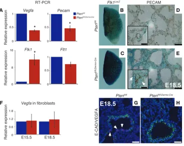
Mesodermal Pten deficiency affects vasculogenesis. Differentiated
endothelial cells, identified by PECAM, are derived from
meso-dermally derived progenitor cells, which express Flk1 (Vegfr2).
These progenitors can be identified in embryonic lungs start-ing as early as E10.5 (13). Their formation is tightly associated with mesenchymal and epithelial development during branching morphogenesis (1). As defective pulmonary vascular formation can cause hypoxemia, we examined PECAM in E15.5 lungs by
IHC (Figure 2, A and B). In Ptenfl/fl;Dermo-Cre lungs, there was
sig-nificant reduction in distal capillary network density compared
with Ptenfl/fl controls, the latter exhibiting normal dense capillary
plexus adjacent to the developing airway epithelium. In E18.5 control lungs (Figure 2C), red blood cell–containing capillaries were found in close spatial proximity to the alveolar-air interface. This coupling is critical for normal gas exchange. In contrast, the
capillary network in Ptenfl/fl;Dermo-Cre lungs was distinctly abnormal,
with clear uncoupled capillary/airway network, rendering pul-monary units incapable of efficient gas exchange (Figure 2D). Statistical analysis of the distance between the capillaries and the lumen of airways showed significant increase in the mutant
versus control lungs (Figure 2E, 3.3 μm ± 0.3 μm vs. 0.9 μm ±
0.1 μm, P < 0.01). More detailed study of the lung ultrastructure
showed mutant alveolar spaces were frequently lined by cuboi-dal cells with “immature” lamellar bodies, while the type 2 cells in control lungs contained surfactant. To better understand the mechanism underlying this phenotype, we assessed the levels of
Vegfa, Flt1 (Vegfr1), and Flk1 (Vegfr2) at E18.5 by qRT-PCR. Vegfa
Table 1
Ptenfl/fl;Dermo1-Cre mice suffer embryonic lethality
Total embryos Ptenfl/fl;Dermo1-Cre
E12.5 65 19 (29%)
E.15.5 63 16 (25%)
E18.5 311 67 (21%)
P21 121 0 (0%)
Ptenfl/fl;Dermo-Cre mice display embryonic and neonatal mortality.
Genotyp-ing of Ptenfl/fl;Dermo-Cre mice at different gestational ages. At E12.5 and
E15.5, the mutants accounted for, respectively, 29% (19 out of 65) and 25% (16 out of 63) of the total embryos, while at E18.5, their number was 21% (67 out of 311), indicating embryonic lethality between E15.5 and E18.5. At P21, no mutant mice were detected (total 121). Ptenfl/fl:Dermo-Cre
was reduced in mutant lungs compared with controls (Figure 3A,
0.39 ± 0.03 vs. 1, P < 0.01) as was Flt1 (Figure 3A, 0.73 ± 0.08 vs.
1, P < 0.05). There was also a quantifiable increase in Flk1 mRNA
in Ptenfl/fl;Dermo-Cre lungs (Figure 3A, 11.61 ± 2.6 vs. 1, P < 0.05).
Fur-thermore, Pecam mRNA was decreased in the mutant lungs versus
controls (Figure 3A, 0.47 ± 0.1 vs. 1, P < 0.01), supporting the IHC
data shown in Figure 2, A and B.
Finally, qRT-PCR for Vegfa, using mRNA from E15.5 and E18.5
isolated fibroblasts, revealed no statistically significant differ-ence between control and mutant cells (Figure 3F, E15.5: 1.067
± 0.5 vs. 1, P > 0.05; E18.5: 1.16 ± 0.6 vs. 1, P > 0.05). Thus, as the
mesenchyme does not appear to be involved, the data suggest the
epithelium as the compartment in which the decrease in Vegfa
occurs. This conclusion was validated via immunofluorescent (IF) for VEGFA plus E-CAD, which showed markedly decreased VEGFA in E-CAD–positive cells in the mutant lungs (compare Figure 3, H and G).
One possibility for the changes in Flk1 and Flt1 levels is that
meso-dermal-specific Pten inactivation affects endothelial cell
differentia-tion even though Dermo1-Cre as shown in this and other reports is
not active in this cell lineage (1, 2). Therefore, we examined the dif-ferentiation of endothelial cells by generating triple-transgenic mice
consisting of the Ptenfl/fl;Dermo-Cre embryos
in a Flk1LacZ reporter background (14).
In these mice, LacZ is under the control
of the endogenous Flk1 promoter. Flk1
is an early marker of angioblasts, and its expression is maintained in mature
endothelial cells together with Flt1
(14). Significant increase in Flk1-driven
LacZ activity was found throughout the entire mutant lungs compared with controls (Figure 3, C vs. B). Ablation
of mesodermal Pten therefore does not
interfere with specification and ampli-fication of angioblasts. However, analy-sis of PECAM, a marker for mature endothelial cells in Ptenfl/fl;Dermo-Cre lungs,
revealed impaired differentiation of angioblasts into mature endothelial cells and blood vessels when compared with controls. We therefore analyzed sections
from E18.5 Ptenfl/fl;Dermo-Cre;Flk1LacZ and
Ptenfl/fl;Flk1LacZ lungs and examined them
for PECAM. The mutant lungs showed increased FLK1-positive/PECAM-neg-ative cells (Figure 3, E vs. D), confirm-ing the conclusion that angioblasts are indeed increased in the mutant lungs. Finally, we examined the tridimensional structure of lung vasculature by inject-ing FITC leptin in the left ventricle of mutant and control hearts, observing again a significant defect in vasculogen-esis in mutant versus controls (Supple-mental Video 1). In sum, the results strongly support the conclusion that lack of PTEN in the mesodermal lineag-es inhibits differentiation of angioblasts into mature endothelial cells.
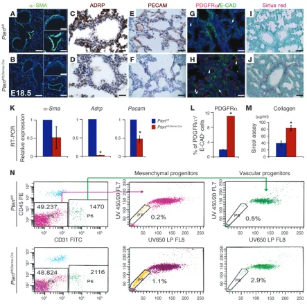
Dermo1-Cre–mediated Pten inactivation affects ontogeny of mesodermal
cell lineages. To determine the impact of mesodermal Pten
inactiva-tion on the emergence and differentiainactiva-tion of lung mesenchymal cell lineages, expression of a number of cell markers was examined by IHC and qRT-PCR. Immunofluorescence showed decreased
α-SMA, a smooth muscle differentiation marker in E18.5 mutant
lungs (Figure 4, B versus A). Adipose differentiation-related pro-tein (ADRP), a lipid droplet–associated propro-tein expressed early during adipose differentiation and a marker of lipofibroblasts
in the lung, was decreased in Ptenfl/fl;Dermo-Cre lungs compared with
Ptenfl/fl (Figure 4, D vs. C). Finally, in E18.5 lungs, the capillary
network was misaligned with corresponding respiratory airways (i.e., airway/capillary uncoupling or dysplasia) in the mutants, as shown by IHC for PECAM (Figure 4, F vs. E). Interestingly, double
IHC for PDGFrα (marker for fibroblasts) and E-CAD (marker for
epithelial cells) and Sirius red staining for collagen, a product of
fibroblasts, showed increased PDGFrα-positive/E-CAD–negative
cells (Figure 4, H vs. G, statistical analysis: Figure 4L, 2% ± 0.2% vs.
11% ± 0.02%, P < 0.01). Collagen deposition was also increased in
the Ptenfl/fl;Dermo-Cre lungs compared with controls (Figure 4, J vs. I).
These IHC results were validated by qRT-PCR for the different cell lineage markers, which showed a trend toward reduced expression
Figure 1
Absence of Pten in the mesenchyme does not affect lung morphogenesis, but leads to increased
mesenchymal cell proliferation. (A–D) Histological analysis of E18.5 lungs. Ptenfl/fl and Ptenfl/fl;Dermo-Cre
lungs were H&E stained. Representative WT and KO sections are shown. No macroscopic
differenc-es were detected between the WT and mutant embryos except for increased cell density (D versus
C). Scale bars: 100 μm (A and B); 50 μm (lower magnification), 20 μm (higher magnification) (C and
D). (E and F) Increased cell proliferation in Pten conditional KO (cKO) mice compared with controls. IF for E-CAD and PH3, showing increased proliferation in Ptenfl/fl;Dermo-Cre mesenchyme (F)
com-pared with Ptenfl/fl (E). Scale bars: 50 μm. (G) Total number of E-CAD–negative cells was counted.
Data are expressed as mean total lung cells ± SEM for 3 mice/group. *P < 0.01, Student’s t test. (H) Increased proliferation (PH3-positive cells) of total cell numbers in the E-CAD–negative population of the cKO mice. Data expressed as percentage of PH3-positive cells and E-CAD–negative over the total E-CAD–negative cells in 3 mice/group. (I) Statistical analysis of the SatO2 in control and mutant
of α-Sma (Figure 4K, 0.52 ± 0.3 vs. 1, P > 0.05) and Adrp (Figure
4K, 0.03 ± 0.00001 vs. 1, P < 0.01) in primary cultures of E18.5
mutant fibroblasts compared with controls. Pecam was also
reduced in the mutant versus control lungs (Figure 4K, 0.47 ±
0.1 vs. 1, P < 0.01). Collagen 1 by Sircol assay was increased in the
mutant lungs versus controls (Figure 4M, 83.13 μg/ml ± 7.2 vs.
39.23 μg/ml ± 6.3, P < 0.01).
Using flow cytometry to confirm these observations, we gated the mesenchymal progenitor cells by Hoechst staining in E17.5
mutant and control lungs (n = 14 for each). In the embryonic
lung, 2 subtypes of side populations are thought to exist (15).
One is identified as CD45–CD31+E-SP cells, believed to
differ-entiate into endothelial cells. The other is identified by markers
CD45–CD31–E-SP, with a gene profile consistent with a smooth
muscle precursor (15). At E17.5, we observed more than a 5-fold
increase in the number of CD45–CD31+E-SP and CD45–CD31–
E-SP cell populations in Ptenfl/fl;Dermo-Cre lungs versus controls
(respectively 1.1% versus 0.2% and 2.9% versus 0.5%) (Figure 4N). Therefore, mesenchymal PTEN controls the size of the 2 putative lung mesenchymal progenitor cell populations.
Early mesodermal Pten inactivation leads to increased FGF10 signaling.
AKT-mediated phosphorylation of β-catenin (β-CAT) on Ser552
drives nuclear localization of β-CAT and activation of WNT
tar-get genes. To determine whether mesodermal Pten inactivation
increases phosphorylation of β-CAT on Ser552 in the
mesen-chyme, we performed IHC for E-CAD and β-CATSer552 (Figure
5, G and H), the form of β-CAT specifically phosphorylated by
the PTEN/AKT pathway (16). We found significantly increased
E-CAD–negative/ β-CATSer552–positive cells in mutant lungs
com-pared with controls (Figure 5J, 1.3% ± 0.1 vs. 0.3% ± 0.1, P < 0.01).
Co-IF for β-CAT and mesenchymal markers including PECAM or
PDGFRα showed that these cells are negative for these markers
(data not shown). We did not observe β-CATSer552–positive cells
in the epithelium. In our model, absence of mesenchymal Pten
affected lipofibroblast, VSMC, and parabronchial SMC (PSMC)
differentiation likely due to increased levels of Ser552β-CAT, a
rec-ognized positive regulator of stem cell homeostasis downstream of the AKT pathway.
The pulmonary mesenchyme is the source of many signaling factors, the most important of which is FGF10, which interacts
with receptor tyrosine kinases (RTKs). Fgf10 expression by the
mesenchyme is positively regulated by WNT and FGF9 signal-ing and is negatively regulated by sonic hedgehog (SHH) (2, 4).
Deletion of Fgf10 leads to lung agenesis, suggesting a key role
in mediating mesenchymal-epithelial interactions that are nec-essary for lung morphogenesis (17, 18). FGF10 is the major
FGFR2b ligand during the pseudoglandular stage. FGF10 is secreted exclusively by the mesenchyme and acts on the epithe-lium through FGFR2b.
Because of the increase in β-CATSer552, Fgf10 expression was
ana-lyzed by in situ hybridization (ISH). Fgf10 mRNA was increased
in mutant lungs compared with controls (Figure 5, B vs. A).
qRT-PCR analysis for Fgf10 confirmed the ISH results (Figure 5I, 2.09
± 0.48 vs. 1, P < 0.05). A downstream pathway activated by FGF10
in the epithelium is β-CAT signaling (3). To examine whether the
observed increased FGF10 and increased mesenchymal β-CATSer552
stimulates β-CAT signaling, we generated triple transgenic mice
composed of Ptenfl/fl;Dermo-Cre mice and Batgal, the latter a WNT
sig-naling reporter (19), and analyzed the embryos at E15.5 (Figure 5,
C–F). β-CAT signaling was increased in lung epithelium and
mes-enchyme of the Ptenfl/fl;Dermo-Cre;Batgal lungs compared with controls
(Figure 5, D and F vs. C and E).
Mesenchymal Pten inactivation leads to expansion of the distal epithelial
progenitor cell domain characterized by ID2 and SPC expression. Since
activated β-CAT is recognized as controlling stem cell
homeosta-sis in lung epithelial cells, we performed IF for epithelial markers in E18.5 control and mutant lungs (Figure 6, C–H). Double IF with antibodies to E-CAD and FGFR2 showed increased FGFR2
in E18.5 Ptenfl/fl;Dermo-Cre lungs compared with controls (Figure 6, B
vs. A). qRT-PCR confirmed this observation by showing a 4-fold
increase of Fgfr2b mRNA in mutant lungs (Figure 6I, 4.1 ± 0.3
vs. 1, P < 0.01). qRT-PCR showed increased expression for Fgf9
and Fgf7, 2 additional growth factors made by the lung
mesothe-lium/epithelium (Fgf9) and by the mesenchyme (Fgf7), (Figure 6I,
Fgf9: 2.6 ± 0.5 vs. 1, P < 0.05; Fgf7: 5.7 vs. 1 ± 1.6, P < 0.05). To validate these results, we also examined the expression of FGF10/ FGFR2b downstream and positive regulator targets by qRT-PCR.
As expected, compared with the controls, Wnt2b, Spry2, Etv4, Etv5,
and Bmp4 were increased, while Shh, Ptch1, and Gli1 decreased in
the mutant lungs (Figure 6I, Wnt2b: 3.03 ± 1.2 vs. 1, NS; Spry2:
2.46 ± 0.58 vs. 1, P < 0.05; Etv4: 1.26 ± 0.15 vs. 1, NS; Etv5: 1.56 ±
0.24 vs. 1, P < 0.05; Bmp4: 1.26 ± 0.06 vs. 1, P < 0.01; Figure 6I, Shh: 0.6 ± 0.3 vs. 1, P < 0.05; Ptch1: 0.47 ± 0.1, P < 0.05; Gli1: 0.5% ± 0.06 vs. 1, P < 0.01).
As we observed increased FGF10 activity in the epithelium of the mutant lungs, we investigated whether this change affected epithelial differentiation along the pulmonary proximal and
dis-tal domains (20). IHC for SPC, T1α, and CC10 markers for
alve-olar type 2 (distal domain), alvealve-olar type 1 (distal domain) and Clara cells (proximal domain), respectively, showed an increase of alveolar type 2 (SPC-positive) cells, while the Clara cells
[image:5.585.42.374.83.215.2](CC10-positive) and type 1 cells (T1α-positive) were unchanged
Figure 2
in the Ptenfl/fl;Dermo-Cre lungs compared with Ptenfl/fl controls
(Fig-ure 6, C–H). Western blot analysis demonstrated a statistically significant increase in SPC expression (marker for alveolar type 2 cells) in the mutant compared with controls (Figure 6J, SPC:
0.2 ± 0.003 vs. 0.09 ± 0.003, in arbitrary units, P < 0.05). In
addi-tion, comparison of CC10 and T1α expression showed no
statis-tical difference between the 2 groups (data not shown). Finally, we performed IHC for TTF1 (NKX2.1), the first transcriptional factor present in the lung during embryogenesis (21), and for ID2, a marker of distal lung progenitor cells (22). The increase in TTF1-positive cells (Figure 6, L vs. K) and ID2/E-CAD–double-positive cells (Figure 6, N vs. M) in the mutant lungs compared with the control confirmed the expansion of the distal domain. Western blot results further confirmed the latter results,
show-ing an increase of TTF1 (0.13 ± 0.006 vs. 0.23 ± 0.006, P < 0.01) in
the mutants compared with controls (Figure 6J). Overall, these data suggest that the increase in FGF10 signaling in the
epithe-lium, due to Pten mesenchymal inactivation, is the likely cause
of distal epithelial domain expansion.
Mesenchymal Pten inactivation
phenocopies ACD. As the phenotype
of the lungs with mesenchymal
Pten inactivation resembled what
has been described in newborn infants as ACD, we examined the hypothesis that the PTEN/PI3K/ AKT pathway is activated in the lungs of ACD patients. For this purpose, we performed PTEN and p-AKT IHC on 5 different and independent lung samples from newborn human infants who died with ACD diagnosis and compared these to lungs of newborns who died of causes unrelated to ACD. ACD patients showed a decrease of PTEN stain-ing and a correspondstain-ing increase in p-AKT–positive cells compared with lungs from control patients. While PTEN staining was read-ily detectable (Figure 7, B vs. A), the number of p-AKT–positive cells was limited in the control lungs (Figure 7, D vs. C). Statisti-cal analysis showed an increase in the percentage of p-AKT–positive cells in the ACD lungs compared with controls (Figure 7F, 1.4 ± 0.4 vs. 0.07 ± 0.05, P < 0.05).
ACD has been associated with mutations in Forkhead box
pro-tein F1 (FOXF1) in humans and
heterozygous Foxf1 and Foxc2
loss of function in mice (23, 24).
Accordingly, Foxf1 and Foxc2
mRNA were measured by qRT-PCR. We found decreased expres-sion of both genes in mutant compared with control lungs
(Figure 7E, Foxf1: 0.59 ± 0.015 vs. 1, P < 0.01; Foxc2: 0.57 ± 0.13
vs. 1, P < 0.05). These data support the involvement of the PTEN/
PI3K/AKT pathway in the pathogenesis of ACD and justify further analysis of this pathway in our newly described mouse model of a lethal human congenital lung disease.
Discussion
Mesodermal Pten inactivation disrupts lung vasculogenesis. A
well-orga-nized and functional capillary network is essential for air-blood gas exchange and therefore for oxygenation of tissues and organs. Impaired development of the vascular capillary network in the lung can cause life-threatening deficiencies in pulmonary func-tion present in neonatal diseases such as BPD and ACD (25, 26).
The results of the present work show that inactivation of Pten
in the mesodermal lineages that contribute to lung morpho-genesis disrupts normal capillary plexus formation as well as mesenchymal and epithelial differentiation.
The role of PTEN in mesodermal lineage formation during lung
[image:6.585.45.412.83.374.2]development had not been hitherto adequately examined. Pten
Figure 3
Defect in angioblast differentiation in Ptenfl/fl;Dermo-Cre lungs. (A) Altered vascular gene expression in E18.5
mutant lungs. qRT-PCR of mRNA using 4 different lungs/group showed decreased Vegfa, Pecam, and
Flt1 and increased Flk1. GAPDH or β-actin served as target reference. *Statistically significant: P < 0.05. (B–E) β-gal staining of E18.5 Ptenfl/fl;Flk1LacZ/+ and Ptenfl/fl;Dermo-Cre;Flk1LacZ/+ lungs showing increased
Flk1-positive cells, a marker for angioblasts in the mutant lungs. (B and C) Whole-mount β-gal staining of the left lobe. Increased Flk1-positive cells in the mutant lungs compared with control was observed. (D and
was previously shown to be important in regulating proliferation
of endothelial cells (10). Endothelial-specific inactivation of Pten
using the Tie2-Cre driver mice led to early embryonic lethality due
to fatal vascular remodeling defects secondary to an impaired recruitment of VSMCs. Hamada and colleagues (10) showed also that the absence of PTEN in endothelial cells causes hypersensitiv-ity to VEGFs, resulting in enhanced angiogenesis and bleeding due
to a failure in vascular remodeling. Deleris et al. (11) found that PTEN is also expressed and active in VSMCs controlling the level of PIP3 and therefore potentially controlling VSMC proliferation. Another report (27) showed that absence of PTEN in VSMCs leads to vascular remodeling and increased progenitor cell recruitment.
However, to date, the specific deletion of Pten in lung mesenchyme
[image:7.585.66.509.80.522.2]during development has not been reported.
Figure 4
Arrested mesenchymal cell differentiation in Ptenfl/fl;Dermo-Cre lungs. (A–F) IHC for α-SMA, ADRP, and PECAM in E18.5 lungs showing reduced
expression in mutant (B, D, and F) compared with control (A, C, and E) lungs. n = 4 lungs per group. Scale bars: 50 μm (lower magnification), 25 μm (higher magnification) (A and B). (G and H) IF for PDGFRα (fibroblast marker) and E-CAD (epithelial marker) showing increased PDGFRα; E-CAD– cells (white arrows) in mutant lungs. (I and J) Sirius red staining showing increased collagen in Ptenfl/fl;Dermo-Cre lungs. Scale bars: 25 μm
(C–J).(K) qRT-PCR showing decreased mesenchymal differentiation markers, α-Sma, Adrp in mutant fibroblasts and Pecam in mutant total lung. *Statistically significant: Adrp, P≤ 0.01; Pecam, P≤ 0.01; number of Pdgfra-positive/E-cadherin–negative cells, P≤ 0.01; collagen, P≤ 0.01. (L) Quantification of PDGFRα and E-CAD– cells. (M) Quantification of collagen 1. (N) Pten deficiency induced increased embryonic mesenchymal
progenitor side populations (E-SP). Representative flow cytometric profiles of side population in E17.5 lungs. Upper panels, WT cells; lower pan-els, mutant cells. Side populations were identified by Hoechst 33342, CD45, and CD31 antibodies. The percentages of side populations (boxed areas) in total lung are indicated in each panel. E-SP–CD45–CD31– and E-SP–CD45–CD31+ cells identify smooth muscle and vascular progenitors
In the present work, we used the Dermo1-Cre driver line to
inac-tivate Pten from the onset of lung morphogenesis (i.e., E9.5) in all
mesodermally derived cells (refs. 12, 28, and Supplemental Fig-ure 1G) except for endothelial progenitors. The resulting mutant mice exhibited perinatal lethality that was largely due to vascular defects and mesenchymal hypercellularity. The newborn mice were clearly hypoxemic and could not oxygenate their blood. Detailed examination of their lungs revealed decreased capillary forma-tion, but more importantly, misalignment of the existing capil-lary network with the airways, due also to the increased number of mesenchymal cells in the parenchyma. Interestingly, our genetic manipulation, which enabled the embryos to survive until birth, is significantly different from the one resulting from
endotheli-al-specific deletion of Pten (10). Thus, the use of the Dermo1-Cre
driver line to delete Pten in all mesodermally derived cells except
for endothelial cells provides new insight into the key role played by PTEN in mesodermal lineage formation.
To investigate the underlying mechanisms for the observed
vas-cular defects in Ptenfl/fl;Dermo-Cre mutants, we measured a number of
relevant parameters. Vegfa was markedly reduced in the mutant
lungs at E18.5. Vegfa is first expressed in the distal subepithelial
mesenchyme, but begins to be expressed also in the epithelium at
E14.5 (1). Thereafter, Vegfa becomes progressively restricted to the
epithelium (1). It is therefore likely that the main source of Vegfa,
whose secretion is decreased in our mutants, is the epithelium. Interestingly, the decrease in VEGFA occurs in spite of increased FGF10 signaling to the epithelium, which we had previously
shown to be a positive regulator of Vegfa, using Fgf10
hypomorph-ic mhypomorph-ice (3). However, in these mutants, this decrease is likely the consequence of reduced epithelial cell numbers rather than being
caused by a direct transcriptional regulation of Vegfa by FGF10.
In support of this observation, the decrease in TTF1 expression
(marker of lung epithelium) in the lungs of Fgf10 hypomorphic
embryos was also apparent within the same range as was observed
for Vegfa (3). The change in Vegfa therefore is indirect and may
reflect changes in the epithelial/mesenchymal ratio, which was
drastically changed in favor of the mesenchyme in Ptenfl/fl;Dermo-Cre
lungs (higher FGF10 signal leads to only modest epithelial
pro-genitor cell increase, whereas mesodermal Pten inactivation causes
a marked increase in mesenchymal proliferation). This, in turn,
will automatically lead to a global decrease in Vegfa expression in
the lung. Reduced VEGFA will undoubtedly cause correspond-ing defects in vascular formation. In addition, SHH and FGF9 are reported to be critical for vascular development (1). Therefore, as
Shh is decreased, vascular (endothelial) development is also
expect-ed to be impairexpect-ed in Ptenfl/fl;Dermo-Cre lungs.
Interestingly, the lack of differentiation in the epithelial com-partment was associated with increased angioblasts in the
mes-enchyme. This was supported by the increase in Flk1 (marker of
vascular progenitors), but also by concomitant decrease in Flt1
and Pecam (both markers of differentiated endothelial cells)
mRNAs. As previously demonstrated, absence of Flt1 leads to
disorganized angiogenesis due to the role of this receptor in vas-cular commitment (29). Moreover, PTEN may not be necessary for differentiation of angioblasts from the ventral mesoderm and for their migration within the lung, as has been shown pre-viously (10). Accordingly, we observed that the absence of PTEN did not affect the formation of the major vessels or the angioblast population. Nevertheless, our studies identified an important
role for mesenchymal Pten in the differentiation of angioblasts to
endothelial cells. As Pten is not deleted in endothelial cells, this role
is likely indirect, being an outcome of alterations in mesenchymal-endothelial or mesenchymal-epithelial-mesenchymal-endothelial crosstalk medi-ated by as yet unknown signaling molecules.
[image:8.585.63.503.83.259.2]In the course of this study, we also noticed that PTEN in the lung mesenchymal cells was present both in the cytoplasm and in the nuclei, while in the epithelium, PTEN was confined only to the cytoplasm. A recent report investigating the role of PTEN
Figure 5
Increase in mesenchymal progenitor cells is due to increased β-CAT and FGF10 signaling. (A and B) Whole-mount ISH for Fgf10 at E15.5 show-ing increased expression in the mutant lungs. (C–F) β-gal staining in Batgal background. Increased WNT signaling in mutant lungs compared with controls. (C and D) Whole-mount images of left lobes. (E and F) Double α-SMA, β-gal staining showed increased WNT signaling in epithelial and
mesenchymal (not α-SMA–positive) cells. (G and H) Double staining for E-CAD/Ser552β-CAT showing increased WNT signaling in mesenchymal
cells. Scale bars: 50 μm (lower magnification), 25 μm (higher magnification) (E and F); 25 μm (G and H).(I) qRT-PCR for Fgf10 showing increased
Fgf10 in mutant lungs versus control. (J) Statistical analysis of β-CATSer552–positive/E-CAD–negative cells in mutant versus control lungs, n = 3/
in cancer cells showed that nuclear-localized PTEN interacts with APC/C (anaphase promoting complex) and promotes its asso-ciation with Fizzy-related protein 1 (FZR1), thereby enhancing tumor-suppressive activity of the APC/C-FZR1 complex in a phos-phatase-independent manner (30). In this way, both proliferation and senescence are affected by reduction of APC/C activity upon
Pten loss. Whether the lack of PTEN in the nuclei or in the
cyto-plasm of mesenchymal cells is responsible for some aspect of the
phenotype observed in Ptenfl/fl;Dermo-Cre lungs remains to be clarified
by future studies.
PTEN controls lung mesenchyme differentiation. The mesenchyme is
essential during lung development, as it contains the progenitors for several mesenchymal cell types that include the PSMCs, the VSMCs, the lipofibroblast, the alveolar myofibroblasts, and the stromal fibroblasts (31, 32). The proper generation of these cells relies on the controlled amplification of progenitors followed by the exit of these cells from the cell cycle and their timely differen-tiation into appropriate progeny (2). Several studies (9, 33) have
shown that epithelial inactivation of Pten in the lung leads to an
increase in epithelial progenitor cells and impaired epithelial cell
differentiation. In our model, absence of mesenchymal Pten
affect-ed lipofibroblast, VSMC, and PSMC differentiation, likely via an
increase in β-CAT Ser552–mediated WNT signaling, a recognized
regulator of epithelial stem cell homeostasis. Indeed, we have
previ-ously shown that mesenchymal-specific deletion of β-CAT results
in lack of amplification and premature differentiation of PSMC progenitor cells (2). Moreover, we have recently shown that reac-tivation of Wnt signaling in mature PSMC after airway epithelial injury results in their dedifferentiation and adoption of a PSMC
progenitor-like state and reexpression of Fgf10 (34). Interestingly,
in contrast to our findings, others have reported increased SMC differentiation associated with increased WNT signaling (35). One difference is that in the latter study, SMC-specific targeting
was employed, while in our study, Dermo1-Cre driver line targeted
[image:9.585.47.535.80.430.2]the mesenchymal progenitor cells. In addition, treatment of lung explants in the pseudoglandular stage with LiCl to increase Wnt
Figure 6
Analysis of epithelial differentiation suggests increased FGF signaling. (A–H) IHC in E18.5 lungs showing increased SPC and FGFR2 in epithe-lium of mutant lungs. CC10 and T1α staining showed no differences. (I) qRT-PCR for Fgfr2b, Fgf9, Fgf7, Wnt2b, Spry2, Etv4, Etv5, Bmp4, Shh,
Ptch1, and Gli1 showed increased FGF10/FGFR2b signaling in epithelium of the Pten cKO lungs compared with the WT. (J) Western blot for epithelial markers showed increased progenitor markers (SPC and TTF1) in the Ptenfl/fl;Dermo-Cre lungs. Data represent 5 lungs/group. *Statistically
significant: Fgf9, P≤ 0.05; Fgf7, P≤ 0.05; Fgfr2b, P≤ 0.01; Spry2, P≤ 0.05; Etv5, P≤ 0.05; Bmp4, P≤ 0.01; Shh, P≤ 0.05; Ptch1, P≤ 0.05; Gli1,
signaling in both epithelium and the mesenchyme was associated
with increased α-SMA around bronchi and blood vessels (35).
Pre-liminary data from our group show that after Dermo1-Cre deletion
of the negative regulator of WNT signaling, adenomatous
pol-yposis coli (leading to increased β-CAT signaling) differentiation
of α-SMA cells was blocked (P. Minoo et al., unpublished data).
These data are consistent with our findings in the present
manu-script. Thus, lack of β-CAT in the mesenchyme led to premature
differentiation of PSMC and lack of amplification of the progeni-tors, while increased WNT signaling blocked differentiation and
increased mesenchymal progenitors. Finally, Pten deletion during
embryonic stages led to increased collagen production. This could occur either by induction of collagen expression in mesenchymal progenitors or by promotion of fibroblast differentiation. Interest-ingly, several studies have reported decrease or absence of PTEN in fibrotic foci in idiopathic pulmonary fibrosis (IPF) (36).
Mesodermal Pten inactivation affects epithelial differentiation via
increased FGF10 and WNT signaling. Epithelial-mesenchymal
inter-actions mediated via key signaling factors during lung develop-ment are critical for proper patterning of the epithelium and mes-enchyme along the proximal-distal axis. The mesmes-enchyme is the source of important signaling factors, such as FGF10, required for airway epithelial development and branching. Also, the
epi-thelium produces signaling molecules essential for mesenchymal differentiation and proliferation, such as bone morphogenetic protein 4 (BMP4), SHH (18, 37, 38), and FGF9 (1). The
obser-vations in this study showed that mesodermal Pten inactivation
caused increased Fgf9, Fgf10, and Fgf7 expression and decreased
expression of Shh, Ptch1, and Gli1. We further found that increase
in FGF10 caused increased FGFR2b signaling, supported by
upregulation of FGF10 downstream targets Fgfr2b, Bmp4, and
Spry2. Such changes in Ptenfl/fl;Dermo-Cre lungs would undoubtedly
have an impact on lung development and cell differentiation.
Thus, mesenchymal-specific inactivation of Pten led to arrested
differentiation of mesoderm-derived cells. Concomitantly, we observed an increase in the number of SMC progenitors, which
include the Fgf10-positive cells. The latter have been described as
giving rise to PSMCs. The increased number of Fgf10-expressing
cells is likely responsible for increased expression of FGF10
down-stream targets Fgfr2b, Bmp4, and Spry2 in the mutant lungs. These
[image:10.585.47.539.83.407.2]cells proliferate and give rise to an increased number of distal SPC-positive cells. IHC analysis for ID2, a distal cell marker, sug-gested alterations in proximal-distal differentiation of epithelial cells; increased numbers of ID2-positive cells were found to be localized proximally in the mutant lung. Interestingly, we did not detect any defect in epithelial differentiation in the proximal
Figure 7
Human newborn ACD lungs display decreased PTEN and increased p-AKT. (A and B) IHC for PTEN in human ACD and control lungs showing
decreased PTEN in the ACD patients. (C and D) IF for p-AKT showing increased p-AKT–positive cells. (E) qRT-PCR using mRNA from Ptenfl/fl and Ptenfl/fl;Dermo-Cre lungs showed decreased Foxf1 and Foxc2 in the mutant lungs. (F) Statistical analysis showed increased p-AKT–positive cell
Ptenfl/fl;Dermo-Cre) and Dermo1-cre;Ptenfl/+ (heretofore Ptenfl/+;Dermo-Cre) on a
C57BL/6 background. Ptenfl/fl mice were used as control. Genotyping of
the Dermo1-Cre mice containing Ptenfl, PtenΔ, and PtenWT alleles was as
previ-ously described (12, 40).
Blood saturation. Heart rate and transcutaneous oxygen saturation were monitored every 10 seconds for 1 minute in newborn mice via a pulse oximeter (MouseOx; STARR Life Sciences Corporation) by placing nonin-vasive sensors on the back of the newborn mice.
Tissue collection. Lungs from embryos were collected at E15.5 and E18.5 and fixed overnight, dehydrated through increasing ethanol concentra-tions, embedded in paraffin, and prepared as 5-μm sections.
Fibroblast isolation. E18.5 lungs (6 controls and 6 mutants) were mechani-cally disrupted and incubated at 37°C with 1:1 Trypsin 0.025%/EDTA: DMEM/F12. Dissociated cells were passed through a 19.5- to 27-gauge needle, incubated in DMEM/F12 with 10% FBS (2 hours), and washed 10 times with DMEM/F12. Adherent fibroblasts were grown for 24 hours, detached by scraping, and centrifuged (at 1,700 g for 5 minutes). Cell pel-lets were stored at –80°C.
Statistics. Data were presented as means ± SEM unless stated otherwise. The data were statistically analyzed using 2-tailed Student’s t tests to com-pare the 2 groups. P < 0.05 was considered significant.
Study approval. All animal experiments were approved by the Uni-versity of Southern California Animal Use and Care Committee. Post mortem unidentified human ACD tissue was contributed by P. Sen and C. Langston (Baylor College of Medicine, Houston, Texas, USA) (IRB number, H-8712).
Acknowledgments
This work was supported by NIH HL56594 (to P. Minoo), HL086322 (to S. Bellusci), HL074832 (to S. Bellusci), the Excel-lence Cluster Cardio-Pulmonary System, LOEWE (to S. Bellusci), HL092967 (to S. De Langhe), the Hastings Foundation (to P. Minoo), the CIRM Clinical Fellowship (to C. Tiozzo and G. Carraro), NIH K12-HD-052954 (to B. Chan), and the American Lung Association as well as the American Heart Association (to D.A. Alam). P. Minoo is Hastings Professor of Pediatrics. S. Bel-lusci holds the chair of Lung Matrix Remodelling at Justus Liebig University. Z. Borok is Edgington Chair in Medicine. The authors also thank the ACD Association and P. Sen and C. Langston (Bay-lor College of Medicine) for providing human ACD lung samples. We thank Hiroyuki Shimada (Children’s Hospital, Los Angeles) for discussion and technical support, Esteban Fernandez for help with tridimensional reconstruction of lung vasculature, and Clar-ence Wigfall for help with the processing of the videos. C. Tiozzo acknowledges C. Fagan and L. Barrow.
Received for publication October 7, 2011, and accepted in revised form August 2, 2012.
Address correspondence to: Parviz Minoo, General Lab Building, Women’s and Children’s Hospital, 1801 E. Marengo St., Los Ange-les, California 90033, USA. Phone: 323.226.4340; Fax: 323.226.5049; E-mail: minoo@usc.edu. Or to: Saverio Bellusci, Department of Internal Medicine II, University of Giessen Lung Center, Excellence Cluster in Cardio-Pulmonary Systems, Aulweg 130, Giessen 35392, Germany. Phone: 49.0.641.99.46.730; Fax: 49.0.641.99.46.739; E-mail: Saverio.Bellusci@innere.med.uni-giessen.de.
Caterina Tiozzo’s present address is: Nassau University Medical Center, Pediatrics Department, East Meadow, New York, USA.
lung; CC10 and T1α staining indicated a normal pool of Clara
and type 1 cells in both mutants and control lungs. Therefore, absence of PTEN in the mesenchyme leads to the expansion of the pool of distal epithelial progenitor cells likely secondary to increased FGF10/FGFR2b signaling in the epithelium.
Mesodermal Pten inactivation leads to ACD-like phenotype. Our
obser-vations suggested vascular similarities in phenotype between Ptenfl/fl;Dermo-Cre lungs and the lungs of human neonates with ACD.
FGF9 and SHH pathways were previously shown to be important for vasculogenesis, and disruption of their function leads to ACD-like phenotypes (1). Consistent with the latter findings,we found
alterations in both FGF9 and SHH pathways in Ptenfl/fl;Dermo-Cre
lungs. qRT-PCR revealed increased Fgf9 mRNA, while Shh and its
downstream targets, Ptch1 and Gli1, were decreased in the mutant
lungs (Figure 6I). Thus, both the vascular phenotype and the
molecular alterations in Ptenfl/fl;Dermo-Cre lungs are consistent with
the features associated with human ACD. Furthermore, a
signifi-cant decrease in Foxf1 and Foxc2 mRNAs (Figure 7E) was also
doc-umented in the Ptenfl/fl;Dermo-Cre lungs, also consistent with previous
findings in human and murine ACD phenotype (23, 24). Finally, to directly ascertain the relevance of our findings in human tis-sues, we conducted IHC for PTEN and p-AKT in 5 independent human lung samples from ACD patients and newborns who died of causes other than ACD. The absence of PTEN staining and the increase in p-AKT–positive cells in ACD lungs demonstrated that the PI3K/AKT pathway is activated and that loss of PTEN
may be relevant to this activation. It has been reported that Foxf1
expression is directly inhibited by AKT in liver myofibroblasts (39). Therefore, it is possible that a similar situation occurs also
in the lung where Foxf1 could be a direct transcriptional target for
PTEN signaling in the lung mesenchyme through AKT activation. Whether loss of PTEN can be recognized as primum movens in the pathogenesis of ACD and whether activation of PTEN can be used as a therapeutic target remains to be determined.
In conclusion, mesodermal-specific inactivation of Pten by
Dermo1-Cre leads to a phenotype resembling ACD, a dramatic
neonatal disease with 100% mortality at birth. The cellular mecha-nisms underlying the phenotype are connected with an increase in mesenchymal progenitor cells and a reduced differentiation of these cells into PSMCs and lipofibroblasts. This also leads indi-rectly to an increase in the angioblast pool as well as defective differentiation of these cells into endothelial cells. We propose a model whereby increased FGF10-expressing PSMC progeni-tor cells activate the FGF10/FGFR2b pathway in the epithelium, leading to an expansion of the distal progenitor pools containing ID2-positive cells. Lack of epithelial differentiation also leads to
decreased Vegfa during early embryonic stages that in turn causes
defects in capillary development around the primitive alveoli and, therefore, a lack of oxygenation at birth, with subsequent perinatal lethality. Finally, blocked differentiation of mesenchymal progeni-tors to SMC and lipofibroblasts drives progenitor cells toward an alternative fibroblast phenotype, leading to increased collagen production (Figure 7G).
Methods
Transgenic mice. Ptenfl/fl (C57BL/6 background; stock no. 006440), Flk1LacZ/+
(stock no. 002938), Dermo1-Cre (stock no. 008712), and Batgal (stock no. 005317) mice were from Jackson Laboratory. Pten knockouts were obtained by mating Ptenfl/fl females with Dermo1-Cre male mice (C57BL/6
1. White AC, Lavine KJ, Ornitz DM. FGF9 and SHH regulate mesenchymal Vegfa expression and devel-opment of the pulmonary capillary network. Devel-opment. 2007;134(20):3743–3752.
2. De Langhe SP, et al. Formation and differentiation of multiple mesenchymal lineages during lung development is regulated by beta-catenin signal-ing. PLoS One. 2008;3(1):e1516.
3. Ramasamy SK, et al. Fgf10 dosage is critical for the amplification of epithelial cell progenitors and for the formation of multiple mesenchymal lineages during lung development. Dev Biol. 2007;307(2):237–247.
4. Del Moral PM, et al. VEGF-A signaling through Flk-1 is a critical facilitator of early embryonic lung epithelial to endothelial crosstalk and branching morphogenesis. Dev Biol. 2006;290(1):177–188. 5. Stenmark KR, Abman SH. Lung vascular
devel-opment: implications for the pathogenesis of bronchopulmonary dysplasia. Annu Rev Physiol. 2005;67:623–661.
6. Ng PC, Lewindon PJ, Siu YK, To KF, Wong W. Congenital misalignment of pulmonary vessels: an unusual syndrome associated with PPHN. Acta Paediatr. 1995;84(3):349–353.
7. Cho KW, et al. ERK activation is involved in tooth development via FGF10 signaling. J Exp Zool B Mol Dev Evol. 2009;312(8):901–911.
8. Stiles B, Groszer M, Wang S, Jiao J, Wu H. PTENless means more. Dev Biol. 2004;273(2):175–184. 9. Tiozzo C, et al. Deletion of Pten expands lung
epithelial progenitor pools and confers resistance to airway injury. Am J Respir Crit Care Med. 2009; 180(8):701–712.
10. Hamada K, et al. The PTEN/PI3K pathway governs normal vascular development and tumor angio-genesis. Genes Dev. 2005;19(17):2054–2065. 11. Deleris P, et al. SHIP-2 and PTEN are expressed and
active in vascular smooth muscle cell nuclei, but only SHIP-2 is associated with nuclear speckles.
J Biol Chem. 2003;278(40):38884–38891. 12. Yu K, et al. Conditional inactivation of FGF
recep-tor 2 reveals an essential role for FGF signaling in the regulation of osteoblast function and bone growth. Development. 2003;130(13):3063–3074. 13. Parera MC, et al. Distal angiogenesis: a new concept
for lung vascular morphogenesis. Am J Physiol Lung Cell Mol Physiol. 2005;288(1):L141–L149. 14. Shalaby F, et al. Failure of blood-island formation
and vasculogenesis in Flk-1-deficient mice. Nature.
1995;376(6535):62–66.
15. Summer R, Kotton DN, Liang S, Fitzsimmons K, Sun X, Fine A. Embryonic lung side population cells are hematopoietic and vascular precursors.
Am J Respir Cell Mol Biol. 2005;33(1):32–40. 16. He L, et al. Co-existence of high levels of the
PTEN protein with enhanced Akt activation in renal cell carcinoma. Biochim Biophys Acta. 2007; 1772(10):1134–1142.
17. Sekine K, et al. Fgf10 is essential for limb and lung formation. Nat Genet. 1999;21(1):138–141. 18. Bellusci S, Grindley J, Emoto H, Itoh N, Hogan BL.
Fibroblast growth factor 10 (FGF10) and branch-ing morphogenesis in the embryonic mouse lung.
Development. 1997;124(23):4867–4878.
19. Maretto S, et al. Mapping Wnt/beta-catenin signaling during mouse development and in colorectal tumors.
Proc Natl Acad Sci U S A. 2003;100(6):3299–3304. 20. Nyeng P, Norgaard GA, Kobberup S, Jensen J.
FGF10 maintains distal lung bud epithelium and excessive signaling leads to progenitor state arrest, distalization, and goblet cell metaplasia. BMC Dev Biol. 2008;8:2.
21. Minoo P, Su G, Drum H, Bringas P, Kimura S. Defects in tracheoesophageal and lung morpho-genesis in Nkx2.1(–/–) mouse embryos. Dev Biol. 1999;209(1):60–71.
22. Rawlins EL, Clark CP, Xue Y, Hogan BL. The Id2+ distal tip lung epithelium contains individual mul-tipotent embryonic progenitor cells. Development. 2009;136(22):3741–3745.
23. Stankiewicz P, et al. Genomic and genic deletions of the FOX gene cluster on 16q24.1 and inactivat-ing mutations of FOXF1 cause alveolar capillary dysplasia and other malformations. Am J Hum Genet. 2009;84(6):780–791.
24. Yu S, Shao L, Kilbride H, Zwick DL. Haploin-sufficiencies of FOXF1 and FOXC2 genes asso-ciated with lethal alveolar capillary dysplasia and congenital heart disease. Am J Med Genet A. 2010;152A(5):1257–1262.
25. Coalson JJ. Pathology of bronchopulmonary dys-plasia. Semin Perinatol. 2006;30(4):179–184. 26. deMello DE. Pulmonary pathology. Semin Neonatol.
2004;9(4):311–329.
27. Nemenoff RA, et al. Targeted deletion of PTEN in smooth muscle cells results in vascular remodeling and recruitment of progenitor cells through induc-tion of stromal cell-derived factor-1alpha. Circ Res. 2008;102(9):1036–1045.
28. Morimoto M, Liu Z, Cheng HT, Winters N, Bader D, Kopan R. Canonical Notch signaling in the developing lung is required for determination of arterial smooth muscle cells and selection of Clara versus ciliated cell fate. J Cell Sci. 2010; 123(pt 2):213–224.
29. Fong GH, Zhang L, Bryce DM, Peng J. Increased hemangioblast commitment, not vascular disorga-nization, is the primary defect in flt-1 knock-out mice. Development. 1999;126(13):3015–3025. 30. Song MS, et al. Nuclear PTEN regulates the
APC-CDH1 tumor-suppressive complex in a phosphatase-independent manner. Cell. 2011;144(2):187–199. 31. Mailleux AA, et al. Fgf10 expression identifies
parabronchial smooth muscle cell progenitors and is required for their entry into the smooth muscle cell lineage. Development. 2005;132(9):2157–2166. 32. Gebb SA, Shannon JM. Tissue interactions mediate
early events in pulmonary vasculogenesis. Dev Dyn. 2000;217(2):159–169.
33. Yanagi S, et al. Pten controls lung morphogen-esis, bronchioalveolar stem cells, and onset of lung adenocarcinomas in mice. J Clin Invest. 2007; 117(10):2929–2940.
34. Volckaert T, et al. Parabronchial smooth muscle constitutes an airway epithelial stem cell niche in the mouse lung after injury. J Clin Invest. 2011; 121(11):4409–4419.
35. Cohen ED, Ihida-Stansbury K, Lu MM, Panettieri RA, Jones PL, Morrisey EE. Wnt signaling regu-lates smooth muscle precursor development in the mouse lung via a tenascin C/PDGFR pathway.
J Clin Invest. 2009;119(9):2538–2549.
36. White ES, et al. Negative regulation of myofibro-blast differentiation by PTEN (Phosphatase and Tensin Homolog Deleted on chromosome 10). Am J Respir Crit Care Med. 2006;173(1):112–121. 37. Litingtung Y, Lei L, Westphal H, Chiang C. Sonic
hedgehog is essential to foregut development. Nat Genet. 1998;20(1):58–61.
38. Pepicelli CV, Lewis PM, McMahon AP. Sonic hedge-hog regulates branching morphedge-hogenesis in the mammalian lung. Curr Biol. 1998;8(19):1083–1086. 39. Godichaud S, et al. The grape-derived polyphenol res-veratrol differentially affects epidermal and platelet-derived growth factor signaling in human liver myo-fibroblasts. Int J Biochem Cell Biol. 2006;38(4):629–637. 40. Lesche R, et al. Cre/loxP-mediated inactivation of