INTRODUCTION
Smicl is a protein that interacts with transcription factors and forms a complex with the Cleavage and polyadenylation specificity factor (CPSF) (Collart et al., 2005a). We have previously shown that Smicl regulates the expression of Chordinin the early Xenopusembryo, but Chordinis unlikely to be the only target of Smicl, because ectopic expression of Chordincannot rescue completely the effects of an antisense morpholino oligonucleotide (MO) directed against Smicl (Collart et al., 2005b). In this paper we use microarray analysis to search for additional targets of Smicl, both in an effort to understand its role in early development as well as to obtain more insights into its mode of action. This analysis has identified many genes whose onset of expression at the midblastula transition (Newport and Kirschner, 1982a; Newport and Kirschner, 1982b; Schier, 2007) requires Smicl activity and whose activation is correlated with the translocation of a tagged form of Smicl from cytoplasm to nucleus. Analysis of one of these targets, Xiro1, shows that Smicl influences 3⬘-end processing of the Xiro1transcript.
The effects of Smicl on zygotic gene expression are first detected at the midblastula transition (MBT), which in Xenopusoccurs after 12 cell divisions and involves many coordinated changes in cell behaviour. In particular, the cell cycle loses its synchrony and, with the introduction of G1 and G2 phases, becomes longer (Kimelman et al., 1987; Lemaitre et al., 1998). In addition, several proteins translocate from cytoplasm to nucleus (Dreyer, 1987), maternal mRNA is degraded (Bushati et al., 2008; Giraldez et al., 2006), the apoptosis programme is engaged (Peng et al., 2007), cells become motile (Kimelman et al., 1987) and karyomeres (individual
chromosomes surrounded by a nuclear envelope) disappear (Lemaitre et al., 1998). Perhaps most significantly, the MBT marks the onset of bulk transcription in the embryo, driven by RNA polymerase II (RNA polII) (Newport and Kirschner, 1982a; Newport and Kirschner, 1982b).
RNA polII consists of two large subunits and several smaller subunits. The largest subunit, Rpb1, contains an unusual C-terminal domain (CTD) that contains repeats of the heptapeptide YSPTSPS, which can be phosphorylated on serines 2 and 5 (Hirose and Ohkuma, 2007). The CTD acts as a docking platform for factors involved in co-transcriptional processes, and the positions of phosphorylated serines determine which transcription factors and RNA processing factors are recruited (Egloff and Murphy, 2008). Importantly, before the MBT in Xenopus, serine 2 of the CTD heptapeptide is not phosphorylated, but it becomes hyperphosphorylated thereafter (Palancade et al., 2001). Serine 2 phosphorylation is important for overcoming an early elongation block in Xenopus(Peterlin and Price, 2006) as well as for splicing and polyadenylation (Hirose and Manley, 1998; Hirose et al., 1999). CPSF is required for pausing and termination of transcription, and it does this by binding through its 30 kDa subunit (CPSF30) to the body of Rbp1 (Nag et al., 2007). Because Smicl affects 3⬘-end processing and has a zinc finger domain similar to that of CPSF30, we asked whether it could form a complex with Rpb1. Our work shows that Smicl interacts with Rpb1 and is required for phosphorylation of the Rpb1 CTD, at least between MBT and mid-gastrula stages. These observations link the onset of zygotic gene expression in the Xenopusembryo with the translocation of Smicl from cytoplasm to nucleus, the phosphorylation of Rpb1 and the 3⬘ -end processing of newly transcribed mRNAs.
MATERIALS AND METHODS Constructs and cloning
The open reading frame (ORF) of Venus was cloned 5⬘of Smicl-HA in pCS2 (Collart et al., 2005b) using restriction enzymes ClaI and EcoRI, to generate a full-length Smicl construct with N-terminal Venus and C-terminal HA tags.
Smicl is required for phosphorylation of RNA polymerase II
and affects 3
⬘
-end processing of RNA at the midblastula
transition in
Xenopus
Clara Collart1, Joana M. Ramis1, Thomas A. Down2and James C. Smith1,3,*
Smicl (Smad-interacting CPSF 30-like) is an unusual protein that interacts with transcription factors as well as with the cleavage and polyadenylation specificity factor (CPSF). Previous work has shown that Smicl is expressed maternally in the Xenopusembryo and is later required for transcription of Chordin. In this paper we search for additional targets of Smicl. We identify many genes whose onset of expression at the midblastula transition (MBT) requires Smicl and is correlated with the translocation of Smicl from cytoplasm to nucleus. At least one such gene, Xiro1, is regulated via 3⬘-end processing. In searching for a general mechanism by which Smicl might regulate gene expression at the MBT, we have discovered that it interacts with the tail of Rpb1, the largest subunit of RNA polymerase II. Our results show that Smicl is required for the phosphorylation of the Rpb1 tail at serine 2 of the repeated heptapeptide YSPTSPS. This site becomes hyperphosphorylated at the MBT, thus allowing the docking of proteins required for elongation of transcription and RNA processing. Our work links the onset of zygotic gene expression in the Xenopusembryo with the translocation of Smicl from cytoplasm to nucleus, the phosphorylation of Rpb1 and the 3⬘-end processing of newly transcribed mRNAs.
KEY WORDS: Xenopus, MBT, Smicl, Polyadenylation, RNA polymerase II phosphorylation, CCCH zinc finger
Development 136, 3451-3461 (2009) doi:10.1242/dev.027714
1Wellcome Trust/CR-UK Gurdon Institute and Department of Zoology, and 2Wellcome Trust/CR-UK Gurdon Institute and Department of Genetics, University of
Cambridge, Tennis Court Road, Cambridge CB2 1QN, UK. 3Medical Research
Council National Institute for Medical Research, The Ridgeway, Mill Hill, London NW7 1AA, UK.
*Author for correspondence ([email protected])
Accepted 13 August 2009
D
E
V
E
LO
P
M
E
N
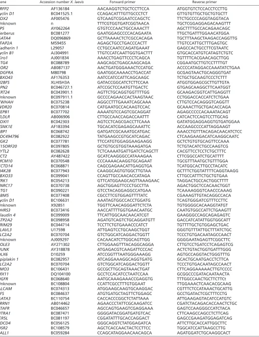
Table 1. Primers used in this study
Gene Accession number X. laevis Forward primer Reverse primer
SRFP2 AF136184 AACAAGGTCTGCTCCTTCCA ATGGTGTCTCCACCTCCTTG
cyclin D1 BC041525.1 CCAGACATTTGTTGCCCTCT GTTGTGTTGCTGCTGTGCTT
SOX2 AF005476 GTCAAGTCGGAATCCAGCTC TTCTGCCCCAGGTAGGTACA Unknown n TTTCGTGGTGATCGGTAACA TGCTCGGAGGAGACAAGTTT
SP5 AY062264 GTGTCCCAACTGCCAAACTT AGCTTTTGCCACAGAACAGC
Cerberus BC081277 GAATGGAGCCCCACAGAATA TTGCTGATTTGGAACATGGA
GATA4 DQ096869 GCTTAAAACTCTCGCCACAGA TGCTTTAAGCTAAGACCAGGTTG
TFAP2A M59455 AGAGCTGCCTGACCCACTT TTGTCCATTGCTTTGAGTGC
Cadherin 1 L29057 CCTGCCAATCCAGATGAAAT GAGCCACTGCCTTCGTAATC
cyclin B1 AJ304991 TTGTCCATCAATTGGTGACTTT GTGCACCATGTCATAGTCTGTC
Xiro1 AJ001834 AAACCTGAGTTCCCTCAGCA TGTTTTCACGAACAGCTTGG
BTG4 BC088789 AAGCAGCTGAGCAAGCAGA CGGAATGCTTGTCCTTTTGT
ROR2 AB087137 AACTGATGGGAAACTCCATGC ACCCCATAGGACCAAATATCTGAA
PDGFRA M80798 GAATGGCAAAACCTGACCAT GCGAGTAACTGCAGGGTGAT
FBXO43 AF176353 AATCCATCCATTCAGCAAGC TCTGCTGCAAGTCCCTCTTT
H2BFS XLHISH3A GATACCGGCATCTCTTCCAA ATGGTGGAGCGCTTGTTGTA
RPN2 BC046727.1 ATCCGCTCCAATGTTGACTC GTGAGCAAGGCTTCAATGGT
KRT24 BC043901.1 AGTTCTGCAGGTGGTTTTGG GCAAGACGGTCATTGAGGTT
Unknown BC097911.1 GCCCCAGAACCACTAAGTAAC CCTGGACCACCATCTCTGAA
YWHAH BC075238 AGGCCTTTGAAATCAGCAAA CTTGTCCACAGGGTCAGGTT
WDR20 BC070814 CATGAATGCCACAAGTCCAC GCAAACTTGCTGACACCAGA
CEPB1 BC077702 AAAATGTCCAGTCGCAGGAT AGAGCCCCCACAAATACAGT
VLDLR AB006906 CTTGCCAACCAGACCAATTT CATCACTCCAGTCCTTGCAG
FOXI1 BC042303 AGTCCTCAGCGACCTTCAAA GATATGGAGGAGGTGTGATGGAA
CSNK1E AF183394 TGCACATCGAGAGCAAATTC ACCAAGCCCCATTAGGAAGT
RBP1 BC068742 GATGATCGCAAATGCATGAC AAACCTGTTTACAGACAACATCTCC
LOC494796 BC082922 TATGAAGCCGTGCATCAGAC CTCAAGAAGACATCAAGGCAATC
PGK2 BC077781 TTCCATGTGGAGGAAGAAGG GCTCTGTGTGCAGTTCCAAA
C15ORF20 BC097805 GCTGTGCGTGGTAAAGATGA TCTGTACATCTGCCAAGTCG
SYTL2 BC082628 TCTCAAAATGATTGATCTCAGCTT CACGTTCCTCCTCTGCTTTC
ECT2 AY487422 GCATCAAGGGCCATAAAGAA CTTCGGCCATCTGCATTTT
MCM10 BC070548 CCCAAAACAAGGTGCAGAAT TGCGTTTAATGCTGTTTGGA
KCTD14 BC068871 CAGCGAGAACATTGAGGTGA TCATGGCACTTGCCTACATC
PI4K2B BC077943 CAAGGCAGTGTGGCTTGTAA GCTTTCTGGTATTTTCAGGTAAGG
FBXO33 BC099041 CCAGTTGCCAACACCATAGA CTTGCCATTTCTGCTGTGAA
VRK1 BC054213 GTTCATGGAAGCAGTTAAAGAAC TAGGACTGCCACTGGCTTTT
TNRC17 BC070738 AGCTGGAGTTCCCTGCCTTA AGACTGGCTCCACAACTGGT
BTG1 BC090221 GTCCTACAGGAGGCCATGAA TCAAAAGGGTCAACCCAAAG
RBM5 BC077408 CGCCTTCGTGGAGTTTTATC CGAAAGTTGTACAGGCCACA
cyclin D1 BC106631 AAATAGTGGCCACCTGGATG TCAGTGGGATCGTTTCCTTC
Unknown X92851 TGGTTCAACAGGGATTCTCTA TGTGGGGCACAAGGTATGT
DHRS3 BC073416 AACCATTTTGGTGAACAATGC CAATGTGGCCATTCTGAAGTT
Claudin 4 BC099009 TTCATTGGCAACAACATCGT GAAGGGCCAGCAGAGAGTC
ATP2A2 BC098958 AGATGTCAGTCTGCAGGATGTT GACCATCATATTGGTGGCATT
TRIM29 BC044714 TCCTTCTGTGAAACCCATCT TGTTTTGCTGTGGAGCTGAC
ELAVL3 U17598 ATTGAGTCCTGCAAGCTGGT GGGTGTTTATTGCTTTATCTGC
SLC2A2 BC070704 GTCTGGCATCAGGACTGGTT TCCCTGTGACAATAGCCAACT
Unknown AJ009297 CACAACATCTTGGCAGTTGG GGGGAATAGAGTTCGGCTTC
POLE3 AY271302 CTTGGAAGTTTACAGGCAGGA CTTGTCCTGATCCTCAGAGTCG
HUNK AY318878 ATGAGACGTCAAGATTCCCAC ACTCTGTACTGGTGATTTGGG
DLX6 D10259 ATCCGGTTTAATGGGAAAGG AGTGCCAGGTACTGGGTTTG
Epiplakin 1 BC082957 ATCAGGAAAGGCAGGTGATG GCACTGCAATGACCTCTTCA
SLC2A2 BC070704 GTCTGGCATCAGGACTGGTT TCCCTGTGACAATAGCCAACT
LMO3 BC106431 GCCGCTTGCAGTAAACTGAT CTTCAGGAAAAACTTGTCCCC
MX11 DQ104100 GCCTCCACATCCTAATCCAA GCGGCCCGATACAATAACTA
NGFR BC068640 AATGCAAAGAAACCGAGGTG TTTGGCCAACTGCTTCTTCt Unknown BC108804 CCATTCGCCTTTGTGGAAT TTGGAAACTCAACACGCAAG
ALCAM BC074313 ATGGAAGCAAGTGCAAGGAC CGTTTCTCCATAAACTGCATTG
EFS BC084637 ATGTGATGCTAGTTCTGGAGC GCCTGATACTCGCTTTCCTG
GATA3 BC110754 CACCACCCGGCTCTATTAAA ATTGAAGAGTACATCCATGTC
LRRN1 AB014462 AGAACCCTATTCGCAAGATCC CGATCTACAGACACCAACTCTGC
PTAFR BC046657 AGCCAGTGAAGTCGAGGAAA GAGTCCAAGGGCCATCTACA
HTRA1 BC087471 GGGGATACGGATGATGTCAC CTTCAAGCCAGCCTCTTCAG
IER5L BC081197 CGGATATTTGCACCAGGACT GAGCCGAAGATGGAGATCAG
PDCD4 BC056125 GGGCAGGTCTATGGAGATCA ATTCTTGCACCATTGGCTTC
OSR2 BC108579 AGCTCACCAACTACTCCTTCC TGGCATCCATTAAGCCTTG
SALL1 BC059284 CCAGCATAGGAACAACAGCA AGATGGATCTGCAAGGCACT
Table continued on next page.
D
E
V
E
LO
P
M
E
N
Smicl-HA in pCS2 was also digested using restriction enzymes ClaI and SacI to generate a similarly tagged Smicl construct containing just the last 338 amino acids of the ORF (which include the zinc fingers). FLAG-Rpb1 and FLAG-Rpb1ΔCTD constructs were as described (Rosonina and Blencowe, 2004).
Xenopusembryos and microinjection
Embryos of Xenopus laeviswere obtained by artificial fertilisation. They were maintained in 10% normal amphibian medium (NAM) (Slack, 1984) and staged as described (Nieuwkoop and Faber, 1975). Embryos were injected at the one-cell stage with 80 ng Smicl antisense morpholino oligonucleotide (Smicl MO) or control morpholino (CoMO) (Collart et al., 2005b), C-terminally HA-tagged XtSmicl in pCS2 (Collart et al., 2005b) was linearised with Asp718 and sense RNA was generated with SP6 RNA polymerase. RNA injections were performed in embryos at the one-cell stage. Use of a fluorescein-labelled Smicl MO confirmed that injected MOs distribute evenly within the embryo (data not shown).
Microarray analysis
Microarray analysis was carried out essentially as described, using a microarray that represents about one-third of the genes expressed during early development (Chalmers et al., 2005; Ramis et al., 2007). Microarrays were scanned using an Axon 4000B scanner and GenePix Pro software (Axon). The microarray results were imported into Acquity analysis software (Axon) and normalised using Lowess normalisation. Data files were then created as described (Ramis et al., 2007). The complete datasets were deposited in the NCBI Gene Expression Omnibus (GEO) data repository (http://www.ncbi.nlm.nih.gov/geo/), with Accession number GSE4952.
Real-time RT-PCR
Differential expression was validated by real-time RT-PCR using the LightCycler 480 (Roche). Reverse transcription was carried out using Transcriptor First Strand cDNA Synthesis Kit (Roche) followed by real-time PCR using the LightCycler 480 SYBR Green I Master kit (Roche) following the manufacturer’s instructions. Primers specific for Ornithine decarboxylase (ODC) and Chordin were as described (Piepenburg et al., 2004). Primers to amplify Xiro1intron sequences are: forward, 5⬘-CAGCTAAGTTCAGCCCAAGG-3⬘; reverse, 5⬘ -GCGTT-TATCGGACAACGATT-3⬘. Primers to amplify Chordin intron sequences are: forward, 5⬘-CACTGTTGAAGCCAAGCAAA-3⬘; reverse, 5⬘-GAGGCTGCATTGCTCTTCTC-3⬘. Details of other primers are provided in Table 1. The normalised target concentrations (in arbitrary units) were calculated from the real-time PCR efficiencies and the crossing point of the target and of the reference gene ODC, as
described (Pfaffl, 2001). Correlations between microarray and real-time RT-PCR results were performed using the Windows SPSS version 11.5 software (SPSS, Chicago, USA).
Polyadenylation assays
An aliquot of 4 μg of total RNA was ligated for 30 minutes at 37°C with 0.4 μg of a 3⬘amino 5⬘phosphorylated oligonucleotide P1 (5⬘ -P-GGTCAC-CTTGATCTGAAGC-NH2-3⬘) in a volume of 10 μl using T4 RNA ligase (New England Biolabs). The reaction was placed at 70°C for 15 minutes. The whole 10 μl ligation reaction was used in a 50 μl reverse transcription reaction using Superscript III (Invitrogen), according to the manufacturer’s directions using 0.4 μg primer P2 (5⬘ -GCTTCAGATCAAGGTGAC-CTTTTT-3⬘) as described (Graindorge et al., 2006). Of the resulting cDNA, 1 μl was used in a 50 μl PCR reaction. As a reverse primer, P2 was used in all reactions. As forward primers, we used P3, specific for our positive control Eg3: 5⬘-AAGTGACTATGCAATTTGAGCTAGAAGTAT-3⬘ (Graindorge et al., 2006), and designed primers specific for MCM10(P4: 5⬘ -ACAGCATTGCAGAATCATGG-3⬘), FBXO43 (P5: 5⬘ -TCTTGCACCT-GATGTTTGTG-3⬘) and Xiro1 (P6: 5⬘ -CCGTGTTCCATTTCAGACCT-3⬘). The PCR reaction mixture contained 1X buffer (Invitrogen), 0.2 μM of each primer (forward and reverse), 200 μM dNTPs, 1U of Platinum Taq Polymerase (Invitrogen) and 1.5 mM MgCl2, in the presence (P4) or absence (P3, P5, P6) of 10% DMSO. The amplification programme consisted of a preincubation step for denaturation of the template cDNA (95°C for 5 minutes), followed by either 40 (P3), 45 (P4) or 50 (P5, P6) cycles consisting of a denaturation step (95°C for 30 seconds), an annealing step (56°C for 30 seconds for P3, P4, P5 or 59°C for 30 seconds for P6) and an extension step (72°C for 30 seconds), and then one final extension step (72°C for 7 minutes). The amplified products were digested with PvuII (P3), BstUI (P6) or PsiI (P4 and P5) to verify the specificity of the PCR products. PCR reactions and digests were separated on a 12% non-denaturing polyacry-lamide gel and stained with ethidium bromide.
Co-immunoprecipitation experiments
Transiently transfected HEK293T cells were solubilised in lysis buffer containing 1% NP40, 150 mM NaCl, 20 mM Tris pH 7.5, 2 mM EDTA, 50 mM NaF, 1 mM sodium pyrophosphate, supplemented with protease inhibitors (Roche Molecular Biochemicals). Cell lysates were cleared by centrifugation and precipitations were performed by overnight incubation at 4°C with anti FLAG M2 agarose affinity gel (Sigma). Unbound proteins were removed by washing four times with cold lysis buffer. Bound proteins were harvested by boiling in sample buffer, and they were resolved by SDS-polyacrylamide gel electrophoresis. Flag-tagged and HA-tagged proteins were visualised after western blotting using rat monoclonal anti-HA-peroxidase-Table 1. Continued
Gene Accession number X. laevis Forward primer Reverse primer
Unknown BC110732 ATCAGCTACAGCCTGTCTCC AGAGCACAAAAGGTGGCACTA
ZFHX1B BC084972 TGGACCACTCCAGGAGCA TAAGTCCAAGGGCTCAGCTT
Unknown AJ009297 GCTGAGAGGAGGCAGCTAGA GGGGAATAGAGTTCGGCTTC
RBM12 BC059291 TCTGAAGCGTAACCGAATG TTAGTTGCTGCATCAAAGGAG
STAG2 AF255018 GAGAATTTGCCATGCTTACCA CCCAAGACAATATGTCGTAGCTT
HNRPU BC046700 AGAGCTTTGCACTTCTCTGG GCAACACCATTGCAACATTT
Unknown BC045031 TGGAAGCCTGGTACAGAGGA GTTTCTGCCAAAGTGCCTTC
IGSF4 BC108832 GCTGGTTCAAGGGAAACAAA CAGCTGGATGGTCAACAAGA
DYNLL1 BC073042 GGAAGGAATTTTGGCAGCTA GAAGGTTTGTTCCCTTGGTT
NUDT22 BC068937 TACCTGCAAGGAGGTCCTGA GGCTGATGGACACAGTTCCT
GATA6 BC082349 CCTTTCTGACTTTTGCACAGC GGCAAAGTCTGTTGGATGGT
MEIS1 BC084920 CGATTCCACCTGTTGGAGTTA ACCCGGTGTACCTGAGTGAG
SALL1 AF310007 GAACAGCACACCTGCAAGAA CCAGCCATTGGAGTAATGC
CYP26A1 BC073518 GGAGAGACTCTGCAAATGGTG GTGCAGGTTGGACAGACAGC
MGC61598 BC084778 GACCAACTGCGAGTTTAACG GCCCCAAACTTTACCAACAG
ZFP36L1 BC100162 AGCTTGTTTGCTCCAAGCAT CTGGAACTGCTCAGGTAGCC
ZNF503 BC046863 ACTGTTCTCCCCCTGGATCT TTTTCTTGGCATCCAGCTCT
KIAA1324L BC077391 CCAAGGGGACAAAATACTACCA CCTTTGCTGTCTGATGGGATA
P4HB BC077772 TGCCAAGATGGATTCTACAGC CATCTCCCTCCTCCAAATCA
Sequences are shown 5⬘to 3⬘.
D
E
V
E
LO
P
M
E
N
[image:3.612.50.566.68.277.2]coupled high-affinity antibody (3F10) (Roche) and goat polyclonal anti-FLAG-peroxidase coupled antibody (Abcam) and the SuperSignal West Dura Extended Duration Substrate kit from Thermo Scientific (Pierce).
Whole-mount antibody staining
Embryos were fixed overnight in MEMFA (3.7% formaldehyde, 100 mM MOPS, 2 mM EGTA, 1 mM MgSO4, pH 7.4) at 4°C, and the vitelline membrane was removed. After gradually dehydrating and rehydrating the embryos in methanol, they were washed in phosphate buffered saline (PBS) and bleached in 2 ⫻SSC with 2% formamide and 1.5% H2O2. Embryos were washed 2 ⫻10 minutes in PBS and 2 ⫻30 minutes in PBS with 0.1% BSA and 0.2% Triton X-100 (PBSbt) at room temperature. They were blocked for 1 hour at room temperature in PBSbt with 10% serum (PBSbts) and incubated overnight at 4°C in a 1/250 dilution of the anti-HA-peroxidase-coupled rat monoclonal antibody (Roche) in PBSbts. Embryos were washed for 1 hour in PBSbts, 4 ⫻1 hour in PBSbt and 10 minutes in PBS and incubated in Tris-buffered saline (TBS) with 0.066% 3, 3⬘-Diaminobenzidinetetrahydrochloride (DAB) (SIGMA) for 10 minutes. H2O2was added to a final concentration of 0.024% and staining was observed after 1 minute.
Anti-Rpb1 antibodies
To detect Rpb1 and phospho S2 Rpb1, we used mouse monoclonal antibody 8WG16 (ab817) and a rabbit polyclonal antibody (ab5095) from Abcam.
RESULTS
Isolation of novel Smicl targets
Smicl maintains expression of Chordinin the early gastrula of Xenopus laevis(Collart et al., 2005b), but it is unlikely that this gene is the only embryonic target of Smicl, because injection of Chordin mRNA cannot completely rescue the phenotype of embryos injected with a Smicl antisense MO (Collart et al., 2005b). Further insight into the role and mode of action of Smicl requires the identification of additional targets, and to this end we performed microarray analysis on RNA derived from control embryos at the early gastrula stage and from embryos injected with Smicl antisense MOs. The microarray slides used in our experiments were designed from transcript sequences derived from a large-scale Xenopus tropicalisEST project (Gilchrist et al., 2004). These X.tropicalis microarrays also work in X.laevis(Chalmers et al., 2005; Ramis et al., 2007).
Xenopus laevisembryos from three different spawnings were injected at the one-cell stage with either Smicl MO, which inhibits Smicl function, or the control CoMO (Collart et al., 2005b) and were cultured to the early gastrula stage before RNA isolation. Some embryos were cultured to later stages to confirm that their development was impaired as described previously (Collart et al., 2005b). RNA from each spawning was hybridised with dye-swapped technical replicates, making six microarray slides in total. Oligonucleotides were considered to be differentially expressed when: (1) they showed at least a twofold difference (sample versus control) in expression levels in four out of the six microarrays; and (2) they were significantly different (q=0). In embryos in which Smicl was downregulated, 95 oligonucleotides fulfilled these criteria (Table 2): 33 genes were upregulated and 62 were downregulated.
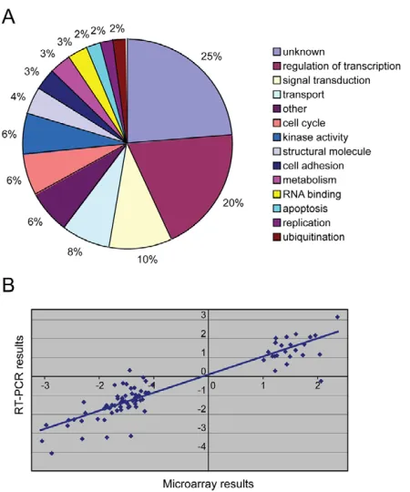
The X.laevishomologues of the X. tropicaliscDNAs recognised by the oligonucleotides were identified by BLAST searches (Table 2). Genes were manually classified according to the annotation of their human homologues (NCBI databases, http://www.ncbi.nih.gov/), because gene ontology annotation for Xenopusspecies is not yet available (Fig. 1A). Apart from the unknowns, the largest group is involved in regulation of transcription (20%).
Microarray results were validated by real-time RT-PCR using the same samples that were used for microarray experiments. Primers were designed for the X.laevishomologues of all differentially
expressed transcripts, with the exception of nine cDNAs for which X.laevishomologues could not be identified (Table 2). Of the genes tested, 76% (66) were confirmed as being differentially expressed (Table 2). Bilateral correlation analysis of the results obtained by microarray hybridisation and those obtained by real-time RT-PCR showed a Pearson correlation of 0.909 (P<0.0005) (Fig. 1B).
The temporal regulation of gene expression by Smicl starts at the MBT
[image:4.612.327.552.57.327.2]Smiclis expressed both maternally and zygotically during Xenopus development (Collart et al., 2005b). Direct targets of Smicl are likely to be among the first to be up- or downregulated by loss of Smicl function. To find such targets, we studied the temporal expression patterns of 78 of the genes identified in our microarray analysis in control embryos and in embryos injected with a Smicl MO. We found that the genes could be divided into four categories, an example of each of which is shown in Fig. 2; for the complete results, see Fig. S1 in the supplementary material. In category 1 (41 genes), normal embryos exhibited low maternal mRNA levels that increased at the onset of zygotic transcription (between stages 7 and 8.5). Loss of Smicl function caused a reduction in zygotic transcripts (Fig. 2A,E). In category 2 (19 genes), maternal transcript levels normally decreased after the MBT, but the degradation of these gene products was delayed in Smicl-depleted embryos (Fig. 2B). Category 3 (four genes) comprise genes with expression levels in Smicl-depleted embryos that were first upregulated and later downregulated compared with control embryos (Fig. 2C). And category 4 (14 genes) consists of the genes with RNA expression levels in this experiment that proved to be only slightly affected by depletion of Smicl (Fig. 2D). We do not yet understand the transient Fig. 1. Microarray experiment searching for targets of Smicl.
(A) Classification of Smicl targets according to the annotation of their human homologues. (B) Correlation between microarray and PCR results. The x- and y-axes represent the log2(sample/control) for the
microarray and RT-PCR, respectively.
D
E
V
E
LO
P
M
E
N
upregulation observed in the four category 3 genes, which include Chordin. One possibility is that loss of Smicl causes the downregulation of a repressor of Chordinexpression.
Smicl translocates from the cytoplasm to the nucleus at MBT
The expression profiles of the genes in our categories 1 and 2 indicate that absence of Smicl affects the time of onset of zygotic gene expression and of the degradation of maternal mRNAs. These events usually occur at the MBT, and, interestingly, we observed that an HA-tagged form of Smicl translocated from the
cytoplasm to the nucleus at precisely this time (Fig. 3A-F). Similar behaviour was observed in dissociated animal pole cells using a Venus-tagged form of Smicl and confocal microscopy: at the MBT the distribution of Smicl changed from diffuse and cortical to well defined and nuclear (Fig. 3G,H). A similar transition was observed in a construct consisting of just the Smicl zinc finger domain (Fig. 3I).
To ask whether the nuclear translocation of Smicl at the MBT requires new transcription, we injected embryos with α-amanitin as well as with RNA encoding HA-tagged Smicl. Such embryos divided normally to stage 9, but the transcription of GS17and Siamoiswas prevented, confirming that transcription was blocked (Fig. 3J). This inhibition of transcription had no effect on the nuclear translocation of Smicl after the MBT (Fig. 3K,L). This is discussed below.
Smicl affects 3⬘-end processing of at least one of its target genes, Xiro1
[image:5.612.53.267.193.505.2]Our previous work in tissue culture identified Smicl as a molecule that may be involved in 3⬘-end processing (Collart et al., 2005a). To address this point we studied three Smicl targets identified in our
[image:5.612.312.550.306.542.2]Fig. 2. Smicl targets fall into four categories. (A-D)Xenopus embryos were injected at the one-cell stage with 80 ng CoMO or Smicl MO. They were harvested at stages 7, 8.5, 9, 9.5 and 10.25, and the RNA expression levels of putative Smicl target genes were measured by quantitative RT-PCR. RNA expression levels relative to Ornithine decarboxylase(ODC) in CoMO embryos are indicated in red, and levels in Smicl-MO-injected embryos are indicated in green. We define four categories of Smicl targets and one example of each type of target is shown here. The others are presented in Fig. S1 in the supplementary material, with the above stages represented as numerals 1 to 5. (A) In normal embryos, RNA expression levels of category 1 genes are upregulated when zygotic transcription starts. Their transcription is downregulated in Smicl-depleted embryos. (B) RNA expression levels of category 2 genes diminish after MBT during normal development. Their decline is slower in embryos lacking Smicl. (C) In the course of normal development, category 3 genes are upregulated at the onset of zygotic transcription. Depletion of Smicl causes an early elevation of expression that is followed, in comparison to normal embryos, by a decrease in transcription. (D) RNA expression levels of genes in category 4 are similar in coMO- and Smicl-MO-injected embryos. The time of the MBT is indicated by an arrow in the four graphs. (E) The temporal RNA expression pattern of Xiro1assayed by quantitative RT-PCR and normalised with respect to ODC.
Fig. 3. Subcellular localisation of Smicl before and after the MBT.
(A-F)Xenopusembryos were injected at the one-cell stage with 1.5 ng RNA encoding HA-tagged XtSmicl or with H2O (inset). Embryos were
harvested 3 (A), 4 (B), 5 (C), 5.5 (D), 6 (E) and 6.5 (F) hours after fertilisation, with the MBT occurring at about 5.5 hours after
fertilisation at room temperature, at the onset of transcription of GS17. Developmental stages are indicated at the bottom right and higher-magnification insets show nuclear translocation between 5 and 5.5 hours after fertilisation. Overexpressed Smicl protein was detected by whole-mount antibody staining. (G-I) Confocal microscopy images of Venus-tagged full-length Smicl (G,H) and a version comprising just the zinc finger domain (I). Note that nuclear staining is not visible at 4.5 hours after fertilisation (G) but is detectable at 7 hours (H). The zinc finger domain is also nuclear at 7 hours (I). (J-L) Nuclear localisation of HA-tagged Smicl 6.5 hours after fertilisation in embryos injected with
α-amanitin (50μg/embryo). (J) Quantitative RT-PCR shows that this concentration of α-amanitin inhibits activation ofGS17and Siamois. HA-tagged Smicl is nuclear in control embryos (K) and also in embryos
injected with α-amanitin (L).
D
E
V
E
LO
P
M
E
N
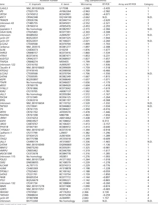
Table 2. Analysis of the genes identified by microarray experiments
Accession number Log2(sample/control)
Gene X. tropicalis X. laevis Microarray RT-PCR Array and RT-PCR Category
ELAVL3 NM_001030326 U17598 –3.040 –3.435 + 1
SP5 CT025179 AY062264 –2.963 –2.582 + 1
KRT24 CX745012 BC043901 –2.862 –4.071 + 1
MX11 CR942348 DQ104100 –2.662 N.D. – N.D.
TRIM29 CR926196 BC044714 –2.572 –2.620 + 1
Unknown 43 CT485755 BC045031 –2.552 –2.321 + 2
SOX2 CR760314 AF005476 –2.453 –2.293 + 1
Epiplakin 1 No homology BC082957 –2.315 –2.366 + N.D.
KIAA1324L CT025403 BC077391 –2.303 –3.308 + 1
Unknown BX689202 AJ009297 –2.277 –1.371 + N.D.
cyclin D1 BQ522031 BC041525 –2.247 –1.944 + 1
cyclin D1 BQ522031 BC106631 –2.021 –2.260 + N.D.
SLC2A2 CT030568 BC070704 –1.899 –1.703 + 1
Cerberus NM_203515 BC081277 –1.897 –2.308 + 1
DLX6 CX836573 D10259 –1.876 –1.677 + 1
DHRS3 CR848157 BC073416 –1.860 –1.534 + 3
HTRA1 BC073416 BC087471 –1.853 –3.231 + 1
NUDT22 CR942377 BC068937 –1.827 –1.657 + 1
TFAP2A CR848274 M59455 –1.799 –1.689 + 1
Unknown 122 CX516192 AJ009297 –1.761 –1.936 + 1
Claudin 4 NM_001016663 BC099009 –1.759 –1.518 + 1
ATP2A2 DT418145 BC098958 –1.709 –0.466 – 1
SLC2A2 CT030568 BC070704 –1.706 –1.550 + N.D.
GATA6 CT030595 BC082349 –1.667 –1.815 + 1
NGFR BQ393897 BC068640 –1.657 –1.071 + 1
PTAFR No homology BC046657 –1.656 –1.179 + 1
MEIS1 CR760178 BC084920 –1.604 –1.418 + 1
DYNLL1 CR761866 BC073042 –1.603 –1.619 + 3
ROR2 CF219705 AB087137 –1.592 –1.781 + 1
SALL1 No homology BC059284 –1.572 –1.167 + 1
PDCD4 CF151949 BC056125 –1.572 –1.264 + 4
IER5L CX363316 BC081197 –1.559 –0.368 – 1
Unknown NM_001016654 BC110732 –1.520 –1.332 + N.D.
ZNF503 CF219041 BC046863 –1.512 –1.034 + N.D.
EFS CR761735 BC084637 –1.494 –0.416 – 4
SRFP2 CR761702 AF059570 –1.478 –1.446 + 3
PDGFRA CR761598 M80798 –1.463 –1.656 + 1
LRRN1 CX374253 AB014462 –1.438 –1.597 + 1
IGSF4 DR833927 BC108832 –1.435 0.313 – 4
LMO3 CX874767 BC106431 –1.413 –3.157 + 1
ZFHX1B DT447181 BC084972 –1.413 –0.640 – 1
CYP26A1 NM_001016147 BC073518 –1.394 –0.918 – 4
Cadherin 1 CF217799 L29057 –1.382 –1.296 + 1
Xiro1 DT434125 AJ001834 –1.370 –1.289 + 1
STAG2 BX775788 AF255018 –1.349 –0.609 – 1
SALL1 CX490174 AF310007 –1.336 –1.465 + N.D.
GATA4 NM_001016949 DQ096869 –1.334 –1.136 + 1
RBM12 DN075293 BC059291 –1.325 –0.981 – 1
HNRPU CR855462 BC046700 –1.319 –1.617 + 1
MGC61598 CX372678 BC084778 –1.316 –1.285 + 1
Unknown 115 No homology X92851 –1.276 –1.093 + 1
POLE3 NM_001017264 AY271302 –1.264 –1.018 + 4
OSR2 DN038055 BC108579 –1.239 –1.278 + 1
ALCAM AL797115 BC074313 –1.229 –0.776 – 1
HUNK CX465369 AY318878 –1.198 –1.228 + 1
ZFP36L1 CT025463 BC100162 –1.188 –0.059 – 4
GATA3 CF221701 BC110754 –1.159 –1.850 + 1
P4HB BX695354 BC077772 –1.144 –0.897 – 4
BTG1 BQ526679 BC090221 –1.142 –0.865 – 4 Unknown 132 CF241734 BC108804 –1.140 –0.260 – 1
RBM5 NM_001017278 BC077408 –1.090 –0.874 – 4
H2BFS NM_001017251 X03018 –1.015 –0.465 – 1
FBXO43 CT010561 AF176353 2.366 3.130 + 2
YWHAH CR848634 BC075238 2.066 –0.259 – 4
cyclin B1 DT407498 AJ304991 2.043 1.157 + 2
[image:6.612.52.560.78.737.2]Unknown BX772260 No homology 2.020 N.D. – N.D.
Table continued on next page.
D
E
V
E
LO
P
M
E
N
microarray experiments. We chose Xiro1, a category 1 gene that is among the earliest zygotically transcribed genes and is therefore likely to be a direct target (Fig. 2A,E), and two category 2 genes, FBXO43 and MCM10, with regulation that is more likely to be indirect. To investigate lengths of poly (A) tails we used an RNA ligation assay (Graindorge et al., 2006) with minor modifications (Fig. 4A). The Eg3 gene, with RNA that undergoes deadenylation between fertilisation and the 64-cell stage (Graindorge et al., 2006), was used as a positive control. Fig. 4B confirms that the assay could detect the deadenylation of Eg3mRNA during early cleavage stages, and Fig. 4C shows that overexpression of Smicl lengthened the Xiro1mRNA poly(A) tail whereas depletion of Smicl, by injection of an antisense MO, shortened it. To facilitate comparison of the PCR products produced in the polyadenylation assay, the assay was performed at the very onset of transcription of Xiro1, when transcript levels in the three different samples are still similar. This activation of Xiro1occurred slightly earlier than that of GS17, the onset of expression of which in our experiments defined the MBT. Smicl had no effect on the lengths of the poly(A) tails of FBXO43or MCM10. These experiments show that Smicl regulates 3⬘-end processing of Xiro1mRNA.
Smicl also affects levels of unprocessed Xiro1 transcripts
To ask whether the early reduction in transcript levels caused by loss of Smicl is associated with this decrease in polyadenylation, perhaps by affecting the stability of the mature gene product, we studied transcript levels of Xiro1 using primers specific for the mature mRNA and for the unprocessed mRNA. We have previously shown that levels of Chordinare reduced in Smicl morpholino-injected embryos and we therefore used Chordinas a positive control.
Our results show that transcript levels of Xiro1 as well as Chordin were downregulated in embryos injected with Smicl MO and, significantly, that this is true for unprocessed as well as processed transcripts at the early gastrula stage (Fig. 5A). These observations suggest that downregulation also occurs at the level of transcription, and that the lower levels of Xiro1mRNA observed are not solely a consequence of RNA instability resulting from a shortening of its poly(A) tail.
The same assay was used to analyse levels of Xiro1 and Chordin after overexpression of Smicl. Although Smicl causes upregulation of Chordin(Collart et al., 2005b), it had no effect on levels of Xiro1at the early gastrula stage (Fig. 5B). Xiro1proves to not be unusual in this respect: of all the Smicl target genes, shown in Fig. S1 in the supplementary material, only Chordinand FoxI1mRNA levels were upregulated in response to Smicl (data not shown).
Smicl does not bind to CBTF and NF-Y but does interact with Rpb1
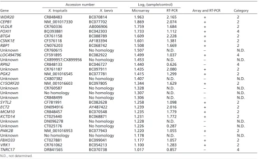
[image:7.612.49.566.70.389.2]We have identified 41 category 1 targets, all of which require Smicl for the onset of zygotic gene expression. To ask whether all 41 are regulated by a common mechanism, we first asked whether Smicl is brought to their promoters by a common transcription factor. To this end we analysed sequences 500 base pairs 5⬘of the transcription start sites of the genes and searched for motifs from the JASPAR CORE database (http://jaspar.genereg.net/). We found the best match for each motif in each sequence using the nmscan tool from the NestedMICA suite (Down and Hubbard, 2005) and then compared the distribution of scores for each motif to that from 1000 randomly selected Xenopusgenes. In this way we found that the NF-Y motif (core sequence CCAAT) was Table 2. Continued
Accession number Log2(sample/control)
Gene X. tropicalis X. laevis Microarray RT-PCR Array and RT-PCR Category
WDR20 CR848483 BC070814 1.963 2.165 + 2
CEPB1 NM_001017330 BC077702 1.869 2.074 + 2
VLDLR CR760336 AB006906 1.759 1.684 + 2
FOXI1 BQ393881 BC042303 1.733 1.112 + 4
BTG4 CR761158 BC088789 1.609 2.228 + 2
CSNK1E CF376118 AF183394 1.601 1.381 + 2
RBP1 CN076203 BC068742 1.508 1.669 + 2 Unknown CR760615 No homology 1.507 N.D. – N.D.
LOC494796 CF591895 BC082922 1.499 1.037 + 4
Unknown CX899957;CX899956 No homology 1.453 N.D. – N.D.
RPN2 CR848133 BC046727 1.440 0.626 – 4 Unknown CR761187 BC097911 1.435 2.080 + 2
PGK2 NM_001016545 BC077781 1.415 1.299 + 2 Unknown CX807382 No homology 1.407 N.D. – N.D.
C15ORF20 NM_001016603 BC097805 1.344 1.629 + 2
Unknown CR760587 No homology 1.328 N.D. – N.D. Unknown No homology No homology 1.307 N.D. – N.D. Unknown CR848499 No homology 1.306 N.D. – N.D.
SYTL2 CF781991 BC082628 1.258 1.098 + 2
ECT2 DN094916 AY487422 1.239 2.016 + 2
MCM10 CR848457 BC070548 1.235 1.779 + 2
KCTD14 CT025440 BC068871 1.231 1.772 + 2
Unknown DN096278 No homology 1.228 N.D. – N.D. Unknown CT025176 No homology 1.226 0.287 – N.D.
PI4K2B NM_001016953 BC077943 1.220 1.055 + 2
Unknown No homology No homology 1.178 N.D. – N.D.
FBXO33 CT027881 BC099041 1.177 1.057 + 2
VRK1 CR761062 BC054213 1.100 1.283 + 2
TNRC17 DR841565 BC070738 1.017 0.857 – 4
N.D., not determined.
D
E
V
E
LO
P
M
E
N
significantly over-represented (P=0.007: an empirical figure based on re-sampling of scores from the random genes). This sequence is the binding site for the transcription factors NF-Y and CBTF in early frog embryos. NF-Y consists of three subunits and is the predominant Y-box binding protein in Xenopus oocyte nuclei (Li et al., 1998). CBTF is the main CCAAT binding factor at midblastula stages (Ovsenek et al., 1991). It is expressed maternally, and its activity is regulated by its p122 subunit (CBTFp122). This protein is perinuclear during early embryogenesis, but moves from cytoplasm to nucleus at stage 9, before the detection of CBTF activity in the nucleus (Orford et al., 1998). These observations suggest that Smicl might regulate gene expression at the MBT by interacting with NF-Y or CBTFp122, but our co-immunoprecipitation experiments have revealed no such interaction under our experimental conditions (data not shown).
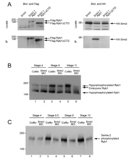
In a further attempt to identify a general mechanism by which Smicl might affect gene expression at the MBT, we noted that CPSF was required for pausing and termination of transcription, and that it did this by binding, through its 30 kDa subunit, to the body of RNA polymerase II (Nag et al., 2007). Because Smicl affects transcription as well as 3⬘-end processing, and because it is similar to the 30K subunit of CPSF, we asked whether it could form a complex with Rpb1, the largest subunit of RNA polymerase II. Co-immunoprecipitation experiments in HEK293T cells indicate that HA-tagged Smicl interacted with full-length Flag-tagged Rpb1 (Fig. 6A). In contrast to the 30 kDa
subunit of CPSF (Nag et al., 2007), Smicl did not interact with a tagged Rpb1 construct that lacked the C-terminal domain (Rpb1ΔCTD).
Smicl affects the phosphorylation of Rpb1 after the MBT
The CTD of Rpb1, which is required for interaction with Smicl, is a docking platform for factors involved in various co-transcriptional events (Egloff and Murphy, 2008). Different proteins are recruited to the Rpb1 CTD at different stages of the transcription cycle in response to changes in the phosphorylation state of this domain (Egloff and Murphy, 2008). Before the MBT
in Xenopus, two forms of Rpb1 are present: one is
hypophosphorylated in the CTD and the other is phosphorylated, but not at serine 2 of the repeated heptapeptide YSPTSPS (Palancade et al., 2001). Phosphorylation of serine 2 usually occurs at the MBT, and this hyperphosphorylated form of Rpb1 can be detected on a western blot as a band that migrates slightly more slowly than the embryonic phosphorylated form, as well as by a specific antibody (Palancade et al., 2001).
[image:8.612.56.397.58.432.2]Phosphorylation of serine 2 at the CTD is required for elongation of transcription and for recruitment of factors involved in cleavage and polyadenylation (Egloff and Murphy, 2008). Smicl is also involved in cleavage and polyadenylation, and it interacts with Rpb1 in a manner that requires the CTD. We therefore asked whether Smicl is required for the phosphorylation of the CTD of Rpb1.
Fig. 4. Analysis of polyadenylation. (A) Schematic of the polyadenylation assay. Primer P1 is ligated to the poly(A) tail of mRNAs. cDNA is synthesised with primer P2, complementary to P1, and gene-specific products are obtained by PCR using a forward gene-specific primer (G) and reverse primer P2. The specificity of the products was tested by digestion with a restriction enzyme that cuts uniquely between the gene-specific primer and the beginning of the poly(A) tail (PvuII for Eg3, PslI for MCM10and FBXO043, and BstUI for Xiro1). The products are analysed on a polyacrylamide gel. The PCR products from the polyadenylation assay, which contain a piece of the 3⬘-end untranslated region as well as the poly(A) tail itself, were split in half. On each gel (see B,C), the undigested halves, which are loaded on the left-hand lanes, reveal differences in the lengths of the poly(A) tails. The digested samples, which are loaded on the right-hand side, show fragments of DNA between the annealing site of the gene-specific region and the restriction site indicated above, together with other fragments, which include the poly(A) tail of unknown length. The lengths of the former can be calculated. They confirm the specificity of the PCR products and are indicated on the figure. (B) Control experiment to validate the polyadenylation assay. Lanes 1 and 2: products of an Eg3polyadenylation assay performed on RNA derived from unfertilised eggs (UFE) and from 64-cell stage embryos. Lanes 3 and 4: restriction digest of the polyadenylation products. M, DNA size marker. (C) Lengths of the poly(A) tails of three Smicl targets. Xenopusembryos were injected at the one-cell stage with 80 ng of Smicl MO, 80 ng of CoMO or 1.5 ng of Smicl RNA, and harvested at stage 7. The
polyadenylation assay described in A was performed on RNA derived from these embryos for the genes MCM10, FBXO43and Xiro1. Lanes 1 to 3 show products of the polyadenylation assays. Lanes 4 to 6 show the restriction digests of these products.
D
E
V
E
LO
P
M
E
N
Injection of our control antisense MO proved to have no effect on the phosphorylation of the Rpb1 CTD, but injection of our Smicl MO inhibited serine 2 phosphorylation at the MBT (Fig. 6B,C). This and our other results are discussed below.
DISCUSSION
Smicl (Smad-interacting CPSF30-like) is an unusual protein that not only interacts with transcription factors (Collart et al., 2005a; Collart et al., 2005b) but also with the CPSF complex. In addition, it has a zinc finger domain that is similar to that of CPSF30 (Collart et al., 2005a). These observations suggest that Smicl may control gene expression by modulating 3⬘-end processing. In an attempt to test this idea we used microarray analysis to identify novel Smicl targets and have shown that at least one of these, the homeobox-containing gene Xiro1, is indeed regulated by 3⬘-end processing. In the course of searching for a general mechanism by which Smicl might function, we discovered that it can form a complex with Rpb1, the largest subunit of RNA polymerase II, and that it is required for serine 2 phosphorylation of the Rpb1 CTD. This modification is required for the elongation of transcription, for splicing and for polyadenylation (Egloff and Murphy, 2008); in the Xenopusembryo it normally occurs at the MBT, and it coincides with the translocation of Smicl from cytoplasm to nucleus. We discuss these observations below, and speculate on the mechanism by which Smicl affects the Rpb1 CTD.
Smicl affects gene expression at the MBT
Our previous work identified Chordinas a target of Smicl, but it seemed unlikely that this gene was the only Smicl target because overexpression of Chordindoes not completely rescue the loss of
[image:9.612.52.267.54.266.2]Smicl function (Collart et al., 2005b). We therefore used microarray analysis to compare gene expression in wild type and in Smicl-depleted embryos in an effort to identify additional Smicl targets (Fig. 1). Quantitative RT-PCR analysis at different stages of development revealed that these putative Smicl targets fell into four categories (Fig. 2A-D). Most fell within category 1, in which Smicl prevented or reduced the onset of gene expression at the MBT, or Fig. 5. Regulation of Xiro1and Chordinby Smicl.(A) Quantitative
RT-PCR using primers specific for exon or intron sequences in Xiro1and Chordinshow that downregulation of Smicl causes loss of immature transcripts as well as mature mRNA. Embryos were injected at the one-cell stage with 80 ng CoMO or Smicl MO and then cultured to stage 10.5. RNA was prepared from these embryos, and cDNA was
synthesised by reverse transcription using a mixture of oligo(dT) primers and random hexamers. Intron and exon sequences from the Smicl targets Xiro1and Chordinwere amplified by quantitative RT-PCR as indicated. (B) Transcriptional regulation of Chordinand Xiro1by Smicl differs. Embryos were injected at the one-cell stage with H2O or RNA
encoding HA-tagged Smicl. The assay described in A was repeated. Note that Smicl upregulates expression of Chordinbut not of Xiro1.
Fig. 6. Smicl interacts with Rpb1 and is required for its hyperphosphorylation at the MBT. (A) Smicl interacts with the largest subunit of RNA polymerase II (Rpb1), but not with Rpb1 lacking the C-terminal domain (Rpb1ΔCTD). HA-tagged Smicl and Flag-tagged Rpb1 and Rpb1ΔCTD were overexpressed in HEK293T cells as indicated. Samples were immunoprecipitated with anti-Flag coupled beads and the presence of Flag-tagged Rpb1 constructs and of HA-Smicl was determined by western blotting. Note that Smicl interacts with Rpb1 but not with Rpb1ΔCTD (lower right panel). (B) Smicl is required for the hyperphosphorylation of Rpb1 that occurs at the MBT. Xenopusembryos at the one-cell stage were injected with 80 ng CoMO or Smicl MO. They were harvested at stages 4, 9 and 11. Proteins derived from whole embryo lysates were separated by SDS PAGE and phosphorylation of Rpb1 was analysed by western blotting using an antibody that recognises all forms of Rpb1. Lanes 1 and 2 show the presence of the hypophosphorylated form and the embryonic form in embryos at stage 4 in CoMO- (lane 1) and Smicl-MO- (lane 2) injected embryos. Levels of the
hyperphosphorylated form increase after MBT in CoMO-injected embryos at stages 9 and 11 (lanes 3 and 5) but not in Smicl-MO-injected embryos (lanes 4 and 6). (C) A similar assay to that described in B, but embryos were harvested at stages 4, 8.5, 9 and 10, and the western blot was probed with an antibody that recognises Rpb1 that is phosphorylated on Ser2 of the C-terminal heptapeptide. Lanes 3, 5 and 7 show increased levels of this phosphorylated form of Rpb1 in CoMO-injected embryos at stages 8.5, 9 and 10. This increase does not occur in Smicl-MO-injected embryos (lanes 4, 6 and 8).
D
E
V
E
LO
P
M
E
N
[image:9.612.309.560.56.364.2]within category 2, in which loss of Smicl caused a delay in the degradation of maternal transcripts at the MBT. Interestingly, the four criteria we define can also be used to classify zebrafish genes that respond to depletion of the TATA-binding protein (TBP) (Ferg et al., 2007), and indeed 27% of our Smicl targets are present in the list of 1927 genes that are regulated by TBP. This observation is consistent with the fact that transcription and 3⬘-end processing are coordinated processes.
We suspect that our list of Smicl targets is not complete, and indeed Xlim5, classified as a target in the course of additional experiments (not shown), was not identified in our microarray experiment. This failure to identify all Smicl targets might have occurred because our microarray does not represent all genes expressed during early development, because our selection criteria were too stringent, or because our Smicl MO does not completely inhibit phosphorylation of Rpb1, and different genes may show different sensitivities to levels of phosphorylation.
Consistent with the observation that Smicl regulates gene expression at the MBT, we note that Smicl becomes concentrated in the nuclei of blastomeres at this stage (Fig. 3A-I), although we cannot exclude the possibility that there is some protein present in the nucleus before the MBT. We do not know how this nuclear accumulation occurs, but we have demonstrated that it does not require new transcription, and can therefore be uncoupled from this manifestation of the MBT (Fig. 3J-L).
Smicl affects 3⬘-end processing of Xiro1
To investigate the ability of Smicl to regulate 3⬘-end processing, we turned to Xiro1, one of the earliest transcribed category 1 Smicl targets and therefore one more likely to be a direct target of Smicl. In our first experiments, use of a modified polyadenylation assay (Fig. 4A,B) indeed showed that depletion of Smicl shortens, and overexpression of Smicl lengthens, the poly(A) tail of zygotically expressed Xiro1(Fig. 4C). These results suggest that Smicl might stabilise Xiro1transcripts around the MBT.
We also investigated two category 2 genes, FBXO43 and MCM10, in which loss of Smicl activity causes a delay in the degradation of maternal transcripts at the MBT. For the mRNA products of these genes we saw no effect of gain or loss of Smicl on the lengths of their poly(A) tails. The delay in degradation of their maternal transcripts may therefore be indirect, resulting from a failure to activate one or more category 1 genes: zygotic transcription is required for degradation of maternal mRNA (Bushati et al., 2008; Giraldez et al., 2006).
Smicl affects expression levels of Xiro1
Consistent with the idea that Smicl regulates Xiro1, we note that the expression patterns of the two genes overlap, both being activated most strongly in dorsal tissues and later in the neural plate (Collart et al., 2005b; Gâomez-Skarmeta et al., 1998). As discussed above, this regulation is likely to occur through 3⬘-end processing of Xiro1 mRNA (Fig. 4), although our data also show that our Smicl MO causes the loss of unprocessed Xiro1transcripts as well as mature Xiro1mRNA (Fig. 5A). This, together with data showing that Smicl affects serine-2 phosphorylation of the CTD of Rpb1 (Fig. 6), which is required for overcoming an early block in transcriptional elongation (Peterlin and Price, 2006), suggests that Smicl may indirectly affect transcription ofXiro1. However, it is possible that Smicl is required to prevent degradation of the unprocessed Xiro1transcript, and we also note that overexpression of Smicl does not elevate expression of Xiro1(Fig. 5B). Indeed, of all the genes with expression controlled by
Smicl, only Chordinand FoxI1are upregulated in response to its overexpression (data not shown). We continue to investigate the role of Smicl in transcription.
Smicl interacts with, and is required for phosphorylation of, the CTD of Rpb1
In searching for a general mechanism by which Smicl regulates all its category 1 and 3 target genes, we first reasoned that Smicl might be brought to the promoter of its targets by a specific transcription factor, influencing 3⬘-end processing by substituting for the 30 kDa subunit of CPSF. It proved that the binding sites of the transcription factors CBTF and NF-Y are over-represented in the promoters of category 1 Smicl targets, but Smicl did not co-immunoprecipitate with either protein (data not shown). However, we then asked whether Smicl interacts with RNA polymerase II itself and were able to show that it forms a complex with Rpb1, the largest subunit of RNA polII (Fig. 6A). In contrast to CPSF30, which interacts with the body of Rpb1 (Nag et al., 2007), it is the tail of Rbp1 that is required for complex formation with Smicl (Fig. 6A).
Smicl is also required for phosphorylation of the Rpb1 CTD at serine 2 of the repeated heptapeptide YSPTSPS, a modification that normally occurs at the midblastula transition in Xenopus(Fig. 6B,C). By regulating phosphorylation of the Rpb1 CTD, Smicl changes the docking platform for proteins involved in the elongation of transcription and RNA processing, and it therefore influences, albeit indirectly, 3⬘-end formation at the MBT. We do not yet know how Smicl causes the phosphorylation state of Rpb1 to change, although we note that the activity of RNA polymerase II can be regulated by small non-coding RNAs (Barrandon et al., 2008). Smicl binds and degrades small RNAs with a stem loop secondary structure (Collart et al., 2005a), and it is possible this activity is related to phosphorylation of the CTD and the modulation of RNA polymerase activity.
Acknowledgements
We thank our colleagues for helpful discussions through the course of this work, and especially James Smith, Martin Roth, Mike Gilchrist and Rick Livesey for their advice concerning microarray construction and analyses. We also thank Mike Chesney for his comments on the manuscript. This work is supported by the Wellcome Trust and the EU Network of Excellence ‘Cells into Organs’. Deposited in PMC for release after 6 months.
Supplementary material
Supplementary material for this article is available at http://dev.biologists.org/cgi/content/full/136/20/3451/DC1
References
Barrandon, C., Spiluttini, B. and Bensaude, O.(2008). Non-coding RNAs regulating the transcriptional machinery. Biol. Cell 100, 83-95.
Bushati, N., Stark, A., Brennecke, J. and Cohen, S. M.(2008). Temporal reciprocity of miRNAs and their targets during the maternal-to-zygotic transition in Drosophila. Curr. Biol. 18, 501-506.
Chalmers, A. D., Goldstone, K., Smith, J. C., Gilchrist, M., Amaya, E. and Papalopulu, N.(2005). A Xenopus tropicalis oligonucleotide microarray works across species using RNA from Xenopus laevis. Mech. Dev. 122, 355-363.
Collart, C., Remacle, J. E., Barabino, S., van Grunsven, L. A., Nelles, L., Schellens, A., Van de Putte, T., Pype, S., Huylebroeck, D. and Verschueren, K.(2005a). Smicl is a novel Smad interacting protein and cleavage and polyadenylation specificity factor associated protein.Genes Cells10, 897-906.
Collart, C., Verschueren, K., Rana, A., Smith, J. C. and Huylebroeck, D.
(2005b). The novel Smad-interacting protein Smicl regulates Chordin expression
in the Xenopus embryo. Development 132, 4575-4586.
Down, T. A. and Hubbard, T. J.(2005). NestedMICA: sensitive inference of over-represented motifs in nucleic acid sequence. Nucleic Acids Res. 33, 1445-1453.
Dreyer, C.(1987). Differential accumulation of oocyte nuclear proteins by
embryonic nuclei of Xenopus. Development 101, 829-846.
Egloff, S. and Murphy, S.(2008). Cracking the RNA polymerase II CTD code.
Trends Genet. 24, 280-288.
D
E
V
E
LO
P
M
E
N
Ferg, M., Sanges, R., Gehrig, J., Kiss, J., Bauer, M., Lovas, A., Szabo, M., Yang, L., Straehle, U., Pankratz, M. J. et al.(2007). The TATA-binding protein regulates maternal mRNA degradation and differential zygotic transcription in zebrafish. EMBO J. 26, 3945-3956.
Gâomez-Skarmeta, J. L., Glavic, A., de la Calle-Mustienes, E., Modolell, J. and Mayor, R.(1998). Xiro, a Xenopus homolog of the Drosophila Iroquois complex genes, controls development at the neural plate. EMBO J. 17, 181-190.
Gilchrist, M. J., Zorn, A. M., Voigt, J., Smith, J. C., Papalopulu, N. and Amaya, E.(2004). Defining a large set of full-length clones from a Xenopus tropicalis EST project. Dev. Biol. 271, 498-516.
Giraldez, A. J., Mishima, Y., Rihel, J., Grocock, R. J., Van Dongen, S., Inoue, K., Enright, A. J. and Schier, A. F.(2006). Zebrafish MiR-430 promotes
deadenylation and clearance of maternal mRNAs. Science312, 75-79.
Graindorge, A., Thuret, R., Pollet, N., Osborne, H. B. and Audic, Y.(2006). Identification of post-transcriptionally regulated Xenopus tropicalis maternal mRNAs by microarray. Nucleic Acids Res. 34, 986-995.
Hirose, Y. and Manley, J. L.(1998). RNA polymerase II is an essential mRNA polyadenylation factor. Nature395, 93-96.
Hirose, Y. and Ohkuma, Y.(2007). Phosphorylation of the C-terminal domain of RNA polymerase II plays central roles in the integrated events of eucaryotic gene expression. J. Biochem. 141, 601-608.
Hirose, Y., Tacke, R. and Manley, J. L.(1999). Phosphorylated RNA polymerase II stimulates pre-mRNA splicing. Genes Dev. 13, 1234-1239.
Kimelman, D., Kirschner, M. and Scherson, T.(1987). The events of the midblastula transition in Xenopus are regulated by changes in the cell cycle.Cell
48, 399-407.
Lemaitre, J. M., Geraud, G. and Mechali, M.(1998). Dynamics of the genome during early Xenopus laevis development: karyomeres as independent units of replication. J. Cell Biol. 142, 1159-1166.
Li, Q., Herrler, M., Landsberger, N., Kaludov, N., Ogryzko, V. V., Nakatani, Y. and Wolffe, A. P.(1998). Xenopus NF-Y pre-sets chromatin to potentiate p300 and acetylation-responsive transcription from the Xenopus hsp70 promoter in vivo. EMBO J. 17, 6300-6315.
Nag, A., Narsinh, K. and Martinson, H. G.(2007). The poly(A)-dependent transcriptional pause is mediated by CPSF acting on the body of the polymerase.
Nat. Struct. Mol. Biol. 14, 662-669.
Newport, J. and Kirschner, M.(1982a). A major developmental transition in early Xenopus embryos: I. Characterization and timing of cellular changes at the midblastula stage. Cell 30, 675-686.
Newport, J. and Kirschner, M.(1982b). A major developmental transition in early Xenopus embryos: II. Control of the onset of transcription. Cell30, 687-696.
Nieuwkoop, P. D. and Faber, J.(1975). Normal Table of Xenopus laevis (Daudin). Amsterdam: North Holland.
Orford, R. L., Robinson, C., Haydon, J. M., Patient, R. K. and Guille, M. J.
(1998). The maternal CCAAT box transcription factor which controls GATA-2 expression is novel and developmentally regulated and contains a double-stranded-RNA-binding subunit. Mol. Cell. Biol. 18, 5557-5566.
Ovsenek, N., Karn, H. A. and Heikkila, J. J.(1991). Analysis of CCAAT box transcription factor binding activity during early Xenopus laevis embryogenesis.
Dev. Biol. 145, 323-327.
Palancade, B., Bellier, S., Almouzni, G. and Bensaude, O.(2001). Incomplete RNA polymerase II phosphorylation in Xenopus laevis early embryos.J. Cell Sci.
114, 2483-2489.
Peng, A., Lewellyn, A. L. and Maller, J. L.(2007). Undamaged DNA transmits and enhances DNA damage checkpoint signals in early embryos. Mol. Cell. Biol.
27, 6852-6862.
Peterlin, B. M. and Price, D. H.(2006). Controlling the elongation phase of transcription with P-TEFb. Mol. Cell23, 297-305.
Pfaffl, M. W.(2001). A new mathematical model for relative quantification in real-time RT-PCR. Nucleic Acids Res. 29, e45.
Piepenburg, O., Grimmer, D., Williams, P. H. and Smith, J. C.(2004). Activin redux: specification of mesodermal pattern in Xenopus by graded
concentrations of endogenous activin B. Development 131, 4977-4986.
Ramis, J. M., Collart, C. and Smith, J. C.(2007). Xnrs and activin regulate distinct genes during Xenopus development: activin regulates cell division. PLoS ONE2, e213.
Rosonina, E. and Blencowe, B. J.(2004). Analysis of the requirement for RNA polymerase II CTD heptapeptide repeats in pre-mRNA splicing and 3⬘-end cleavage. RNA10, 581-589.
Schier, A. F.(2007). The maternal-zygotic transition: death and birth of RNAs.
Science316, 406-407.
Slack, J. M.(1984). Regional biosynthetic markers in the early amphibian embryo.
J. Embryol. Exp. Morphol. 80, 289-319.