Familial CD8 deficiency due to a mutation in the
CD8
aa
gene
Oscar de la Calle-Martin, … , Jose L. Rodriguez-Sanchez,
Teresa Espanol
J Clin Invest.
2001;
108(1)
:117-123.
https://doi.org/10.1172/JCI10993
.
CD8 glycoproteins play an important role in both the maturation and function of MHC class
I-restricted T lymphocytes. A 25-year-old man, from a consanguineous family, with recurrent
bacterial infections and total absence of CD8
+cells, was studied. Ab deficiencies and
ZAP-70 and TAP defects were ruled out. A missense mutation (gly90
®
ser) in both alleles of the
immunoglobulin domain of the
CD8
a
gene was shown to correlate with the absence of CD8
expression found in the patient and two sisters. Conversely, high percentages of
CD4
–CD8
–TCR
ab
+T cells were found in the three siblings. A novel autosomal recessive
immunologic defect characterized by absence of CD8
+cells is described. These findings
may help to further understanding of the role of CD8 molecules in human immune response.
Article
Find the latest version:
Introduction
CD8 T cells recognize processed peptides associated with class I molecules of the MHC (1). Recognition of such peptide-MHC complexes by T cell receptors (TCRs) leads to cytotoxic T lymphocyte (CTL) acti-vation and lysis of the cell presenting the ligand. This mechanism enables CTLs to recognize and eliminate infected cells (2), tumor cells (3), and allo-geneic graft cells (4). CD8 molecules are expressed on the cell surface either as an αα homodimer or as an αβ heterodimer (5, 6), but surface expression of CD8βis dependent on expression of CD8αbecause CD8β polypeptides are otherwise retained in the endoplasmic reticulum and degraded (7). Both chains (α and β) are composed of a single extracel-lular Ig superfamily (IgSF) V domain, a membrane proximal hinge region, a transmembrane domain, and a cytoplasmic tail (6). Comparison of CD8α sequences from different animal species indicates that the basic structure of this molecule has been preserved during more than 400 million years of evo-lution (8). CD8 serves as a coreceptor for TCR recog-nition of MHC class I–associated peptides (5) and supports CTL activation by binding to the MHC, while making no direct contact with the peptide (9). The ability of CD8 to act as a TCR coreceptor lies in its capacity to interact with MHC class I and β2 -microglobulin (β2m) (9–12). CD8 associates with
β2m and the α2 and α3 domains of MHC class Ia molecules using its A/B β strands and the comple-mentary determining regions (CDRs) within the extracellular IgSF V domain. This association
increases the adhesion/avidity of the T cell receptor with its class I target. In addition, CD8 associates with the scrtyrosine protein kinase p56lckthrough a conserved binding motif within its cytoplasmic tail (13–15). This latter event leads to the rapid activa-tion of the cytotoxic T lymphocyte by internal sig-naling events. Expression of CD8 is characteristic of CTLs and is critical for their progression through the process of positive selection during differentia-tion in the thymus (16). An essential role for CD8α during thymocyte development was demonstrated by gene targeting, as selection of cytotoxic T cells was greatly reduced in CD8α–/– mice (17). Reduced thymic maturation but normal effector function of CD8+T cells has been demonstrated in CD8β gene-targeted mice (18, 19). Although CD8 knockout mice have been derived and their immunological characteristics published (17–22), no CD8 deficien-cy in humans has been described to date.
The case of a patient from a consanguineous fami-ly, with repeated respiratory bacterial infections and total absence of CD8+cells, is presented. Ab deficien-cies and ZAP-70 and TAP defects were ruled out, and molecular and genetic studies of CD8 were performed in the proband and his family. Family studies yielded two asymptomatic sisters with the same defect. Sequence analysis detected a homozygous missense mutation in the immunoglobulin domain of the CD8αmolecule in three out of ten family members studied. The findings reported in this new CD8 defect may help to further understanding of the role of CD8 molecules in human immune response.
Familial CD8 deficiency due to a mutation in the CD8
α
gene
Oscar de la Calle-Martin,
1Manuel Hernandez,
2Jose Ordi,
3Natalia Casamitjana,
1Juan I. Arostegui,
1Isabel Caragol,
2Monserrat Ferrando,
2Moises Labrador,
3Jose L. Rodriguez-Sanchez,
1and Teresa Espanol
21Department of Immunology, Hospital de la Santa Creu i Sant Pau, Barcelona, Spain 2Immunology Unit, and
3Department of Internal Medicine, Hospitals Vall d’Hebron, Barcelona, Spain
Address correspondence to: Oscar de la Calle-Martin Department of Immunology, Hospital de la Santa Creu i Sant Pau, 08025 Barcelona, Spain. Phone: 34-93-2919017; E-mail: [email protected].
Oscar de la Calle-Martin and Manuel Hernandez contributed equally to this work.
Received for publication August 8, 2000, and accepted in revised form May 21, 2001.
CD8 glycoproteins play an important role in both the maturation and function of MHC class I-restricted T lymphocytes. A 25-year-old man, from a consanguineous family, with recurrent bacter-ial infections and total absence of CD8+cells, was studied. Ab deficiencies and ZAP-70 and TAP
defects were ruled out. A missense mutation (gly90→ser) in both alleles of the immunoglobulin domain of the CD8αgene was shown to correlate with the absence of CD8 expression found in the patient and two sisters. Conversely, high percentages of CD4–CD8–TCRαβ+T cells were found in the
three siblings. A novel autosomal recessive immunologic defect characterized by absence of CD8+
cells is described. These findings may help to further understanding of the role of CD8 molecules in human immune response.
Methods
Patient and family studied. A 25-year-old male from a con-sanguineous Spanish Gypsy family was admitted with respiratory distress, weight loss, and general malaise of 1 month’s duration. He had suffered repeated bouts of bronchitis with productive cough and otitis media from the age of five. Chest x-ray and computed tomog-raphy (CT) revealed disseminated bronchiectases. Spu-tum culture was positive for Haemophilus influenzae.
Functional respiratory tests revealed severe mixed ven-tilatory disturbance. Clinical status improved after intravenous antibiotic therapy. He has required further admissions because of respiratory reinfections. Although bacterial infections and bronchiectasis sug-gested an Ab deficiency, immunoglobulin levels and IgG subclasses were normal. Natural Ab’s were in the low normal range and Ab’s to different antigens — tetanus, toxoplasma, Mycoplasma pneumoniae, cytomegalovirus (CMV), herpes zoster, herpes simplex, rubella — were present. Serologies to HIV and Epstein-Barr virus (EBV), Legionella pneumophila, Aspergillus, and
Brucella were negative. Autoantibodies were negative. Complement levels and function and oxidative capaci-ty of neutrophils were also normal. The XY karyocapaci-type was normal. Lymphocyte phenotyping detected total absence of CD8+cells, both CD3+and CD3–. CD4 T cell, B cell, and natural killer (NK) cell percentage and absolute numbers were normal. The patient is the fourth of nine siblings. None of the immediate family members had a relevant medical history. All the family members showed a normal immunophenotype, except two asymptomatic younger sisters who also presented total absence of CD8+cells (Table 1). A differential diagnosis of immunodeficiency with low CD8 cells such as ZAP-70 (23) and TAP (24, 25) deficiencies was made: ZAP-70 protein expression and PBMC prolifera-tive responses to different stimuli were normal in the patient. HLA class I molecule expression and NK cell cytotoxic activity against the class I-negative K562 cell line were also normal in the CD8-deficient patient.
The assay performed to study the CTL activity of the proband was a mixed lymphocyte reaction (MLR) (as described in ref. 26). Briefly, PBMCs from a blood-bank donor were used as stimulator/target cells. CTLs were derived from the patient (II-4) by incubating irra-diated stimulator cells with responder, patient’s, cells in three different concentrations of responders/stim-ulators in sensitization medium in 24-well microtiter plates for 7 days at 37°C with 5% CO2, in an incubator. Separately, previously phytohemagglutinin-activated (PHA-activated) target cells were labeled with 51Cr for 45 minutes in an incubator. 51Cr-labeled target cells were added to effector cells in triplicate at different effector/target ratios, in 96-well microtiter plates for 5 hours in an incubator. Control target cells from a dif-ferent donor and control effector cells (nonsensitized) were also used in different curves at the same effec-tor/target ratios. Two healthy controls were tested for CTL activity in the same plate.
Flow cytometric analysis.Whole blood samples from the patient, the parents, and seven siblings were stained with an extensive panel of anti-CD8α mAb’s (Becton Dickinson, San Jose, California, USA; Coul-ter-Immunotech, Miami, Florida, USA; Serotec Ltd., Oxford, United Kingdom; OncoScience, Frankford, Germany; Santa Cruz Biotechnology Inc., Santa Cruz, California, USA; and Ortho Clinical Diagnostic Sys-tems, Tokyo, Japan), anti-CD8β (5H7; Coulter-Immunotech), and anti-TCR Vβ (anti-Vβ5, anti-Vβ7.1, anti-Vβ12, anti-Vβ13, and anti-Vβ17 from Endogen Inc., Woburn, Massachusetts, USA; anti-Vβ3.1, anti-Vβ6.7, and anti-Vβ8 from Innogenetics, Inc., Gante, Belgium). Indirect immunofluorescence with anti-CD8α (109-2D4, 143-44, and 138-17 donated by R. Vilella, Department of Immunology, Hospital Clinic, Barcelona, Spain) and anti-CD8β (5F2; Coulter-Immunotech) was also performed. The same analysis was applied to transfected cells. Fluorescence-stained cells were analyzed by a FACSCalibur (Becton Dickin-son Immunocytometry Systems, San Jose, California, USA) or an EPICS XL (Coulter Electronics Inc., Miami, Florida, USA).
[image:3.576.303.535.78.372.2]Western blot analysis.PBMCs were lysed in 1% Nonidet P-40 lysis buffer. Proteins from the lysates were resolved in reducing conditions by SDS-PAGE and transferred onto nitrocellulose membranes (Bio-Rad Laboratories Inc., Hercules, California, USA) using a semidry blotter
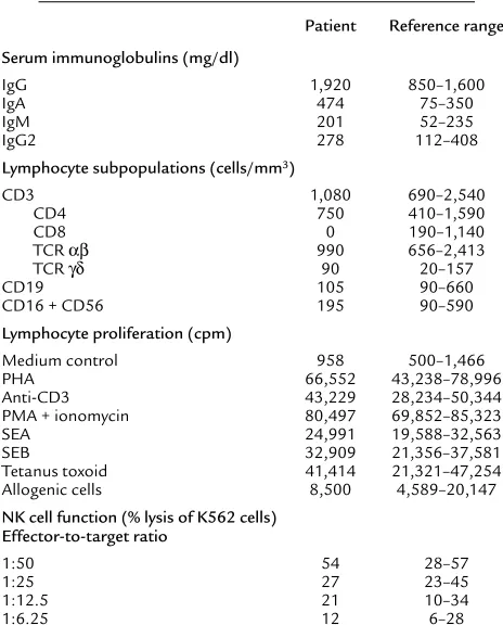
Table 1
Immunological profile of CD8-deficient patient
Patient Reference range
Serum immunoglobulins (mg/dl)
IgG 1,920 850–1,600
IgA 474 75–350
IgM 201 52–235
IgG2 278 112–408
Lymphocyte subpopulations (cells/mm3)
CD3 1,080 690–2,540
CD4 750 410–1,590
CD8 0 190–1,140
TCR αβ 990 656–2,413
TCR γδ 90 20–157
CD19 105 90–660
CD16 + CD56 195 90–590
Lymphocyte proliferation (cpm)
Medium control 958 500–1,466
PHA 66,552 43,238–78,996
Anti-CD3 43,229 28,234–50,344
PMA + ionomycin 80,497 69,852–85,323
SEA 24,991 19,588–32,563
SEB 32,909 21,356–37,581
Tetanus toxoid 41,414 21,321–47,254
Allogenic cells 8,500 4,589–20,147
NK cell function (% lysis of K562 cells) Effector-to-target ratio
1:50 54 28–57
1:25 27 23–45
1:12.5 21 10–34
1:6.25 12 6–28
(Bio-Rad Laboratories Inc.). Incubation was made with a polyclonal antiserum anti–ZAP-70 (Santa Cruz Biotechnology Inc.), mAb anti-CD8β(2ST8-5H7, kind-ly donated by E.L. Reinherz, Dana-Faber Cancer Insti-tute, Boston, Massachusetts, USA; and 5F2 from Coul-ter-Immunotech), and anti–β-actin (Sigma Chemical Co., St. Louis, Missouri, USA). Antigen-Ab complexes were visualized using the enhanced chemiluminescence detection system (ECL; Amersham International, Amer-sham, United Kingdom).
Amplification of CD8α and β mRNA.Total RNA was extracted from peripheral blood lymphocytes by standard methods (Ultraspec RNA; Biotech Labora-tories, St. Louis, Misouri, USA) and then subjected to RT-PCR amplification. CD8α (5′-GTC ATG GCC TTA CCA GTG AC-3′and 5′-GCA CGA AGT GGC TGA AGT AC-3′)and CD8β(α 5′-CAG CTG ACA GTT CTC CAT GG-3′ and 5′-CGG CAC ACT CTC TTC TTG AG-3′) primers were annealed at 55°C, and 25–30 cycles of amplification were performed. Levels of β-actin were used as RT-PCR control. The PCR products were visualized on 1.5% agarose gels.
Sequence analysis.One microgram of total RNA was reverse-transcribed into first-strand cDNA using Superscript II RT (Life Technologies Inc., Pisley, Unit-ed Kingdom). One-tenth of the cDNA was amplifiUnit-ed by PCR with the Expand High-Fidelity PCR system (Boehringer Mannheim Biochemicals Inc., Mannheim, Germany) using the following primers: forward 5′-CGA AAA GGA GGG TGA CTC-3′; reverse 5′-CGC CCC CAC TAA AAT AAT-3′(27). The PCR products were visualized on 1.5% agarose gels and cloned into PCR2.1 TOPO vec-tor (Invitrogen Corp., Carlsbad, California, USA). Sequencing was performed with the ALFexpress Auto-Cycle Sequencing Kit (Amersham Pharmacia Biotech, Piscataway, New Jersey, USA) and an ALFexpress sequencer (Amersham Pharmacia Biotech). For analy-sis of the point mutation in exon 2 (immunoglobulin domain), genomic DNA was prepared from fresh mononuclear blood cells from the patient, siblings, and their parents. The PCR products were amplified with the following primers: forward 5′-GTC ATG GCC TTA CCA GTG AC-3′; reverse 5′-GTT GAG GTG AAC CCC AAG CC-3′(intron 2) (28). The 550-bp product was purified and directly sequenced from both strands with the Applied Biosystems PRISM dye terminator cycle-sequencing kit (Perkin Elmer Applied Biosys-tems, Foster City, California, USA) and an ABI DNA sequencer (Perkin-Elmer).
Mutagenesis and transfection studies. Wild-type and mutated CD8 were subcloned into the XbaI/SacI sites of pCDL-SRα296 vector (29). Chimeric CD8 molecules (CD8 MUT/WT and WT/MUT) were constructed and subcloned into the pCDL-SRα296 vector. CD8 single-point mutants were generated by site-directed mutage-nesis of the wild-type (WT CD8 > CD8ser90or CD8arg90) and mutated CD8 (mutant CD8 > CD8gly90), using the QuikChange Site-Directed Mutagenesis Kit (Strata-gene, La Jolla, California, USA).
Results
Total absence of CD8 and high percentage of CD4–CD8–TCRαβ+
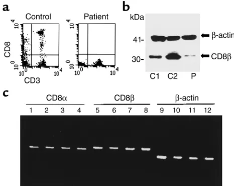
[image:4.576.304.537.433.618.2]T cells. Flow cytometric analysis in a patient with repeat-ed sino-pulmonary infections showrepeat-ed a complete lack of CD8+cells in peripheral blood (Figure 1a and Table 1). Since the parents were consanguineous, the members of his family were analyzed. Two sisters were also found to have a complete absence of CD8 cells. Parents (father: I-1; mother: I-2), CD8-deficient siblings (patient: II-4; sis-ters: II-5, II-7), and other brothers and sisters (II-1, II-2, II-3, II-8, II-9) were studied further. Membrane and intra-cellular (data not shown) expression of CD8αmolecules in T and NK lymphocytes and serum sCD8 concentra-tion were below detectable limits in the three affected siblings. Mean fluorescence intensity of CD8α expres-sion on cell surface and sCD8 were decreased in the par-ents and two brothers (II-2 and II-8), but normal in the remaining siblings. Weak expression of CD8β was detected by Western blot analysis in PBMCs of the patient (Figure 1b) and two CD8-deficient sisters; how-ever, CD8βmembrane expression was absent (data not shown). Messenger RNAs for CD8αand β were detected by RT-PCR at similar levels in the CD8-negative individ-uals, their relatives, and normal controls (Figure 1c). The three CD8-deficient siblings showed a higher percentage of CD4– CD8–T cells than normal donors. This increase was due to the rise in CD4–CD8–TCRαβ+T cells (α/β DN T cells) (II-4 = 16%, II-5 = 10%, and II-7 = 4%; reference range = 0.1–2%) with a wide Vβrepertoire. The percent-age of CD4–CD8–TCRγδ+T cells was normal (II-4 = 6%, II-5 = 4%, and II-7 = 3%; reference range = 1–8%). The
Figure 1
CD8 expression analysis. (a) Expression of CD8 on T lymphocytes and CD3–cells in the CD8-deficient patient and a healthy donor. (b) CD8βexpression studied by Western blot analysis in two controls (C1 is a primary immunodeficient patient with low CD8 T cells and C2 is a healthy adult) and in the CD8-deficient patient. Weak intra-cellular expression of this molecule was detected in the patient;
predominant immunophenotype found in these α/β DN T cells was CD3hi CD5+CD2+CD45RA+CD57+ CD11b+ CD28–and IFN-γ+IL-2–IL-4–(with in vitro stim-ulation) (data not shown), suggestive of effector cytotoxic cells (30) .
To demonstrate the cytotoxic activity of these cells an allogeneic CTL test was performed with the method described, but unfortunately the results were not conclusive, although tests were repeated. No fur-ther samples are available at the present time. NK cytotoxicity was shown to be normal despite the absence of CD8 in these cells.
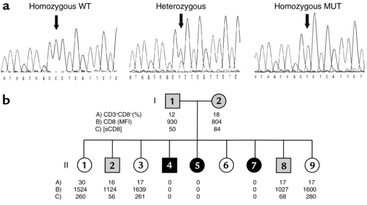
[image:5.576.86.273.56.208.2]Autosomal recessive familial CD8 deficiency. Genetic stud-ies of CD8αmolecule were conducted in view of the above-mentioned results. Primers were chosen to gen-erate a product covering the entire coding region and the 5′and 3′untranslated regions (UTRs) for CD8αby RT-PCR. PCR products were subcloned in plasmid vec-tors and sequenced. Sequences from recombinant clones derived from different PCR products showed a nucleotide substitution in the three siblings, but not in the controls. The G→A mutation found produces a change from glycine to serine residue at position 90 (Figure 2).The glycine at position 90 (gly90) is located in the immunoglobulin domain of the CD8αchain and is conserved in all reported species, and also in the corresponding region of the closely related molecules CD8βand CD7 (8, 31).
Intrafamilial segregation of the mutant allele was analysed to confirm its role in the CD8 deficiency. Genomic analysis of the patient’s and his relatives’ DNA, including the two parents, was performed by direct sequencing of genomic PCR products to establish the inheritance of the mutant allele. The three CD8-deficient siblings were indeed homozygous for the mutated allele (CD8ser90). The parents and two brothers found to have lower CD8 expression were heterozygous carriers, whereas three sisters with normal CD8 levels were homozygous for the wild-type alleles. The levels of CD8 as detected by flow cytometry (membrane-bound CD8) and ELISA (soluble CD8) correlated strictly with the genotype of the family members (Figure 3).
Highly conserved gly90 is responsible for CD8 expression.
[image:5.576.122.481.458.659.2]The presence of the single amino acid substitution that clearly segregates in a classic Mendelian fashion with
Figure 2
Mutation in the messenger RNA-coding region of the CD8αgene. Sequence of the CD8α cDNA reveals a nucleotide substitution (G→A) at position 331 (numbered from the ATG sequence initiat-ing the codinitiat-ing region), leadinitiat-ing to the replacement of a glycine by a serine at position 90.
Figure 3
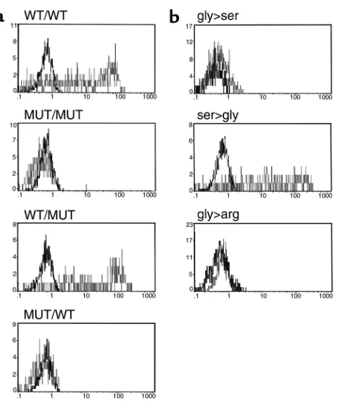
the different levels of CD8 expression in the family does not rule out coexistence with other alterations in the regulatory elements of the CD8α gene. Hence, transfection studies were performed to establish a direct correlation between the mutation found in the CD8 immunoglobulin domain and the absence of any detectable CD8 antigen in the homozygous members of the family. The full coding regions amplified from the patient, a normal (homozygous) sister, and a con-trol were subcloned in a mammalian expression vector. Two different chimeric CD8 molecules were also gen-erated and subcloned in the same vector. The first had the extracellular mutated domain together with wild-type transmembrane and cytoplasmic portions (MUT/WT), which were inverted in the second (WT/MUT). The different constructs were transiently transfected in COS-7 cells and analysed 48 hours later by immunofluorescence. The results demonstrate (Fig-ure 4a) that the presence of serine at position 90 (CD8ser90in the MUT/MUT and the MUT/WT con-struct) precludes CD8 expression.
Further analysis included the generation of single mutants and their transfection into COS-7 cells. The
wild-type CD8 used for the expression experiments was engineered to produce two single mutants (CD8ser90and CD8arg90), whereas the mutant CD8 prepared from the patient reverted to CD8gly90. The presence of serine or another amino acid (arginine) prevents CD8 expression (Figure 4b), thereby sug-gesting that the presence of the evolutionary con-served glycine at this position may be important for appropriate folding of the protein. Moreover, the single reversion of the serine to a glycine in the mutant gene restored full CD8 expression, hence showing that this unique mutation is sufficient to avoid a proper CD8 expression. Cotransfection experiments with CD8β also showed complete absence of CD8αβ heterodimers (data not shown), as already observed in the affected individuals. Taken together, these data demonstrate that substitution of gly90 in the immunoglobulin domain is responsi-ble for the complete absence of CD8 expression observed in the affected members of this family and may be related to the clinical status of the patient.
Discussion
An autosomal recessive familial CD8 deficiency due to a single mutation in the CD8α gene is described. CD8+cells were shown to be absent in a patient with repeated bacterial infections in whom other known immunodeficiencies were ruled out. Familial studies showed the same defect in two sis-ters from the same consanguineous parents.
CD8αchain was found to be completely absent in three members of this family, whereas scarce expres-sion of CD8βchain was detected in their cytoplasm. CD8 glycoproteins are expressed predominantly on MHC class I–restricted T cells as a disulfide-linked
αβ-heterodimer, but can also be expressed as an αα -homodimer in NK cells and intraepithelial T cells (6, 32, 33). In the absence of CD8α, CD8βis retained and destroyed within the cell (7), whereas CD8αcan be expressed at the cell surface without CD8βas CD8αα homodimers. These data, together with normal mRNA expression for CD8αand β, led us to postulate that the defect could more probably involve the CD8αgene. Therefore, genetic analysis was performed, and a mis-sense mutation (gly90→ser) in the CD8α gene was found to be present in both alleles of the CD8-deficient individuals. In all family members, the genotype corre-lated strictly with the CD8 phenotype observed. No CD8 molecules could be detected in the cells or sera of the homozygous individuals with any of the anti-CD8 Ab’s tested, whereas heterozygous subjects showed impaired CD8 expression both on the cell surface of CD8+lymphocytes and in serum.
[image:6.576.65.305.50.338.2]A serine in the mutant allele replaced the glycine residue, which is well preserved in the F strand of the immunoglobulin domain of all known CD8α mole-cules from fish to humans (over 400 million years) (8). This particular glycine is also conserved in the closely related molecules CD8βand CD7 (31). The importance
Figure 4
of this particular mutation in CD8 was also established by our in vitro experiments. Mutant CD8 constructs that lack gly90 cannot be detected on the surface or in cytoplasm of transiently transfected COS-7 cells. The relevance of gly90 was further demonstrated by the transfection of chimeric CD8 molecules and by site-directed mutagenesis-generated molecules. Taken together, these experiments demonstrated the need for gly90 in the immunoglobulin domain of CD8α, since its substitution by another amino acid, arginine, result-ed in an identical lack of detection. Furthermore, these results showed that substitution of the glycine residue was sufficient to avoid CD8 expression, thereby ruling out the possibility that a mutation in noncoding sequences, that is, regulatory sequences, could be responsible for the lack of CD8 found in this family. Although the glycoprotein CD8 plays an important role in the maturation and function of MHC class I–restricted T lymphocytes, as clearly shown by either CD8αor CD8βknockouts (17–19), its presence does not appear to be essential for either CD8 lineage commit-ment or peripheral cytolytic function. It has been report-ed that TCR transgenic thymocytes from CD8α -defi-cient mice were able to restore positive selection of CD8 lineage cells (as shown by CD8β expression) in the absence of CD8, thereby compensating for the lack of CD8 expression by increasing the affinity of TCR for the positively selecting ligand (7, 34, 35). Recent reports addressing the mechanism of CD4/CD8 lineage com-mitment are consistent with a model in which recogni-tion of class I or class II MHC directs thymocytes to the appropriate lineage (instructive model) (36–38). The presence or absence of coreceptor-related signals (CD4, CD8, or p56lck) can further modulate the selection process, but lineage commitment can take place in the absence of the appropriate coreceptor. Polyclonal α/β DN T cells found in the CD8-deficient individuals were most probably committed to being CD8 cytotoxic T cells, because several findings strongly suggest: (a) a high percentage of α/βDN T cells in our patient were CD11b+ CD57+CD45RA+CD28–IFN+IL-2– IL-4–, a phenotype associated with effector CD8+cytotoxic T lymphocytes (30); (b) the absence of CD4 expression in a substantial number of peripheral T cells (α/βDN T cells) in these CD8-deficient individuals suggests a specific CD4 anti-gen downregulation event; and (c) transcripts for CD8α and CD8βwere detected at similar levels in the CD8-neg-ative individuals, as in their heterozygous relCD8-neg-atives or normal controls. These findings indicate that CD8 tran-scription can continue without direct binding of CD8 to MHC I molecules during thymic selection.
The clinical manifestations present in the affected member of this family, as in the murine counterpart (CD8αmice) (20–22), are not severe. This syndrome, as TAP1 and TAP2 deficiencies, is compatible with life, but seems to be less aggressive than the HLA class I deficiencies. Probably, the absence of CD8 is more crit-ical for the development of the cytotoxic T cell reper-toire than for the effector function in the periphery.
We believe that the absence of classic CTL CD8+may be partially compensated for by the cytolytic function of α/βDN T cells and the NK cell activity. However, as the impact of the CD8 absence in CTL function could not be properly addressed in this patient, further stud-ies on CD8 cell function, such as allogeneic cytotoxici-ty or cytotoxicicytotoxici-ty to recall antigens and DN T cell reper-toire, are required before the significance of this CD8 defect can be better interpreted.
The high Ab titers to many viral infections (CMV, herpes zoster, herpes simplex, rubella) in the patient would seem to demonstrate that he has been in con-tact with these viruses and immunocompetent enough to overcome these infections. Although analysis of the recent infections in the proband showed only those of bacterial nature, viral infections suffered at an early age might have been responsible for the alveolar lesions that have later become over-infected and have produced bronchiectases, as reported in TAP-deficient patients (24–25). The diagnosis of pelvic inflammato-ry disease (PID) in adults is more frequently reported (39–41), and it remains to be seen whether the CD8-negative sisters will develop symptoms in the future. Examples of poor correlation between genotype and clinical symptoms in other PID patients are described in the literature (24, 42–44), and factors such as poly-morphisms in other host defense molecules associat-ed with monogenic disorders (45) could also be involved in phenotypic differences in PID with the same genetic defect.
The prevalence of this particular CD8 defect with such a dramatic effect on lymphocyte phenotype should be extremely low, since CD8 expression has been widely determined without any similar case being reported. Nevertheless, mutations in CD8 that affect MHC I binding or signal transduction capabilities without substantial impairment of CD8 expression may be more frequent than expected and should be investigated in patients with repeated infections that resemble Ab deficiencies.
A novel immunological defect is described and a point mutation in the CD8αgene is demonstrated to be responsible for this autosomal recessive familial CD8 deficiency. We believe that many lessons on the role of the CD8 molecule in the maturation and func-tion of MHC class I–restricted T lymphocytes can be learned from the study of this natural human model of immunologic defect.
Acknowledgments
Note added in proof: The accession number for the mutant CD8 alpha allele described here is AY039664.
1. Townsend, A., and Bodmer, H. 1989. Antigen recognition by class I-restricted T lymphocytes. Annu. Rev. Immunol.7:601–624.
2. Zinkernagel, R.M., and Doherty, P.C. 1979. MHC-restricted cytotoxic T cells: studies on the biological role of polymorphic major transplanta-tion antigens determining T cell restrictransplanta-tion-specificity functransplanta-tion and responsiveness. Adv. Immunol.27:51–77.
3. Tanaka, K., Yoshioka, T., Bieberich, C., and Jay, G. 1998. Role of the major histocompatibility complex class I antigens in tumor growth and metastasis. Annu. Rev. Immunol.6:359–380.
4. Mason, D.W., and Morris, P.J. 1986. Effector mechanisms in allograft rejection. Annu. Rev. Immunol.4:119–145.
5. Janeway, C.A. 1992. The T cell receptor as a multicomponent signalling machine: CD4/CD8 coreceptors and CD45 in T cell activation. Annu. Rev. Immunol.10:645–674.
6. Zamoyska, R. 1994. The CD8 coreceptor revisited: one chain good, two chains better. Immunity.1:243–246.
7. Goldrath, A.W., Hogquist, K.A., and Bevan, M.J. 1997. CD8 lineage com-mitment in the absence of CD8. Immunity.6:633–642.
8. Hansen, J.D., and Strassburger, P. 2000. Description of an ectodermic TCR coreceptor, CD8α, in rainbow trout. J. Immunol.164:3132–3139. 9. Gao, G.F., et al. 1997. Crystal structure of the complex between human
CD8ααand HLA-A2. Nature.387:630–634.
10. Salter, R.D., et al. 1990. A binding site for the T-cell receptor CD8 on the alpha 3 domain of HLA-A2. Nature.345:41–46.
11. Sanders, S.K., Fox, R.O., and Kavathas, P. 1991. Mutations in CD8 that affect interactions with HLA class I and monoclonal anti-CD8 antibod-ies. J. Exp. Med.174:371–379.
12. Sun, J., Leahy, D.J., and Kavathas, P. 1995. Interaction between CD8 and major histocompatibility complex (MHC) class I mediated by multiple contact surfaces that include the α2 and α3 domains of MHC class I.
J. Exp. Med. 182:1275–1280.
13. Turner, J.M., et al. 1990. Interaction of the unique N-terminal region of tyrosine kinase p56lck with cytoplasmic domains of CD4 and CD8 is mediated by cystein motifs. Cell. 60:755–765.
14. Chapupny, N.J., Ledbetter, J.A., and Kavathas, P. 1998. Association of CD8 with p56lck is required for early T cell signalling events. EMBO J.
10:1201–1207.
15. Veillette, A., Bookman, M.A., Horak, E.M., and Bolen, J.B. 1988. The CD4 and CD8 T cell surface antigens are associated with the internal mem-brane tyrosine-protein kinase p56lck. Cell. 55:301–308.
16. Zamoyska, R. 1998. CD4 and CD8: modulators of T-cell receptor recog-nition of antigen and of immune responses? Curr. Opin. Immunol.
10:82–87.
17. Fung-Leung, W.P., et al. 1991. CD8 is needed for development of cyto-toxic T cells but not helper T cells. Cell. 65:443–449.
18. Nakayama, K.I., et al. 1994. Requirement for CD8βchain in positive selection of CD8-lineage T cells. Science. 263:1131–1133.
19. Fung-Leung, W.P., et al. 1993. The lack of CD8αcytoplasmic domain resulted in a dramatic decrease in efficiency in thymic maturation but only a moderate reduction in cytotoxic function of CD8+ T lymphocytes.
Eur. J. Immunol. 23:2834–2840.
20. Fung-Leung, W.P., Kündig, T.M., Zinkernagel, R.M., and Mak, T.W. 1991. Immune response against lymphocytic choriomeningitis virus infection in mice without CD8 expression. J. Exp. Med. 174:1425–1429. 21. Huber, M., Timms, E., Mak, T.W., Rollinghoff, M., and Lohoff, M. 1998.
Effective and long-lasting immunity against the parasite Leishmania major in CD8-deficient mice. Infect. Immun. 66:3968–3970.
22. Xing, Z., Wang, J., Croitoru, K., and Wakeham, J. 1998. Protection by CD4 or CD8 T cells against pulmonary Mycobacterium bovis bacillus Calmette-Guerin infection. Infect. Immun. 66:5537–5542.
23. Chan, A.C., et al. 1994. ZAP-70 deficiency in an autosomal recessive form
of severe combined immunodeficiency. Science.264:1599–1601. 24. De la Salle, H., et al. 1994. Homozygous human TAP peptide transporter
mutation in HLA class I deficiency. Science.265:237–241.
25. De la Salle, H., et al. 1999. HLA class I deficiencies due to mutations in subunit 1 of the peptide transporter TAP1. J. Clin. Invest. 103:R9–R13. 26. Wunderlich, J., Shearer, G., and Livingstone, A. 1997. Induction and measurement of cytotoxic T lymphocyte activity. In Current protocols in immunology. Volume 1. J.E. Coligan, D.H. Konisbeck, E.M. Sheach, and W. Strober, editors. John Wiley and Sons, Inc. USA. 3.11.1–3.11.20. 27. Littman, D.R., Thomas, Y., Maddon, P.J., Chess, L., and Axel, R. 1985. The
isolation and sequence of the gene encoding T8: a molecule defining functional classes of T lymphocytes. Cell. 40:237–246.
28. Nakayama, K., Tokito, S., Okumura, K., and Nakauchi, H. 1989. Struc-ture and expression of the gene encoding CD8αchain (Leu-2/T8).
Immunogenetics.30:393–397.
29. Takebe, Y., et al. 1988. Srαpromoter: an efficient and versatile mam-malian cDNA expression system composed of the simian virus 40 early promoter and the R-U5 segment of human T-cell leukemia virus type 1 long terminal repeat. Mol. Cell. Biol. 8:466–472.
30. Hamann, D., Roos, M.Th.L., and Lier, R.A.W. 1999. Faces and phases of human CD8+ T-cell development. Immunol. Today. 20:177–179. 31. Barclay, A.N., Brown, M.H., Law, S.K.A., McKnight, A., and Tomlinson,
M. 1997. The leucocyte antigen factsbook. 2nd edition. Academic Press Inc. London, United Kingdom. 149–151.
32. Moebius, U., Kober, G., Griscelli, A.L., Hercend, T., and Meuer, S.C. 1991. Expression of different CD8 isoforms on distinct human lymphocyte subpopulations. Eur. J. Immunol. 21:1793–1800.
33. Terry, L.A., DiSanto, J.P., Small, T.N., and Flomenberg, N. 1990. Differ-ential expression and regulation of the human CD8αand CD8βchains.
Tissue Antigens. 35:82–91.
34. Sebzda, E., Choi, M., Fung-Leung, W.P., Mak, T.W., and Ohashi, P.S. 1997. Peptide-induced positive selection of TCR transgenic thymocytes in a coreceptor-independent manner. Immunity. 6:643–653.
35. Bachmann, M.F., Oxenius, A., Mak, T.W., and Zinkernagel, R.M. 1995. T cell development in CD8–/– mice: thymic positive selection is biased toward the helper phenotype. J. Immunol. 155:3727–3733.
36. Hernandez-Hoyos, G., Sohn, S.J., Rothenberg, E.V., and Alberola-Ila, J. 2000. Lck activity controls CD4/CD8 T cell lineage commitment. Immu-nity.12:313–322 .
37. Yasutomo, K., Doyle, C., Miele, L., and Germain, R.N. 2000. The dura-tion of antigen receptor signalling determines CD4+ versus CD8+ T-cell lineage fate. Nature.404:506–510.
38. Itano, A., and Robey, E. 2000. Highly efficient selection of CD4 and CD8 lineage thymocytes supports an instructive model of lineage commit-ment. Immunity. 12:383–389.
39. Kornfeld, S.J., et al. 1996. A novel mutation (Cys145 Stop) in Bruton´s tyrosine kinase is associated with newly diagnosed X-linked agamma-globulinemia in a 51-year-old male. Mol. Med. 2:619–623.
40. Shapiro, B.L., Newburger, P.E., Klempner, M.S., Dinauer, M.C. 1991. Chronic granulomatous disease presenting in a 69-year-old male. N. Engl. J. Med. 325:1786–1790.
41. Ozshahin, H., et al. 1997. Adenosin deaminase deficiency in adults. Blood.
89:2849–2855.
42. Arnaiz-Villena, A., et al. 1992. Brief report: primary immunodeficiency caused by mutations in the gene encoding the CD3-γsubunit of the T-lymphocyte receptor. N. Engl. J. Med. 327:529–533.
43. Kornfeld, S.J., et al. 1997. Extreme variation in X-linked agammaglobu-linemia phenotype in a three-generation family. J. Allergy Clin. Immunol.
100:702–706.
44. Sneller, M.C., et al. 1997. Clinical, immunological and genetic features of an autoimmune lymphoproliferative syndrome associated with abnormal lymphocyte apoptosis. Blood. 89:1341–1348.