Copyright © 1998, American Society for Microbiology. All Rights Reserved.
Comparison of Genomic Methods for Differentiating Strains of
Enterococcus faecium: Assessment Using Clinical
Epidemiologic Data
CONNIE SAVOR,
1MICHAEL A. PFALLER,
2JULIE A. KRUSZYNSKI,
3RICHARD J. HOLLIS,
2GARY A. NOSKIN,
1,3,4ANDLANCE R. PETERSON
1,3,4*
Medical Microbiology Division, Department of Pathology, University of Iowa, Iowa City, Iowa,
2and Department of
Medicine, Northwestern University,
1and Department of Pathology, Clinical Microbiology Division,
3and Department of Medicine, Division of Infectious Diseases,
4Northwestern
Memorial Hospital and Northwestern University, Chicago, Illinois
Received 6 April 1998/Returned for modification 6 August 1998/Accepted 18 August 1998
Genomic DNA extracted from 45 vancomycin-resistant Enterococcus faecium (VRE) isolates was cleaved with
HindIII and HaeIII and subjected to agarose gel electrophoresis. The ability of this method (restriction
endo-nuclease analysis [REA]) to distinguish strains at the subspecies level was compared with results previously
de-termined by pulsed-field gel electrophoresis (PFGE). Chart reviews were performed to provide a clinical
corre-lation of possible epidemiologic relatedness. A likely clinical association was found for 29 patients as part of
two outbreaks. REA found 21 of 21 isolates were the same type in the first outbreak, with PFGE calling 19 strains
the same type. In the second outbreak with eight patient isolates, HindIII found six were the same type and two
were unique types. HaeIII found three strains were the same type, two strains were a separate type, and three
more strains were unique types, while PFGE found three were the same type and five were unique types. No
sin-gle “ideal” method can be used without clinical epidemiologic investigation, but any of these techniques is
help-ful in providing focus to infection control practitioners assessing possible outbreaks of nosocomial infection.
Accurate epidemiologic investigation requires an assessment
of relatedness between individuals with similar infections in
order to determine if person-to-person spread has occurred. In
order to accomplish this, one rapid laboratory approach taken
has been to determine the presence or absence of genetic
identity between microbial strains of the same genus and
spe-cies affecting persons who may have had a common exposure.
For this to be useful, it is desirable to rapidly compare different
isolates of an organism in a simple and accurate manner that
can demonstrate the presence or absence of important
epide-miologic associations (clonality).
Enterococci (especially those carrying vancomycin resistance
genes) are now important causes of clinical infections,
includ-ing endocarditis, urinary tract infection, and superinfection in
persons who have received antimicrobial agents (14). Although
enterococci are part of normal human gastrointestinal flora
and can cause infection from this endogenous source, these
organisms can also be spread nosocomially (13, 31). In the
past, epidemiologic evaluation of enterococcal infection has
been somewhat limited by the lack of a simple and sufficiently
discriminatory typing system (2, 11, 13, 16, 31). Recently,
how-ever, pulsed-field gel electrophoresis (PFGE) and restriction
endonuclease analysis (REA) of genomic DNA were shown to
be useful for epidemiologic evaluations of nosocomial
entero-coccal infections (2, 16). Gordillo et al. compared ribotyping
with an rRNA probe derived from Escherichia coli to PFGE for
differentiating strains of Enterococcus faecalis and found that
PFGE was the superior technique, showing 25 clearly different
patterns plus 6 related variants versus 7 ribopattern types (9).
We have used REA of total genomic DNA with success in
epidemiologic study of other organisms (6) and have applied
this technique to type enterococcal isolates (2). The purposes
of this study are (i) to describe our technique, (ii) to report
the cataloging of REA types by using two different restriction
enzymes from the first 45 vancomycin-resistant Enterococcus
faecium isolates at Northwestern Memorial Hospital, and (iii)
to compare the results with those previously obtained by
PFGE. The comparison of each method’s utility for focusing
infection control interventions was assessed in view of the
clinical correlation determined by epidemiologic data obtained
from comprehensive chart review of the patients involved.
(This report was presented in part at the 35th Annual
Meet-ing, Infectious Diseases Society of America, San Francisco,
California, September 1997 [abstract 345].)
MATERIALS AND METHODS
Bacterial isolates.Forty-five vancomycin-resistant E. faecium isolates from
various sites that were obtained from 42 patients hospitalized at Northwestern Memorial Hospital during a 15-month period between July 1992 and October 1993 were recovered from storage at270°C for this study.
REA typing.Genomic DNA from the enterococcal isolates was prepared by a
modification of the method described by Pitcher and colleagues (21). Colonies from 24-h growth on a blood agar plate were suspended in sufficient 10/1 TE buffer (10 mM Tris, 1 mM EDTA [pH 8.0]) to equal that of a no. 2 McFarland standard, centrifuged, resuspended in 0.1 ml of 50-mg/ml lysozyme (Sigma, St. Louis, Mo.) in 10/1 TE buffer, and incubated for 30 min at 37°C. The DNA was harvested by the guanidine thiocyanate-EDTA-Sarkosyl (GES) method. RNase T1(Gibco BRL, Gaithersburg, Md.) was added to the suspensions. Quantitation
of the DNA was made with a Lambda-Bio spectrophotometer and corrected for dilution. Samples were stored at 4°C.
For restriction endonuclease digestion, genomic DNA (10 to 20ml) was incu-bated with restriction endonuclease and digested according to the manufactur-er’s instructions (Gibco BRL). All strains were restricted with two enzymes, one used in each of two separate assessments of bacterial relatedness. HindIII was used in one assessment series, and HaeIII was used in the other series. The restricted DNA fragments were separated by agarose gel electrophoresis with 0.6% agarose (Sigma) in TBE buffer (1 M Tris, 0.9 M boric acid, 0.01 M EDTA) at 44 V for 16.5 h. Gels were stained for 2 h in SYBR Green I (Molecular Probes, Eugene, Oreg.) and photographed under UV illumination.
The DNA band patterns for each new isolate digested with a common
restric-* Corresponding author. Mailing address: NMH Infection Control
and Prevention Project, Galter Carriage House, no. 913, Northwestern
Memorial Hospital, 215 East Chicago Ave., Chicago, IL 60611. Phone:
(312) 926-2885. Fax: (312) 926-0051. E-mail: [email protected].
3327
on May 15, 2020 by guest
http://jcm.asm.org/
tion enzyme were systematically compared according to the method described first by Clabots and colleagues (6). The first isolate in this analysis with a new DNA band pattern was arbitrarily designated a reference REA type. Gels were run so that the molecular weight ladder covered the top 6 cm (60 mm) of the electrophoresis gel from the origin. This was then the portion of the gel used for analysis. Similarities between the new and reference REA types were scored by visual comparison of each 1-mm segment of the top 60 mm of the DNA band patterns run on the same gel. The presence or absence of a DNA band within each segment was assessed. The actual intensity of the band is not part of the similarity scoring system. A similarity index was calculated from the number of identical 1-mm segments expressed as a percentage of the total number of 1-mm segments measured. A pattern with greater than six differences in the 1-mm segments had a similarity index of less than 90% and was designated a new REA type that was used for all future comparisons. For any epidemiologic investiga-tion involving more than 10 isolates of apparently similar types, it is routine to repeat the REA analysis of purified DNA on the same gel to improve pattern matching. Any REA pattern with a similarity index of greater than 90% was included within a type. The types were designated by letters, and a distinct REA pattern within a type (similarity index of.90% but,100%) was designated by a subscript Arabic number indicating a subtype (A0, A1, A2, etc.). For this
analysis, all strains within a given type were considered as being possibly related by the typing method.
PFGE typing.PFGE was performed with the same 45 enterococcal isolates
described above at the University of Iowa by the method of Pfaller et al. (20). Restriction digestion of chromosomal DNA was performed with SmaI (New England Biolabs, Inc.). The resultant restriction fragments were resolved in a 1% agarose gel with a CHEF-DRII system (Bio-Rad Laboratories, Richmond, Cal-if.). The pulse time ramped from 5 to 30 s over 23 h at 13°C and 6 V/cm. PFGE patterns were considered identical if they shared every band, similar (subtype) if they differed from one another by one to three clearly visible bands, and distinct if they differed by over three bands.
Chart reviews.Detailed review of each of the 42 patients’ charts was
com-pleted for the duration of the hospitalization during which they had a culture positive for vancomycin-resistant enterococci (VRE). Data were collected about date of admission and discharge, in-hospital transfers, dates of VRE-positive cultures and body site(s), patient location (nursing unit) within the medical center, any diagnostic testing procedures (location and date), and date(s) seen by various consulting services. Any potentially significant clinical findings such as diarrhea and urinary incontinence were also recorded. Simultaneous location on the same ward, same-day visits by consulting services, same-day common proce-dures, or presence in the same room within 3 days of another patient with VRE constituted potential relatedness based on clinical assessment. If none of these association criteria were fulfilled, then the patient was not considered epidemi-ologically related to any other patient. For this report, the grouping into two distinct clusters making up separate potential outbreaks and one group of uniquely unrelated patients was fully based on the epidemiology from the chart review data.
RESULTS
Of these 45 vancomycin-resistant E. faecium isolates, 17
were obtained from rectal swabs as part of ongoing
surveil-lance; 12 were from urine; 6 were from blood; 2 each were
obtained from abscesses, catheter tips, and decubitus ulcers;
and 1 each was obtained from a surgical wound, a T-tube
drainage, hand surveillance, and a rectal biopsy.
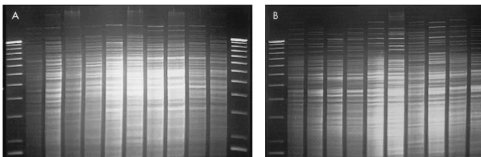
REA with HindIII provided 20 distinct patterns (subtypes)
that were categorized into 9 unique types. Isolates cleaved with
HindIII yielded between 25 and 35 bands per strain in the 60
[image:2.612.57.540.70.228.2]mm of the DNA profiles analyzed. REA typing with HaeIII
provided 21 subtypes that were categorized into 19 types.
Iso-lates cleaved with HaeIII yielded a similar number of bands per
strain in the top 60 mm of the DNA profiles. When these
iso-lates were previously subjected to PFGE, they were found to
have 27 distinct subtypes belonging to 21 types. PFGE gave
approximately half the number of bands for analysis per strain
(typically 12 to 15 bands). Representative isolates are shown
that were analyzed by the REA technique with HindIII (Fig.
1A) and HaeIII (Fig. 1B) and by the PFGE technique (Fig. 2).
A likely clinical association was found for 29 patients as part
of two distinct outbreaks. REA with HindIII and HaeIII found
21 of 21 isolates were the same type in the first outbreak, with
FIG. 1. Representative isolates of each type analyzed by the REA technique (HindIII [A] and HaeIII [B]). The lanes, from left to right, represent a 1-kb DNA molecular weight ladder; strains EF18 (HindIII type B2, HaeIII type B2), EF20 (HindIII type C1, HaeIII type D0), EF23 (HindIII type B5, HaeIII type E0), EF27
(HindIII type B6, HaeIII type G0), EF32 (HindIII type B3), EF33 (HindIII type E0, HaeIII type H0), EF36 (HindIII type B4, HaeIII type B4), EF39 (HindIII type D0, HaeIII type J0), EF3 (HindIII type B0, HaeIII type B1), and EF45 (HindIII type C1, HaeIII type D0); and another 1-kb DNA molecular weight ladder standard.
FIG. 2. Representative isolates of each type analyzed by PFGE. The lanes, from left to right, represent a 48.5-kb lambda DNA molecular weight ladder; a
Staphylococcus aureus control digested with SmaI; strains EF18 (type B5), EF20
(type D), EF23 (type E), EF27 (type G), EF32 (type I), EF33 (type J), EF36 (type M), EF39 (type O), EF3 (type B1), and EF45 (type U); another lambda
molecular weight ladder; and another S. aureus control.
on May 15, 2020 by guest
http://jcm.asm.org/
[image:2.612.310.547.507.674.2]PFGE identifying 19 strains as the same type and 2 isolates as
unique types (Table 1). In the second outbreak, represented by
eight patient isolates, HindIII found six were the same type and
two were unique types. Here, HaeIII found three were the same
type, three strains were a separate type, and two more strains
were unique types. PFGE found three strains were the same
type and five strains were unique types (Table 2). In the
seven discrepant isolates from the two outbreaks, HindIII
found four types, HaeIII found five types, and PFGE found
seven types. Of these seven strains, two appeared clonal by
both REA enzymes and clinical association but were not
re-lated by PFGE.
The clinically unrelated patients (Table 3) presented the
most diverse genomic groupings. Here, the various methods
found from 8 to 14 unique types. Also, there were only two
pa-tients who were designated a B type by all three methods,
rep-resenting a suggestion of clonality in only two (12.5%) of the
strains.
DISCUSSION
Numerous typing methods have been used by investigators
to augment the epidemiologic evaluation of nosocomial
infec-tions. Typing methods for enterococci that have been
exam-ined include ribotyping (9), biotyping (7, 10, 27), bacteriocin
typing (11, 12, 23), phage typing (4, 5, 11–12, 22, 27), and
sero-typing (25–27). Antimicrobial agent susceptibility testing and
determination of plasmid content with or without plasmid
digestion patterns have also been used (13, 18, 29–31). None
of these methods, however, have proven optimal for typing
enterococci. Bacteriophage typing requires access to special
reagents and performance of a large number of tests (11).
Several investigators have experienced inconsistent plasmid
patterns and irreproducible results when using total plasmid
content for typing enterococci (9, 16). Recently, PFGE has
been shown to be useful for epidemiologic evaluations of
nos-ocomial enterococcal infections (9, 16).
PFGE is used by many different investigators and has shown
a great deal of diversity among patterns of epidemiologically
unrelated strains (15–17, 19). PFGE has an advantage over
traditional agarose gel electrophoresis in that it is possible to
separate even very large DNA molecules with as many as 10
7 [image:3.612.50.290.89.338.2]nucleotide pairs (1). Ordinary gel electrophoresis fails to
sep-arate these molecules because the pores in the gel are too
small for the large fragments. The constant electric field can
also stretch them into elongated configurations that travel
lin-early at a rate relatively independent of size. However,
fre-quent alterations in the direction of the electric field force the
molecules to reorient in order to move, allowing separation of
the large fragments with good resolution. Therefore,
restric-tion endonucleases that have few recognirestric-tion sites can be used
to cleave the DNA, producing fewer fragments that generate
more readily visible and easily comparable patterns. The
pri-mary disadvantage of PFGE is the relatively lengthy and
cum-bersome specimen preparation required before running the
gel. The equipment required is modest in cost.
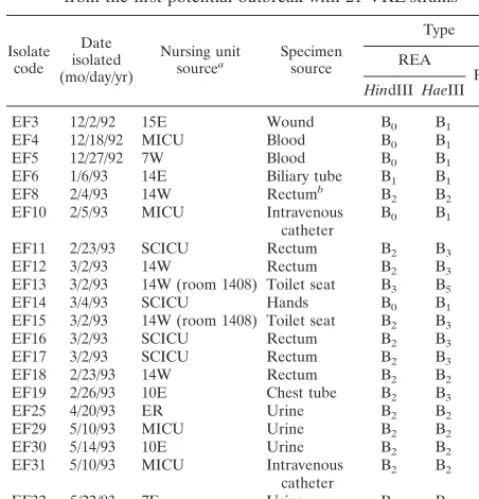
TABLE 1. Description of isolate sources and genomic typing results
from the first potential outbreak with 21 VRE strains
Isolate code
Date isolated (mo/day/yr)
Nursing unit
sourcea Specimensource
Type REA
PFGE
HindIII HaeIII
EF3 12/2/92 15E Wound B0 B1 B1
EF4 12/18/92 MICU Blood B0 B1 B1
EF5 12/27/92 7W Blood B0 B1 B1
EF6 1/6/93 14E Biliary tube B1 B1 B1
EF8 2/4/93 14W Rectumb B
2 B2 B1
EF10 2/5/93 MICU Intravenous
catheter B0 B1 B1 EF11 2/23/93 SCICU Rectum B2 B3 B3
EF12 3/2/93 14W Rectum B2 B3 B4
EF13 3/2/93 14W (room 1408) Toilet seat B3 B5 B5
EF14 3/4/93 SCICU Hands B0 B1 B1
EF15 3/2/93 14W (room 1408) Toilet seat B2 B3 B5
EF16 3/2/93 SCICU Rectum B2 B3 B1
EF17 3/2/93 SCICU Rectum B2 B3 B1
EF18 2/23/93 14W Rectum B2 B2 B5
EF19 2/26/93 10E Chest tube B2 B3 B1
EF25 4/20/93 ER Urine B2 B2 B1
EF29 5/10/93 MICU Urine B2 B2 B1
EF30 5/14/93 10E Urine B2 B2 B1
EF31 5/10/93 MICU Intravenous
catheter B2 B2 B1 EF32 5/22/93 7E Urine B3 B3 I
EF36 6/2/93 8E Rectal biopsy B4 B4 M aE and W, East and West Wings, respectively; MICU, Medical Intensive Care
Unit; SCICU, Spinal Cord Intensive Care Unit; ER, Emergency Room.
[image:3.612.307.547.89.276.2]bSurveillance isolates.
TABLE 2. Description of isolate sources and genomic typing results
from the second potential outbreak with eight VRE strains
Isolate
code Date isolated(mo/day/yr)
Nursing unit sourcea
Specimen source
Type REA
PFGE
HindIII HaeIII
EF20
3/21/93
8E
Urine
C1
D0
D
EF21
3/15/93
8W
Urine
B2
B2
B6
EF22
4/7/93
SICU
Rectum
bB2
B2
B7
EF23
4/7/93
SICU
Rectum
B5
E0
E
EF24
4/10/93
8E
Urine
D0
F0
F
EF26
4/22/93
SICU
Blood
B2
B2
B1
EF27
4/21/93
14E
Rectum
B6
G0
G
EF28
4/21/93
14W
Rectum
B7
G1
H
aFor definitions of abbreviations, see Table 1. bSurveillance isolates.
TABLE 3. Description of isolate sources and genomic typing
results with 16 clinically unrelated VRE strains
Isolate code
Date isolated (mo/day/yr)
Nursing unit
sourcea Specimensource
Type REA
PFGE
HindIII HaeIII
EF1 7/1/92 14E Blood A0 A0 A
EF2 9/3/92 14W Blood B0 B0 B1
EF7 1/27/93 Home Health Urine B2 B2 B2
EF9 2/1/93 6W Urine C0 C0 C
EF33 5/18/93 14W Urine E0 H0 J
EF34 5/18/93 14W Rectumb E
0 H1 K
EF35 5/18/93 14E Rectum B2 B2 L
EF37 6/8/93 14E Rectum E1 G0 N
EF38 6/24/93 14E Blood F0 I0 B1
EF39 7/22/93 11E Rectum D0 J0 O
EF40 7/21/93 SCICU Rectum A1 K0 P
EF41 8/10/93 11E Urine D1 L0 Q
EF42 7/27/93 10W Urine F0 I0 R
EF43 8/23/93 9W Skin ulcer G0 M0 S
EF44 9/27/93 12W Foot ulcer H0 N0 T
EF45 10/7/93 10W Urine C1 D0 U aFor definitions of abbreviations, see Table 1.
bSurveillance isolates.
on May 15, 2020 by guest
http://jcm.asm.org/
[image:3.612.50.290.587.709.2]Genomic REA analyzes the entire DNA content of a
mi-crobe by cleaving the chromosomal DNA and any plasmid
DNA into fragments small enough to be separated by
elec-trophoresis on an agarose gel, producing a greater number
of bands than PFGE. Although this method is very specific,
one disadvantage is that DNA extracted from different
iso-lates needs to be run on the same gel to facilitate pattern
comparison because of the large number of bands requiring
comparison, and this becomes difficult if an extraordinarily
large collection of isolates must be tested. The presence of 30
to 50 bands typically found with REA makes reading of these
gels difficult to automate, since no available image analysis
system can adequately assess this large number of bands
(au-thor’s unpublished observation). The principal advantages of
REA are the ease and rapidity of specimen preparation and
the minimal amount of equipment required. This technique is
also reported to be among the most specific methods of
epi-demiologic fingerprinting available (1, 28).
One limitation of these genomic digestion techniques is that
the degree of relatedness between strains cannot be calculated
by the absolute number of bands in common or different. One
may not know how to interpret isolates that differ by only a few
fragments. Such differences could arise within a single
individ-ual from inversions, deletions, or other rearrangements of the
chromosome or from the acquisition or loss of a prophage,
transposon, insertion sequence, or plasmid. On the other hand,
such differences could indicate that isolates are more distantly
related (16). In the converse, it also has been illustrated that
chromosomal patterns the same as those in tested bacteria can
be found in epidemiologically unrelated individuals (8, 24).
In this study, we have analyzed the chromosomal digestion
patterns of 45 isolates of VRE cleaved with HindIII and HaeIII
and compared these results to those obtained previously by
PFGE. On initial assessment, a somewhat surprising
diver-sity appears to exist among the three methods. The two REA
studies were discordant in detecting clonality, with HaeIII
pro-ducing 19 unique clonal types versus 9 produced with HindIII.
The same observation was seen when comparing PFGE results.
Interestingly, by chart review, the methods were much more
concordant in providing an overall epidemiologic
interpreta-tion. None of the enzymes produced completely concordant
clinical correlation. For example, EF23 was identified as a new
type by HaeIII and PFGE, but clinically may have
represent-ed nosocomial transmission, because the patients with strains
EF22 and EF26 were in rooms adjacent to this patient during
the same time period. Conversely, there is no clinical evidence
that EF40 and EF1 or EF41 and EF24 should be related, as
suggested by HindIII patterns, but not by HaeIII or PFGE.
There were also cases in which PFGE categorized two isolates
into different types that clinically and by REA (with both
en-zymes) were the same. For example, EF27 and EF28 were
isolated from patients on the same ward on the same day who
also had common managing and consulting services and who
had even had a Portacath placement within a day of each
other. Another such example occurred with strains EF33 and
EF34. They were isolated from the same person on the same
day, and although from two different sources, they most likely
represent the same organism. HindIII found these to be
iden-tical, HaeIII classified them as the same type but different
subtypes, and PFGE determined them to be different types.
Overall, many isolates that were identified as clonal by PFGE
and REA had strong clinical data supporting this finding.
Ap-parent discrepancies could be due to errors in visual
interpre-tation of patterns by the investigators and/or poor resolution of
some of the bands, or they could be due to actual differences in
DNA patterns that are recognized differently by the restriction
enzymes used.
Taking a broader view of our two potential outbreak groups
and the group of clinically unrelated patients provides an
in-teresting observation. In the 21-patient cluster (Table 1), each
method found only one to three types and suggested an
epi-demiological association in 90 to 100% of cases, indicating a
careful infection control investigation would be worthwhile. In
potential outbreak 2 (Table 2), the methods identified from
three to six types (from a total of eight specimens) and
sug-gested that the largest single clonal group included an
associ-ation ranging from 38 to 75% of cases. Here, an infection
control investigation appears moderately indicated as useful
from the typing data. The unrelated patient group was also the
most diverse based on all three typing methods. From these 16
specimens, the methods found from 8 to 14 types, with the
largest genomic clone (type B) representing only 19% (3 of 16)
of the strains by any single method. This result would suggest
little likelihood of the ongoing spread of a single, clonal VRE
strain between these patients. Therefore, for a clinical
appli-cation, the three typing approaches were quite concordant in
indicating a high, moderate, or low probability of nosocomial
spread of clonal VRE from interpretations based on the
ge-nomic typing data alone. Supporting our conclusion is the
re-cent report by Bonten and colleagues, who found little genetic
variation of VRE within individual patients and that when used
as an epidemiologic tool, genetic typing found most strains
were either very similar or very different, readily separating
re-lated from unrere-lated isolates (3). They too concluded that
typ-ing can be a very powerful tool to evaluate VRE epidemiology.
We believe the data presented show that a genomic typing
approach for gathering clonality assessment information can
be very useful in focusing the efforts of infection control
prac-titioners when deciding which episodes of nosocomial infection
likely represent patient-to-patient spread of a pathogenic
mi-crobe. Our results indicate that there is no single “ideal”
meth-od that can stand alone without clinical epidemiologic
in-vestigation, but all of these techniques are very helpful when
reproducibly performed and carefully applied in a timely
man-ner to assess possible outbreaks of nosocomial infection.
ACKNOWLEDGMENTS
This investigation was supported by Northwestern Memorial
Hos-pital and Northwestern University Medical School, Chicago, Ill.
REFERENCES
1. Alberts, B., D. Bray, J. Lewis, M. Raff, K. Roberts, and J. Watson. 1994. Molecular biology of the cell, 3rd ed. Garland Publishing, Inc., New York, N.Y.
2. Bodnar, U. R., G. A. Noskin, T. Suriano, I. Cooper, B. E. Reisberg, and L. R.
Peterson.1996. Use of in-house molecular epidemiology and full species
identification for controlling spread of vancomycin-resistant Enterococcus
faecalis isolates. J. Clin. Microbiol. 34:2129–2132.
3. Bonten, M. J. M., M. K. Hayden, C. Nathan, T. W. Rice, and R. A. Weinstein. 1998. Stability of vancomycin-resistant enterococcal genotypes isolated from long-term-colonized patients. J. Infect. Dis. 177:378–382.
4. Brandis, H., P. Plecas, and L. Andries. 1985. Typing of Streptococcus faecalis and Streptococcus faecium strains by means of bacteriophages. Zentralbl. Bakteriol. Parasitenkd. Infektionskr. Hyg. Abt. 1 Orig. Reihe A 260:206–215. 5. Caprioli, T., F. Zaccour, and S. S. Kasatiya. 1975. Phage typing scheme for group D streptococci isolated from human urogenital tract. J. Clin. Micro-biol. 2:311–317.
6. Clabots, C. R., S. Johnson, K. M. Bettin, P. A. Mathie, M. E. Mulligan, D. R.
Schaberg, L. R. Peterson, and D. N. Gerding.1993. Development of a rapid
and efficient restriction endonuclease analysis typing system for Clostridium
difficile and correlation with other typing systems. J. Clin. Microbiol. 31:
1870–1875.
7. Coudron, P. E., C. G. Mayhall, R. R. Facklam, A. C. Spadora, V. A. Lamb,
M. R. Lybrand, and H. P. Dalton.1984. Streptococcus faecium outbreak in a
neonatal intensive care unit. J. Clin. Microbiol. 20:1044–1048.
on May 15, 2020 by guest
http://jcm.asm.org/
8. Denning, D. W., C. J. Baker, N. J. Troup, and L. S. Tompkins. 1989. Restriction endonuclease analysis of human and bovine group B streptococci for epidemiologic study. J. Clin. Microbiol. 27:1352–1356.
9. Gordillo, M. E., K. V. Singh, and B. E. Murray. 1993. Comparison of ribotyping and pulsed-field gel electrophoresis for subspecies differentiation of strains of Enterococcus faecalis. J. Clin. Microbiol. 31:1570–1574. 10. Hussain, Z., M. Kuhn, R. Lannigan, and T. W. Austin. 1988. Microbiological
investigation of an outbreak of bacteremia due to Streptococcus faecalis in an intensive care unit. J. Hosp. Infect. 12:263–271.
11. Kuhnen, E., F. Richter, K. Richter, and L. Andries. 1988. Establishment of a typing system for group D streptococci. Zentralbl. Backteriol. Parasitenkd. Infektionskr. Hyg. Abt. 1 Orig. Reihe A 267:322–330.
12. Kuhnen, E., K. Rommelsheim, and L. Andries. 1987. Combined use of phage typing, enterococcinotyping and species differentiation of group D strepto-cocci as an effective epidemiological tool. Zentralbl. Bakteriol. Parasitenkd. Infektionskr. Hyg. Abt. 1 Orig. Reihe A 266:586–595.
13. Luginbuhl, L. M., A. Rotbart, R. R. Facklam, M. H. Roe, and J. A. Elliot. 1987. Neonatal enterococcal sepsis: case-control study and description of an outbreak. Pediatr. Infect. Dis. J. 6:1022–1030.
14. Murray, B. E. 1990. The life and times of the enterococcus. Clin. Microbiol. Rev. 3:46–65.
15. Murray, B. E., H. A. Lopardo, E. A. Rubeglio, M. Frosolono, and K. V. Singh. 1992. Intrahospital spread of a single gentamicin-resistant, b-lactamase-pro-ducing strain of Enterococcus faecalis in Argentina. Antimicrob. Agents Chemother. 36:230–232.
16. Murray, B. E., K. V. Singh, J. D. Heath, B. R. Sharma, and G. M. Weinstock. 1990. Comparison of genomic DNAs of different enterococcal isolates using restriction endonucleases with infrequent recognition sites. J. Clin. Micro-biol. 28:2059–2063.
17. Murray, B. E., K. V. Singh, S. M. Markowitz, H. A. Lopardo, J. E. Patterson,
M. J. Zervos, E. Rubeglio, G. M. Eliopoulos, L. B. Rice, F. W. Goldstein, S. G. Jenkins, G. M. Caputo, R. Nasnass, L. S. Moore, E. S. Wong, and G.
Weinstock.1991. Evidence for clonal spread of a single strain of
b-lacta-mase-producing Enterococcus faecalis to six hospitals in five states. J. Infect. Dis. 163:780–785.
18. Patterson, J. E., B. L. Masecar, C. A. Kauffman, D. R. Schaberg, W. J.
Hierholzer, Jr., and M. J. Zervos.1988. Gentamicin resistance plasmids of
enterococci from diverse geographic areas are heterogeneous. J. Infect. Dis.
158:212–216.
19. Patterson, J. E., A. Wanger, K. K. Zscheck, M. J. Zervos, and B. E. Murray.
1990. Molecular epidemiology ofb-lactamase-producing enterococci. Anti-microb. Agents Chemother. 34:302–305.
20. Pfaller, M. A., R. J. Hollis, and H. J. Sader. 1994. Chromosomal restriction fragment analysis by pulsed-field gel electrophoresis, p. 10.5c.1–10.5.c.12. In H. D. Isenberg (ed.), Clinical microbiology procedures handbook, vol. 2, suppl. 1. American Society for Microbiology, Washington, D.C.
21. Pitcher, D. G., N. A. Saunders, and R. J. Owen. 1989. Rapid extraction of bacterial genomic DNA with guanidium thiocyanate. Lett. Appl. Microbiol.
8:151–156.
22. Pleceas, P. 1979. A phage typing system for Streptococcus faecalis and
Strep-tococcus faecium, p. 264–265. In M. T. Parker (ed.), Pathogenic
streptococ-ci—Proceedings of the VIIth International Symposium on Streptococci and Streptococcal Disease. Reedbooks, Chertsey, England.
23. Pleceas, P., C. Bogdan, and A. Vereanu. 1972. Enterocine-typing of group D streptococci. Zentralbl. Bakteriol. Parasitenkd. Infektionskr. Hyg. Abt. 1 Orig. Reihe A 221:173–181.
24. Prado, D., B. E. Murray, T. G. Cleary, and L. K. Pickering. 1987. Limitations of using the plasmid pattern as an epidemiological tool for clinical isolates of
Shigella sonnei. J. Infect. Dis. 155:314–316.
25. Sharpe, M. E. 1964. Serological types of Streptococcus faecalis and its vari-eties and their cell wall type antigen. J. Gen. Microbiol. 36:151–160. 26. Sharpe, M. E., and P. M. F. Shattock. 1952. The serological typing of group
D streptococci associated with outbreaks of neonatal diarrhea. J. Gen. Mi-crobiol. 6:150–165.
27. Smyth, C. J., H. Matthews, M. K. Halpenny, H. Brandis, and G. Colman. 1987. Biotyping, serotyping and phage typing of Streptococcus faecalis iso-lated from dental plaque in the human mouth. J. Med. Microbiol. 23:45–54. 28. Tompkins, L. S. 1992. The use of molecular methods in infectious diseases.
N. Engl. J. Med. 327:1290–1297.
29. Uttley, A. H. C., R. C. George, J. Naidoo, N. Woodford, A. P. Johnson, C. H.
Collins, D. Morrison, A. J. Gilfillan, L. E. Fitch, and J. Heptonstall.1989.
High-level vancomycin-resistant enterococci causing hospital infections. Epi-demiol. Infect. 103:173–181.
30. Woodford, N., E. McNamara, E. Smyth, and R. C. George. 1992. High-level resistance to gentamicin in Enterococcus faecium. J. Antimicrob. Chemother.
29:395–403.
31. Zervos, M. J., C. A. Kauffman, P. M. Therasse, A. G. Bergman, T. S.
Mikesell, and D. R. Schaberg.1987. Nosocomial infection by
gentamicin-resistant Streptococcus faecalis: an epidemiologic study. Ann. Intern. Med.
106:687–691.