
Heterozygous deficiency of hypoxia-inducible
factor–2
aa
protects mice against pulmonary
hypertension and right ventricular dysfunction
during prolonged hypoxia
Koen Brusselmans, … , Désiré Collen, Peter Carmeliet
J Clin Invest.
2003;
111(10)
:1519-1527.
https://doi.org/10.1172/JCI15496
.
Chronic hypoxia induces pulmonary vascular remodeling, leading to pulmonary
hypertension, right ventricular hypertrophy, and heart failure. Heterozygous deficiency of
hypoxia-inducible factor–1a (HIF-1a), which mediates the cellular response to hypoxia by
increasing expression of genes involved in erythropoiesis and angiogenesis, has been
previously shown to delay hypoxia-induced pulmonary hypertension. HIF-2a is a
homologue of HIF-1a and is abundantly expressed in the lung, but its role in pulmonary
hypertension remains unknown. Therefore, we analyzed the pulmonary response of WT
and viable heterozygous HIF-2a–deficient (
Hif2
a
+/–) mice after exposure to 10% O
2
for 4
weeks. In contrast to WT mice,
Hif2
a
+/–mice were fully protected against pulmonary
hypertension and right ventricular hypertrophy, unveiling a critical role of HIF-2a in
hypoxia-induced pulmonary vascular remodeling. Pulmonary expression levels of endothelin-1 and
plasma catecholamine levels were increased threefold and 12-fold respectively in WT but
not in
Hif2
a
+/–mice after hypoxia, suggesting that HIF-2a–mediated upregulation of these
vasoconstrictors contributes to the development of hypoxic pulmonary vascular remodeling.
Article
Vascular biology
Find the latest version:
Introduction
Pulmonary hypertension is a formidable health prob-lem, as it often leads to right ventricular (RV) hyper-trophy and heart failure (1, 2). Current treatment includes the administration of oxygen, bronchodila-tors, vasodilators (e.g., prostacyclin, NO, and endothe-lin-1 antagonists), and, eventually, mechanical ventila-tion (2–4). However, since oxygenaventila-tion and vasodilatation merely delay the progression of this dis-ease, a better understanding of its pathogenesis is required (2). The pathophysiology of hypoxic pul-monary hypertension is complex and poorly under-stood. It is characterized by increased levels of the vaso-constrictors angiotensin II and endothelin-1 (ET-1),
impaired production of the vasodilators NO and prostacyclin, and an unbalanced production of factors affecting growth, migration, and differentiation of VSMCs, including FGF-2, PDGF-B, TGF-β, IGF-I and -II, and EGF (5, 6). As a result, peripheral, normally nonmuscular arteries become muscularized, and the media and adventitia enlarge (5–7). Loss of eNOS (8) or prostacyclin receptor (9) aggravates hypoxic pulmonary vascular disease, whereas ET-1 receptor blockade (4), overexpression of prostacyclin (10), and gene transfer of iNOS (11) reduce hypoxia-induced pulmonary hypertension. In addition, deficiency of the serotonin transporter, a pulmonary VSMC mitogen that is upreg-ulated during hypoxia, also attenuates hypoxic pul-monary hypertension (12). Furthermore, serine elas-tase, plasminogen, and MMPs have been implicated in growth and migration of VSMCs via degradation of the ECM and release of mitogens or differentiation factors (13). Proteinases are involved in the pathology of pul-monary hypertension, since mice deficient in plas-minogen or urokinase-type plasplas-minogen activator are partially protected against pulmonary vascular remod-eling (14). Very recently, gene transfer of VEGF was also shown to reduce pulmonary hypertension in rats (15). Hypoxia-inducible factor–1α(HIF-1α) is a key regu-lator in the cellular adaptation to hypoxia (16). During hypoxia, HIF-1αupregulates the expression of a num-ber of genes involved in erythropoiesis, glycolysis, and angiogenesis by binding, as a heterodimer with HIF-1β,
Heterozygous deficiency of hypoxia-inducible factor–2
α
protects mice against pulmonary hypertension and right
ventricular dysfunction during prolonged hypoxia
Koen Brusselmans,
1Veerle Compernolle,
1Marc Tjwa,
1Michael S. Wiesener,
2Patrick H. Maxwell,
3Désiré Collen,
1and Peter Carmeliet
11Center for Transgene Technology and Gene Therapy, Flanders Interuniversity Institute for Biotechnology,
Katholieke Universiteit Leuven, Leuven, Belgium
2Department of Nephrology and Medical Intensive Care, Klinikum Charité, Campus Virchow, Humboldt University,
Berlin, Germany
3Renal Section, Imperial College of Science, Technology and Medicine, Hammersmith Campus, London, United Kingdom
Chronic hypoxia induces pulmonary vascular remodeling, leading to pulmonary hypertension, right ventricular hypertrophy, and heart failure. Heterozygous deficiency of hypoxia-inducible factor–1α
(HIF-1α), which mediates the cellular response to hypoxia by increasing expression of genes involved in erythropoiesis and angiogenesis, has been previously shown to delay hypoxia-induced pulmonary hypertension. HIF-2αis a homologue of HIF-1αand is abundantly expressed in the lung, but its role in pulmonary hypertension remains unknown. Therefore, we analyzed the pulmonary response of WT and viable heterozygous HIF-2α–deficient (Hif2α+/–) mice after exposure to 10% O2for 4 weeks.
In contrast to WT mice, Hif2α+/–mice were fully protected against pulmonary hypertension and right
ventricular hypertrophy, unveiling a critical role of HIF-2αin hypoxia-induced pulmonary vascular remodeling. Pulmonary expression levels of endothelin-1 and plasma catecholamine levels were increased threefold and 12-fold respectively in WT but not in Hif2α+/–mice after hypoxia, suggesting
that HIF-2α–mediated upregulation of these vasoconstrictors contributes to the development of hypoxic pulmonary vascular remodeling.
J. Clin. Invest.111:1519–1527 (2003). doi:10.1172/JCI200315496.
Received for publication March 21, 2002, and accepted in revised form March 11, 2003.
Address correspondence to:Peter Carmeliet, Center for Transgene Technology and Gene Therapy, Flanders Interuniversity Institute of Biotechnology, Katholieke Universiteit Leuven, Gasthuisberg, Herestraat 49, B-3000 Leuven, Belgium.
Phone: 32-16-34-57-72; Fax: 32-16-34-59-90; E-mail: [email protected].
Conflict of interest:The authors have declared that no conflict of interest exists.
to a hypoxia-response element (HRE) in the promoter of these target genes (16, 17). Loss of HIF-1αor HIF-1β
impaired gene expression in response to hypoxia and/or hypoglycemia and caused embryonic lethality around embryonic day 10.5 (16, 17). Recently, a novel homologue, HIF-2α(also known as EPAS1 [ref. 18], HLF [ref. 19], or HRF [ref. 20]), was identified, which also binds as a heterodimer with HIF-1βto the HRE in the promoter of target genes. Gene-inactivation stud-ies revealed a role of HIF-2αin cardiovascular develop-ment and angiogenesis in the embryo (21, 22), but its role in adult pathologies remains unknown.
HIF-1αwas recently demonstrated to be involved in the pulmonary response to chronic hypoxia, since pul-monary hypertension was delayed in heterozygous deficient Hif1α+/–mice (23). In addition, Hif1α+/–
pul-monary arterial myocytes showed impaired electro-physiological responses to chronic hypoxia (24). Although HIF-2αis abundantly expressed in the lung (19, 20, 25), its role in pulmonary hypertension has thus far not been studied. We previously inactivated the Hif2αgene in embryonic stem cells (26) and used them to generate transgenic mice (25). Since homozy-gous deficient Hif2α–/–mice died during gestation or
immediately after birth (21, 22, 25), viable heterozy-gous Hif2α+/–mice were used in the present study to
analyze the role of HIF-2αduring pulmonary hyper-tension and vascular remodeling.
Methods
Animal protocol. Animal experiments were approved by the institutional review board and were performed as previously described (14), according to the guidelines for animal experiments of the NIH. Eight-week-old mice (littermates; mixed-background Swiss/129Sv) were weighed and placed in a tightly sealed chamber under normobaric hypoxia (10% O2), which was
main-tained by a continuous inflow of 2 l/min N2and 2
l/min normal air (21% O2). Control mice were kept in
normal air (21% O2). After exposure to hypoxia for the
indicated period, mice were weighed and immediately used for determination of RV hypertrophy, hematocrit, plasma catecholamine levels, gene expression, and his-tology. For the hemodynamic measurements, mice were first equilibrated to room air for 1 hour.
Hemodynamic measurements after exposure to chronic hypoxia. Hemodynamic measurements were performed as previously described (14). Mice were first equilibrat-ed by returning them to room air for 1 hour, to avoid acute vasomotor responses (11, 23), and were then anes-thetized with urethane (1.4 mg/kg). While the mice were ventilated (Minivent 845; Hugo Sachs Elektronik, March-Hugstetten, Germany; 150 strokes per minute, volume 250 µl), RV pressures were measured via transthoracic puncture using a 1.4F high-fidelity pres-sure micromanometer (SPR-671; Millar Instruments Inc., Houston, Texas, USA) at controlled normal body temperatures. Manometer reference settings were cali-brated before each measurement. Correct manometer
position in the right ventricle was verified by pressure readings and confirmed by postmortem examination. After hemodynamic stabilization, pressure measure-ments were amplified (Siemens Pressure Amplifier 863; Siemens-Elema AB, Solna, Sweden) and unfiltered digi-talized (Dataq DA Convert DI-205; Dataq Instruments Inc., Akron, Ohio, USA) (sampling rate 2,000 Hz). Digi-tal files were recorded and analyzed (WinDaq software; Dataq Instruments Inc.) as previously described (14). For each measurement, an average of ten sequential beats was calculated. Contractile performance indices includ-ed maximal isovolumetric rate of development of ven-tricular pressure (dP/dtmax) and contractility (dP/dtmaxP).
We calculated τ, the time constant of ventricular relax-ation during isovolumic diastole, by fitting the decay of RV pressure (from dP/dtmin) to a monoexponential
equa-tion assuming a zero asymptote: P(t) = P(0).e–t/τ(27).
Functional assessment of the right ventricle in the set-ting of chronic pulmonary hypertension was performed as described previously (28).
Hemodynamic responses of normoxic mice to acute hypoxia and to agonists. Normoxic mice were anesthetized with urethane, ventilated, and maintained at controlled nor-mal body temperature. Calibration of 1.4F micro-manometer reference settings was performed before each measurement. Introduction of the manometer was performed as previously described (29). Briefly, the pressure catheter was introduced into the RV cavity via the right jugular vein. Continuous recording of pres-sure parameters was performed during initial ventila-tion with room air (15 minutes), and during ventilaventila-tion with a hypoxic gas mixture (7% O2, 93% N2; 20 minutes)
or after agonist administration. Acute hypoxic vaso-constriction was defined as the change in RV systolic pressure, expressed as percentage of base-line. The vasodepressor response was evaluated as previously described (30). Briefly, vasoconstriction was induced by i.v. injection of prostaglandin F2α(0.3 µg/kg). After 3
minutes, bradykinin (1 µg/kg) was injected i.v., and vasodepressor responses, defined as the RV systolic pressure at injection of bradykinin minus the RV sys-tolic pressure 3 minutes later, were measured.
Hematocrit and plasma catecholamine analysis. Blood samples were collected from the vena cava, and hemat-ocrit was analyzed using an automated cell counter (Cell-Dyn 1330; Abbott Laboratories, Abbott Park, Illi-nois, USA) (14). Plasma norepinephrine levels were quantified by HPLC in the University Hospital Labo-ratory (Katholieke Universiteit Leuven, Leuven, Bel-gium) as previously described (22).
Histological analysis. As described previously (14), we performed tissue preparations; Hart’s elastin staining, for visualization of internal and external elastic lamina (IEL and EEL); staining of smooth muscle cells (SMCs) with mouse antibodies against murine SMC α-actin; and staining of endothelial cells with rabbit antibodies against thrombomodulin. To assess pulmonary vascu-lar remodeling, we counted the number of nonmuscu-larized (only IEL), partially muscunonmuscu-larized (IEL plus incomplete EEL), and fully muscularized (IEL and complete EEL) peripheral vessels that were located dis-tally to the bronchi. Vessel numbers were quantified using the Quantimet Q600 imaging system (Leica Imaging Systems Ltd., Cambridge, United Kingdom) and expressed per 100 alveoli (14, 31). Wall thickness of completely muscularized pulmonary arterioles (diam-eter less than 80 µm) was calculated as the percentage of the diameter of the EEL minus the diameter of the IEL divided by the diameter of the EEL (14, 31). Capil-lary density and intercapilCapil-lary distance were measured as previously described (14), using the Quantimet Q600 imaging system.
Gene expression analysis. Protein extraction and Western blot analysis were performed as previously described (17, 26). Briefly, frozen tissues were homogenized in extrac-tion buffer, and extracts were quantified with the Bio-Rad DC protein assay (Bio-Bio-Rad Laboratories GmbH, Munich, Germany). Fifty micrograms of total protein was separated by 6% SDS-PAGE, transferred onto Immobilon-P membranes (Millipore Corp., Bedford, Massachusetts, USA), and incubated with antibodies against eNOS (Calbiochem-Novabiochem Corp., San Diego, California, USA) or against HIF-1αor HIF-2α
(previously generated [ref. 32]). Detection was performed by chemiluminescence (SuperSignal West Dura; Pierce Chemical Co., Rockford, Illinois, USA) with HRP-conju-gated secondary antibodies. ET-1 protein levels were determined by ELISA (Immuno-Biological Laboratories Hamburg, Hamburg, Germany). RNA extractions from lungs and hearts and TaqMan quantitative real-time RT-PCR (Applied Biosystems, Lennik, Belgium) were performed as previously described (26). Gene expression was determined as the amount of mRNA copies per 100 mRNA copies of the HPRT gene (hypoxanthine-guanine phosphoribosyl transferase), which was used as an inter-nal control. We used the following forward (F) and reverse (R) primers, and probes (P) labeled with fluores-cent dyes (FAM or JOE) and quenchers (TAMRA): for
HPRT, F, 5′-TTATCAGACTGAAGAGCTACTGTAATGATC -3′, R, 5′-TTACCAGTGTCAATTATATCTTCAACAATC-3′, and P, 5′-JOE- TGAGAGATCATCTCCACCAATAACTTTTATGTC-CC-TAMRA-3′; for eNOS, F, 5′- AGCCCGGGACTTCAT-CAATC-3′, R, 5′-TGAAGCCGCTGCTCATGAG-3′, and P, 5′ -FAM-ACTATAACTCCATCAAAAGGAGTGGCTCCCAG-TAM RA-3′; for ET-1, F, 5′-CTTCTGCCACCTGGACATCA-3′, R, 5′-CTCCCAGTCCATACGGTACGA-3′, and P, 5′
-FAM-CTGGGTCAACACTCCCGAGCGC-TAMRA-3′; for PDGF-B, F, 5′-CGGTCCAGGTGAGAAAGATTG-3′, R, 5′- CGTCTTG-GCTCGCTGCTC-3′, and P, 5′-FAM-
CCCATCTTCAAGAAG-GCCACAGTGACCT-TAMRA-3′; for HIF-1α, F, 5′- TG-AGCTCACATCTTGATAAAGCTTCT-3′, R, 5′- GGGCTTT-CAGATAAAAACAGTCCAT-3′, and P, 5′-FAM- AGACCACCG-GCATCCAGAAGTTTTCTCA-TAMRA-3′, for HIF-2α, F, 5′-CCTGGCCATCAGCTT-3′, R, 5′- CTGGTCGGCCTCA-GCTTC-3′, and P, 5′-FAM- CACATAAGCTCCTCAGTCT-GCTCTGA-TAMRA-3′.
Statistical analysis. Data are expressed as mean ± SEM. Comparisons of values were made using a nonpara-metric Mann-Whitney Utest.
Results
Generation of heterozygous HIF-2α–deficient (Hif2α+/–) mice. To study the role of HIF-2αin vivo, we previ-ously inactivated the HIF-2αgene in embryonic stem cells by homologous recombination (26) with dele-tion of the second exon, encoding the basic helix-loop-helix domain, which is essential for DNA bind-ing and dimerization (16, 18, 22). Transgenic mice lacking a single HIF-2αallele (Hif2α+/–) were
generat-ed using diploid embryo aggregation and subsequent breeding of high-percentage chimeras as previously described (25). Hif2α+/–mice were viable and healthy
and, upon macroscopic and microscopic analysis, did not exhibit obvious vascular abnormalities in the heart (see below) and kidneys (afferent and efferent arterioles per longitudinal kidney section: 170 ± 4 in
HIF-2α+/+mice — hereafter referred to as WT mice —
and 168 ± 10 in Hif2α+/–mice; n = 3–5; P = NS). Half of
the Hif2α–/–embryos died during gestation, while the
remaining Hif2α–/–mice died at birth, consistent with
previous reports (21, 22, 25).
Expression of HIF-1αand HIF-2αwas analyzed in WT and Hif2α+/– mice. HIF-2α transcript levels,
expressed as mRNA copy number per 100 HPRT
mRNA copies, were reduced by 50% in Hif2α+/–mice in
the lungs (23 ± 4 and 20 ± 2 in WT mice vs. 12 ± 2 and 11 ± 1 in Hif2α+/– mice in normoxia and hypoxia,
respectively; n = 6; P < 0.05) and in the heart (3.3 ± 0.7 and 3.0 ± 0.5 in WT mice vs. 1.7 ± 0.3 and 1.6 ± 0.4 in
Hif2α+/–mice in normoxia and hypoxia, respectively; n = 6; P < 0.05). Heterozygous loss of HIF-2αwas not compensated by HIF-1α, since HIF-1αmRNA levels (mRNA copy number per 100 HPRTmRNA copies) were comparable in both genotypes in the lungs (16 ± 2 and 12 ± 1 in WT mice vs. 13 ± 1 and 13 ± 3 in
Hif2α+/–mice in normoxia and hypoxia, respectively; n = 6; P = NS) and in the heart (55 ± 3 and 39 ± 5 in WT mice vs. 48 ± 9 and 44 ± 3 in Hif2α+/–mice in
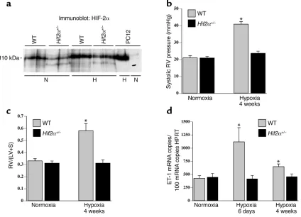
normox-ia and hypoxnormox-ia, respectively; n = 6; P = NS). Western blot analysis revealed that both normoxic and hypox-ic pulmonary HIF-2αprotein levels were significantly decreased in Hif2α+/–mice compared with WT mice
(Figure 1a). HIF-1αprotein was undetectable in both normoxic and hypoxic lungs (data not shown), consis-tent with previous reports (23).
Hif2α+/–mice survive long-term exposure to severe hypoxia.
housed under normobaric hypoxia (10% O2) for 4
weeks. Control groups of both genotypes were main-tained in normal air (21% O2). At the start of the
exper-iment, mice of both genotypes appeared healthy, active-ly moved in their cages, and had comparable body weights (about 26 g). After 4 weeks in normoxia, WT mice gained more weight than Hif2α+/–mice (22% ± 3%
for WT mice vs. 10% ± 3% for Hif2α+/–mice; n = 6–8; P = 0.04). When exposed to hypoxia, most WT mice became immobile and lost 29% ± 2% of their body weight, but none of the mice died after 4 weeks of hypox-ia. In contrast, Hif2α+/–mice did not exhibit any signs of
discomfort, behaved normally, moved around actively, and lost only 16% ± 2% of their body weight after 4 weeks of hypoxia (P < 0.001 vs. WT mice; n = 15–17).
Hif2α+/–mice are protected against pulmonary hyperten-sion. No genotypic differences in systolic RV pressure were observed in normoxia (Figure 1b). Chronic hypoxia increased the systolic RV pressure in WT mice but, remarkably, not in Hif2α+/–mice (Figure 1b).
Additional hemodynamic measurements demon-strated that exposure of normoxic mice to acute hypoxia (7% O2) for 20 minutes caused a similar
increase in systolic RV pressure in WT mice (25% ± 9%;
n = 5) and Hif2α+/–mice (29% ± 7%;n = 4;P = NS). In
addition, bradykinin-induced vasodepressor re-sponses were also comparable in WT and Hif2α+/–
mice (2.0 ± 0.2 mmHg in WT mice vs. 2.2 ± 0.2 mmHg in Hif2α+/–mice;n = 4;P = NS). These data indicate
that acute vasoconstriction and vasodepressor responses were comparable in WT and Hif2α+/–mice
and that protection of Hif2α+/– mice against
[image:5.576.84.508.53.359.2]pul-monary hypertension was not attributable to differ-ences in acute vasomotor responses. Taken together, these results suggest that any possible additional increase of RV pressure due to hypoxia-induced vaso-constriction may indeed have also contributed to the RV hypertrophy in WT mice but had, obviously, no effect on pulmonary vascular remodeling and RV hypertrophy in Hif2α+/–mice.
Figure 1
(a) Western blot analysis showed that pulmonary HIF-2αprotein levels were decreased in Hif2α+/–mice compared with WT mice after
nor-moxia (N) and after 1 week of hypoxia (H). Extracts from PC12 cells, exposed to nornor-moxia or hypoxia (1% oxygen, 4 hours) as previously described (17, 26), were used as controls. (b) Systolic RV pressures in WT and Hif2α+/–mice under normoxia or after hypoxic exposure.
Hemodynamic analysis revealed no differences in RV pressure under normoxia; however, WT mice showed increased RV pressure after 4 weeks of hypoxia, but Hif2α+/–mice did not. (c) RV hypertrophy analysis in WT and Hif2α+/–mice revealed no differences under normoxic
conditions. After hypoxic exposure for 4 weeks, the ratio of the mass of the right ventricle to the mass of the left ventricle plus septum [RV/(LV+S)] was increased in WT mice but not in Hif2α+/–mice. (d) Pulmonary ET-1 transcript levels were increased in WT mice after
expo-sure to chronic hypoxia for 6 days and were still elevated after 4 weeks of hypoxia. In contrast, chronic hypoxia failed to induce ET-1 expres-sion in lungs of Hif2α+/–mice. Data represent means ± SEM of systolic RV pressure (mmHg; n = 4–7) (b), of the ratio of the mass of the right
ventricle to the mass of the left ventricle plus septum [RV/(LV+S); n = 6–7] (c), and of the ET-1 mRNA copy number per 100 copies of HPRT
RV contractility, measured by the maximal dP/dt cor-rected for the RV pressure (dP/dtmaxP), was comparable
in both genotypes in normoxia (180 ± 32 s–1 in WT
mice vs. 160 ± 29 s–1in Hif2α+/–mice; n = 4–5; P = NS).
After 4 weeks of hypoxia, RV contractility tended to be impaired in WT mice (110 ± 27 s–1; n = 4; P = NS vs.
nor-moxic WT mice), while it was improved in Hif2α+/–mice
(290 ± 41 s–1; n = 6; P < 0.02 vs. normoxic WT and
Hif2α+/– mice). RV diastolic function, evaluated by
measuring the relaxation time constant τ, was compa-rable in both genotypes in normoxia (τ= 9 ± 1 ms in WT mice vs. 7 ± 1 ms in Hif2α+/–mice; n = 4–5; P = NS).
In contrast, after 4 weeks of hypoxia, RV relaxation was significantly impaired in WT but not in Hif2α+/–mice
(τ= 17 ± 5 ms in WT mice vs. 6 ± 1 ms in Hif2α+/–mice; n = 4–6; P = 0.01). Heart rates were comparable in WT and Hif2α+/–mice in normoxia and after exposure to
hypoxia (data not shown).
Hif2α+/–mice are protected against RV hypertrophy. To
evaluate RV hypertrophy, the weights of the RV and of the left ventricle plus septum (LV+S) were determined. No genotypic differences were observed in the RV/(LV+S) weight ratio in normoxia (Figure 1c). After 4 weeks of hypoxia, the RV/(LV+S) weight ratio increased in WT but not in Hif2α+/–mice (Figure 1c).
Similar findings were obtained when the RV weight was normalized for the total body weight (B). Whereas no differences in the RV/B weight ratio were observed in normoxia (0.25 ± 0.02 mg/g in WT mice vs. 0.24 ± 0.01 mg/g in Hif2α+/–mice; n = 7–10; P = NS), the
RV/B weight ratio was higher in WT than in Hif2α+/–
mice after hypoxia (0.44 ± 0.03 mg/g in WT mice vs. 0.27 ± 0.02 mg/g in Hif2α+/–mice; n = 6–7; P = 0.001).
As a result of the hypoxia-induced RV hypertrophy in WT mice, significant changes in the coronary vas-culature developed in the RV myocardial wall: the intercapillary distance enlarged (11 ± 0.3 µm in nor-moxia vs. 14 ± 0.1 µm in hypoxia; n = 5; P < 0.05), while the capillary density decreased (number of capillar-ies/mm2: 7,200 ± 240 in normoxia vs. 5,600 ± 390 in
hypoxia; n = 5; P < 0.05). Since there was no compen-satory angiogenesis, as evidenced by the normal cap-illary/cardiomyocyte ratio (1.2 ± 0.08 in normoxia vs. 1.1 ± 0.04 in hypoxia; n = 5; P = NS), the hypertrophic
cardiomyocytes may have suffered some degree of ischemia. In contrast, no such signs of vascular insuf-ficiency were detectable in Hif2α+/–mice
(intercapil-lary distance: 12 ± 0.3 µm in normoxia vs. 11 ± 0.3 µm in hypoxia; capillaries/mm2: 7,000 ± 310 in
normox-ia vs. 7,600 ± 410 in hypoxnormox-ia; capillary/cardiomyocyte ratio: 1.1 ± 0.05 in normoxia vs. 1.2 ± 0.08 in hypox-ia; n = 5; P = NS).
Lack of pulmonary vascular remodeling in Hif2α+/–mice.
Since pulmonary hypertension is often associated with vascular remodeling of pulmonary arteries (5, 6), we investigated whether HIF-2αaffected pulmonary vascu-lar remodeling. Counting of vessels at the level of alveoli and alveolar ducts after elastin staining revealed that the densities of nonmuscularized vessels (containing only an IEL) or partially muscularized vessels (containing an IEL plus an incomplete EEL) were comparable in WT and
Hif2α+/–mice in normoxia (Table 1). After 4 weeks of
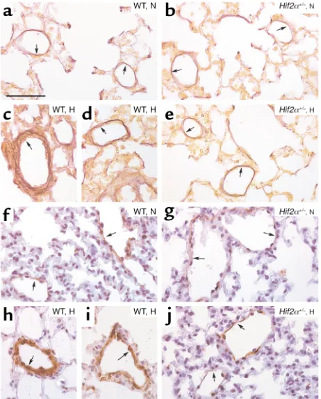
hypoxia, WT mice had more thick-walled muscularized vessels containing an IEL and a complete EEL (Table 1). In contrast, vascular remodeling failed to occur in hypox-ic Hif2α+/–mice, in which the fraction of muscularized
vessels with an IEL and a complete EEL was comparable to that of normoxic WT and Hif2α+/–mice (Table 1;
Fig-ure 2, a–e). The wall thickness of completely muscular-ized pulmonary arterioles, measured as the percentage of the EEL diameter minus the IEL diameter divided by the EEL diameter, was comparable in both genotypes in nor-moxia (7.6% ± 0.5% in WT mice vs. 8.0% ± 0.5% in Hif2α+/–
mice; n = 5; P = NS). Hypoxia increased the wall thick-ness of completely muscularized arterioles in WT mice but not in Hif2α+/–mice (16% ± 0.4% in WT mice vs.
7.4% ± 0.2% in Hif2α+/–mice; n = 5; P = 0.02). The
geno-typic differences in vascular remodeling were confirmed by immunostaining for SMC α-actin on lung sections. Compared with Hif2α+/–mice, WT mice contained
sig-nificantly more fully muscularized vessels that were com-pletely surrounded by SMCs after hypoxic exposure. In
Hif2α+/–mice, most of the pulmonary vessels were only
partially surrounded by SMCs after hypoxic exposure (Table 1; Figure 2, f–j).
Normal hematocrit levels in Hif2α+/–mice. Hematocrit
levels were comparable in WT and Hif2α+/–mice in
[image:6.576.62.534.610.702.2]nor-moxia (36% ± 1% in WT mice vs. 34% ± 1% in Hif2α+/–
Table 1
Hypoxia-induced pulmonary vascular remodeling
Normoxia Hypoxia
WT Hif2α+/– WT Hif2α+/–
Single IEL 1.9 ± 0.09 2.0 ± 0.11 0.98 ± 0.06A 1.9 ± 0.07
IEL + incomplete EEL 0.98 ± 0.06 1.0 ± 0.04 1.1 ± 0.06 0.88 ± 0.09 IEL + complete EEL 0.19 ± 0.03 0.15 ± 0.01 0.73 ± 0.11A 0.17 ± 0.02
Total 3.0 ± 0.06 3.2 ± 0.14 2.8 ± 0.16 2.9 ± 0.13
Partial SMC coverage 1.3 ± 0.05 1.3 ± 0.10 1.2 ± 0.10 1.2 ± 0.04
Complete SMC coverage 0.65 ± 0.06 0.60 ± 0.12 1.4 ± 0.09A 0.66 ± 0.02
mice; n = 6; P = NS). After 4 weeks of hypoxia, hemat-ocrit levels were comparably increased in both geno-types (46% ± 1% in 12 WT mice vs. 46% ± 1% in 16
Hif2α+/–mice; P = NS). These findings indicate that the
reduced pulmonary hypertension in Hif2α+/–mice was
not attributable to a lower vascular resistance caused by reduced erythropoiesis in hypoxia.
Role of HIF-2αin pulmonary and cardiac gene expression. ET-1 may contribute to pulmonary vasoconstriction and pulmonary vascular remodeling by stimulating growth and proliferation of VSMCs (33), while ET-1 receptor blockade ameliorates pulmonary hyperten-sion (4). Moreover, the promoter of the ET-1 gene har-bors a HIF-binding site, and ET-1 is upregulated by hypoxia in vitro and in hypoxic lungs in vivo (4, 16). Therefore, pulmonary expression of ET-1 was deter-mined by quantitative RT-PCR. No genotypic differ-ences were observed in pulmonary ET-1 transcript lev-els in normoxia. After 6 days and 4 weeks of hypoxia,
pulmonary ET-1 transcript levels were increased in WT but not in Hif2α+/–mice (Figure 1d). Measurements of
ET-1 protein levels by ELISA confirmed that hypoxic upregulation of pulmonary ET-1 protein was impaired in Hif2α+/–mice (pg ET-1/mg protein in normoxia and
hypoxia, respectively: 10 ± 2 and 31 ± 2 in WT mice vs. 11 ± 3 and 19 ± 3 in Hif2α+/–mice; n = 4; P < 0.05 in
hypoxia). Because NO has a critical role in vasodilation of pulmonary vessels and inhibits VSMC growth (34), we also determined eNOS expression in the lungs. Pul-monary eNOS transcript levels were comparable in both genotypes under normoxia and after 6 days of hypoxia (mRNA copy number per 100 copies of HPRT
mRNA in normoxia and hypoxia, respectively: 4.3 ± 0.5 and 3.7 ± 0.1 in WT mice vs. 3.2 ± 0.4 and 3.1 ± 0.4 in
Hif2α+/–mice; n = 6; P = NS). Western blot analysis (not
shown) revealed that pulmonary eNOS protein was increased by hypoxia in WT mice (consistent with pre-vious findings; ref. 35); pulmonary eNOS protein was upregulated to the same degree in Hif2α+/–mice (data
not shown), indicating that pulmonary eNOS expres-sion in hypoxia is not regulated by HIF-2α.
ET-1 has also been implicated in cardiac hypertrophy (36). Cardiac ET-1 transcript levels were comparable under normoxia (mRNA copy number per 100 copies of
HPRTmRNA: 90 ± 10 in WT mice vs. 100 ± 10 in Hif2α+/–
mice; n = 6; P = NS) but were upregulated to a higher level in WT than in Hif2α+/–mice after 4 weeks of hypoxia
(240 ± 20 in WT mice vs. 160 ± 30 in Hif2α+/–mice; n = 5; P < 0.05). ELISA measurements demonstrated that hypoxic upregulation of cardiac ET-1 protein was also impaired in Hif2α+/–mice (normoxia: 3.9 ± 0.3 pg/mg
protein in WT mice vs. 4.4 ± 1.4 pg/mg protein in
Hif2α+/–mice; n = 4; P = NS; hypoxia: 7.1 ± 0.3 pg/mg
pro-tein in WT mice vs. 5.1 ± 1.5 pg/mg propro-tein in Hif2α+/–
mice; n = 4; P < 0.05). Cardiac PDGF-B expression was also elevated by hypoxia in WT but not in Hif2α+/–mice
(mRNA copy number per 100 copies of HPRTmRNA in normoxia: 0.5 ± 0.1 in WT mice vs. 0.5 ± 0.1 in Hif2α+/–
mice; n = 6; P = NS; mRNA copy number per 100 copies of HPRTmRNA in hypoxia: 1.2 ± 0.3 in WT mice vs. 0.5 ± 0.1 in Hif2α+/–mice; n = 5; P < 0.05).
Role of HIF-2αin catecholamine homeostasis. Expression of tyrosine hydroxylase, an essential gene for catecholamine biosynthesis, is upregulated by hypoxia (16). In addition, plasma catecholamines are increased during hypoxia and in patients with chronic obstructive pulmonary dis-ease and/or pulmonary hypertension (37, 38). Since loss of HIF-2αimpairs fetal catecholamine production (22) and norepinephrine (NE) stimulates vasoconstriction of isolated pulmonary arteries (39) and pulmonary microvessels in rat lung explants (40), plasma NE levels were determined. Under normoxia, plasma NE levels were comparable in both genotypes (0.5 ± 0.1 ng/ml in WT mice vs. 0.6 ± 0.1 ng/ml in Hif2α+/–mice; n= 5; P = NS). Chronic hypoxia for 4 weeks elevated plasma NE 12-fold in WT mice but only 3.5-fold in Hif2α+/–mice
(5.8 ± 1.5 ng/ml in WT mice vs. 2.2 ± 0.5 ng/ml in
[image:7.576.60.291.54.341.2]Hif2α+/–mice; n = 5; P = 0.022). After 4 days of hypoxia,
Figure 2
(a–e) Hart’s elastin staining revealed the presence of vessels, located distally to the bronchi, at the level of alveoli and alveolar ducts, that contained only an IEL (or an IEL plus an incomplete EEL) (arrows) in lungs of normoxic (N) WT (a) and Hif2α+/–mice (b). Lungs of
hypox-ic (H) WT mhypox-ice showed the presence of thhypox-ick-walled vessels contain-ing both an IEL and a complete EEL (arrows) (cand d), whereas no hypoxia-induced vascular remodeling occurred in Hif2α+/– mice
(arrows) (e). (f–j) SMC α-actin staining shows the presence of partially muscularized peripheral vessels (arrows) in lungs of normoxic WT (f) and Hif2α+/–mice (g). Chronic hypoxia caused pulmonary vascular
remodeling in WT mice, as revealed by the presence of fully muscular-ized vessels (arrows) (hand i), but not in Hif2α+/–mice (arrows) (j).
when no pulmonary vascular remodeling was observed in WT mice (data not shown), plasma NE levels were already increased 3.5-fold in WT mice (1.8 ± 0.3 ng/ml), suggesting that the increased plasma NE levels in WT mice occurred prior to pulmonary vascular remodeling.
Discussion
The principal finding of this study is that heterozygous deficiency of HIF-2αprotects mice against pulmonary hypertension, RV hypertrophy, and dysfunction upon exposure to prolonged hypoxia. These findings unveil a critical role for HIF-2αin pulmonary vascular remod-eling, possibly by upregulation of ET-1 and NE.
Pulmonary vascular remodeling. Hypoxia caused a sig-nificant increase in neomuscularization of the pul-monary resistance vessels in WT mice, as evidenced by the increased fraction of pulmonary vessels that were completely covered by SMCs and contained both an IEL and a complete EEL. Since pulmonary veins exhib-it minimal structural changes during chronic hypoxia (31), the increase in thick-walled pulmonary vessels likely corresponded to an increase in muscularized arterioles. Remarkably, vessel muscularization in
Hif2α+/–mice after hypoxia was comparable to that in Hif2α+/–or WT mice after normoxia. Similar to
het-erozygous deficient Hif1α+/–mice (23), heterozygous
deficient Hif2α+/–mice were also protected against
pul-monary vascular remodeling. Although our data demonstrate that pulmonary hypertension during chronic hypoxia is mediated by HIF-2α, it remains to be determined whether the vascular remodeling is the result of a direct effect of HIF-2αin the vessel wall or is caused, indirectly, by a HIF-2α–mediated pressure increase in the pulmonary arterioles. Another intrigu-ing question is why the reduced HIF-2α levels in
Hif2α+/– mice were sufficient to sustain embryonic
development but not to induce pulmonary hyperten-sion, suggesting that different thresholds of HIF-2α
expression may be required for distinct biological processes. A similar phenomenon has been previously observed in angiogenesis, where placental growth fac-tor seems to affect pathological angiogenesis but not vascular development (41).
Pulmonary vascular remodeling is a complex, poor-ly understood process. Pulmonary vessels react to local hypoxia with constriction via upregulation of vasoactive substances as part of a natural response to normalize the oxygenation/perfusion ratio. When hypoxia is sustained and generalized, pulmonary ves-sels undergo significant structural changes involving neomuscularization and medial thickening of termi-nal bronchiolar and alveolar duct arterioles. Inhibitors of NAPDH oxidoreductases, which block HIF-1α (16), reduce pulmonary vasoconstriction (42), suggesting that hypoxia-induced pulmonary vasoconstriction may be mediated by HIFs. Further-more, pulmonary expression levels of ET-1, a well-known HIF-1α target gene (16), are increased in patients with pulmonary hypertension (4, 7), and
inhibition of ET-1 or its receptor prevents hypoxia-induced pulmonary hypertension and vascular remodeling (4). By causing vasoconstriction, ET-1 may initially increase the shear stress and initiate pul-monary vascular remodeling (7). Since ET-1 is known to stimulate growth of VSMCs (4, 33), it may also contribute to the muscularization of pulmonary ves-sels during hypoxia. As pulmonary ET-1 levels were markedly upregulated during hypoxia in WT but not in Hif2α+/–mice, HIF-2αmay regulate the
develop-ment of pulmonary hypertension at least in part via upregulation of ET-1. While ET-1 is a well-known tar-get gene of HIF-1α(16), our findings now demon-strate that expression of ET-1 is also inducible by HIF-2α. This is not entirely surprising, since both HIF-1α and HIF-2α bind to the same HRE in the ET-1 promoter (18, 19) and HIF-2αis abundantly expressed in the lung (19, 20). Whereas our findings suggest an involvement of HIF-2α in pulmonary hypertension by regulation of ET-1 expression, they obviously do not exclude that HIF-2αregulates the expression of additional vasoactive substances.
RV heart hypertrophy and dysfunction. Pulmonary hyper-tension leads to RV hypertrophy and dysfunction, ulti-mately progressing to RV failure (cor pulmonale) (1, 2). When WT mice were exposed for prolonged periods to hypoxia, RV pressures were significantly increased and caused RV hypertrophy. RV pressures might even have been underestimated, as they were measured after equi-libration of the mice to room air for 1 hour, which like-ly reduced or eliminated a hypoxia-induced vasocon-strictor response. WT mice also suffered signs of systolic and diastolic RV dysfunction, likely represent-ing an insufficient myocardial adaptation to the pro-longed increase in hemodynamic stress. In contrast, RV diastolic function was preserved, while RV systolic function improved in hypoxic Hif2α+/–mice as
com-pared with normoxic mice. This is remarkable since RV loading remained normal in hypoxic mutant mice, as suggested by the lack of remodeling of pulmonary vas-cular resistance vessels and the normal RV pressures. Possibly, cardiac function is enhanced in these mice to compensate for the hypoxemia and impaired oxygena-tion of peripheral tissues when maintained for pro-longed periods in a low-oxygen environment. Expres-sion of ET-1 and PDGF-B, previously implicated in cardiac hypertrophy (36, 43), was increased in hyper-trophic hearts of hypoxic WT mice, suggesting that these molecules may have contributed, at least in part, to the hypertension-induced cardiomyocyte hypertro-phy. In addition, ET-1 is known to induce hypertrophy with enhanced expression of muscle-specific genes in cultured rat cardiomyocytes in vitro (36). While HIF-2α
Catecholamine homeostasis. HIF-2αhas been implicat-ed in the regulation of catecholamine biosynthesis dur-ing development (22), but a possible role durdur-ing adult life has not been reported thus far. Hypoxia upregulat-ed plasma NE levels 12-fold in WT mice but only 3.5-fold in Hif2α+/–mice. Plasma NE levels are upregulated
in patients with chronic obstructive pulmonary disease and/or pulmonary hypertension (37, 38). Since NE stimulates vasoconstriction of isolated pulmonary arteries (39) and of pulmonary microvessels in adult rat lung explants (40), and since NE is also known to stim-ulate growth of pulmonary SMCs (44), the impaired upregulation of NE under hypoxic conditions may also have contributed to the impaired pulmonary vascular remodeling and hypertension in Hif2α+/– mice.
Al-though we cannot exclude that NE levels were upregu-lated due to an increase in pulmonary pressure, plasma NE levels were already elevated after 4 days of hypoxia in WT mice, i.e., prior to onset of significant pul-monary vascular remodeling. The observation that both WT and Hif2α+/–mice had a similar increase in
systolic RV pressure during acute hypoxia indicates that the acute hypoxic vasoconstriction response is unlikely to be mediated by HIF-2α. Instead, HIF-2αis essential for upregulation of ET-1 and NE during chronic hypoxic conditions and thereby mediates SMC proliferation and pulmonary vascular remodeling.
Hematocrit. Yu et al. documented a delay in the devel-opment of polycythemia in Hif1α+/–mice (23). We did
not observe any difference in hematocrit levels in
Hif2α+/– mice after 4 weeks of hypoxia. Possibly,
hypoxic induction of erythropoietin expression might be more dependent on HIF-1α than on HIF-2α, at least under the present conditions. This contrasts with the significant role of HIF-2αin the hypoxic upregulation of ET-1 and NE and in the induction of pulmonary hypertension and vascular remodeling. While we cannot exclude that partial loss of HIF-2α
might have delayed polycythemia in the initial stage, the comparable hematocrit levels in combination with the impaired vascular remodeling in Hif2α+/–
mice after 4 weeks of hypoxia suggest that the lower vascular resistance in these mice was not attributable to the reduced hematocrit levels.
Weight loss. Both genotypes had comparable body weights at the start of the experiment (about 26 g). Changes in body weight were more pronounced in WT than in Hif2α+/–mice — both the weight gain in
nor-moxia and the weight loss in hypoxia. As a result, WT mice weighed 18 g after 4 weeks of hypoxia and 32 g after 4 weeks of normoxia, while corresponding body weights of Hif2α+/–mice were 22 g in hypoxia and 29 g
in normoxia. Another remarkable observation is that both Hif1α+/–and Hif2α+/–mice gained less weight
dur-ing normoxia, while Hif1α+/– mice, unlike Hif2α+/–
mice, lost more weight than WT mice during hypoxia (23). These findings suggest that both HIFs have dis-tinct roles. This may relate to their different cell-spe-cific expression. Possibly, deficiency of HIF-1αimpairs
glycolysis during hypoxia, thereby reducing the over-all metabolism in Hif1α+/–mice. In contrast, we
recent-ly showed that HIF-2αplays a less important role than HIF-1αin hypoxic upregulation of glycolytic genes (26), suggesting that HIF-1αcould play a more impor-tant role than HIF-2αin hypoxic regulation of partic-ular processes influencing weight gain. Weight loss has also been documented in patients suffering from chronic obstructive pulmonary disease (45).
Medical significance. The role of HIF-2αin the devel-opment of pulmonary hypertension and RV dysfunc-tion may have clinical relevance. Overall, Hif2α+/–mice
tolerated hypoxic stress much better than did WT mice, as they lost less weight, remained more active, and showed no pulmonary vascular remodeling or hyper-tension. Therefore, our data would encourage evalua-tion of the possible therapeutic potential of HIF-2α
inhibitors for this disease. Retrovirally mediated inhi-bition of HIFs, via blocking of the interaction of the cofactor p300 with HIF-1αand HIF-2α, has been pre-viously demonstrated to suppress tumor growth (46). Considering the limitations of current treatments of pulmonary hypertension (2, 3), inhibition of HIFs might be considered to prevent or reduce hypoxia-induced pulmonary hypertension. Since heterozygous deficiency of HIF-2αalready protected mice, partial inhibition of hypoxia-inducible factors may be thera-peutically sufficient.
Acknowledgments
The authors thank S. Janssens (Center for Transgene Technology and Gene Therapy) for helpful discussion and A. Bouché, K. Bijnens, E. Gils, S. Jansen, L. Kieck-ens, A. Manderveld, K. Maris, T. Vancoetsem, A. Van-denhoeck, B. Vanwetswinkel, and S. Wyns (all from the Center for Transgene Technology and Gene Therapy) for technical assistance. This work was supported by a European BIOMED grant (no. PL963380) and a grant from the Interuniversitaire Attractiepolen.
1. Barbera, J.A., Peinado, V.I., and Santos, S. 2000. Pulmonary hypertension in COPD: old and new concepts. Monaldi Arch. Chest Dis. 55:445–449. 2. Hida, W., Tun, Y., Kikuchi, Y., Okabe, S., and Shirato, K. 2002.
Pul-monary hypertension in patients with chronic obstructive pulPul-monary disease: recent advances in pathophysiology and management. Respirol-ogy. 7:3–13.
3. Barnes, P.J. 2000. Chronic obstructive pulmonary disease. N. Engl. J. Med.
343:269–280.
4. Chen, Y.F., and Oparil, S. 2000. Endothelin and pulmonary hyperten-sion. J. Cardiovasc. Pharmacol. 35(4 Suppl. 2):S49–S53.
5. Durmowicz, A.G., and Stenmark, K.R. 1999. Mechanisms of structural remodeling in chronic pulmonary hypertension. Pediatr. Rev.
20:e91–e102.
6. Stenmark, K.R., and Mecham, R.P. 1997. Cellular and molecular mech-anisms of pulmonary vascular remodeling. Annu. Rev. Physiol. 59:89–144. 7. Voelkel, N.F., and Tuder, R.M. 2000. Hypoxia-induced pulmonary vas-cular remodeling: a model for what human disease? J. Clin. Invest.
106:733–738.
8. Fagan, K.A., et al. 1999. The pulmonary circulation of homozygous or heterozygous eNOS-null mice is hyperresponsive to mild hypoxia. J. Clin. Invest. 103:291–299.
9. Hoshikawa, Y., et al. 2001. Prostacyclin receptor-dependent modulation of pulmonary vascular remodeling. Am. J. Respir. Crit. Care Med.
164:314–318.
11. Budts, W., et al. 2000. Aerosol gene transfer with inducible nitric oxide synthase reduces hypoxic pulmonary hypertension and pulmonary vas-cular remodeling in rats. Circulation. 102:2880–2885.
12. Eddahibi, S., et al. 2000. Attenuated hypoxic pulmonary hypertension in mice lacking the 5-hydroxytryptamine transporter gene. J. Clin. Invest.
105:1555–1562.
13. Carmeliet, P., and Collen, D. 1998. Vascular development and disorders: molecular analysis and pathogenic insights. Kidney Int. 53:1519–1549. 14. Levi, M., et al. 2001. Deficiency of urokinase-type plasminogen activator-mediated plasmin generation impairs vascular remodeling during hypox-ia-induced pulmonary hypertension in mice. Circulation. 103:2014–2020. 15. Campbell, A.I., Zhao, Y., Sandhu, R., and Stewart, D.J. 2001. Cell-based gene transfer of vascular endothelial growth factor attenuates monocro-taline-induced pulmonary hypertension. Circulation. 104:2242–2248. 16. Semenza, G.L. 1999. Regulation of mammalian O2 homeostasis by
hypoxia-inducible factor 1. Annu. Rev. Cell Dev. Biol. 15:551–578. 17. Carmeliet, P., et al. 1998. Role of HIF-1αin hypoxia-mediated
apopto-sis, cell proliferation and tumour angiogenesis. Nature. 394:485–490. 18. Tian, H., McKnight, S.L., and Russell, D.W. 1997. Endothelial PAS domain protein 1 (EPAS1), a transcription factor selectively expressed in endothelial cells. Genes Dev. 11:72–82.
19. Ema, M., et al. 1997. A novel bHLH-PAS factor with close sequence sim-ilarity to hypoxia-inducible factor 1αregulates the VEGF expression and is potentially involved in lung and vascular development. Proc. Natl. Acad. Sci. U. S. A. 94:4273–4278.
20. Flamme, I., et al. 1997. HRF, a putative basic helix-loop-helix-PAS-domain transcription factor is closely related to hypoxia-inducible factor-1αand developmentally expressed in blood vessels. Mech. Dev. 63:51–60. 21. Peng, J., Zhang, L., Drysdale, L., and Fong, G.H. 2000. The transcription
factor EPAS-1/hypoxia-inducible factor 2αplays an important role in vascular remodeling. Proc. Natl. Acad. Sci. U. S. A. 97:8386–8391. 22. Tian, H., Hammer, R.E., Matsumoto, A.M., Russell, D.W., and McKnight,
S.L. 1998. The hypoxia-responsive transcription factor EPAS1 is essen-tial for catecholamine homeostasis and protection against heart failure during embryonic development. Genes Dev. 12:3320–3324.
23. Yu, A.Y., et al. 1999. Impaired physiological responses to chronic hypox-ia in mice parthypox-ially deficient for hypoxhypox-ia-inducible factor 1α. J. Clin. Invest. 103:691–696.
24. Shimoda, L.A., Manalo, D.J., Sham, J.S., Semenza, G.L., and Sylvester, J.T. 2001. Partial HIF-1αdeficiency impairs pulmonary arterial myocyte elec-trophysiological responses to hypoxia. Am. J. Physiol. Lung Cell. Mol. Phys-iol. 281:L202–L208.
25. Compernolle, V., et al. 2002. Loss of HIF-2αand inhibition of VEGF-sig-naling impair fetal lung maturation, while VEGF treatment prevents fatal respiratory distress in premature mice. Nat. Med. 8:702–710. 26. Brusselmans, K., et al. 2001. Hypoxia-inducible factor-2α(HIF-2α) is
involved in the apoptotic response to hypoglycemia but not to hypoxia.
J. Biol. Chem. 276:39192–39196.
27. Hoit, B.D. 1998. Invasive hemodynamic studies in open and closed chest mice. In Cardiovascular physiology in the genetically engineered mouse. B.D. Hoit and R.A. Walsh, editors. Kluwer Academic Publishers. Norwell, Massachusetts, USA. 111–124.
28. Chen, E.P., Craig, D.M., Bittner, H.B., Davis, R.D., and Van Trigt, P. 1998. Pharmacological strategies for improving diastolic dysfunction in the setting of chronic pulmonary hypertension. Circulation. 97:1606–1612.
29. Steudel, W., et al. 1998. Sustained pulmonary hypertension and right ventricular hypertrophy after chronic hypoxia in mice with congenital deficiency of nitric oxide synthase 3. J. Clin. Invest. 101:2468–2477. 30. Champion, H.C., et al. 1999. Gene transfer of endothelial nitric oxide
synthase to the lung of the mouse in vivo. Circ. Res. 84:1422–1432. 31. Roberts, J.D., Jr., Roberts, C.T., Jones, R.C., Zapol, W.M., and Bloch, K.D.
1995. Continuous nitric oxide inhalation reduces pulmonary arterial structural changes, right ventricular hypertrophy, and growth retarda-tion in the hypoxic newborn rat. Circ. Res. 76:215–222.
32. Wiesener, M.S., et al. 2002. Widespread, hypoxia-inducible expression of HIF-2αin distinct cell populations of different organs. FASEB J.
17:271–273.
33. Wort, S.J., Woods, M., Warner, T.D., Evans, T.W., and Mitchell, J.A. 2001. Endogenously released endothelin-1 from human pulmonary artery smooth muscle promotes cellular proliferation: relevance to pathogen-esis of pulmonary hypertension and vascular remodeling. Am. J. Respir. Cell Mol. Biol. 25:104–110.
34. Nathan, C., and Xie, Q.W. 1994. Nitric oxide synthases: roles, tolls, and controls. Cell. 78:915–918.
35. Le Cras, T.D., and McMurtry, I.F. 2001. Nitric oxide production in the hypoxic lung. Am. J. Physiol. Lung Cell. Mol. Physiol. 280:L575–L582. 36. Ito, H., et al. 1991. Endothelin-1 induces hypertrophy with enhanced
expression of muscle-specific genes in cultured neonatal rat cardiomy-ocytes. Circ. Res. 69:209–215.
37. Zaloga, G.P., et al. 1984. Increased circulating plasma norepinephrine concentrations in noncardiac causes of pulmonary hypertension. Crit. Care Med. 12:85–89.
38. Bratel, T., Wennlund, A., and Carlstrom, K. 2000. Impact of hypoxaemia on neuroendocrine function and catecholamine secretion in chronic obstructive pulmonary disease (COPD). Effects of long-term oxygen treatment. Respir. Med. 94:1221–1228.
39. Jin, N., Packer, C.S., English, D., and Rhoades, R.A. 1993. Inositol trisphosphate is involved in norepinephrine- but not in hypoxia-induced pulmonary arterial contraction. Am. J. Physiol. 264:L160–L164. 40. Davies, P., Maddalo, F., and Reid, L. 1984. The response of microvessels
in rat lung explants to incubation with norepinephrine. Exp. Lung Res.
7:93–100.
41. Carmeliet, P., et al. 2001. Synergism between vascular endothelial growth factor and placental growth factor contributes to angiogenesis and plas-ma extravasation in pathological conditions. Nat. Med. 7:575–583. 42. Jones, R.D., Thompson, J.S., and Morice, A.H. 2000. The NADPH
oxi-dase inhibitors iodonium diphenyl and cadmium sulphate inhibit hypoxic pulmonary vasoconstriction in isolated rat pulmonary arteries.
Physiol. Res. 49:587–596.
43. Suzuki, J., et al. 1999. Immunohistochemical analysis of platelet-derived growth factor-B expression in myocardial tissues in hypertrophic car-diomyopathy. Cardiovasc. Pathol. 8:223–231.
44. Nakaki, T., Nakayama, M., Yamamoto, S., and Kato, R. 1990. Alpha 1-adrenergic stimulation and beta 2-adrenergic inhibition of DNA syn-thesis in vascular smooth muscle cells. Mol. Pharmacol. 37:30–36. 45. Gross, N.J. 2001. Extrapulmonary effects of chronic obstructive
pul-monary disease. Curr. Opin. Pulm. Med. 7:84–92.

![N,N [(Z,Z) 7,7 Diphenylbicyclo[4 1 0]hept 3 ene 2,5 diylidene]dibenzenesulfonamide](data:image/gif;base64,R0lGODlhAQABAIAAAP///wAAACH5BAEAAAAALAAAAAABAAEAAAICRAEAOw==)