0095-1137/95/$04.0010
Copyrightq1995, American Society for Microbiology
Identification and Ribotypes of Staphylococcus caprae Isolates
Isolated as Human Pathogens and from Goat Milk
FRANC¸ OIS VANDENESCH,1* SUSANNAH J. EYKYN,2MICHE` LE BES,1HE´ LE` NE MEUGNIER,1
JEAN FLEURETTE,1
ANDJEROME ETIENNE1
De´partement de Recherche en Bacte´riologie Me´dicale EA1655, Faculte´ de Me´decine Alexis Carrel, Lyon, France,1
and Department of Microbiology, St. Thomas’ Hospital, London, United Kingdom2
Received 14 September 1994/Returned for modification 4 January 1995/Accepted 19 January 1995
We report five cases of human infection withStaphylococcus caprae. Two were community acquired (one case
each of endocarditis and urinary tract infection); the other three were acquired in a hospital (two cases of bacteremia associated with intravenous access and one case of urinary tract infection). Analysis of human isolates and goat isolates from eight herds showed that they could be misidentified by some commercial
identification systems but were clearly identified asS. capraeby ribotyping, according to their species-specific
ribotype. Phylogenetic methods applied to the ribotypes did not reveal two distinct lineages corresponding to the goat and human origins of the isolates, although human ribotypes were clearly distinguishable by the presence of a core of four specific bands. The latter observation may reflect some degree of evolutionary change within the species between human and goat isolates.
The coagulase-negative (CN) species Staphylococcus caprae was described in 1983 (6), and S. caprae strains have been isolated from goat milk (5, 21) but never from cow or sheep milk (5, 17). Two human clinical isolates of S. caprae have been reported: one from the pus of a patient with dermatitis and the other from the urine of a patient hospitalized ‘‘with urinary tract infection’’. There was no convincing evidence that these strains caused true infection (10). S. caprae has also been isolated from 6% of 1,500 clinical isolates of CN staphylococci in a Japanese hospital (no further details were given), suggest-ing that this species can inhabit the human body and is quite widely distributed in hospitals (10). Nevertheless, its actual pathogenic potential for humans has not been clarified. The relationship between human and animal isolates and the exis-tence of cross-contamination between humans and animals has not been established.
We report five cases of human infections with S. caprae (one case of infective endocarditis, two cases of intravenous [i.v.] treatment-associated bacteremia, and two cases of urinary tract infection) and compare the bacteriological characteristics and ribotypes of the human isolates with a series of unrelated isolates from goat milk. We showed that human isolates of S.
caprae were clearly distinguishable by ribotyping of goat
iso-lates, but phylogenetic methods applied to the ribotypes did not produce concordant delineation into two distinct lineages corresponding to the animal and human origins of the isolates.
CASE REPORTS
Patient 1.A 30-year-old woman underwent medullary
de-compression for a rhabdomyosarcoma in January 1988. The operation was followed by radiotherapy and chemotherapy. In November 1988 the patient was readmitted to the hospital for a relapse of the rhabdomyosarcoma with metastases, and 2 days later she developed a fever (388C). Her leukocyte (WBC) count was 3.0 3 109/liter. Microscopy of centrifuged urine showed pyuria and cocci. Culture of the urine yielded S. caprae
(107CFU/ml) in pure growth. The patient was not catheter-ized. She was given oral cotrimoxazole for 8 days, and although she became apyrexial, she died 4 days later from cancer.
Patient 2.An 8-year-old previously healthy girl was admitted
to a hospital in January 1991 with an abdominal pain and a fever of 408C that had persisted for 4 days. Her WBC count was 123109/liter with 76% polymorphs, and the erythrocyte sedimentation rate was 109 mm/h. Microscopy of centrifuged urine showed pyuria and cocci, and culture yielded pure growth of S. caprae (107CFU/ml). Duplex ureters were found by ultrasonography. The patient was successfully treated for 2 days with i.v. ceftriaxone.
Patient 3.A 46-year-old man was admitted to a hospital in
August 1992 with a 6- to 8-week history of malaise and night sweats. He had no known cardiac disease and had been deemed fit by a medical examination for diving. On admission, he was febrile (38.28C), with a hemoglobin level of 12.5 g/liter, WBC count of 13.33109/liter, and erythrocyte sedimentation rate of 80 mm/h. He had no clinical stigmata of endocarditis and was hemodynamically stable. Echocardiography showed a lesion suggestive of an atrial myxoma adjacent to the mitral valve, but since CN staphylococci (later identified as S. caprae) were then isolated from 9 of 10 blood culture bottles, the clinicians were persuaded that the ‘‘myxoma’’ was a large veg-etation on the mitral valve and decided to excise it. i.v. vanco-mycin was started then, both as treatment and as prophylaxis for the surgery. The mitral valve was not replaced. The excised vegetation grew S. caprae. The patient received a total of 2 weeks of i.v. vancomycin and made an excellent recovery.
Patient 4.A 67-year-old woman underwent an ovariectomy
for carcinoma in July 1992, and a permanent i.v. device (Port-ocath) was implanted for chemotherapy. In November, she became febrile (398C), when chemotherapy was begun. Her WBC count was 6.73109/liter with 69% polymorphs, and the erythrocyte sedimentation rate was 100 mm/h. S. caprae was recovered from three blood cultures drawn through the Port-ocath. The patient was treated with i.v. co-amoxiclav for 2 days followed by oral co-amoxiclav for 8 days. A similar episode occurred in December 1992, and three blood cultures were again positive for S. caprae. No blood peripheral cultures were ever taken. The Portocath was removed, and culture of the
* Corresponding author. Mailing address: Laboratoire de Bacte ´ri-ologie, Hoˆpital Louis Pradel, BP Lyon Montchat, 69394 Lyon cedex 03, France. Phone: (33) 72 35 72 52. Fax: (33) 72 35 73 35.
888
on May 15, 2020 by guest
http://jcm.asm.org/
catheter tip grew S. caprae. The patient received a further 10-day course of co-amoxiclav but died 4 months later from cancer.
Patient 5.In August 1993, a neonate with aortic coarctation
and interventricular communication was transferred from a hospital for ventilation and consideration of surgical treat-ment. He was treated by dinoprostone to close the aortic co-arctation. Four days later, he was extubated, as there was hemodynamic improvement, but within 24 h he became febrile (398C) and dyspneic with abdominal distension. There were no peripheral pulses, and ventilation was restarted. The WBC count was 13.53109/liter. The umbilical catheter was removed and yielded.100 colonies of S. caprae by culture. S. caprae was also isolated from two blood cultures. The patient was treated with i.v. vancomycin and amikacin for 8 days followed by oral oxacillin for a further 8 days and made a complete recovery. He later underwent successful surgical treatment of the cardiac abnormalities.
MATERIALS AND METHODS
Bacterial isolates.Four of the above five S. caprae infections were reported to the French Centre National de Re´fe´rence des Staphylocoques in Lyon by clini-cians at different hospitals in France between 1988 and 1993. The fifth patient was admitted to St. Thomas’ Hospital (London, United Kingdom) with commu-nity-acquired endocarditis. The two human clinical isolates of S. caprae described by Kanda et al. (10) were kindly provided by E. Tateda-Suzuki and K. Hiramatsu (Juntendo University, Tokyo, Japan). Seven S. caprae isolates from seven unre-lated herds were cultured from the milk of goats with mastitis and were provided by B. Poutrel (Institut National de la Recherche Agronomique, Nouzilly, France). Eight further milk isolates from different goats of the same herd were provided by Y. Richard (Ecole Ve´te´rinaire, Marcy-l’Etoile, France). The patient and animal isolates and the reference strain S. caprae CCM3573 (6) were iden-tified and typed in Lyon (Table 1).
Phenotypic characterization.Conventional identification of the isolates was performed by using a biochemical gallery (ID32 Staph; bioMe´rieux,
Marcy-l’Etoile, France) (2). The results were interpreted by using both the API database and published references (12). All isolates were tested for coagulase with rabbit plasma (bioMe´rieux), for fibrinogen affinity factor (clumping factor) by the agglutination procedure (Staphyslide agglutination test; bioMe´rieux), and for production of heat-stable DNase by a diagnostic kit (Sanofi Diagnostics Pasteur, Marnes-la-Coquette, France). Hemolytic activity was detected on Trypticase soy agar plates supplemented with 5% defibrinated sheep blood (bioMe´rieux); test strains were streaked perpendicularly to, but not touching, a streak of a beta-hemolysin-producing strain of S. aureus RN4220 (14), and plates were incubated for 24 h at 378C aerobically. The following characteristics were used to describe the hemolysis observed: alpha-hemolysin produced a wide zone of complete hemolysis with blurred edges, beta-hemolysin gave a wide zone of partial hemo-lysis which increased after further incubation at 48C for 4 to 6 h, and delta-hemolysin formed a narrow zone of complete hemolysis and produced a com-plete clearing within the zone of incomcom-plete hemolysis produced by the beta-hemolysin of S. aureus RN4220. The MICs of a range of antistaphylococcal antibiotics were estimated for all strains by an agar dilution technique (16).
Ribotyping.Whole-cell DNA was isolated from the staphylococci by standard procedures (18), restricted by EcoRI according to the manufacturer’s instruc-tions (Boehringer Mannheim, Meylan, France), and separated on a 0.8% agarose gel (Bioprobe, Montreuil-sous-bois, France) in 0.089 M boric acid–0.0002 M EDTA at 2.5 V/cm for 18 h. Subsequently, the DNA was vacuum transferred to nylon membranes (Amersham, Les Ulis, France) and cross-linked with UV light. Hybridization was performed at 608C with a commercially available 16S-23S rRNA probe labeled with acetylaminofluorene (Eurogentec, Lie`ge, Belgium). Immunoenzymatic detection was carried out according to the manufacturer’s instructions (Eurogentec). Raoul I (Applige`ne, Illkirch, France) was used as the size marker in all blots. Migration distances of the hybridizing bands were measured and were kindly analyzed by P. A. D. Grimont using the Taxolab software (Institut Pasteur, Paris, France). The Dice coefficient (7) was used to compare rRNA gene profiles by scoring positive or negative matches for all bands, and clustering was based on the unweighted pair group arithmetic average algorithm (UPGMA).
RESULTS
Microbiological findings.Strains were identified as S. caprae
if they were gram-positive, catalase-positive, oxidase-negative cocci, and if the results of the biochemical tests were char-acteristic of S. caprae (Table 2). Twenty-three isolates were identified as S. caprae by the API database, one isolate was identified as S. intermedius because of the production ofb
-ga-TABLE 1. Origins and characteristics of S. caprae strains
Strain Origin and/or characteristics Reference and/or source
CCM3573 Reference strain 6
JA21 Clinical, Japan 19, E. Tateda-Suzukia JA187 Clinical, Japan; mecA 19, E. Tateda-Suzuki N940142 Goat milk, herd 1 B. Poutrela N940143 Goat milk, herd 2 B. Poutrel N940144 Goat milk, herd 3 B. Poutrel N940145 Goat milk, herd 4 B. Poutrel N940146 Goat milk, herd 5 B. Poutrel N940147 Goat milk, herd 6 B. Poutrel N940148 Goat milk, herd 7 B. Poutrel N910326 Milk, goat Ab Y. Richarda N910327 Milk, goat Bb
Y. Richard N920115 Milk, goat Cb Y. Richard N920116 Milk, goat Db
Y. Richard N920117 Milk, goat Eb Y. Richard N920119 Milk, goat Fb
Y. Richard N920120 Milk, goat Gb Y. Richard N920121 Milk, goat Hb Y. Richard
N880526 Patient 1 (urine) Hoˆpital P. Wertheimerc N910072 Patient 2 (urine) Hoˆpital E. Herriotc N920271 Patient 3 (cardiac valve) St. Thomas’ Hospitala N930093 Patient 4 (blood culture) Institut Paoli-Calmettesd N930094 Patient 4 (blood culture) Institut Paoli-Calmettes N930095 Patient 4 (indwelling catheter) Institut Paoli-Calmettes N930298 Patient 5 (blood culture) Hoˆpital L. Pradelc N930299 Patient 5 (blood culture) Hoˆpital L. Pradel
aSee Materials and Methods for location. bHerd 8.
[image:2.612.58.298.82.340.2]cLyon, France. dMarseille, France.
TABLE 2. Results of biochemical tests for patient and goat isolates, strains JA21 and JA187, and the type
strain of S. caprae, CCM3573
Testa Result for CCM3573
No. positive
Patient isolates [n58]
Goat isolates [n515]
JA21 and JA187
Total [n526]
(%)
Thermostable DNaseb 1
8 14 0 23 (88)
Nitrate reduction 1 6 11 2 20 (77)
Urease 2 8 12 2 22 (85)
Arginine dihydrolase 1 8 15 1 25 (96)
Pyrrolydonyl arylamidase 1 8 12 0 21 (81)
b-Galactosidase 2 0 1 0 1 (4)
Alkaline phosphatase 1 8 15 2 26 (100)
Acetoin production 1 8 15 2 26 (100)
Aerobic acid
Glucose 1 8 15 2 26 (100)
Fructose 1 8 15 2 26 (100)
Mannose 1 8 14 0 23 (88)
Maltose 2 8 7 2 17 (65)
Lactose 1 4 12 2 19 (73)
Trehalose 1 8 14 2 25 (96)
Mannitol 1 8 10 2 20 (77)
a
No positive results were obtained for tube coagulase (rabbit plasma), fibrin-ogen affinity factor (clumping factor), resistance to novobiocin or nitrofurantoin, arginine arylamidase, ornithine decarboxylase,b-glucuronidase, esculin hydroly-sis, or aerobic acid from raffinose, sucrose, N-acetylglucosamine, turanose, or arabinose.
b
The positive results for CCM3573, 3 of the 8 patient isolates, and the 14 goat isolates were weak.
on May 15, 2020 by guest
http://jcm.asm.org/
[image:2.612.314.556.99.297.2]lactosidase, and the two isolates from Japan could not be identified. Atypical results for one to three tests (usually man-nitol acidification) were indicated by the computer program for most of these isolates. An excellent ($99.9%) or a very good ($99%) identification rate was given by the API system for 13 strains only. Examination of the biochemical results according to Kloos’ recommendations (12) and analysis of the ribotypes (see below) definitely assigned all 26 isolates to the group of S.
caprae. Results of hemolysin assays showed that none of the
isolates produced a beta-like hemolysin but 11 isolates (3 of 10 human isolates and 8 of 16 animal isolates) produced an alpha-like hemolysin and 20 isolates (8 of 10 human isolates and 12 of 16 animal isolates) produced a delta-like hemolysin. A del-ta-like hemolysin was also detected for the reference strain S.
caprae CCM3573.
The MICs of the antibiotics tested were usually low for human as well as for animal isolates. All isolates were resistant to fosfomycin and susceptible to the other antibiotics, except for the isolates from patients 3 and 5, which were resistant to penicillin by production ofb-lactamase.
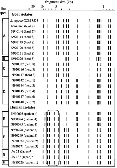
Ribotyping.The 26 isolates analyzed produced eight distinct
EcoRI ribotypes, designated A to H, consisting of 13 to 16
hybridizing bands, with six being common to the eight patterns (Fig. 1). The ribotype of the reference strain CCM3573 con-tained all the bands previously observed by de Buyser et al. for the same strain with a 16S rRNA-specific probe (4). Since we
used a 16S-23S rRNA-specific probe, the additional bands present in our case can be attributed to hybridization to 23S rRNA gene fragments. It is interesting that isolate N940146, which could not be identified by conventional methods, showed a ribotype identical to that of the reference strain (ribotype A). Goat isolates produced four distinct ribotypes (A to D) which were all distinct from those observed with the human isolates (E to H). These human ribotypes were easily distinguishable from the goat ribotypes by a core of common bands: two bands of 6.6 and 5.2 kb, strictly specific to the human profiles, and two bands of 7.2 and 4.5 kb also present in some goat ribotypes (Fig. 1). Ribotypes E, G, and H were associated with isolates from patient 4, Japanese patients, and patient 1, respectively. Ribotype F was common to isolates from patients 2, 3, and 5. Among the goat isolates, certain ribotypes (B and C) were observed with isolates from one herd only (herd 8) while other ribotypes (D and A [the ribotype of the reference strain CCM3573]) were scattered among different herds (herds 1, 3, 4, 6, and 7 and herds 2, 5, and 8, respectively).
The UPGMA algorithm was applied on a matrix of Dice’s coefficient to estimate the phylogeny of the eight ribotypes. The resulting dendrogram (Fig. 2) did not show a clear-cut distinction between human and goat ribotypes. Ribotype E (patient 4) appeared closer to the goat ribotype D than to the other human ribotypes. Conversely, human ribotypes F, G, and H formed a homogeneous cluster related to ribotypes A and C from goats.
DISCUSSION
Since the 1960s, certain species of CN staphylococci, espe-cially S. epidermidis, S. haemolyticus, and S. saprophyticus, have emerged as pathogens (13). Other, less frequently isolated species have recently been recognized as important pathogens, for instance S. lugdunensis in infective endocarditis (22). Al-though S. caprae has been occasionally isolated from human clinical specimens (10), minimal information has been avail-able about these patients and it is not possible to know whether such isolates represent true clinical infection. This study has unequivocally demonstrated that S. caprae is a human patho-gen capable of causing severe clinical disease both in the com-munity and in the hospital. Our patient 3 had the first reported case of definite infective endocarditis caused by S. caprae. The diagnosis was based on persistent positive blood cultures and macroscopic evidence of an intraventricular vegetation at sur-FIG. 1. Schematic representation of the eight EcoRI ribotypes (A to H) for
[image:3.612.65.300.73.410.2]the 26 isolates of S. caprae studied.p, species could not be determined with the ID32 Staph gallery. The four bands common to all human isolates are outlined.
FIG. 2. Dendrogram based upon UPGMA analysis of eight ribotypes of S. caprae isolated from goats (A to D) and humans (E to H).
on May 15, 2020 by guest
http://jcm.asm.org/
[image:3.612.319.555.73.232.2]gery from which S. caprae was recovered by culture, thus sat-isfying the Duke criteria for definite diagnosis of endocarditis (8). Like other species of CN staphylococci, S. caprae causes bacteremia associated with i.v. devices and it also appears to be a urinary pathogen capable of causing symptomatic infection in the absence of a urethral catheter.
The discovery of human infections with a species of staph-ylococcus originally isolated from goat milk raised the question of possible transmission of these bacteria from animals to humans. Although none of our five patients was known to have had contact with goats, our observations do not rule out this hypothesis, as phylogenetic methods applied to the ribotypes did not reveal two distinct lineages corresponding to the goat and human origins of the isolates. On the other hand, this result may illustrate the limits of the UPGMA algorithm in obtaining the correct phylogenetic tree on the basis of restric-tion fragment data, and this model can be inconsistent if the number of independent data analyzed is small and if there is one branch evolving at a faster rate than the others (3). When our data were analyzed with a different algorithm (Ward’s algorithm), a different result, which differentiated the goat ribotypes from the human ribotypes (analysis kindly provided by M. Struelens [data not shown]), was obtained. Visual anal-ysis of the ribotypes (Fig. 1) clearly distinguished the human isolates on the basis of a constant four-band pattern, suggest-ing a recent common ancestor for these strains. Other authors have found that S. caprae human isolates can be distinguished from some goat isolates on the basis of SmaI macrorestriction profiles and, to some extent, on the basis of their cellular fatty acids (9, 13). These observations suggest a parallel and inde-pendent evolution of the goat isolates and the human isolates. Nothing is known about the normal habitat of human strains of
S. caprae, but circumstantial evidence from the association of S. caprae with nosocomial i.v.-access-associated bacteremias
strongly suggests that it must be part of the commensal skin flora. Such information requires accurate identification of CN staphylococci in clinical laboratories. Kawamura et al. (11) reported 80 isolates recognized as S. caprae by a DNA-DNA hybridization but initially misidentified as S. haemolyticus, S.
warneri, S. hominis, and S. epidermidis by conventional
meth-ods or commercial kits. In our study only the two isolates from Japan and one goat isolate were not correctly recognized by the API database. The major discrepancy was observed with mannitol acidification, which was positive for 77% of our iso-lates (Table 2), contrasting with 1% positivity in the API da-tabase. When conventional methods for identification were not convincing, the ribotyping was of considerable help since the taxon-specific pattern of S. caprae (1, 4) was recognized for these isolates.
When our data are compared with those reported by Kloos (12) and the API database, key characters which help with the identification of S. caprae can be defined. S. caprae was CN, fibrinogen affinity factor negative, novobiocin susceptible, al-kaline phosphatase positive, acetoin positive, and sucrose neg-ative. The thermostable DNase was often positive (88%) but usually weak. All the S. caprae strains described by Devriese et al. (6) produced weak hemolysis on sheep blood agar similar to the effect of the S. aureus beta-hemolysin. We, however, found that even the hemolysis detected with the reference strain had a synergistic effect with the beta-hemolysin of S. aureus RN4220 and was considered to be of a delta type. Most isolates of S. caprae produced a delta-like hemolysin which was some-times associated with an alpha-like hemolysin.
S. caprae isolates were usually susceptible to most
anti-staphylococcal antibiotics including penicillin for three of the five patients. All 26 strains were uniformly resistant to
fosfo-mycin, a characteristic not reported previously for S. caprae. Resistance to methicillin (shown by the presence of the mecA gene and the mec regulator genes) has been detected only in Japanese human S. caprae strains (15, 19, 20). The 10 isolates from goats studied by Devriese et al. (6) were also largely susceptible to antibiotics, but one was resistant to penicillin, whereas all our goat isolates were penicillin sensitive.
The recognition of S. caprae as a human pathogen has prob-ably been thwarted by difficulties in its identification. Even with the current availability of commercial kits, not all clinical lab-oratories identify CN staphylococci, even strains isolated from severe infections, and ‘‘S. epidermidis’’ is the term still widely used to refer to such organisms. A larger collection of human isolates of S. caprae should allow a modification of the identi-fication scheme defined by the conventional method or com-mercial kits. This should lead to a greater understanding of the ecology of S. caprae and its clinical significance in human and veterinary medicine.
ACKNOWLEDGMENTS
We thank the clinicians and microbiologists in Japan (E. Tateda-Suzuki and K. Hiramatsu), in the United Kingdom (G. E. Venn), and in France (B. Poutrel, Y. Richard, B. Lina, and M. Miquel) for permission to study their patients and/or for submitting organisms; P. A. D. Grimont and F. Grimont (Institut Pasteur) and M. Struelens (Hoˆpital Erasme, Brussels, Belgium) for useful scientific advice and computer assistance; and V. Delorme and C. Mouren for technical assistance.
REFERENCES
1. Bialkowska-Hobrzanska, H., V. Harry, D. Jaskot, and O. Hammerberg. 1990. Typing of coagulase-negative staphylococci by Southern hybridization of chromosomal DNA fingerprints using a ribosomal RNA probe. Eur. J. Microbiol. Infect. Dis. 9:588–594.
2. Brun, Y., M. Bes, J. M. Boeufgras, D. Monget, J. Fleurette, R. Auckenthaler, L. A. Devriese, M. Kocur, R. R. Marples, Y. Piemont, B. Poutrel, and F. Schumacher-Perdreau.1990. International collaborative evaluation of the ATB 32 Staph gallery for identification of the Staphylococcus species. Int. J. Med. Microbiol. 273:319–326.
3. DeBry, R. W. 1992. The consistency of several phylogeny-inference methods under varying evolutionary rates. Mol. Biol. Evol. 9:537–551.
4. De Buyser, M. L., A. Morvan, S. Aubert, F. Dilasser, and N. El Solh. 1992. Evaluation of a ribosomal RNA gene probe for the identification of species and subspecies within the genus Staphylococcus. J. Gen. Microbiol. 138:889– 899.
5. Deinhofer, M., and A. Pernthaner. 1993. Differentiation of staphylococci from sheep and goat milk samples. Dtsch. Tiera¨rztl. Wochenschr. 100:234– 236.
6. Devriese, L. A., B. Poutrel, R. Kilpper-Ba¨lz, and K. H. Schleifer. 1983. Staphylococcus gallinarum and Staphylococcus caprae, two new species from animals. Int. J. Syst. Bacteriol. 33:480–486.
7. Dice, L. R. 1945. Measures of the amount of ecological association between species. Ecology 26:297–302.
8. Durack, D. T., A. S. Lukes, D. K. Bright, and the Duke Endocarditis Service. 1994. New criteria for diagnosis of infective endocarditis: utilization of spe-cific echocardiographic findings. Am. J. Med. 96:200–209.
9. George, C. G., and W. E. Kloos. 1994. Comparison of the SmaI-digested chromosomes of Staphylococcus epidermidis and the closely related species of Staphylococcus capitis and Staphylococcus caprae. Int. J. Syst. Bacteriol. 44: 404–409.
10. Kanda, K., K. Suzuki, K. Hiramatsu, T. Oguri, H. Miura, T. Ezaki, and T. Yokota.1991. Identification of a methicillin-resistant strain of Staphylococcus caprae from a human clinical specimen. Antimicrob. Agents Chemother. 35:174–176.
11. Kawamura, H., S. Adnan, N. Li, H. Miura, Y. Hashimoto, H. Yamamoto, and T. Ezaki.1992. Identification of staphylococci by quantitative hybridization and selection of 18 biochemical tests useful for their rapid identification. Presented at the Conference on Taxonomy and Automated Identification of Bacteria, Prague.
12. Kloos, W. E. 1990. Systematics and the natural history of staphylococci. J. Appl. Bacteriol. 19(Suppl.):25S–37S.
13. Kloos, W. E., and T. L. Bannerman. 1994. Update on clinical significance of coagulase-negative staphylococci. Clin. Microbiol. Rev. 7:117–140. 14. Kreiswirth, B. N., S. J. Projan, P. M. Schlievert, and R. P. Novick. 1989.
Toxic shock syndrome toxin 1 is encoded by a variable genetic element. Rev.
on May 15, 2020 by guest
http://jcm.asm.org/
Infect. Dis. 11:S83–S89.
15. Murakami, K., W. Minamide, K. Wada, E. Nakamura, and H. Teraoka. 1991. Identification of methicillin-resistant strains of staphylococci by poly-merase chain reaction. J. Clin. Microbiol. 29:2240–2244.
16. National Committee for Clinical Laboratory Standards. 1985. Reference agar dilution procedure for antimicrobial susceptibility testing for bacteria that grow aerobically. Approved standard M7-A. National Committee for Clinical Laboratory Standards, Villanova, Pa.
17. Rampone, H., C. Bogni, J. Giraudo, and A. Calzolari. 1993. Identification of staphylococci from bovine milk in Argentina. Zentralbl. Bakteriol. 279:537– 543.
18. Renaud, F., J. Etienne, A. Bertrand, Y. Brun, T. B. Greenland, J. Freney, and J. Fleurette.1991. Molecular epidemiology of Staphylococcus haemolyticus
strains isolated in an Albanian hospital. J. Clin. Microbiol. 29:1493–1497. 19. Suzuki, E., K. Hiramatsu, and T. Yokota. 1992. Survey of
methicillin-resis-tant clinical strains of coagulase-negative staphylococci for mecA gene dis-tribution. Antimicrob. Agents Chemother. 36:429–434.
20. Suzuki, E., K. Kuwahara-Arai, J. F. Richardson, and K. Hiramatsu. 1993. Distribution of mec regulator genes in methicillin-resistant Staphylococcus clinical strains. Antimicrob. Agents Chemother. 37:1219–1226.
21. Valle, J., S. Piriz, R. de la Fuente, and S. Vadillo. 1991. Staphylococci isolated from healthy goats. Zentralbl. Veterina¨rmed. 38:81–89.
22. Vandenesch, F., J. Etienne, M. E. Reverdy, and S. J. Eykyn. 1993. Staphylo-coccus lugdunensis endocarditis: report of 11 cases and review. Clin. Infect. Dis. 17:871–876.