Neutralizing antiidiotypic antibodies to factor
VIII inhibitors after desensitization in patients
with hemophilia A.
J G Gilles, … , J Vermylen, J M Saint-Remy
J Clin Invest.
1996;
97(6)
:1382-1388.
https://doi.org/10.1172/JCI118558
.
Hemophilia A patients producing antibodies towards FVIII are usually treated with infusions
of high doses of FVIII in an attempt to "desensitize" them. To examine the mechanisms by
which such desensitization operates, sequential plasma samples of two unrelated inhibitor
patients were analyzed for anti-FVIII and antiidiotypic antibodies before and during
infusions of high doses of FVIII. Anti-FVIII antibodies were separated from antiidiotypic
antibodies by immunoaffinity chromatography before analysis. We show in the present
study that the concentration of anti-FVIII antibodies did not change during a successful
desensitization and that antibodies maintained their capacity to inhibit the procoagulant
function of FVIII, even though the number of Bethesda units in plasma was reduced to
undetectable levels. Using a competition assay with mAbs, we further show that the
specificity of human antibodies did not vary significantly during therapy. Finally, we show
that the treatment elicited antiidiotypic antibodies, which neutralized the inhibitory capacity
of anti-FVIII antibodies. Inhibitor antibodies can therefore not be accurately evaluated in
plasma, as their function appears to be neutralized by antiidiotypic antibodies. These
findings could have implications for the design of new therapies for hemophilia A patients
with inhibitors.
Research Article
Find the latest version:
J. Clin. Invest.
© The American Society for Clinical Investigation, Inc. 0021-9738/96/03/1382/07 $2.00
Volume 97, Number 6, March 1996, 1382–1388
Neutralizing Antiidiotypic Antibodies to Factor VIII Inhibitors after Desensitization
in Patients with Hemophilia A
Jean Guy Gilles,* Benoît Desqueper,* Harald Lenk,‡ Jozef Vermylen,* and Jean-Marie Saint-Remy*
*Center for Molecular and Vascular Biology, Katholieke Universiteit Leuven, 3000 Leuven, Belgium; and ‡Universitätes-Kinderklinik, 4317 Leipzig, Germany
Abstract
Hemophilia A patients producing antibodies towards FVIII are usually treated with infusions of high doses of FVIII in an attempt to “desensitize” them. To examine the mecha-nisms by which such desensitization operates, sequential plasma samples of two unrelated inhibitor patients were an-alyzed for anti-FVIII and antiidiotypic antibodies before and during infusions of high doses of FVIII. Anti-FVIII an-tibodies were separated from antiidiotypic anan-tibodies by im-munoaffinity chromatography before analysis. We show in the present study that the concentration of FVIII anti-bodies did not change during a successful desensitization and that antibodies maintained their capacity to inhibit the procoagulant function of FVIII, even though the number of Bethesda units in plasma was reduced to undetectable lev-els. Using a competition assay with mAbs, we further show that the specificity of human antibodies did not vary signifi-cantly during therapy. Finally, we show that the treatment elicited antiidiotypic antibodies, which neutralized the in-hibitory capacity of FVIII antibodies. Inhibitor anti-bodies can therefore not be accurately evaluated in plasma, as their function appears to be neutralized by antiidiotypic antibodies. These findings could have implications for the design of new therapies for hemophilia A patients with in-hibitors. (J. Clin. Invest. 1996. 97:1382–1388.) Key words:
Factor VIII • inhibitors • desensitization • antiidiotypic
an-tibodies • tolerance
Introduction
More than 20% of hemophilia A patients under Factor VIII (FVIII)1 infusion therapy develop an antibody response
to-ward FVIII (1, 2). These antibodies, also called FVIII inhibi-tors, are routinely detected by the Bethesda method, an assay system carried out in plasma and in which the capacity of
anti-bodies to neutralize the procoagulant activity of FVIII is mea-sured (3).
Patients presenting with inhibitors are usually treated by regular infusions of high doses of FVIII, a treatment which is successful in 680% of patients with low titers of inhibitors (, 10 Bethesda units [BU]/ml) but only 60% in patient with high ti-ters (. 10 BU/ml; 4). Although the precise mechanism of ac-tion of such a “desensitizaac-tion” therapy is not elucidated, its aim is to reduce antibody titers , 1 BU, which is deemed to reflect the suppression of anti-FVIII antibody production.
The detection of anti-FVIII antibodies in plasma is, how-ever, rendered difficult by interference due to other serum proteins, such as antiidiotypic antibodies. The latter are found in the plasma of hemophilia A patients, and it has been sug-gested by us (5) and others (6, 7) that antiidiotypic antibodies could not only inhibit the binding of antibodies to FVIII but also play a role in the down-regulation of anti-FVIII antibody production. We have therefore developed a method by which anti-FVIII antibodies can be separated from possible antiidio-typic antibodies by affinity chromatography (8) and a series of assay systems by which anti-FVIII and antiidiotypic antibodies can be evaluated separately. Our aims were twofold: to deter-mine whether antiidiotypic antibodies could indeed interfere with the evaluation of anti-FVIII antibodies in plasma, and to determine whether infusions of FVIII boosted the production of antiidiotypic antibodies.
We report here the results obtained by analyzing sequen-tial plasma samples of two unrelated patients with inhibitors, before and during a successful desensitization treatment with high doses of FVIII. Our findings show that successful desensi-tization does not correspond to a reduction in FVIII anti-body titers. Instead, such treatment induces the production of specific antiidiotypic antibodies. This is, to our knowledge, the first longitudinal description of the evolution of anti-FVIII and corresponding antiidiotypic antibodies in hemophilia A pa-tients under therapy.
Methods
Reagents and buffers. Human recombinant FVIII (rFVIII) was ob-tained from Hyland (Glendale, CA) as material for laboratory use only; this preparation contained vWf but no stabilizer or other pro-tein. N-hydroxysuccinimidyl-LC (NHS-LC) biotin was purchased from Pierce (Rockford, IL), and Tween-20 was from Technicon In-struments Corp. (Tarrytown, NY). Buffers used were: glycine-buff-ered saline (GBS), 270 mM, pH 9.2; PBS, 8 mM, pH 7.4, containing 0.5% BSA (PBS-BSA) or 0.1% Tween (PBS-Tween); tris-hydroxy-aminomethane, 10 mM, pH 7.3, containing 0.5% casein (Aldrich Chemical Co., Milwaukee, WI) and 0.02% thimerosal (Sigma Chemi-cal Co., St. Louis, MO), pH 7.2 (Tris-casein).
Patient description and treatment. Patient RS, born in 1984, was diagnosed as severe hemophiliac at the age of 5 mo, with a residual FVIII level , 1%. He was treated by infusions of FVIII cryoprecipi-tate for bleeding episodes. An inhibitor (36 BU) was first detected at Address correspondence to J.-M. Saint-Remy, M.D. Ph.D., Center
for Molecular and Vascular Biology, Katholieke Universiteit Leuven, Herestraat 49, 3000-Leuven, Belgium. Phone: 32-16-345.791; FAX: 32-16-345.990.
Received for publication 25 April 1995 and accepted in revised form 18 December 1995.
the age of 3 yr. 14 mo later, the patient underwent a low-dose desensi-tization trial with 25 U FVIII cryoprecipitate/kg body wt adminis-tered three times a week; this resulted in a rise in antibody titer from 2 to 2,640 BU after 4 wk of treatment, followed by a gradual decline to 610 BU. At the age of 6, a high-dose infusion treatment (300 FVIII U/kg body wt per d) was initiated using Profilate SD 1000 (Al-pha Therapeutic, Langen, Germany) and followed without interrup-tion. Profilate SD shows a specific activity of 14.2 IU FVIII/mg pro-tein and contains 6200 mg IgG/ml. The antibody titer was 8 U before the start of the treatment and peaked at 900 U 4 wk afterwards. At the end of the fourth month, the level of inhibitor was , 1 BU, but it took a year to normalize FVIII recovery. The patient is currently on regular prophylactic treatment with daily infusions of 10 U FVIII/kg body wt (Profilate SD).
Patient TB was born in 1979 and diagnosed as severe hemophiliac (FVIII , 1% of normal value) during early infancy. He received infu-sions of FVIII cryoprecipitate when required for bleeding episodes. An inhibitor was detected at the age of 6. He was started on a low-dose desensitization treatment at the age of 10, using 25 U FVIII cryoprecipitate/kg body wt, administered three times a week. The in-hibitor titer increased to 360 U within 4 wk and then gradually de-clined to levels between 10 and 20 BU. At the age of 12, the patient was started on a high-dose continuous daily regimen (300 FVIII U/kg body wt per d) using Profilate SD. The inhibitor titer rose from 18 to 150 BU. 9 mo of therapy were necessary to reduce the level of inhibi-tors to , 1 BU. 18 mo later, the FVIII recovery time was normalized. The patient is currently on daily infusions of 30 U FVIII/kg body wt using Profilate SD. Both patients were HIV seronegative.
Preparation of human anti-FVIII antibodies. Plasma samples were taken at defined time points during the second desensitization trial: just before the start of FVIII infusions, when inhibitor titer was at its highest, after a significant decrease in titer had been observed, and roughly 1 and 2 yr after the start of infusions. All samples were ob-tained 12 or 24 h after the last infusion. Human anti-FVIII antibodies were prepared from serum by salt precipitation, gel filtration chroma-tography, and adsorption on insolubilized FVIII (8). For the latter, rFVIII was insolubilized on a hydrazide-activated solid phase (Affi-gel-Hz; Bio-Rad Laboratories, Richmond, CA). Specific antibodies were recovered from the column by sequential elution with acid and alkaline buffers.
The amount of IgG antibodies recovered by affinity purification and their isotype distribution was measured by specific ELISA as de-scribed previously (8). IgG subclasses were determined on plates coated with an anti–mouse IgG1 rat monoclonal IgG (UCL, Brussels, Belgium), followed by a mouse IgG1 specific to either human IgG1 (Oxoid Ltd., Basingstoke, UK), IgG2, or IgG3 (both from Calbio-chem Corp., La Jolla, CA). For the determination of IgG4, the corre-sponding mouse antibody (Calbiochem Corp.) was insolubilized di-rectly on the plate.
Assay for FVIII inhibitors. The titers of inhibitors were deter-mined in plasma using the Bethesda method (3). The capacity of af-finity-purified antibodies to neutralize the function of FVIII was eval-uated by a chromogenic assay in which thrombin-activated FVIII acts as a cofactor to Factor IXa in the conversion of Factor X to Factor Xa (9), using commercially available kits (Merz & Dade AG, Düdingen, Switzerland). The assay was carried out as described previously (5). Briefly, 35 ml of rFVIII diluted at 150 ng/ml in 150 mM NaCl contain-ing 10 mM CaCl2 were mixed with 35 ml human anti-FVIII antibodies
diluted at a concentration varying from 30 to 2.5 mg/ml in the same buffer. The mixture was incubated for 30 min at 378C. 50 ml was then added to a vial containing the chromogenic assay reagents. Control experiments included rFVIII incubated without specific antibodies or with the same concentration of autologous antibodies from which anti-FVIII antibodies had been removed by adsorption (flow-through fractions [FT]).
Assay for anti-vWf antibodies. Microtitration plates were incu-bated for 2 h at room temperature (RT) with 50 ml rabbit F(ab9)2
fragments specific for human vWf. These antibodies were obtained
from Dakopatts (Copenhagen, Denmark) and digested with pepsin by standard procedures. The plates were then washed three times with PBS-Tween before addition of 50 ml of a solution of purified hu-man vWf (obtained by courtesy of Dr. Mirella Ezban, Novo Nordisk, Copenhagen, Denmark), which was diluted to 2 U/ml in casein buffer. The plates were incubated for 2 h at RT and then washed four times with PBS-Tween containing 0.4 M CaCl2 to eliminate possible
residual FVIII contamination. 50 ml of a human IgG sample (1 mg/ml for IgG preparations before adsorption or 10 mg/ml for affinity-puri-fied anti-FVIII antibodies) diluted in casein buffer were then added for an incubation of 2 h at RT. The plates were washed three times with PBS-Tween, and the detection of bound IgG was carried out by sequential addition of 50 ml peroxidase-conjugated goat IgG specific for human IgG (diluted 1/1,000 in casein buffer) and 100 ml o -phe-nylene-diaminedihydrochloride (OPD). Control experiments in-cluded a panel of mouse mAb to FVIII and an mAb to vWf (obtained by courtesy of Dr. J. Arnout).
Protein biotinylation. A 1-ml solution of 360 mg/ml of rFVIII was prepared in 150 mM NaCl containing 10 mM CaCl2 and labeled with
biotin by addition of 100 ml of a 10 mg/ml stock solution of NHS-LC-biotin (8). After an incubation of 2 h at 48C the mixture was dia-lyzed against the same buffer. Affinity-purified human IgG were la-beled with biotin by the same method except for the use of 500 mg protein/ml and of a 150 mM NaCl solution without CaCl2.
Production and characterization of anti-FVIII mouse mAbs. mAbs were produced in BALB/c mice as described previously (8). Hybridomas secreting anti-FVIII antibodies were identified by direct binding to insolubilized rFVIII. Positive hybridomas were cloned by limiting dilution and expanded. Antibody isotypes were determined by specific immunoassays. mAb specificity was evaluated on nitrocel-lulose blots containing native or thrombin-digested FVIII as de-scribed (8; Gilles, J.G.G., K. Peerlinck, J. Arnout, J. Vermylen, and J.-M.R. Saint-Remy, manuscript submitted for publication). The ca-pacity of mAbs to neutralize the function of FVIII was evaluated with the Bethesda method and in the chromogenic assay described above.
Epitope mapping. The FVIII regions recognized by affinity-puri-fied human antibodies were identiaffinity-puri-fied in an assay system in which af-finity-purified human antibodies compete with mAbs for the binding to insolubilized FVIII (8). For this, polystyrene microtitration plates (Nunc, Roskilde, Denmark) were incubated for 2 h at RT with a solu-tion of 2 mg/ml rFVIII in GBS and then washed with PBS-Tween. 50 ml of an anti-FVIII mAb diluted at 0.5 mg/ml in PBS-BSA was mixed with 50 ml of a sample containing human anti-FVIII antibodies di-luted at a concentration varying from 30 to 2.5 mg/ml in the same buffer. 50 ml of the mixture was added to FVIII-coated plates for a 2-h incubation at RT. The plates were washed four times with PBS-Tween before addition of 50 ml biotin-conjugated goat IgG specific to mouse Fcg (Tago Inc., Burlingame, CA), which was diluted 1/4,000 in Tris-casein. The plates were incubated for a further 2 h at RT and then washed four times. 50 ml avidin-peroxidase (Sigma Chemical Co.) diluted at 1 mg/ml in PBS-BSA was then added. After 30 min at RT, 50 ml OPD was added and the absorbance read at 492 nm.
Assays for antiidiotypic antibodies. The detection of antiidiotypic antibodies was carried out using IgG FT fractions. Antiidiotypic anti-bodies were detected by their capacity to recognize the variable part of anti-FVIII mAbs and to inhibit the binding of labeled FVIII to mAbs (5, 8).
For the binding assay, polystyrene microtitration plates (Nunc) were incubated overnight with 50 ml of a given mAb diluted at 2 mg/ ml in GBS. The plates were then washed before addition of 50 ml of an FT sample containing 500 mg IgG/ml in PBS-BSA. After an incu-bation of 2 h at RT, the plates were washed again. The binding of hu-man antibodies was then detected by sequential addition of peroxi-dase-labeled goat IgG specific for human Fcg (Sigma Chemical Co.) and OPD. Control experiments included plates coated with an mAb of the same isotype but of unrelated specificity, and substitution of FT samples by a multidonor pool of g-globulins.
10 mg/ml in Tris-casein was mixed with 50 ml of an FT sample contain-ing antiidiotypic antibodies, previously diluted at either 250 or 500 mg/ml in PBS-BSA. A 50-ml aliquot of the mixture was added to the mAb-coated plates for an incubation of 2 h at RT. After washing, avi-din-peroxidase and OPD were added as described above. Control ex-periments included addition of biotin-labeled FVIII mixed with a pool of human g-globulins.
Reconstitution experiments. 50 ml of a solution containing 2 mg/ml of biotin-labeled affinity-purified anti-FVIII antibodies in Tris-casein were mixed with 50 ml of an FT sample diluted at 500 mg/ml in the same buffer. The mixture was incubated for 2 h at RT before addition to polystyrene plates preincubated with 2 mg/ml rFVIII in GBS. After an incubation of 2 h at RT, the plates were washed and the binding of anti-FVIII antibodies was evaluated by addition of avidin-peroxidase
and OPD as described above. Control experiments include the use of pooled human g-globulins as a substitute for FT samples.
Results
[image:4.612.63.555.82.260.2]Two hemophilia A patients who had developed an anti-FVIII antibody response were treated by infusions of high doses of FVIII in an attempt to desensitize them, i.e., to reduce specific antibodies to undetectable levels as assessed by a functional coagulation assay (Bethesda method). Five plasma samples for each patient were available for analysis, one before the start of desensitization and four while in therapy, including two sam-ples taken when inhibitors were no longer detected in plasma.
Table I. Anti-FVIII Antibodies during FVIII Desensitization
IgG subclasses
Patient Plasma sample Month BU FVIII recovery Anti-FVIII antibodies IgG1 IgG2 IgG3 IgG4
U/ml
% rise of FVIII:c/UI FVIII/
kg body wt mg/10 mg IgG %
RS 1 0 8 0.03 98 63 29 2 6
2 1 608 0.01 72 62 22 6 10
3 5 0.8 0.45 65 26 62 2 10
4 17 ,0.5 2.64 73 40 38 4 18
5 33 ,0.5 1.6 69 42 41 3 14
TB 1 0 18 ND 37 81 10 5 4
2 1 85 ,0.01 82 81 11 5 3
3 6 3 0.59 57 83 10 3 4
4 14 ,1 1.15 49 66 20 6 8
5 25 ,1 1.39 59 80 14 3 3
Evolution of the level of anti-FVIII antibodies during desensitization with high doses of FVIII. The number of BU was evaluated on plasma, The to-tal amount of anti-FVIII antibodies and their isotypic distribution were measured after affinity purification on FVIII-Affigel.
[image:4.612.57.430.496.722.2]The follow-up period covered 33 and 25 mo for patients RS and TB, respectively. The same plasma samples were used for assays described in Table I and in all of the figures.
Evolution of total anti-FVIII antibodies during desensitiza-tion. Antibodies to FVIII were purified by affinity chromatog-raphy from each plasma sample. The amounts of total anti-FVIII antibodies recovered from the immunosorbent together with results of the Bethesda method, which was carried out on plasma, are shown in Table I. It can be seen that there was ei-ther no (patient RS) or only marginal increase (patient TB) in the amount of antibodies 4 wk after the start of the desensitiza-tion treatment, suggesting that the increase in BU did not re-flect a mere increase in antibody production. Moreover, al-though the number of BU gradually declined over time to undetectable values, the yield of antibodies remained practi-cally unchanged over the entire follow-up time period. The isotypic distribution of anti-FVIII antibodies showed a relative increase in IgG2 and IgG4 for patient RS, but no significant changes for patient TB. As the rFVIII used for adsorption con-tained vWf, we checked the eluates for the presence of anti-vWf antibodies, which could interfere in the assay systems used here. No such antibodies were found using a direct bind-ing ELISA with a sensitivity of 10 ng/ml (data not shown).
Specificity of anti-FVIII antibodies. The discrepancy be-tween the results of the Bethesda method and those of total anti-FVIII antibodies prompted us to verify whether affinity-purified anti-FVIII antibodies maintained their capacity to neutralize FVIII during the follow-up period and to examine whether antibodies with novel specificity, but without inhibi-tory capacity, had been produced as a result of the treatment.
The capacity to neutralize the function of FVIII was evalu-ated in a chromogenic assay in which FVIII acts as a cofactor to FIX in the activation of FX. The results are shown in Fig. 1. It can be seen that there is in both cases an increase in the pro-portion of inhibitory antibodies, or of their affinity, which is, however, not proportional to the increase in BU (second plasma samples). Moreover, plasma samples taken at the time when inhibitors were undetectable by the Bethesda method still contained inhibitory antibodies. The concentration of the latter did not change significantly with time for patient TB; for patient RS, a two- to threefold reduction was observed com-pared with initial levels, which was much less than that ex-pected from the Bethesda results.
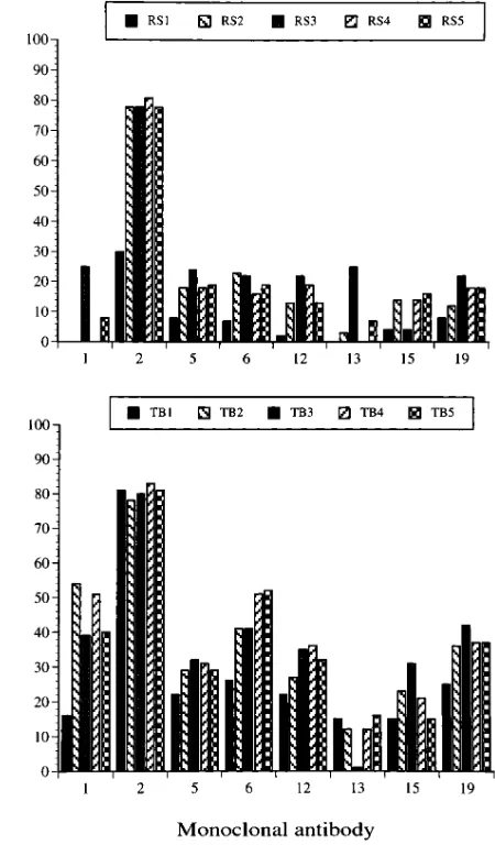
Next, to determine whether the specificity of antibodies had changed with time, we used an assay system in which hu-man antibodies competed with mAbs for the binding to insolu-bilized FVIII. 10 mAbs were used for this analysis; no signifi-cant competition was observed for 2 mAbs (mAbs 7 and 18). Results for the other 8 mAbs are given in Fig. 2, which shows that, for the two patients, the profile of antibody specificity did not change with time. Instead, in a few instances (see mAbs 1 and 12), the capacity of human antibodies to inhibit the bind-ing of mAbs increased, suggestbind-ing that the treatment increased either the proportion or the affinity of antibodies toward these sites. Of particular interest are the results obtained with mAb 15, an anti–heavy chain antibody that completely inhibited the procoagulant activity of FVIII: here again, the level of human antibodies did not change significantly with treatment, in con-trast with the evolution of functional inhibitors assessed by the Bethesda method in plasma. Remarkably, mAb 2 seems to identify an epitope that is precisely recognized by human anti-bodies of both patients; this epitope is located in the A2
do-main in between residues 606 and 740 (as determined by cour-tesy of Dr. Dorothea Scandella, Holland Laboratory, American Red Cross, Rockville, MD).
[image:5.612.327.552.247.631.2]Evolution of antiidiotypic antibodies. As neither the con-centration of anti-FVIII antibodies nor their specificity had changed with the desensitization treatment, we evaluated whether antibodies had been induced toward the idiotypes carried by anti-FVIII antibodies. For this, we used the IgG fractions that were not retained on the FVIII immunosorbent (flow-through, FT). These fractions did not contain residual anti-FVIII antibodies or FVIII that would have leached from the immunosorbent (data not shown). Two assay systems were used to detect antiidiotypic antibodies: a binding assay in which FT Igs reacted with the variable part of specific mAbs and an inhibition assay in which FT Igs inhibited the binding of labeled FVIII to mAbs.
As can be seen from Fig. 3, antibodies that bound to anti-FVIII mAbs became detectable, or their level increased, when infusions of high doses of FVIII were started. This is well illus-trated, for instance, for antibodies binding to mAb 2, which de-fines precisely an epitope recognized by human antibodies. One notable exception, however, concerns mAb 5, a heavy chain–specific mAb that showed partial competition with mAb 2. Control experiments using mAbs of unrelated specific-ity did not show any significant binding of FT Ig fractions (data not shown).
We then determined whether some of these antiidiotypic antibodies could inhibit the binding of labeled FVIII to insolu-bilized mAbs. Fig. 4 shows that it was in no cases possible to detect such antibodies in the FT fractions taken before treat-ment. However, these antibodies were often found as soon as
the treatment started. In some cases (see patient RS mAb 15), an almost complete inhibition of FVIII binding was observed.
[image:6.612.323.553.56.426.2]Reconstitution experiments. The capacity of antiidiotypic antibodies to inhibit the binding of affinity-purified human an-tibodies to FVIII was measured in a reconstitution experi-ment. FT samples taken before and during therapy were com-pared, using anti-FVIII antibodies obtained before treatment. As shown in Fig. 5, an FT fraction prepared from a plasma sample taken when the number of BU had already signifi-cantly decreased (sample 3) inhibited the binding of anti-FVIII antibodies to FVIII-coated plates to a significantly higher ex-tent than the pretreatment FT fraction. Because substitution of FT fractions by pooled g-globulins resulted in no inhibitory effect, this finding was taken as evidence for the induction of antiidiotypic antibodies to human anti-FVIII antibodies.
Figure 3. Binding of human antibodies to the variable part of anti-FVIII mAbs. The presence of antiidiotypic antibodies in human FT fractions was assessed by measuring their capacity to bind directly to the variable regions of anti-FVIII mAbs. Plates were incubated over-night with a given mAb. After washing, FT fractions containing 500 mg IgG/ml were added for 2 h at RT. The binding of human IgG was detected by addition of specific goat IgG. FT samples taken before (1) and at different time intervals (2 to 5) during the desensitization treatment were analyzed. Control experiments showed essentially no binding of FT fractions to mAbs of unrelated specificity and no bind-ing of pooled g-globulins to anti-FVIII mAbs (not shown).
[image:6.612.59.293.61.435.2]Discussion
Hemophilia A patients presenting with antibodies towards FVIII are usually treated with regular infusions of FVIII at doses that vary from one investigator to the other (10–13). The success or failure of such a treatment is decided based upon the capacity of the patient’s plasma to neutralize the procoagu-lant activity of FVIII, with a reduction , 1 BU considered as the objective to achieve. Through the analysis of sequential plasma samples obtained from two unrelated patients, we show that desensitization may in fact not reduce the level of anti-FVIII antibodies, that antibodies maintain their capacity to neutralize the procoagulant activity of FVIII, and that no significant changes occur with regard to antibody specificity.
The amount of anti-FVIII antibodies was evaluated after adsorption on insolubilized FVIII, a method that had been previously standardized (5). Whenever necessary, IgG samples were passed several times on the FVIII immunosorbent until no specific antibodies could be detected in the FT fractions. The isotypic profile of eluted antibodies showed a normal dis-tribution, except for a slightly raised proportion of IgG2 in one of the two patients, a finding that is usual with the adsorption method used here and that is possibly related to the recogni-tion of carbohydrate moieties on FVIII, as already discussed (8). Functional inhibitors were recovered in the eluates at each time point during the study, even when they were not detected in the plasma. It can therefore be concluded that reduction of BU titers , 1 U does not mean that functional inhibitors have been eliminated. It suggests, rather, that an interference, likely to be antiidiotypic in nature, existed in plasma.
That no change in antibody specificity occurred during treatment with FVIII was concluded from an assay system in which a panel of 10 mAbs was used. These mAbs were se-lected out of a large series of anti-FVIII antibodies and are probably representative of the antibody repertoire that BALB/c mice can mount toward human FVIII. Although it can be de-duced from the limited capacity of human antibodies to inhibit the binding of mAbs to FVIII (see Fig. 2) that mAbs do not recognize epitopes identical to those bound by human anti-bodies, we think mAbs can at least identify the main regions to which human antibodies bind. The difference in antibody spec-ificity should nevertheless be kept in mind when trying to iden-tify FVIII epitopes relevant to patients’ immune response. One possible exception concerns mAb 2, which defines an FVIII epitope that is clearly close, if not identical, to an epitope recog-nized by human antibodies. Interestingly, both patients have antibodies toward this epitope, which, however, was not prom-inent in the immune response made by other hemophiliac pa-tients analyzed so far (8). The question of whether the forma-tion of antibodies toward the region of FVIII defined by mAb 2 is related to the use of FVIII cryoprecipitates is currently un-der investigation.
Anti-FVIII mAbs were also used to detect antiidiotypic an-tibodies, and it is shown here that the treatment increased hu-man antibody binding to mAb V regions. As the patients had not received an FVIII preparation that could be contaminated by mAbs, and as the binding was specific for anti-FVIII mAbs, we think it corresponds to an idiotype–antiidiotype interac-tion. In addition, at least some of the human antiidiotypic anti-bodies recognized the antigen-binding site of mAbs, as shown by their capacity to displace FVIII from its binding to mAbs. However, this was observed mainly during the early months of treatment with FVIII, indicating that antiidiotypic specificity varied with time, whereas the total level of antiidiotypic anti-bodies was not reduced (Fig. 3). One might argue that antiidio-typic antibodies were passively given with the FVIII prepara-tion used to desensitize patients. It can indeed be calculated that patients received 630 mg IgG/d, a significant amount but one unlikely to contain a higher proportion of antiidiotypic an-tibodies than that found in healthy blood donors (5), which therefore renders it unlikely to be of physiological relevance or to interfere in our assay systems.
In an attempt to circumvent the difficulties inherent in the use of antibodies of different origins, a reconstitution experi-ment was carried out in which the binding of affinity-purified human antibodies to FVIII was prevented by preincubation with FT fractions taken before and during desensitization with FVIII. Although such experiments are difficult to carry out be-cause of the polyclonal nature of both the FVIII and anti-idiotypic immune responses, we think the results suggest an explanation why inhibitors were no longer detected in the plasma of patients under desensitization. Further insight in this matter would be gained if human mAbs were available; meth-ods to produce them are currently under development in our laboratory.
Our findings indicate that induction of unresponsiveness to FVIII by high-dose FVIII infusions does not involve losing the capacity to produce anti-FVIII antibodies. Instead, an inhibi-tion is exerted at the effector level, most likely because of the presence of antiidiotypic antibodies. A key question, which re-mains unanswered for now, is whether antiidiotypic antibodies can actively down-regulate the production of FVIII
[image:7.612.58.292.58.258.2]bodies. This has been suggested from the observation that an inverse relationship exists between the level of FVIII anti-bodies and that of corresponding antiidiotypic antianti-bodies in patients with acquired inhibitors (14), and it is certainly sup-ported by animal experiments (15). If this happens to be the case in the human immune response to FVIII, then therapeutic strategies by which antiidiotypic antibody production is boosted would be worth designing. One of the main advantages of such treatments would be their high specificity and potential low cost compared with current treatments for FVIII inhibitors.
In conclusion, and with the limitations related to the analy-sis of only two patients, we think that the present results chal-lenge the current view of the mechanism by which desensitiza-tion is effective for patients with FVIII inhibitors, and they might provide clues for novel therapies for such patients. One possible approach has already been reported by us (Gilles, J.G.G., J. Arnout, K. Peerlinck, J. Vermylen, and J.M.R. Saint-Remy, manuscript in preparation), in which patients were treated by injections of immune complexes made of FVIII and autologous specific antibodies, which resulted in a significant reduction in the level of FVIII inhibitors.
Acknowledgments
The authors thank Kathleen Peerlinck for reviewing the manuscript, Yves Delmarcelle for expert technical assistance, and Brigitte Firket for editorial help.
References
1. Schwarzinger, I., I. Pabinger, C. Korninger, F. Hasckeke, M. Kundi, H. Niessner, and K. Lechner. 1987. Incidence of inhibitors in patients with severe and moderate hemophilia A treated with factor VIII concentrates. Am. J. He-matol. 24:241–245.
2. McMillan, C.W., S.S. Shapiro, D. Whitehurst, L.W. Hoyer, A. Vijaya Rao, J. Layerson, and the hemophilia study group. 1988. The natural history of
factor VIII: c inhibitors in patients with hemophilia A: a national cooperative study. II. Observations on the initial development of factor VIII: c inhibitors.
Blood. 71:344–348.
3. Kasper, C.K., L.M. Aledort, R.B. Counts, J.R. Edson, J. Fratantone, D. Green, J.W. Hempton, M.N. Hilgartner, J. Lazarson, P.H. Levin, et al. 1975. A more uniform measurement of factor VIII inhibitors. Thromb. Res. 34:869–872. 4. Mariani, G., A. Ghirardini, and R. Bellocco. 1994. Immune tolerance in hemophilia: principal results from the international registry. Thromb. Haemo-stasis. 72:155–158.
5. Gilles, J.G., and J.M.R. Saint-Remy. 1994. Healthy subjects produce both anti–factor VIII and specific antiidiotypic antibodies. J. Clin. Invest. 94:1496– 1505.
6. Rossi, F., Y. Sultan, and M.D. Kazatchkine. 1988. Anti-idiotypes against autoantibodies and alloantibodies to VIII:C (anti-haemophilic factor) are present in therapeutic polyspecific normal immunoglobulins. Clin. Exp. Immu-nol. 74:311–316.
7. Sultan, Y., M.D. Kazatchkine, P. Maisonneuve, and U.F. Nydegger. 1984. Anti-idiotypic suppression of auto-antibodies to factor VIII (anti-hemophilic factor) by high dose intravenous gammaglobulin. Lancet. ii:765–768.
8. Gilles, J.G.G., J. Arnout, J. Vermylen, and J.M.R. Saint-Remy. 1993. Anti-factor VIII antibodies of hemophiliac patients are frequently directed to-wards nonfunctional determinants and do not exhibit isotypic restriction.
Blood. 82:2452–2461.
9. Svendsen, L., M. Brogli, G. Lindeberg, and K. Stocker. 1984. Differentia-tion of thrombin- and factor Xa-related amidiolytic activity in plasma by means of synthetic thrombin inhibitor. Thromb. Res. 34:457–464.
10. Brackmann, H., F. Etzel, P. Hoffmann, and H. Egli. 1977. The successful treatment of acquired inhibitors against factor VIII. Thromb. Haemostasis. 38: 369–374.
11. Gomperts, E.D., J. Stanley, J.A. Church, R. Sakai, and J. Lemire. 1984. Induction of tolerance to FVIII in a child with a high-titer inhibitor: in vitro and in vivo observations. J. Pediatr. 104:70–75.
12. Van Leeuwen, E., E. Mauser-Bunschoten, P. Van Dijken, A. Kok, E. Sjamsoendin-Visser, and J. Sixma. 1986. Disappearance of factor VIII: C anti-bodies in patients with haemophilia A upon frequent administration of FVIII in intermediate or low dose. Br. J. Haematol. 64:291–297.
13. Mauser-Bunshoten, R., J. Nilsson, and C. Kasper. 1991. Immune toler-ance, a 1990 approach. In Hemophilia and von Willebrand’s Disease in the 1990s. J.M. Lusher and C.M. Kessler, editors. Elsevier Science, New York. 265– 271.
14. Sultan, Y., F. Rossi, and M.D. Kazatchkine. 1987. Recovery from anti-VIIIc (anti-hemophilic factor) autoimmune disease is dependent on generation of anti-idiotypes against anti-VIIIc autoantibodies. Proc. Natl. Acad. Sci. USA.
84:828–831.