A common SCN5A polymorphism modulates
the biophysical effects of an SCN5A mutation
Prakash C. Viswanathan, … , D. Woodrow Benson, Jeffrey
R. Balser
J Clin Invest.
2003;
111(3)
:341-346.
https://doi.org/10.1172/JCI16879
.
Our understanding of the genetic basis of disease has expanded with the identification of
rare DNA sequence variations (“mutations”) that evoke inherited syndromes such as cystic
fibrosis, congenital epilepsy, and cardiac arrhythmias. Common sequence variants
(“polymorphisms”) have also been implicated as risk factors in multiple diseases. Mutations
in SCN5A, the cardiac Na
+channel gene, that cause a reduction in Na
+current may evoke
severe, life-threatening disturbances in cardiac rhythm (i.e., Brugada syndrome), isolated
cardiac conduction disease, or combinations of these disorders. Conduction disease is
manifest clinically as heart rate slowing (bradycardia), syncope, or “lightheadedness”.
Recent electrophysiologic studies reveal that mutations in particular families exhibiting
cardiac conduction disease cause marked effects on several competing voltage-dependent
gating processes, but nonetheless cause a mild “net” reduction in Na
+current. Here we
show that a common SCN5A polymorphism (H558R) in the Na
+channel I-II interdomain
cytoplasmic linker, present in 20% of the population, can mitigate the in vitro effects of a
nearby mutation (T512I) on Na
+channel function. The mutation and the polymorphism were
both found in the same allele of a child with isolated conduction disease, suggesting a
direct functional association between a polymorphism and a mutation in the same gene.
J. Clin. Invest.111
:341–346 (2003). doi:10.1172/JCI200316879.
Article
Find the latest version:
Introduction
Inherited mutations in the family of genes encoding voltage-gated Na+ channel pore-forming subunits
(SCNXA) underlie a wide spectrum of neurologic, mus-culoskeletal, and cardiovascular disorders. Mutations in SCN5A, the cardiac Na+ channel gene, generally
evoke disturbances in cardiac rhythm (1–3); however, it is increasingly clear that a great many single amino acid substitutions within the SCN5Acoding region can evoke a broad spectrum of cardiac rhythm behav-ior. While patients with SCN5Amutations linked to either long QT syndrome or Brugada syndrome expe-rience sudden, life-threatening arrhythmias, patients with isolated conduction disease exhibit heart rate slowing (bradycardia) that manifests clinically as syn-cope, or perhaps only as lightheadedness.
Functional biophysical studies of cardiac Na+
chan-nels with Brugada syndrome mutations invariably reveal defects in either gating function or surface
mem-brane expression that lead to a marked reduction in Na+current (4–6). This causes a disproportionate
has-tening of repolarization in the epicardial cell layer, lead-ing to a transmural electrical gradient that provokes reentrant arrhythmias (7, 8). More recent studies of Na+
channels have identified particular mutations linked to isolated conduction disease; these studies have revealed defects that also lead to a reduction of Na+
cur-rent, and in some cases the ion channel functional defect is mild (3, 9). Whereas even a single Na+channel
mutation may cause multiple changes in gating func-tion, each of which could individually drastically increase or decrease the Na+current, computational
models of cardiac excitability equipped to consider the ensemble of these mutational effects may predict only a mild net decrease in Na+current (3).
Mutations associated with a reduction of Na+current
may evoke Brugada syndrome, conduction disease, or both. While the severity of the Na+channel functional
defect may underlie the ECG phenotype in some cases (3), other unrecognized factors (i.e., humoral regula-tion, auxiliary subunits, transcriptional regulation) are certain to play a role. A recent study identified a new Brugada syndrome locus (10) distinct from SCN5Aand associated with progressive conduction disease, sug-gesting that other genes could also play a role in the manifestation of the disease phenotype.
Most contemporary studies of ion channels have sought to characterize the functional effects of isolat-ed mutations linkisolat-ed to rare diseases. At the same time,
A common SCN5A polymorphism modulates
the biophysical effects of an SCN5A mutation
Prakash C. Viswanathan,
1D. Woodrow Benson,
2and Jeffrey R. Balser
1,31Department of Anesthesiology, Vanderbilt University School of Medicine, Nashville, Tennessee, USA 2Children’s Hospital Medical Center, Cincinnati, Ohio, USA
3Department of Pharmacology, Vanderbilt University School of Medicine, Nashville, Tennessee, USA
Our understanding of the genetic basis of disease has expanded with the identification of rare DNA sequence variations (“mutations”) that evoke inherited syndromes such as cystic fibrosis, congeni-tal epilepsy, and cardiac arrhythmias. Common sequence variants (“polymorphisms”) have also been implicated as risk factors in multiple diseases. Mutations in SCN5A, the cardiac Na+channel gene,
that cause a reduction in Na+current may evoke severe, life-threatening disturbances in cardiac
rhythm (i.e., Brugada syndrome), isolated cardiac conduction disease, or combinations of these dis-orders. Conduction disease is manifest clinically as heart rate slowing (bradycardia), syncope, or “lightheadedness”. Recent electrophysiologic studies reveal that mutations in particular families exhibiting cardiac conduction disease cause marked effects on several competing voltage-dependent gating processes, but nonetheless cause a mild “net” reduction in Na+current. Here we show that a
common SCN5Apolymorphism (H558R) in the Na+channel I-II interdomain cytoplasmic linker,
present in 20% of the population, can mitigate the in vitro effects of a nearby mutation (T512I) on Na+channel function. The mutation and the polymorphism were both found in the same allele of a
child with isolated conduction disease, suggesting a direct functional association between a poly-morphism and a mutation in the same gene.
This article was published online in advance of the print edition. The date of publication is available from the JCI website, http://www.jci.org.J. Clin. Invest.111:341–346 (2003). doi:10.1172/JCI200316879.
Received for publication September 10, 2002, and accepted in revised form December 17, 2002.
Address correspondence to: Jeffrey R. Balser, Vanderbilt University School of Medicine, Room 560, Preston Research Building, 2220 Pierce Avenue, Nashville, Tennessee 37232-6602, USA. Phone: (615) 936-0277; Fax: (615) 936-2980;
E-mail: [email protected].
Conflict of interest: The authors have declared that no conflict of interest exists.
single-nucleotide polymorphisms, DNA sequence vari-ations that are common in the population, have been implicated in phenotypic variability in physiology, pharmacology, and pathophysiology (11–14). Recent-ly, a polymorphism in SCN5A(Y1102) was identified in individuals of African descent and implicated in an ele-vated risk for arrhythmia (15). Surprisingly, studies have not identified polymorphic alleles that modulate disease phenotypes by influencing the effects of inher-ited mutations within the same gene. Here we show that a common SCN5Aallele (H558R), present in one of five people (16), mitigates the gating defect caused by a nearby mutation (T512I) in the cardiac Na+
chan-nel DI-DII linker. The mutation and the polymor-phism were both found in the same allele of a child with conduction disease, and are the first to suggest a direct functional interaction between a common poly-morphism and a rare mutation within the same gene.
Method
Mutagenesis. Site-directed mutagenesis (T512I) was per-formed on SCN5A cDNA cloned in pSP64T as previ-ously described (17, 18). 1795insD mutant and H558R were prepared using the QuikChange Mutagenesis Kit
from Stratagene (La Jolla, California, USA), using hH1/pCGI-WT as template. H558R/T512I and H558R/1795insD double mutants were made by the same mutagenesis using H558R-hH1/pCGI and 1795insD/pCGI as template, respectively, for bicistron-ic expression of the channel protein and GFP reporter in tsa-201 or HEK 293 cells. Cells were cotransfected with an equimolar ratio of Na+channel human β1
sub-unit (provided by Al George, Department of Medicine, Vanderbilt University School of Medicine).
Electrophysiology. All whole-cell Na+ currents were
recorded at 21°C to limit the voltage-clamp error dur-ing activation and inactivation gatdur-ing at higher tem-peratures. The pipette solution contained 10 mM NaF, 110 mM CsF, 20 mM CsCl, 10 mM EGTA, and 10 mM HEPES (pH adjusted to 7.35 with CsOH); the bath solu-tion contained 145 mM NaCl, 4 mM KCl, 1.8 mM CaCl2, 1 mM MgCl2, and 10 mM HEPES (pH 7.35).
Cur-rents were sampled at 20 kHz using a Digidata 1200 analog to digital board (Axon Instruments Inc., Foster City, California, USA) and low-pass filtered at 2 kHz. The data were acquired using pClamp 8.0.1 (Axon Instruments Inc.) and analyzed using Clampfit (Axon Instruments Inc.). Voltage-clamp protocols are provid-ed as insets within each figure. The results are expressprovid-ed as mean ± SEM, and statistical comparisons were made by one-way ANOVA using Origin software (Microcal Software Inc., Northampton, Massachusetts, USA), with P < 0.05 indicating significance. Multiexponential functions were fitted to the data using the nonlinear least-squares method with Origin software.
Results
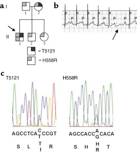
Clinical characteristics and genetic findings. We studied a family who came to medical attention when the proband, a 2-year-old at the time of initial examination (for pedigree see Figure 1a, II-1), was evaluated for an irregular heartbeat. A second-degree atrioventricular (AV) block was detected and treated with a pacemaker. The ECG trace (Figure 1b) shows a background heart rate of approximately 130 beats per minute (normal for a 2-year-old). The QRS duration and QT interval are normal. On conducted beats, the PR interval is rela-tively stable and prolonged to 200–240 ms (normal for age 2 years is less than 150 ms), a pattern suggestive of type 2 conduction system (Purkinje) block. In addition, the ECG shown (arrow) indicates an intermittent blocked beat, with shortening of the PR interval on the beat immediately following block, a feature suggestive of type 1 (AV) block. The parents and siblings have a normal ECG (normal QRS, PR, and QT intervals), and there was no evidence of maternal antibody–associated heart block (19).
SCN5Awas evaluated as a candidate gene, and muta-tions were sought using direct, bidirectional sequenc-ing of the codsequenc-ing region. Sequence analysis revealed a change of cytosine to thymine (C1535T), resulting in an amino acid change from threonine to isoleucine (T512I) at codon 512, and a change of adenine to gua-Figure 1
Genotype and ECG phenotype. (a) Pedigree shows affected
individu-als. Gray boxes represent the polymorphism, H558R, while black boxes represent the T512I mutation. While the father (I-1) was het-erozygous for H558R, the mother (I-2) was hethet-erozygous for H558R and T512I. The proband (arrow, II-1) was homozygous for H558R and heterozygous for T512I while his siblings (II-2 and II-3) were
het-erozygous for H558R. (b) ECG of proband indicating a second-degree
conduction block with normal QT and QRS durations. (c) Sequence
[image:3.576.59.290.52.311.2]nine (A1673G), resulting in an amino acid change from histidine to arginine (H558R) at codon 558 (Figure 1c). The nucleotide change C1535T creates a unique BstF5I restriction site, while the nucleotide change A1673G creates an AciI restriction site, allowing independent confirmation of both sequence changes (T512I and H558R). The father was heterozygous for H558R, while the mother was heterozygous for H558R and T512I with both sequence changes on the same allele. The proband (Figure 1a, II-1) was homozygous for H558R and heterozygous for T512I. His siblings (Figure 1a, II-2, II-3) were heterozygous for H558R and did not carry T512I. To verify that the polymorphism and mutation were on the same allele, exon 12 (from the proband) was PCR amplified and subcloned using the TA cloning method (Invitrogen Corp., San Diego, Cal-ifornia, USA). The four individual clones were then evaluated by restriction digest with BstF5I and AciI; two clones contained both T512I and H558R (inherit-ed from the mother) and two contain(inherit-ed H558R (inher-ited from the father).
T512I and H558R: competing effects on voltage-dependent gating. We expressed SCN5A wild-type, T512I, and H558R channels in tsa-201 cells to allow whole-cell voltage-clamp measurements. Figure 2c shows a typi-cal Na+current (I
Na) recorded during a step
depolariza-tion to –20 mV from a holding potential of –120 mV. We measured peak INaat varying activating voltages
(protocol inset, Figure 2b) and after conditioning steps to a range of inactivating voltages (protocol inset, Fig-ure 2a). The voltage dependence of activation (circles) and inactivation (squares) for wild-type
(filled symbols) and H558R (open symbols) channels were plotted (Figure 2a), and volt-age-dependent parameters were derived by fitting a Boltzmann function to the data (Table 1). Over a broad range of membrane potentials, activation and inactivation of wild-type and H558R channels were similar. In contrast, the voltage-dependence of acti-vation (open triangles in Figure 2b) and inac-tivation (filled triangles in Figure 2b) of T512I was shifted negatively by 8–9 mV (Table 1). Since the proband was heterozy-gous for T512I and homozyheterozy-gous for H558R, we also investigated the gating of the double
mutant H558R/T512I. Figure 2b shows activation (filled diamond) and inactivation (open diamond) of the H558R/T512I construct. Surprisingly, H558R elim-inated the negative shift in activation and inactivation.
Enhanced inactivation and conduction disease. Given that the polymorphism entirely corrected the T512I-induced effects on the voltage dependence of gating, we considered whether residual kinetic changes in H558R/T512I could explain the slow conduction phe-notype observed. Upon depolarization, Na+channels
rapidly open and “fast inactivate” within a few mil-liseconds, resulting in a transient inward current. To determine whether the rate of fast inactivation gating was modified by the mutation, we compared the decay of wild-type and T512I INaupon step depolarization to
test potentials ranging from –40 mV to 0 mV. Figure 2c shows representative INarecordings from wild-type and
T512I channels during a step depolarization from a holding potential of –120 mV to –20 mV. At all voltages tested, the rate of decay was similar.
Recent studies have demonstrated a pathophysiolog-ic role for slower kinetpathophysiolog-ic components of inactivation that develop throughout the time course of the cardiac action potential and delay recovery from inactivation between heartbeats (5, 6, 20). In Brugada syndrome, enhanced “slow” inactivation significantly delays the recovery of Na+channels between stimuli, causing a
[image:4.576.58.329.54.182.2]cumulative loss of function, particularly at rapid heart rates due to the shortened diastolic interval (5). We examined slow inactivation of T512I mutants using a dual-pulse protocol to mimic consecutive stimuli
Figure 2
Steady-state gating parameters. (a) Voltage dependence of
activation and inactivation of wild type and H558R obtained using the protocols shown in the inset and fitted to a
Boltz-mann function. (b) Activation and inactivation parameters
of wild type, T512I, and H558R/T512I fitted to a Boltzmann function. Note the hyperpolarizing shifts in activation and inactivation curves as a result of the mutation as well as their
restoration by H558R. (c) Wild-type and T512I INatransients
obtained during depolarization to –20 mV from a holding potential of –120 mV are normalized to illustrate similarity in fast inactivation.
Table 1
Voltage dependence of activation and inactivation
Inactivation Activation
V1/2 k V1/2 k
Wild type (n= 11) –85.0 ± 1.2 5.2 ± 0.2 –40.7 ± 1.4 5.9 ± 0.4
H558R (n= 10) –83.2 ± 1.4 5.0 ± 0.3 –42.1 ± 1.3 5.7 ± 0.5
T512I (n= 7) –92.5 ± 1.7A 4.8 ± 0.3 –49.4 ± 1.8A 6.6 ± 0.3
H558R/T512I (n= 10) –87.2 ± 1.3B 5.2 ± 0.1 –40.0 ± 1.8C 6.8 ± 0.3
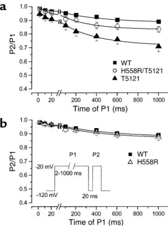
[image:4.576.260.539.598.677.2](inset, Figure 3b). As the duration of the first depolar-ization pulse (P1) was increased, the extent of slow inactivation increased, as indicated by the fractional reduction in peak INaduring the P2 pulse relative to
that recorded in the P1 pulse. A 20-ms repolarization to –120 mV was interposed between P1 and P2 to allow recovery from fast, but not slow, inactivation. Figure 3a shows that with increasing P1 duration, the current elicited by the P2 pulse decreased due to slow inactiva-tion. For the wild type, the extent of slow inactivation was similar to previously published results (21). In con-trast, T512I markedly increased the development of slow inactivation (Figure 3a), whereas H558R did not change the extent to which slow inactivation developed (Figure 3b). Given that H558R appeared to moderate the effect of T512I on the voltage dependence of inac-tivation gating (Figure 2), we hypothesized that H558R may also limit the extent to which slow inactivation develops with T512I. To test this, we assessed the devel-opment of slow inactivation of H558R/T512I (Figure 3a). We found that H558R/T512I significantly enhanced slow inactivation relative to the wild type but to a lesser extent than T512I alone (see Table 2), lend-ing support to our hypothesis.
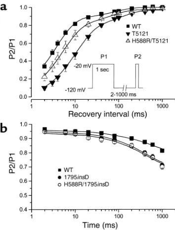
The detailed kinetics of recovery from inacti-vation were evaluated using a paired-pulse pro-tocol wherein the hyperpolarizing interval before the second depolarization pulse (P2) was varied (Figure 4a, inset). Both wild-type and mutant data followed a biphasic time course, reflecting components of recovery attributable to fast and slow inactivation. The kinetics of H558R recovery were indistinguishable from wild type (data not shown, but see Table 3). However, recovery of T512I was significantly slower than the wild type (Figure 4a and Table 3). Once again, the double mutation
H558R/T512I significantly hastened recovery from inactivation compared with T512I alone (P< 0.05, Fig-ure 4a and Table 3). These results also point to a mod-ulatory role of the H558R polymorphism on the slow inactivation gating characteristics of the mutation.
To investigate whether H558R modulation of slow inactivation is localized to companion mutations in the DI-DII linker, we engineered the C-terminal mutation 1795insD and H558R in the same cDNA and expressed them in HEK 293 cells. The 1795insD mutation enhances slow inactivation and elicits Brugada syn-drome, as reported previously (5) (Figure 4b). H558R had no effect on the kinetic features of 1795insD (Figure 4b), suggesting that the modulatory role of H558R can-not be generalized to remote channel loci and may be specific to nearby mutations within the DI-DII linker.
Discussion
Mutations of the human cardiac Na+channel gene
(SCN5A) underlie multiple cardiac diseases, including long QT syndrome and Brugada syndrome. While most of the mutations that cause long QT syndrome result in a gain of Na+channel function, mutations
that cause Brugada syndrome invariably result in a loss of Na+ channel function. Recent studies have
identified yet another class of mutations in the Na+
channel that cause isolated conduction disease. Tan et al. (3) provided the first evidence that a mutation in
SCN5A causes competing shifts in activation and inactivation gating, where the net effect is a small reduction in Na+current — sufficient to cause
[image:5.576.85.259.54.287.2]con-duction slowing, but not Brugada syndrome. While this suggests that manifestation of a particular rhythm phenotype (Brugada syndrome or conduction Figure 3
Development of slow inactivation. (a) Slow inactivation was
evaluat-ed using the two-pulse protocol shown in the inset in b. Plot shows
the ratio of P2/P1 for wild-type, T512I, and H558R/T512I as a func-tion of durafunc-tion of P1 pulse. Data points were fitted using a two-expo-nential function. While T512I dramatically enhanced slow
inactiva-tion, H558R attenuated slow inactivation caused by T512I alone. (b)
Slow inactivation of wild type and H558R. Inset shows the protocol used for evaluation. Slow inactivation of H558R was not different from wild type.
Table 2
Development of slow inactivation
A1 A2 τfast(ms) τslow(ms)
Wild type (n= 9) 0.03 ± 0.005 0.08 ± 0.007 13 ± 5 583 ± 61
T512I (n= 7) 0.07 ± 0.01 0.22 ± 0.01A 7 ± 3 565 ± 59
H558R/T512I (n= 6) 0.04 ± 0.009 0.12 ± 0.01B 10 ± 1 410 ± 83
Data points shown in Figure 3a were fit using a two-exponential function of the form
defect) may depend on the interplay between compet-ing gatcompet-ing lesions, it is also likely that other, unrecog-nized factors contribute to manifestation of a partic-ular disease phenotype. Recent studies have identified a novel gene locus for Brugada syndrome (10) on chromosome 3p22-25, distinct from the known Na+
channel loci and overlapping with a previously report-ed condition arising from locus 3p22-25 — dilatreport-ed cardiomyopathy with conduction disease (22). Although the functional role of this gene locus has not been identified, the study suggests that other as yet unidentified genes could play a role in the mani-festation of disorders in cardiac excitability.
We identified a novel mutation, T512I, in the Na+channel gene of a 2-year-old boy diagnosed
with second-degree AV conduction block. The T512I mutation, when heterologously ex-pressed, caused hyperpolarizing shifts in activa-tion and inactivaactiva-tion, and also enhanced slow inactivation. However, the common polymor-phism H558R also found in this child’s Na+
channel gene, which had no effect on wild-type Na+ current, attenuated the gating effects
caused by mutation T512I. The polymorphism
entirelyrestored the voltage-dependent activa-tion and inactivaactiva-tion voltage shifts caused by the T512I mutation, but only partially restored
the kinetic features of slow inactivation. No other fam-ily member carried the loss-of-function mutation (T512I) except the mother, who also carried the correc-tive H558R variant on the same allele (Figure 1a).
While additional studies will be required to firmly establish causality between conduction block and mod-est changes in slow inactivation, we offer the following hypotheses for linkage between the biophysical obser-vations and the observed phenotypes. The patient described here exhibits AV conduction slowing (pro-longed PR intervals) but no intraventricular or intra-atrial conduction defect (normal P wave and QRS dura-tion). Recent studies have associated similar Na+
channel slow-inactivation gating abnormalities with AV conduction defects in patients not exhibiting evi-dence of other conduction slowing (9). These results contrast markedly with the phenotype of another mutation (G514C) associated with slow AV conduc-tion, as well as delayed conduction throughout the atria and ventricles, including broad P waves, PR inter-val prolongation, and widening of the QRS complex (3). It is possible that enhanced slow inactivation pro-duced by H558R/T512I, which would cause Na+
chan-nels to recover from inactivation more slowly during diastole than wild-type channels do (Figure 3a), pro-vides a mechanism whereby AV conduction is slowed in preference to atrial or ventricular conduction. Muta-tions that preferentially delay recovery from inactiva-tion by enhancing slow inactivainactiva-tion could dispropor-tionately affect cells with longer inherent action potential duration (Purkinje cells). In contrast, as in the case of G514C, mutations targeting the channel activa-tionprocess could affect the myocardium more uni-formly, as has been observed (3). Consistent with this idea, it is noteworthy that in the present study H558R entirely eliminated the T512I effect on activation gat-ing (Figure 2b), but only partly corrected the slow inac-tivation defect. Greater accumulation of Na+channel
slow inactivation upon successive stimuli in Purkinje cells, with their longer action potential duration and smaller consequent diastolic interval (23), could lead to greater loss of Na+channel function in these cells at
[image:6.576.83.260.55.287.2]rapid heart rates (130 beats per minute, basal heart rate in the proband), and thereby produce isolated AV con-duction delay. Moreover, a premature stimulus could Figure 4
Recovery from inactivation. (a) Recovery was evaluated using the
two-pulse protocol shown in the inset. Data points were fitted using a two-exponential function with the fast time constant correspon-ding to recovery from fast inactivation and the slow time constant corresponding to recovery from slow inactivation. Plotted are the
results obtained from wild type, T512I, and H558R/T512I. (b)
Devel-opment of slow inactivation of wild type and 1795insD using the
pro-tocol shown in Figure 3b inset. It is observed that H558R had no
effect in attenuating the slow inactivation caused by 1795insD.
Table 3
Recovery from inactivation
A1 A2 τfast(ms) τslow(ms)
Wild type (n= 7) 0.87 ± 0.01 0.11 ± 0.01 3.8 ± 0.3 85 ± 7
H558R (n= 7) 0.86 ± 0.03 0.12 ± 0.03 4.1 ± 0.4 74 ± 5
T512I (n= 6) 0.82 ± 0.03 0.16 ± 0.03 11.6 ± 1.6A 137 ± 14A
H558R/T512I (n= 7) 0.85 ± 0.01 0.14 ± 0.01 5.5 ± 1.1B 83 ± 17C
Data points shown in Figure 4a were fit using a two-exponential function of the form
also further compromise the Purkinje diastolic interval and lead to dramatic loss of Na+current and result in
all-or-none repolarization and conduction block. In adulthood, the mother’s electrophysiologic pheno-type (by history), ECG, and 24-hour ambulatory moni-toring was entirely normal. It is possible that the slower heart rate of the mother and consequent longer diastolic interval provides sufficient time for recovery from slow inactivation and thereby did not result in a disease phe-notype. While it is unknown whether she displayed ECG abnormalities similar to the proband’s during child-hood, reduced penetrance of abnormalities in adults with SCN5Amutations has been previously described (3, 24). This may also relate to unidentified factors control-ling ion channel function and expression.
There is increasing awareness of the role of common polymorphisms in altering gene function and in sus-ceptibility to disease. Studies have linked gene polymor-phisms to elevated risk for cystic fibrosis, Alzheimer dis-ease (12, 13), and even heart disdis-ease (25, 26). In addition to their role in disease, polymorphisms are also thought to confer sensitivity to drug therapy (27), as well as proar-rhythmic risk from drug therapy (15, 28). This study pro-vides the first example in which a polymorphism in the same gene as a rare mutation alters the biophysical effect of the mutation on the channel protein.
Acknowledgments
This work was supported by the American Heart Associ-ation, Southeast Affiliate (to P.C. Viswanathan), and by the NIH (GM-56307 to J.R. Balser, U01 HL-65962 to P.C. Viswanathan, and HD-39946 to D.W. Benson). The authors thank the members of the subject family for their participation. The studies would not have been possible without the technical assistance of Ping Lu and the assistance of Linda Herrell and Marlene Brabham in collecting patient material.
1. Wang, Q., et al. 1995. SCN5A mutations associated with an inherited cardiac arrhythmia, long QT syndrome. Cell. 80:805–811.
2. Chen, Q., et al. 1998. Genetic basis and molecular mechanism for idio-pathic ventricular fibrillation. Nature. 392:293–296.
3. Tan, H.L., et al. 2001. A sodium-channel mutation causes isolated car-diac conduction disease. Nature. 409:1043–1047.
4. Dumaine, R., et al. 1999. Ionic mechanisms responsible for the electro-cardiographic phenotype of the Brugada syndrome are temperature dependent. Circ. Res. 85:803–809.
5. Veldkamp, M.W., et al. 2000. Two distinct congenital arrhythmias evoked by a multidysfunctional Na(+) channel. Circ. Res. 86:E91–E97. 6. Wang, D.W., Makita, N., Kitabatake, A., Balser, J.R., and George, A.L., Jr. 2000. Enhanced Na(+) channel intermediate inactivation in Brugada syndrome. Circ. Res. 87:E37–E43.
7. Antzelevitch, C., Yan, G.X., and Shimizu, W. 1999. Transmural disper-sion of repolarization and arrhythmogenicity: the Brugada syndrome versus the long QT syndrome. J. Electrocardiol. 32(Suppl):158–165. 8. Alings, M., and Wilde, A. 1999. “Brugada” syndrome: clinical data and
suggested pathophysiological mechanism. Circulation. 99:666–673. 9. Wang, D.W., Viswanathan, P.C., Balser, J.R., George, A.L., Jr., and Benson,
D.W. 2002. Clinical, genetic, and biophysical characterization of SCN5A mutations associated with atrioventricular conduction block. Circula-tion. 105:341–346.
10. Weiss, R., et al. 2002. Clinical and molecular heterogeneity in the Bru-gada syndrome: a novel gene locus on chromosome 3. Circulation.
105:707–713.
11. Hull, J., and Thomson, A.H. 1998. Contribution of genetic factors other than CFTR to disease severity in cystic fibrosis. Thorax. 53:1018–1021. 12. Roses, A.D. 1998. Apolipoprotein E and Alzheimer’s disease. The tip of
the susceptibility iceberg. Ann. NY Acad. Sci. 855:738–743.
13. Saunders, A.M., et al. 2000. The role of apolipoprotein E in Alzheimer’s disease: pharmacogenomic target selection. Biochim. Biophys. Acta.
1502:85–94.
14. Wilkins, M.R., Roses, A.D., and Clifford, C.P. 2000. Pharmacogenetics and the treatment of cardiovascular disease. Heart. 84:353–354. 15. Splawski, I., et al. 2002. Variant of SCN5A sodium channel implicated in
risk of cardiac arrhythmia. Science. 297:1333–1336.
16. Yang, P., et al. 2002. Allelic variants in long-QT disease genes in patients with drug-associated torsades de pointes. Circulation. 105:1943–1948. 17. Wang, D.W., Yazawa, K., George, A.L., Jr., and Bennett, P.B. 1996. Char-acterization of human cardiac Na+ channel mutations in the congenital long QT syndrome. Proc. Natl. Acad. Sci. USA.93:13200–13205. 18. Bezzina, C.R., et al. 1999. A single Na(+) channel mutation causing both
long-QT and Brugada syndromes. Circ. Res.85:1206–1213.
19. Buyon, J.P. 1998. Autoimmune-associated congenital heart block: demo-graphics, mortality, morbidity and recurrence rates obtained from a national neonatal lupus registry. J. Am. Coll. Cardiol. 31:1658–1666. 20. Rivolta, I., et al. 2001. Inherited Brugada and long QT-3 syndrome
muta-tions of a single residue of the cardiac sodium channel confer distinct channel and clinical phenotypes. J. Biol. Chem. 276:30623–30630. 21. Viswanathan, P.C., et al. 2001. Gating-dependent mechanisms for
fle-cainide action in SCN5A-linked arrhythmia syndromes. Circulation.
104:1200–1205.
22. Olson, T.M., and Keating, M.T. 1996. Mapping a cardiomyopathy locus to chromosome 3p22-p25. J. Clin. Invest. 97:528–532.
23. Schram, G., Pourrier, M., Melnyk, P., and Nattel, S. 2002. Differential dis-tribution of cardiac ion channel expression as a basis for regional spe-cialization in electrical function. Circ. Res. 90:939–950.
24. Priori, S.G., Napolitano, C., and Schwartz, P.J. 1999. Low penetrance in the long-QT syndrome: clinical impact. Circulation. 99:529–533. 25. Daley, G.Q., and Cargill, M. 2001. The heart SNPs a beat:
polymor-phisms in candidate genes for cardiovascular disease. Trends Cardiovasc. Med. 11:60–66.
26. Roses, A.D. 2000. Genetic susceptibility to cardiovascular diseases. Am. Heart J. 140:S45–S47.
27. Roses, A.D. 2000. Pharmacogenetics and the practice of medicine.
Nature. 405:857–865.
28. Sesti, F., et al. 2000. A common polymorphism associated with anti-biotic-induced cardiac arrhythmia. Proc. Natl. Acad. Sci. USA.