Human immunoglobulin selection associated
with class switch and possible tolerogenic
origins for C
dd
class-switched B cells
Nai-Ying Zheng, … , J. Donald Capra, Patrick C. Wilson
J Clin Invest.
2004;
113(8)
:1188-1201.
https://doi.org/10.1172/JCI20255
.
Current paradigms of peripheral B cell selection suggest that autoreactive B cells are
controlled by clonal deletion, anergy, and developmental arrest. We report that changes to
the human antibody repertoire likely resulting from these mechanisms both for a
well-characterized autoreactivity from antibodies encoded by the V
H4-34 gene and for other
hallmarks of an autoreactive repertoire are apparent mainly for class-switched B cells and
not for IgM germinal center, IgM memory, or IgM plasma cells. Other possible indicators of
autoreactivity found selected with immunoglobulin class include J
H6 gene segment usage,
increased frequency of B cells with long third hypervariable regions, and distal J
kgene
segment bias. Of particular interest is the finding that B cells with these same characteristics
are selected into the lineage of B cells that have undergone the unusual class switch from
constant region Cµ to C
d
(C
d
-CS). The C
d
-CS population also displays an increased
frequency of charged amino acids localized to the complementarity-determining regions,
further suggesting autoreactivity, and evidence is presented that these B cells had
undergone extensive receptor editing. Thus, the C
d
-CS lineage may be a “sink” for B cells
harboring autoreactive specificities in normal humans. A model for a new tolerizing
mechanism that could account for the C
d
-CS lineage is presented.
Article
Immunology
Find the latest version:
Research article
Introduction
One of the most prolific topics in immunology today is the mech-anism and regulation of Ig class-switch recombination (CSR). In this report, we find that changes to the human Ig repertoire occur mainly in association with class switch. Naive B cells can be induced by antigen encounter and T cell help to form germinal centers (GCs), ultimately leading to the generation of memory and plasma cells (PCs). Antigen-activated GC precursors differentiate to centroblasts, which rapidly proliferate and initiate the somatic hypermutation process (1–3). Centroblasts differentiate to cen-trocytes that can undergo CSR from IgM/IgD to secondary class-es such as IgG and IgA (4). CSR involvclass-es a genetic recombination that deletes all intervening sequence between switch regions, thus permanently juxtaposing the specific variable-diversity-joining (VDJ) region to the various secondary constant (C) regions. Of par-ticular interest to this study is a lineage of B cells that we refer to as “Cδ-CS” for “Cδclass-switched.” Unlike normal Cµ-versus-Cδ usage involving differential splicing of a single VDJ-Cµ-Cδ tran-script, Cδ-CS B cells actually undergo class switch from Cµto Cδ at the genetic level using cryptic switch regions between the Cµ and Cδexons (5, 6). Cδ-CS GC cells can differentiate into IgD-secreting PCs (7) and memory cells (8) and have the unusual char-acteristics that they use more than 90% λlight chains (7) and
accu-mulate more somatic mutations than any other B cell population (9), and we have shown this population has the highest frequency of receptor-edited heavy chain V regions (10).
VH4-34 gene segments encode natural autoantibodies that
rec-ognize the i and/or I (iI) antigens, which are the glycan “scaffold” for a number of common molecules, including the A, B, O, H, and Lewis blood group antigens and keratan sulfate (11–13). Neoplas-tic transformation and infections causing proliferation of VH4-34+
B cells can cause pathological cold agglutinin disease. VH4-34 also
has an enhanced role in a number of autoimmune syndromes, including lupus (14–16). There is also a report of an expanded IgD–CD27highB cell population in patients with lupus in which 20%
of the total cells utilized VH4-34 and another 20% expressed
anti-bodies encoded by the VH4-61 gene segment (17). Despite the
autoimmune potential of VH4-34, it is significantly
over-represent-ed in developing and naive B cells because it has the most efficient recombination signal sequence of all human VHgenes (18),
repre-senting 6–8% of naive and developing B lymphocytes in normal people (14, 19, 20). However, there is a powerful counterselection against VH4-34+B cells in GC cell, memory cell, and PC
popula-tions, and VH4-34+antibodies are difficult to detect in most serum
samples. There is no other naturally encoded antibody or VHgene
that is known to undergo such a profound change in repertoire rep-resentation, thus analysis of VH4-34 usage provides an excellent
model of B cell selection in normal humans.
B cells harboring receptors that bind to our own tissues are nor-mally controlled by clonal deletion or by editing of their B cell receptor through further V gene recombination (21, 22). Various related processes also alter the normal immunogenesis of autore-active B cells, including induction of anergy (23), exclusion from B cell follicles and developmental arrest (24, 25), and autoimmune “ignorance” (26). It has recently been found that 20% of B cells
Human immunoglobulin selection associated
with class switch and possible tolerogenic
origins for Cδ
class-switched B cells
Nai-Ying Zheng,1Kenneth Wilson,1Xiaojian Wang,1Angela Boston,1Grant Kolar,1
Stephen M. Jackson,1Yong-Jun Liu,2Virginia Pascual,3J. Donald Capra,1and Patrick C. Wilson1
1Molecular Immunogenetics, Oklahoma Medical Research Foundation, Oklahoma City, Oklahoma, USA. 2MD Anderson Cancer Center, Houston, Texas, USA. 3Baylor Institute for Immunology Research, Dallas, Texas, USA.
Current paradigms of peripheral B cell selection suggest that autoreactive B cells are controlled by clonal
dele-tion, anergy, and developmental arrest. We report that changes to the human antibody repertoire likely
result-ing from these mechanisms both for a well-characterized autoreactivity from antibodies encoded by the V
H4-34
gene and for other hallmarks of an autoreactive repertoire are apparent mainly for class-switched B cells and not
for IgM germinal center, IgM memory, or IgM plasma cells. Other possible indicators of autoreactivity found
selected with immunoglobulin class include J
H6 gene segment usage, increased frequency of B cells with long third
hypervariable regions, and distal J
κκgene segment bias. Of particular interest is the finding that B cells with these
same characteristics are selected into the lineage of B cells that have undergone the unusual class switch from
con-stant region C
µ
µ
to C
δδ
(C
δδ
-CS). The C
δδ
-CS population also displays an increased frequency of charged amino acids
localized to the complementarity-determining regions, further suggesting autoreactivity, and evidence is
pre-sented that these B cells had undergone extensive receptor editing. Thus, the C
δδ
-CS lineage may be a “sink” for
B cells harboring autoreactive specificities in normal humans. A model for a new tolerizing mechanism that could
account for the C
δδ
-CS lineage is presented.
Nonstandard abbreviations used: antinuclear antigen (ANA); class-switch recombination (CSR); complementarity-determining region (CDR); constant (C); Cδclass-switched (Cδ-CS); diversity (D); germinal center (GC); i and/or I (iI); Ig heavy chain joining region (gene) (JH); Ig heavy chain variable region (gene) (VH); Ig κchain
joining region (gene) (Jκ);Ig κlight chain variable region (gene) (Vκ); joining (J); plasma cell (PC); recombination signal sequence (RSS); third complementarity-determining region (CDR3); variable (V).
Conflict of interest:The authors have declared that no conflict of interest exists.
Citation for this article: J. Clin. Invest.113:1188–1201 (2004). doi:10.1172/JCI200420255.
research article
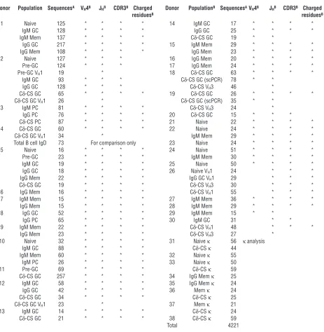
emerging from the bone marrow still retain some degree of autospecificity that must be controlled by peripheral selective mech-anisms (27). We initiated this study to characterize the dynamics of selection of peripheral B cells in normal humans. Our study includes a comprehensive characterization of peripheral B cell selec-tion involving cytometric analysis of 25 normal donors, and 86 V gene libraries from 38 donors totaling over 4,200 V gene sequences from various peripheral B cell differentiations (Table 1). Various libraries isolated from particular donors were analyzed for selection against use of the VH4-34 gene segment in addition to several other
well-characterized hallmarks typical of an autoimmune or
[image:3.585.59.522.248.721.2]coun-terselected repertoire and for evidence that the cells were subjected to receptor editing during development. The most significant find-ing was that changes to the Ig V gene repertoire in the periphery occur mainly in association with the Ig CSR process rather than just with differentiation to GC cells, memory cells, and PCs. Because the various features of the Ig repertoire, and particularly those features associated with autoimmunity, were selected in association with B cell class rather than B cell compartment, we conclude that although avoidance of autoreactive IgM-utilizing B cells must cer-tainly occur, the prevailing gross effect of peripheral selective mech-anisms may be avoidance of secondary class-switched B cells with
Table 1
Donors, populations, sequences, and clones analyzed
Donor Population SequencesA V
H4B JHB CDR3B Charged Donor PopulationB SequencesAVH4B JHB CDR3B Charged
residuesB residuesB
1 Naive 125 * * * * 14 IgM GC 17 * * * * IgM GC 128 * * * * IgG GC 25 * * * * IgM Mem 137 * * * * Cδ-CS GC 19 * *
IgG GC 217 * * * * 15 IgM Mem 29 * * * * IgG Mem 108 * * * * IgG Mem 23 * * * * 2 Naive 127 * * * 16 IgG Mem 20 * * * * Pre-GC 124 * * * 17 IgG Mem 24 * * * * Pre-GC VH1 19 * 18 Cδ-CS GC 63 * * * *
IgM GC 93 * * * Cδ-CS GC (scPCR) 78 * * IgG GC 128 * * * Cδ-CS VH3 46 *
Cδ-CS GC 65 * * * * 19 Cδ-CS GC 26 * * * * Cδ-CS GC VH1 26 * Cδ-CS GC (scPCR) 35 * *
3 IgM PC 81 * * * * Cδ-CS VH3 24 *
IgG PC 76 * * * * 20 Cδ-CS GC 15 * * * Cδ-CS PC 87 * * * * 21 Naive 22 * * * * 4 Cδ-CS GC 60 * * * * 22 Naive 24 * * * * Cδ-CS GC VH1 34 * IgM Mem 29 * * * *
Total B cell IgD 73 For comparison only 23 Naive 24 * * * * 5 Naive 16 * * * * 24 Naive 51 * * * * Pre-GC 23 * * * * IgM Mem 30 * * * * IgM GC 19 * * * * 25 Naive 50 * * * * IgG GC 18 * * * * 26 Naive VH1 24 * * *
IgG Mem 22 * * * * IgG GC VH1 29 * * *
Cδ-CS GC 19 * * * * Cδ-CS VH3 30 * * *
6 IgG Mem 16 * * * * Cδ-CS VH1 55 * * *
7 IgM Mem 15 * * * * 27 IgM Mem 36 * * * * IgG Mem 15 * * * * 28 IgM Mem 29 * * * * 8 IgG GC 52 * * * * 29 IgM Mem 15 * *
IgG PC 65 * * * * 30 IgM GC 31 * * 9 IgM Mem 22 * * * * Cδ-CS VH1 48 * * *
IgG Mem 23 * * * * Cδ-CS VH3 27 *
10 Naive 32 * * * * 31 Naive κ 56 κanalysis IgM GC 88 * * * * Cδ-CS κ 44
IgM Mem 60 * * * * 32 Naive κ 55 IgM PC 26 * * * * 33 Naive κ 50 11 Pre-GC 69 * * * * Cδ-CS κ 59 Cδ-CS GC 257 * * * * 34 IgG Mem κ 25 12 IgM GC 58 * * * * 35 IgG Mem κ 24 IgG GC 42 * * * * 36 Mem κ 24 Cδ-CS GC 34 * * * * Cδ-CS κ 25 Cδ-CS GC VH1 23 * 37 Mem κ 21
13 IgM GC 14 * * * * Cδ-CS κ 24 Cδ-CS GC 21 * * * * 38 Cδ-CS κ 59 Total 4221
ATo ensure accuracy, particular sequences were excluded from certain analyses (Methods).BStudies for which indicated populations were analyzed.
research article
autoreactive specificity. In addition, the Cδ-CS lineage of B cells appears to be a “sink” for autoreactive B cells in normal humans. These findings provide an important step toward understanding the homeostasis between antibody-mediated immunity and dan-gerous autoimmunity in normal humans.
Methods
B lymphocyte isolation and cytometry. B lymphocytes were isolated from human tonsils obtained during routine tonsillectomy following established NIH guidelines with proper institutional board approval or from the buffy coat component from 500 ml of freshly donated peripheral blood purchased from the Oklahoma Blood Institute (Oklahoma City, Oklahoma, USA). B cell purification by magnetic beads, staining, and sorting were performed as described previously (3, 7, 28), except for use of anti-CD27 to sort or identify memory B cells as described (29). The 9G4 rat mAb was purified from hybrido-ma supernatants using protein G–Sepharose columns (Amersham Biosciences, Arlington Heights, Illinois, USA) and was used in con-junction with PharMingen FITC-conjugated anti-rat Vκprotein (PharMingen, San Diego, California, USA).
VH4 library population nvalues are indicated in Table 1. The
populations for which V gene libraries were generated included naive (IgD+IgM+CD38– [tonsil] or CD27– [blood]), pre-GC
(IgD+IgM+CD38+), GC (IgD–CD38+, also CD27–for donors 13 and
14), centroblast (IgD–CD38+CD77+), centrocyte (IgD–CD38+CD77–),
memory (IgD–CD38–CD27+), PC (low density, CD38highCD20low),
and Cδ-CS GC (IgD+IgM–CD38+). Detailed phenotypes and isolation
procedures of all populations have been published previously (3, 4, 7, 9, 30–32). It is notable that the IgM GC (IgD–CD38+) fraction can
contain a minor portion of recent bone marrow immigrants (33), and a potential counterpart to the murine marginal zone B cell has been recently described that would also fall into this category (34).
Immunohistochemistry. Sections 5 µm in thickness from “snap-frozen” tonsils were cut and fixed with acetone. For three-step IgM analysis (Figure 1C), incubation with primary antibody 9G4 (anti–VH4-34) was followed by incubation with
FITC-conjugat-ed rabbit anti-rat IgG (Sigma-Aldrich, St. Louis, Missouri, USA), and then biotinylated goat anti-human IgM (Jackson ImmunoResearch, West Grove, Pennsylvania, USA) followed by Texas red streptavidin (Jackson ImmunoResearch) and finally aminomethylcoumarin acetate–conjugated donkey anti-human IgG (Jackson ImmunoResearch). Analyses were made with a Zeiss Axioplan fluorescent microscope in the Oklahoma Medi-cal Research Foundation imaging facility.
Sequencing Ig VDJ and VJ gene transcripts. For analyses of V gene repertoires presented in Figures 2, 3, 4, and 5 and Table 2, all PCR primer sequences are listed in Supplemental Table 1 (supplemen-tal material available at http://www.jci.org/cgi/content/full/113/ 8/1188/DC1) or as previously described (10, 28). Isolation of RNA, production of cDNA, and generation and screening of the various V gene libraries was done as described previously (10). An alternate VH4 gene primer (VH4P2) was used for several libraries to ensure
changes in the repertoire were not introduced by primer bias. PCR products were cloned using the TA Cloning Kit or the TOPO-TA Cloning Kit (Invitrogen, Carlsbad, California, USA). Individual colonies from the various V gene libraries were ran-domly picked for preparation of plasmid DNA using the miniprep kit (Qiagen, Valencia, California, USA) and sequencing at the Oklahoma Medical Research Foundation Sequencing Facility using either Applied Biosystems ABI-377 or ABI-3730
DNA sequencers. All V gene sequences were analyzed using “in-house” analysis software and the National Center for Biotech-nology Information IgBlast server (http://www.ncbi.nlm.nih. gov/igblast/) or the Immunogenetics server (http://imgt.cines. fr/textes/vquest/) (35). Statistical analyses were performed using Microsoft Excel (Redmond, Washington, USA).
Background mutation frequencies were determined to average 0.79 base mismatches per 1,000 bp based on analysis of a section of C-region gene sequence isolated as part of each cDNA clone ana-lyzed. Sequences of questionable quality for incorporated muta-tions were analyzed for excessive C-region mutamuta-tions above back-ground and if above backback-ground in the C region they were excluded from analyses of charged residues, somatic mutation, and iI anti-gen–binding residue analysis. Sequences in which hybrid V genes or other ambiguities were included for various analyses only if they were judged to have unique long third complementarity-determin-ing region (CDR3) segments; that is, a sequence with an ambiguous J region identity but a clearly unique VDJ junction (CDR3) was included for analysis of V gene usage but not J gene usage. Popula-tions isolated as naive were given greater scrutiny and only accept-ed as “naive” if one or zero mutations were detectaccept-ed in a V gene. Clonally related sequences were included if they differed by more than one mutation and thus could be considered differentially somatically mutated in order to represent the “true” repertoire. In order to determine if inclusion of clonal expansions would affect the outcome of this study, each analysis was also done in which only V genes with unique CDR3s were considered. These analyses gener-ated the same conclusions as the analyses presented that included all clones. Clones with identical CDR3s and differing by only one or no mutations were not included because they may have represent-ed dual cloning of a single PCR amplification product, although inclusion would have been unsubstantial (tested empirically).
Analysis of Vκ4-1 to Jκ2 for receptor editing. Genomic DNA was isolat-ed using DNAzol reagent (Molecular Center Research Inc., Cincin-nati, Ohio, USA) from naive (IgM+IgD+CD38–CD27–), Cδ-CS
(IgM–IgD+CD38+), and memory (IgD–CD38–CD27+) B cells. PCR of
Vκ4-to-Jκ2 rearrangements and recombination signal sequence (RSS) junctions were performed using the primer pairs (Supple-mental Table 1) illustrated in Figure 5B. PCR reactions were done using Qiagen HotStar Taq polymerase with the following cycle con-ditions: 15 minutes at 95°C, then 30 cycles of 95°C for 30 seconds, 60°C for 30 seconds, and 72°C for 1 minute, with a final 10-minute extension at 72°C. Ratios of genomic DNA isolates were first nor-malized for the abundance of Vκ4-to-Jκ2 rearrangements. Normal-ization based on presence of the Vκ4-1–to–Jκ2 rearrangement fre-quency also accounts for any loss of the RSS junctions due to the κ-deleting element rearrangement (36) that often accompanies receptor editing, because both the rearrangement and the junction would be lost with the κlocus. Comparison with memory B cells as well as naive B cells controls for the possibility that the increased RSS junctions are from epigenic (recombination circle) DNA due to previous rearrangement that might be detectable in the naive cells after recent immigration from the bone marrow.
Antibody expression and testing for 9G4 reactivity and cold agglutination. Transcripts of antibody VHgenes were cloned into an expression
vec-tor containing human IgG and were cotransfected with an FS7-κV gene into the 293A fibroblast cell line using Roche FuGene trans-fection reagent following the manufacturer’s suggested protocol (Roche Diagnostics Corp., Indianapolis, Indiana, USA). The FS7 anti-body is a classic cold agglutinin encoded by VH4-34, and the FS7-κ
research article
gene is permissive for expression of 9G4-reactive antibodies (37). Transfected 293A cells were allowed to secrete antibodies in serum-free DMEM supplemented with 1% Nutridoma-SP (Roche) for 5 days. Antibodies were purified using a protein G–Sepharose column (Amersham Biosciences). Antibody concentrations were determined by ELISA and Western blot and were adjusted to 1 µg/ml. Reactivity to the 9G4 antibody was determined by capture ELISA. ELISA plates were coated with anti-rat IgG (Jackson ImmunoResearch), followed by 9G4 mAb, and then were incubated with the various expressed VH/FS7κantibodies in serial threefold dilutions. Bound antibody was detected using biotin–anti-human IgG (Jackson ImmunoResearch) followed by steptavidin-HRP (Jackson ImmunoResearch) and devel-opment with HRP substrate (BioRad, Hercules, California, USA). Analysis of cold agglutination was performed by Coomb’s assay using commercially available reagents following the manufacturer’s protocol (Immucor Inc., Norcross, Georgia, USA).
Single-cell RT-PCR. IgD+IgM–CD38+ Cδ-CS B cells were bulk
sort-ed from the B cell fraction of isolatsort-ed human tonsils using a Cytomation MoFlo cytometer as described above. The bulk-sorted Cδ-CS cells were then re-sorted into 96-well PCR plates. VH4 gene
libraries were then prepared from the unused bulk-sorted cells and single-cell analysis was performed on the plates for comparison. Single-cell RT-PCR was performed as described previously (27). Individual V genes were cloned and sequenced as described above.
Results
Selection of the naturally autoreactive VH4-34 gene segment is evident by
analysis with the 9G4 mAb. In order to gain insight into the dynamics of tolerance to autoantibody-producing B cells in humans, we ana-lyzed VH4-34 gene segment usage in various mature B cell
subpopu-lations. The mAb 9G4 is highly specific for Ig encoded by the VH4-34
gene and no other, but as described below it is highly sensitive to incorporated somatic mutations. The predicted outcome of this
analysis was that B lymphocytes expressing the VH4-34 gene would
be excluded after the highly selective environment of the GC reac-tion. Evidence of this selection in GCs had been previously report-ed using the 9G4 mAb (14). Consistent with previous studies, anal-ysis of B cells from 24 donors (Figure 1A) by flow cytometry found that 6% of all naive B cells utilize the VH4-34 gene (6% ± 2% SD).
Given that there are approximately 50 functional VHgenes, the
VH4-34 gene is used at threefold the random frequency. Also
con-sistent with previous analyses, 3% ± 1% of IgM+memory cells (11
donors) and 3.9% ± 1% of IgM+GCs (2 donors) were 9G4+. Analysis
of 12 donor blood and tonsil specimens found that only 1.8% ± 1% of IgG+memory B cells were “encoded” by V
H4-34, which is
signifi-cantly fewer than the IgM+cells by analysis of the variance of means
between donors by Student’s ttest (P< 0.05). Interestingly, in results averaged from 14 tonsil specimens, 6% ± 3% of the unusual Cδ-CS GC B cells utilized VH4-34. Because this population accumulates the
most somatic hypermutations of all B cells (ref. 9 and Table 3), and, as discussed below, binding by the 9G4 antibody is highly sensitive to somatic mutations, detection of 6% of the cells as VH4-34+likely
indicated a much higher frequency of VH4-34+utilization, which did
indeed turn out to be the case (see below). Thus, this analysis gave the first indication that the Cδ-CS lineage might differentiate from B cells expressing natural autoreactivities in normal humans.
In conclusion, analysis with the 9G4 mAb demonstrated that VH4-34 usage is powerfully counterselected from IgG-utilizing B
cells and, unlike the results in previous reports, is significantly greater in IgM+memory or IgM+GC cells than the IgG+cells. In
addition, analysis with the 9G4 mAb would indicate counterselec-tion between naive and IgM+GC and memory cells. The 9G4 mAb
does not bind to all VH4-34–encoded antibodies (37), and there is
evidence that it is inhibited by accumulation of somatic mutations. Therefore, we were concerned that analyses of VH4-34 usage as
[image:5.585.45.284.74.360.2]detected by 9G4 binding both in our own studies reported above
Figure 1
Immunohistochemical and cytometric analysis of VH4-34+antibody
expression. (A) Cytometric analysis to determine the percentage of naive (24 donors), IgM+GC (2 donors), IgM+memory (Mem) cells (11
donors), IgG+memory cells (12 donors), and Cδ-CS GC cells (14
donors) utilizing VH4-34 (mAb 9G4+). Bars indicate mean. (B) Detection
of VH4-34–encoded antibodies by 9G4 is inhibited by somatic
hyper-mutation. Relative binding by 9G4 was compared by ELISA for an unmutated VH4-34–encoded antibody (FS7) plus six VH4-34–encoded
heavy chains harboring somatic mutations and a non–VH4-34 control
antibody (VH4-39). Dashes adjacent to the clone names indicate
muta-tions to the 9G4 mAb epitope at amino acids W7, A23, V24, and Y25, and replacements are indicated by single-digit amino acid code. The mean and SD of three assays are indicated. (C) Micrograph of a human tonsil section stained with anti-IgM (red), anti-IgG (blue), and anti-VH4-34
(9G4 mAb, green). The field is bisected by a follicular mantle (naive B cells) surrounding a GC at the bottom. Individual naive cells were scored for frequency of VH4-34 usage from 4 donors (7% ± 1% SD were
VH4-34+). (D) Micrograph of a tonsil section stained with 9G4 mAb
(anti–VH4-34, green). Two GCs are evident from the mantles of 9G4+
stained naive cells. The GC on the left is made up of an oligoclonal expansion of VH4-34-utilizing (9G4+) cells with 9G4brightplasmablasts
differentiating within the GC proper, indicating that VH4-34+B cells can
research article
and by other groups previously might underestimate the somati-cally mutated populations. Indeed, expression of six different mutated VH4-34–encoded VDJ transcripts with a “permissive” light
chain were significantly less reactive to 9G4 than to a germline VH
4-34 antibody (Figure 1B). The epitope of the 9G4 mAb includes a tryptophan residue (W) at position 7, and alanine, valine, and tyro-sine residues (AVY) at positions 23–25 (37). Clones having muta-tions directly to the W and AVY posimuta-tions are indicated in Figure 1B by dashes (identity to W AVY = “-_---“), and replacements are iden-tified by replacement of the dashes with the amino acid single-let-ter code. Three of these antibodies harbored mutations directly to the 9G4-bound epitope, and three had no mutations to the epitope directly but were otherwise mutated, suggesting that 9G4 binding is sensitive to mutations both directly and outside of the defined epitope. Assuming that 9G4 affinity translates directly to detection by flow cytometry, this sensitivity could account for the reduced 9G4 binding in all somatically mutated populations reported both here and previously (14). In addition, a comparison of 9G4 detec-tion of VH4-34 usage with VH4 gene repertoires generated by
ran-dom sequencing of the same populations demonstrated that only one half to three quarters of somatically mutated VH4-34
gene–uti-lizing B cells are detectable by 9G4 staining (data incorporated into Figures 1 and 2). In conclusion, there appears to be selection of the VH4-34 gene in association with class switch more than with B cell
compartment. However, because of the caveat that 9G4 antibody binding is highly sensitive to somatic mutations, we initiated a more accurate analysis of VH4-34 selection using random sequencing.
Analysis of the VH4 gene repertoire indicates that selection of the
autore-active VH4-34 gene segment is associated mainly with class switch. VH4-34
gene segment utilization was analyzed relative to that of the other nine VH4 family gene segments by random sequencing to provide
a more accurate and comprehensive analysis of the selection of this gene segment than the cytometric and histological studies provided. A VHgene family is defined as all VHgenes (totaling 50)
having greater than 90% similar nucleotides (38). Comparison of VH4-34 usage only to other VH4 family genes allowed a more
man-ageable and quantitative analysis, as all VH4 cDNA molecules can
be similarly PCR amplified by a single nondegenerate priming site in the VH4 leader region. Sixty-one libraries of VH4 gene family VDJ
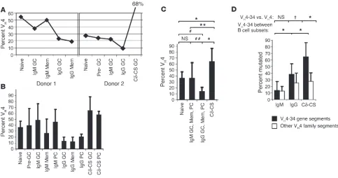
rearrangements were generated by random cloning of RT-PCR– amplified transcripts from various B cell subpopulations sorted by cytometry from 30 subjects. As indicated in Table 1 and Sup-plemental Figure 1, one to six B cell populations were isolated from each donor. Figure 2A depicts the selection of VH4-34 gene
usage in various populations sorted from two donors typical of the selection of this gene. Similar analysis of all donors is provid-ed in Supplemental Figure 1 and means with standard deviation (SD) are provided in Table 2. As depicted by the error bars (SD) of Figure 2, A and B, and by the comparison of all patients for VH4-34 use in Supplemental Figure 1, there was great variation
between individual donors for VH4-34 gene segment utilization
probably due to different immunological histories and different genetic backgrounds. This deviation demonstrates the need for sufficient nvalues to allow consideration of variance in analyses of human B cell repertoires. Despite the deviation in the overall frequency of VH4-34 usage between donors, VH4-34 was typically
used consistently between naive and secondary IgM populations (pre-GC, GC, memory, and PC), but at double the frequency in the IgM B cell subsets over the IgG populations. On average, VH4-34
was over-represented in naive B cells (37% ± 10% from nine donors,
Figure 2B) and was used consistently in all other B cell popula-tions that had not undergone class switch, including 40% ± 29% of VH4 transcripts from IgM+pre-GC cells (three donors), 43% ± 26%
of VH4 transcripts from IgM+GC cells (six donors), 28% ± 24% of
IgM+memory transcripts (ten donors), and 46% ± 21% of IgM+PC
transcripts (two donors). In contrast, B cells utilizing VH4-34 were
stringently counterselected from IgG populations, including IgG GC (mean = 11% ± 8%; six donors), and IgG memory (12% ± 8%; eight donors) or IgG PC (20% ± 5%; two donors). Consistent with the flow cytometry analyses, VH4-34–utilizing B cells were highly enriched
in the unusual Cδ-CS populations, including GC (65% ± 23%; ten donors) and a single Cδ-CS PC population (58%). It is notable that Student’s t-test comparisons did not demonstrate statistically sig-nificant differences between the various IgM naive, GC, memory, and PC populations, but showed they were all significantly differ-ent from the IgG populations with probabilities significant at least at the level of P< 0.05. IgG+GC, memory, and PC
popula-tions were also statistically similar. VH4-34 frequency in the Cδ-CS
lineage was different from that of all other B cell populations to at least the level of P< 0.005. It is notable that loss of VH4-34
uti-lization in the IgG populations was compensated for mainly by increases in VH4-39 and VH4-59, which are the next two J-distal
VH4 family gene segments in the VHlocus (Table 2). Certain other
VH4 genes (VH4-31, VH4-39, VH4-59, and VH4-61) displayed
signif-icant increases or reductions in particular donor populations, but not in a consistent fashion.
Reorganization of VH4-34 usage by Ig class demonstrates the
highly significant association of VH4-34 selection solely with class
switch (see Pvalues, Figure 2C). There is stringent selection against VH4-34+B cells when naive or secondary IgM B cells undergo class
switch to IgG (naive, 37% ± 10%; secondary IgM, 36% ± 25%; IgG, 13% ± 8%), and there is selection of VH4-34–utilizing B cells into the
Cδ-CS lineage (64% ± 22%). In conclusion, a more accurate and extensive analysis of VH4-34 gene usage by cDNA sequencing
indi-cates that peripheral differentiation or progression through a GC reaction of IgM+B cells to any other stage does not effect V
H4-34
usage. Selection of B cells using this naturally autoreactive VHgene
is only apparent in association with Ig class switch.
Correspondence between PCR/sequencing data, immunohistochemistry, cytometry, and single-cell PCR analysis. As described above, the 9G4 antibody binds with high affinity to unmutated VH4-34 gene–
encoded antibodies as found on naive B cells. In order to control for the quantitative capacity of the analyses performed with a dif-ferent technology, we stained tonsil tissue sections from four donors to count naive cells expressing VH4-34–encoded Ig usage
(9G4+) (two donors are presented in Figure 1, C and D). The tissue
section in Figure 1C has a clearly defined B cell follicular mantle stained with anti-IgM (red) arcing through the center of the image, over a GC at the bottom that is purple due to staining of the follicular dendritic cell network coated with captured IgM (red) and IgG (blue) antibodies. It is notable that unlike a previ-ous report in which no GCs could be found that were made up from expansions of VH4-34+cell populations (14), we readily
found 9G4+GCs in tonsil specimens from all donors analyzed. An
example of a VH4-34+GC and a VH4-34–GC in the same
micro-scopic field is presented in Figure 1D. There are also 9G4bright
plas-mablasts (early PCs) that have differentiated within the GC prop-er. Note that this section was stained only with the 9G4 mAb (and FITC-conjugated anti-rat) as secreted antibody captured in the fol-licular dendritic cell network are brightly stained by anti-IgM and
research article
anti-IgG, masking the GC cells that express only low levels of sur-face Ig (Figure 1C). Upon assigning scores for 9G4 staining, we found that 7% ± 1% (average ± SD) of the follicular mantle B cells from four donors are “encoded” by the VH4-34 gene segment.
Analysis by PCR determined that VH4 encodes 37% of VH4
tran-scripts sequenced (Figure 2). Because VH4 genes represent
approx-imately 20% of the total naive pool, based on the genetic frequen-cy of VH4 genes (38), our own analyses (Methods), and those of
others (14, 27, 39), this 36% represents 7.4% of all VHgenes (0.2
multiplied by 36%), corresponding excellently to the immunohis-tochemical analysis. The sequencing data also corresponds well with the cytometric analysis presented in Figure 1A, where 9G4+
cells averaged 6% ± 2% of the total from 24 donor samples. Previ-ous analyses have demonstrated that analysis of V gene repertoires by bulk PCR and random sequencing is similarly quantitative to single-cell PCR (40). However, with the unique conditions of PCR primers used herein, we felt it necessary to verify the bulk PCR data by single-cell PCR. Cδ-CS B cells from two donors were bulk sorted, and then a portion of these cells was distributed as single cells into a 96-well PCR plate by the flow cytometer for single-cell RT-PCR. We found that as long as the single-cell PCR efficiency approached 90%, the bulk–versus–single-cell analysis of VH4 and
JHgene use corresponded excellently for all genes (VH4-34, JH4, and
JH6 correspondence is presented in Table 4).
It is notable that bulk PCR analysis amplifying only VH4 genes
does not provide correspondence of gene expression relative to other VHgene families unless it is known what proportion of the
total cells use VH4 gene segments. For example, 20% of all naive
cells use VH4 gene segments; thus reference to 50% of the naive cells
expressing VH4-encoded antibodies would represent 10% of all
naive cells. However, relative abundance of VH4 genes was not
pre-viously known for the Cδ-CS lineage. As indicated in Table 4, sin-gle-cell analysis of two donors indicates that 46% and 54% of total B cells use VH4 family Ig genes, and VH4-34 accounts for 30% and
42% of all Cδ-CS B cells, respectively, in these donors. Thus, VH4-34
is even more over-represented in the Cδ-CS lineage than as deter-mined by the analyses in Figure 2. As indicated in Table 3 and pre-viously (9), the Cδ-CS lineage B cells harbor more somatic muta-tions than any other B cell lineage, and thus this analysis further demonstrates the sensitivity of the 9G4 mAb to incorporated mutations, as analysis with 9G4 appears to have detected only approximately 20% of the VH4-34+Cδ-CS B cells (Figure 1A).
Selection of VH4-34–utilizing B cells in association with class switch is
dependent on their autospecificity to the iI antigen. Due to the well-char-acterized natural autoreactivity of the VH4-34 gene to the iI glycan,
it is predicted that loss of VH4-34–utilizing B cell clones in the
[image:7.585.44.527.82.336.2]class-switched pools is due to negative selection to avoid autoreactivity in the secondary Ig repertoire. Alternatively, B cells expressing Ig
Figure 2
VH4-34+B cell selection correlates with antibody isotype and is due to iI antigen autoreactivity. VH4 family genes were randomly cloned and
sequenced from various B cell subpopulations sorted by flow cytometry. B cell subpopulation phenotypes are in the text, and Table 1 lists popu-lations isolated from each donor and number of V genes sequenced per population. (A) VH4-34 usage in the various subpopulations of two donors.
Similar analyses of all 29 donors are presented in Supplemental Figure 1. (B) Average use of VH4-34 by the various B cell subpopulations between
donors (means ± SD; Table 2 lists mean use of the remaining VH4 genes). VH4-34 encodes antibodies from the various IgM populations
signifi-cantly more than the IgG populations (Student’s ttest, P< 0.05) and was used in the Cδ-CS populations more than all others (P< 0.01). (C) VH4-34
usage organized by Ig class. Student’s ttest probabilities are indicated. *P< 0.0001; **P= 0.004;#P= 0.002;##P= 0.0008. (D) V
H4-34 is
proba-bly selected due to its autoreactivity to iI glycans. Bars indicate mean ± SD of V genes with mutations at amino acid positions 7, 23, 24, or 25. These amino acids are used for iI glycan binding by VH4-34+antibodies (W7 and AVY23–25). Despite equivalent mutation frequencies between
VH4-34+and other VH4 genes (Table 3), mutations at these amino acid positions are significantly selected only for VH4-34 genes, and only in
research article
encoded by VH4-34 may simply not be positively selected to
partic-ipate in IgG-mediated immune responses. In order to address this issue, we considered the specific binding of VH4-34–encoded
anti-bodies to the iI autoantigen. If indeed B cells utilizing VH4-34 are
negatively selected, it can be predicted that VH4-34+B cells
surviv-ing to selected populations, and particularly the IgG populations in which VH4-34 counterselection is so apparent, might do so because
the well-defined amino acids involved in iI glycan binding were somatically mutated during GC reactions. In Figure 2D, VH4-34+V
genes from the various isotype pools are analyzed for loss of the iI antigen binding site due to accumulated somatic mutations. iI anti-gen specificity is anti-generally independent of the CDR3 or the Ig light chain. Similar to the epitope of mAb 9G4 on VH4-34–encoded
anti-bodies, the specific amino acids important for binding of VH4-34 to
iI antigen are known through mutagenesis studies to be W at posi-tion 7 and AVY at posiposi-tions 23–25 (41, 42). V genes using VH4-34+
segments from IgG+and Cδ-CS B cells have accumulated somatic
mutations causing replacement of amino acid positions 7 and 23–25 at a significantly higher frequency than did V genes encoded by other VH4 family gene segments (for Cδ-CS cells, 67% ± 18% of
VH4-34+vs. 28% ± 16% of other VH4, χ2, P< 0.0001; and for IgG+cells,
39% ± 12% vs. 26% ± 8%, χ2, P< 0.05; Figure 2D). These mutations
are not due to an overall greater frequency of mutations to VH4-34
gene segments which are mutated at a frequency similar to that of V genes using other VH4 family gene segments (Table 3). However,
selection of iI binding site mutations is not evident for somatically
mutated secondary IgM populations (14% ± 8% of VH4-34 and 13%
± 9% of other VH4; analysis only of IgM+clones with >1 mutation).
Thus, there appears to be a profound selection against VH
4-34–uti-lizing B cells due to iI glycan binding, but in class-switched popula-tions only (χ2, P< 0.0001 for Cδ-CS vs. IgG or secondary IgM, and
also χ2, P< 0.0001 for IgG vs. secondary IgM). It is interesting to
note that despite the unexpected selection of VH4-34–utilizing B
cells into the Cδ-CS lineage (Figures 1A and 2, A and B), the cells must still be tolerized against iI glycan binding to survive. In addi-tion, the six recombinant VH4-34–encoded antibodies from
class-switched populations expressed for the analysis of Figure 1B were tested for their ability to agglutinate erythrocytes at low tempera-tures (cold agglutination). Cold agglutination is a hallmark of VH4-34 gene–encoded antibodies and is indicative of their ability to
bind the iI antigen (42, 43). Although three of these antibodies had mutations not directly at amino acid positions 7 and 23–25, only one of the six was able to agglutinate red blood cells in the cold at a low level, but not at a level comparable to that of control sera from a patient with cold agglutinin disease or the FS7 control antibody (data not shown). In conclusion, the selection for specific mutations of amino acids at the iI antigen binding site of VH4-34 genes but not
other VH4 genes is highly suggestive that B cells using VH4-34 are
being counterselected due to their iI antigen autoreactivity. As described in the previous section, consistent with this analysis of iI epitope mutations, mAb 9G4 only detects 20% of the Cδ-CS B cells and half of the already greatly reduced “IgG-encoded” B cell clones. However, based on the low frequency (14%) of iI glycan– binding mutations and thus the 9G4 epitope of mutated IgM clones, we would have expected higher detection of IgM clones by the 9G4 mAb. Because VH4-34 is found at a similar frequency in IgM
cells and naive cells, we would have predicted 9G4 binding to equal the frequency of naive cell detection by 9G4 multiplied by the fre-quency of clones retaining the 9G4 epitopes in the IgM GC and memory cells, or 6% ×0.86 = 5%. However, only 3% of IgM memory cells were actually detected (Figure 1A). Though seemingly minor, this 2% discrepancy is significant because binding of 9G4 mAb to IgM clones in Figure 1A is significantly different from that of naive clones by Student’s ttest (P< 0.01). Figure 1A is an analysis of CD27+IgM memory cells from blood. The autoantigen bound by
VH4-34 is the iI glycan that is found in abundance on erythrocytes.
Both iI antigen binding and the 9G4 epitope found in the FW1 region are affected in some VH4-34–encoded antibodies by the
CDR3 and light chain sequences (37, 42), possibly explaining the discrepancy between the current sequencing analysis and the
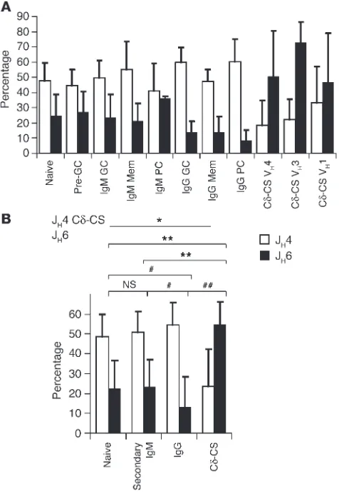
cyto-Figure 3
The autoimmune-associated JH6 gene is counterselected from IgG
pop-ulations and selected into the Cδ-CS lineage. (Aand B) The JH4 gene
segment is normally the dominant JHgene used in human VHgene
rearrangements. The JH6 gene is upregulated in receptor-edited and
autoimmune B cells, linking it to autoimmune repertoires. Only usage of JH4 versus JH6 gene segments is shown and compared, as all other
JHgene segment use is unchanged (other JHrepresentations are in
Table 2). JHanalysis was based on naive B cells (ten donors), IgM/D+
pre-GC cells (three donors), IgM+GC cells (eight donors), IgM+
mem-ory cells (ten donors), IgM+PCs (two donors), IgG+GC cells (seven
donors), IgG+memory cells (eight donors), IgG+PCs (two donors),
Cδ-CS PCs (one donor), and Cδ-CS GC cells (ten, five, and four donors for VH4, VH1, and VH3 libraries, respectively). Bars indicate means of all
donors and error bars, the SD. Student’s ttest Pvalues are indicated. *P< 0.0005 vs. all; **P< 0005;#P< 0.01;##P< 0.0001.
[image:8.585.45.283.84.430.2]research article
metric analysis of this and a previous report from a different group (14). Thus, although the repertoire representation of VH
4-34–uti-lizing B cells in the secondary IgM pools may not be significantly reduced, there could be selection of IgM+V
H4-34+B cell clones with
light chains or CDR3 regions that effectively “edit” the ability to bind iI glycan, which would also affect the 9G4 epitope involving the same region and amino acids of VH4-34–encoded Ig molecules.
Similarly, there may be selection of IgM+V
H4-34+B cell clones with
somatic mutations of the more-mutable CDR-encoded amino acids also indirectly affecting iI-glycan binding and also with the ancil-lary effect on detection by 9G4. A search for other possible mutat-ed sites in the IgM clones that could account for this discrepancy was fruitless. In conclusion, (a) there is selection against VH4-34–
utilizing B cells that retain iI glycan binding from all populations of B cells, including Cδ-CS cells, despite profound selection of VH4-34–utilizing B cells toward this differentiation; and (b)
although not significant at the gross level of repertoire analysis or sequence analysis for iI binding mutations, IgM memory cells may still be selected to avoid this autoreactivity. In the following sections, a number of other features of the human V gene repertoire associ-ated with autoreactivity or autoimmunity are characterized to gen-eralize the findings for VH4-34 gene usage to the total Ig repertoire. Peripheral selection of the autoimmune-associated JH6 gene is also
associ-ated with class switch. JH6 predominance has been detected in a
pop-ulation of receptor-editing human B cells (44–46) and is counters-elected from the pre–B cell repertoire upon progression to naive mature B cells (27), and thus it is already known to be subjected to primary B cell–selective mechanisms. Increased JH6 usage has also
been associated with autoreactive lymphocytic leukemia (47) and Sjogren syndrome (48). Finally, direct antibody expression analy-ses have recently demonstrated that JH6-encoded antibodies are
more likely to “encode” antinuclear antigen (ANA) autoreactivity and polyreactivity (27, 46). As indicated in Figure 3, the JH6
seg-ment encodes about one-quarter of the Ig molecules of the naive and secondary IgM populations (naive = 25% ± 14%; IgM/D+
pre-GC = 27% ± 14%; IgM+GC = 24% ± 15%; IgM+memory = 21% ± 12%;
and IgM+PC = 36% ± 2%; these populations were statistically
simi-lar), but use of JH6 is stringently counterselected from all IgG
pop-ulations (IgG+GC = 14% ± 7%, IgG+memory = 14% ± 11%, and IgG+
PC = 8% ± 7%; these populations are all similar, but each is statisti-cally different from naive and the various IgM populations by Stu-dent’s ttests at a level of P< 0.05). In addition, similar to VH4-34
usage, cells utilizing JH6 appear to preferentially class switch to Cδ,
where 50% ± 30% of the cells have V genes encoded by JH6 (P< 0.001
versus all other populations), with a significant reduction in the use of the normally dominant JH4 gene segment (P< 0.001).
Popu-lations analyzed for the various donors are indicated in Table 1. Reorganization of JHgene segment usage by Ig class in Figure 3B
more pointedly demonstrates the class-related selection of the JH
gene repertoire. Compared with V genes from naive cells (22% ± 14% JH6; 11 donors) and V genes from differentiated secondary IgM B
cells (23% ± 13% JH6; 20 donors), B cells with JH6-utilizing V genes are
significantly counterselected from the IgG populations (13% ± 9%; 18 donors; P< 0.01 by paired Student’s ttests). B cells utilizing the normally dominant JH4 gene are significantly selected into the IgG
Table 2
VH4 and JHgene segment utilization
VH4-04A VH4-28A VH4-30-2A VH4-30-4A VH4-31A VH4-34A VH4-39A VH4-59A VH4-61A VH4-bA
Naive 8 ± 7% 0 ± 0% 0 ± 1% 5 ± 8% 11 ± 9% 37 ± 10% 12 ± 6% 18 ± 11% 10 ± 11% 0 ± 0% Pre-GC 3 ± 4% 0 ± 0% 2 ± 3% 7 ± 2% 19 ± 13% 40 ± 29% 10 ± 4% 16 ± 14% 2 ± 2% 1 ± 3% IgM GC 5 ± 8% 1 ± 3% 1 ± 3% 4 ± 4% 18 ± 15% 43 ± 26% 12 ± 7% 13 ± 9% 2 ± 3% 1 ± 2% IgG GC 5 ± 4% 0 ± 1% 2 ± 2% 2 ± 4% 22 ± 14% 11 ± 8% 22 ± 7% 29 ± 11% 6 ± 7% 1 ± 3% IgM memory 3 ± 5% 1 ± 1% 1 ± 2% 3 ± 6% 8 ± 7% 28 ± 24% 19 ± 15% 21 ± 14% 15 ± 10% 1 ± 2% IgG memory 4 ± 5% 0 ± 0% 3 ± 5% 8 ± 12% 16 ± 12% 12 ± 8% 23 ± 11% 26 ± 14% 6 ± 8% 1 ± 2% IgM PC 9 ± 7% 0 ± 0% 0 ± 0% 4 ± 5% 0 ± 0% 46 ± 21% 1 ± 2% 11 ± 4% 30 ± 39% 0 ± 0% IgG PC 13 ± 2% 1 ± 1% 1 ± 1% 9 ± 12% 14 ± 20% 20 ± 5% 19 ± 3% 21 ± 1% 3 ± 2% 0 ± 0% Cδ-CS GC 0 ± 1% 0 ± 0% 2 ± 4% 4 ± 8% 10 ± 8% 65 ± 23% 4 ± 8% 7 ± 9% 4 ± 8% 3 ± 3% Cδ-CS PC 1% 0% 18% 19% 0% 58% 0% 4% 1% 0%
JH-1A JH-2A JH-3A JH-4A JH-5A JH-6A
Naive 3 ± 4% 3 ± 3% 10 ± 7% 48 ± 12% 12 ± 8% 25 ± 14% Pre-GC 1 ± 1% 4 ± 6% 10 ± 4% 45 ± 11% 13 ± 6% 27 ± 14% IgM GC 1 ± 2% 3 ± 3% 10 ± 7% 50 ± 11% 12 ± 6% 24 ± 15% IgG GC 2 ± 2% 3 ± 2% 10 ± 6% 60 ± 10% 13 ± 5% 14 ± 7% IgM memory 3 ± 3% 4 ± 4% 6 ± 6% 54 ± 19% 15 ± 6% 19 ± 12% IgG memory 3 ± 5% 5 ± 6% 11 ± 5% 47 ± 8% 20 ± 11% 14 ± 11% IgM PC 0 ± 0% 0 ± 0% 7 ± 4% 41 ± 18% 16 ± 12% 36 ± 2% IgG PC 2 ± 1% 5 ± 5% 6 ± 2% 60 ± 15% 19 ± 9% 8 ± 7% Cδ-CSB 1 ± 2% 3 ± 6% 13 ± 22% 23 ± 18% 9 ± 11% 52 ± 29%
AValues represent the mean percent utilization of each gene for the various donors ± the standard deviation.BCombined total mean J
Husage of VH1,
[image:9.585.53.542.108.336.2]VH3, and VH4 rearrangements.
Table 3
Mean mutations in various isotype pools
VHA VH4-34B Sequences
IgM GC, memory, PC 5 5 512 IgG GC, memory, PC 14 14 419 Cδ-CS lineage 23 24 265
AMean mutations per sequence for all V
Hrearrangements.BMean
research article
lineages (P= 0.04). Finally, JH6 is highly over-represented in the
Cδ-CS lineage in 50 ± 18% of the VHDJHtranscripts (17 donors, P< 0.0005 for Cδ-CS vs. naive cells and secondary IgM+B cells; and P< 0.0001 vs. IgG+B cells). Use of the remaining J
Hgenes is listed
in Table 2. Selection of the JH6 gene segment into the Cδ-CS lineage
is also independent of the VHgene used, as similar results were
obtained for V genes formed by rearrangements of VH1 family
genes (46% ± 33%) and VH3 family genes (73% ± 14%) (Figure 3). In
conclusion, JH6 gene usage is an independently selected feature
indicative of an autoimmune antibody repertoire that is selected in the periphery solely associated with class switch.
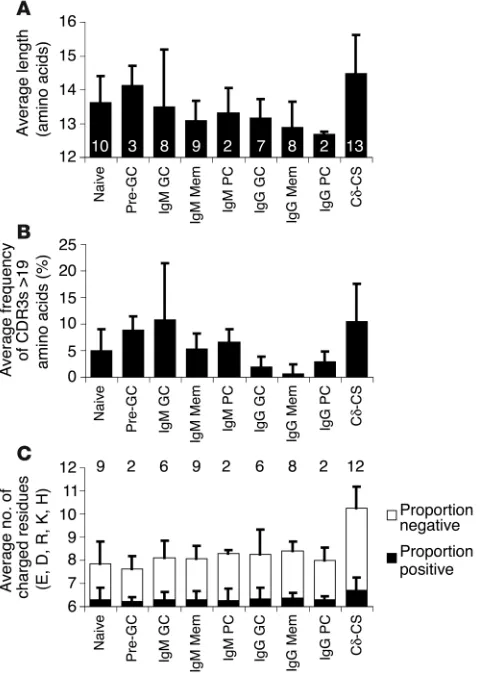
Long CDR3s are a classic hallmark of an autoimmune repertoire that are also selected in mature B cells solely on the basis of Ig class. The correlation of long CDR3s to autoimmunity has been made both for humans (49) and in mouse models of lupus, such as mrl/lpr mice (50, 51). Longer CDR3s have also been demonstrated in receptor-editing human B cells (44–46) and are counterselected during normal human B cell development (27). Finally, direct antibody expression analyses have recently demonstrated that antibodies with long CDR3s are more likely to have ANA autoreactivity and polyreactivity (27, 46). Typical-ly, a population is judged as having longer CDR3 regions if a higher frequency of B cells are found with excessively long CDR3 regions. Average CDR3 lengths are of limited utility because most V genes pre-dominantly have CDR3 lengths between 13 and 14 amino acids with some proportion of outliers. If there is counterselection of these out-liers, then there will still be little effect on the mean. However, as indi-cated in Figure 4A, V genes from B cells of the Cδ-CS lineage average
the longest CDR3s, although only significantly greater than the aver-age lengths of CDR3s of V genes from IgG (Student’s ttest, P< 0.01) and the IgM memory (Student’s ttest, P< 0.01) populations. In regards to difference in mean CDR3 length between IgM and IgG populations, although longer CDR3s are generally found in the V genes of IgM populations, only the means of naive and the IgM+D+
pre-GC populations were significantly increased over the IgG mem-ory and IgG PC populations (not the IgG GC). However, when the mean numbers of clones between the various donors with excessive-ly long CDR3 regions (>19 residues) were considered, the naive, all IgM populations (including IgM pre-GC, GC, memory, and PC), and the Cδ-CS lineage were statistically similar, and all had significantly more clones with CDR3 lengths greater than 19 amino acids than any of the IgG GC, memory, or PC populations at a level of P< 0.05 by Student’s ttest. It is interesting to note the immense variation between the eight donors from which IgM GC populations were ana-lyzed for both average CDR3 lengths and regarding the number of clones with CDR3s longer than 19 amino acids (see Figure 4, A and B, error bars representing SD), suggesting a significant effect of
anti-Figure 4
[image:10.585.46.286.83.421.2]Analysis of CDR3 length and frequency of charged residues in various V gene libraries. V genes with long CDR3s and increased numbers of charged amino acids have been associated with increased autoreactivity and are typical in the repertoires of people and mice with autoimmune dis-ease.The nvalues of donors analyzed per population are indicated on and above the bars, and the particular donors compared are in Table 1. (A) Average length of CDR3s in amino acids of the various donors for the var-ious populations. The average lengths of the Cδ-CS and pre-GC popula-tions are significantly greater then all IgG populapopula-tions and the IgM mem-ory population (ttest, P< 0.01), although the pre-GC cells were from only three donors, one of whom had a particularly large average CDR3 length. (B) Average frequency of V gene clones between donors that are more than 19 amino acids in length.The Cδ-CS frequency of clones with CDR3s longer than 19 amino acids is similar to that of the naive and IgM popula-tions.The IgG populations have on average significantly fewer clones with lengths greater than 19 amino acids than do the naive, IgM, and Cδ-CS populations (ttest, P< 0.05). (C) The mean number of charged residues in the CDR regions averaged among donors for each population where positively charged residues (R, K, H) are the black portion of the bars, and negatively charged (acidic) residues (E and D) are represented by the white portion of the bars. Error bars indicate standard deviations between the various donors for all panels.
Table 4
Single-cell RT-PCR (scRT-PCR) and bulk PCR analysis of Cδ-Cs cells
VH4A VH4-34B JH Sequences
VH4 Total JH4 JH6 VH/JH
Donor 19, scPCR: 46% 66% 30% 18% 75% 76/35C
Donor 19, bulk PCR: 69% 16% 76% 26 Donor 18, scPCR: 54% 79% 42% 14% 72% 78 Donor 18, bulk PCR: 81% 14% 76% 26
APercent of “V
H4 family–encoded” cells from total cells.BPercent
“VH4-34–encoded” rearrangements of VH4 genes and of total B cells.
CJ
Hanalysis was considered for VH4 only for donor 1 because bulk
analysis was only of VH4 genes and there is a VH3 clonal expansion
utilizing JH6. For each donor, 84 cells were analyzed (efficiency: 90%, donor 1; 93%, donor 2).
[image:10.585.309.536.607.688.2]research article
gen selection on average CDR3 length. In summary, as with analyses of VH4-34 and JHgene usage, CDR3 length demonstrates another
repertoire variation that is associated with autoreactive B cells and is counterselected from the IgG populations.
Additional distinctions analyzed for the various B cell subpopulations pro-vided no further evidence of repertoire variation beyond that found in associ-ation with class switch. Separation of naive cells (IgD+IgM+CD38–) from
two of the donors (donors 2 and 24) into CD23–and CD23+subsets
(or Bm1 and Bm2, as previously described) (3) found that these two naive B cell differentiations were the same for all analyses described above, and thus these groups were pooled. Similar differential anal-yses were performed comparing IgM versus IgD V gene libraries for the naive cells of one patient (donor 2, Bm1) and the pre-GC cells (IgM+IgD+CD38+) of two patients (donors 2 and 11). IgM versus IgD
libraries were also equivalent and the data pooled for the various analyses when from a single donor and population. In addition, two of the donor GC cell populations (from donors 2 and 5) were sepa-rated with anti-CD77 into centroblast (CD77+) and centrocyte
(CD77–) pools and were found to be the same. Finally, inclusion of
anti-CD27 (with anti-IgD and anti-CD38) for sorting cells of two tonsil specimens (donors 18 and 19) provided finer distinction of GC (IgD–CD38–CD27+) and memory cells (IgD–CD38+CD27–) but
pro-duced no significant differences from GC or memory populations analyzed from other donors. None of these finer distinctions pro-duced differential V gene repertoires other than the association with Ig class. It is also notable that PCs can arise via GC-independent dif-ferentiation; however, there is no way to sort IgM+PCs into
GC-dependent versus GC-inGC-dependent fractions. However, because naive versus IgM+pre-GC, IgM+GC, and subdivisions of IgM+GC cells
(centroblasts versus centrocytes), or IgM+memory cells from both
blood and tonsil were all similar, we can predict that differentiation of IgM+PCs into GC-dependent or -independent fractions would
not affect selection of VH4-34, JH, or CDR3 length.
B cells selected into the Cδ-CS population have an increased number of charged residues in their CDRs. Charged residues in autoantibody hypervariable regions are a common feature associated with
polyre-Figure 5
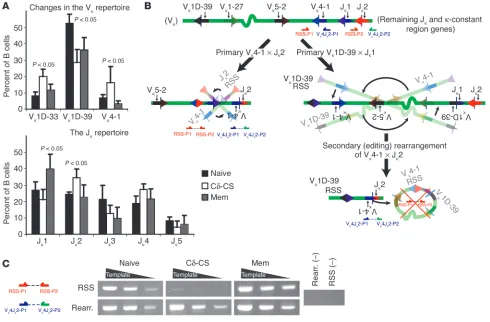
Analysis of the VJκrepertoire and configuration of the Vκloci for evidence of receptor editing. (A) Mean ± SD of Vκand Jκgene segments used
at significantly different frequencies between naive (four donors), Cδ-CS (five donors), and combined total memory (two donors) plus IgG+
mem-ory (two donors). (B) Semi-quantitative PCR was used to determine if increased Vκ4-1 usage provides evidence of receptor editing for Cδ-CS
cells. Vκ4-1 is the most Jκ-proximal Vκgene and is inverted. Thus, primary Vκ4-1 rearrangements retain the RSS junction (left), but following any
other primary inversional rearrangement, a secondary (editing) Vκ4-1 rearrangement will delete the RSS junction (right). Thus, analysis of the
genetic configuration of Vκ4-1–to-Jκ2 rearrangements provides a measure of receptor editing for all Vκgenes. The relative proportion of primary
versus editing rearrangements was determined by PCR using the primer sets indicated to get the ratio of Vκ4-1–to-Jκ2 rearrangements (primers: Vκ4Jκ2-P1, blue arrows; and Vκ4Jκ2-P2, green arrows) compared to RSS junctions (RSS-P1, red arrows; and RSS-P2, orange arrows). (C)
Genom-ic DNA template from naive, Cδ-CS, and memory B cells were normalized for Vκ4-1–to-Jκ2 rearrangements (Rearr.); then the relative proportion
of RSS junctions were determined by PCR at three dilutions of template (wedges). There was ten-fold less Vκ4-1–to-Jκ2 RSS junctions for the
[image:11.585.51.541.81.403.2]research article
activity, ANA autoreactivity, and are particularly important in pro-ducing anti-DNA reactivity (52–54). Charged residues are increased in human immature and naive B cells that are ANA reactive or are polyreactive (27, 46). In addition, increased numbers of charged residues are found among the Ig’s of editing human B cells (44–46). Finally, antibodies with excessive charged residues are counterse-lected during progression from pre-B and immature to naive B cells (27). As indicated in Figure 4C, Cδ-CS lineage populations from the various donors averaged significantly more charged residues in the CDRs than did all other B cell populations studied (ttest, P< 0.05 or more significant for Cδ-CS versus any other population). The Cδ-CS lineage populations averaged 25–30% more positively charged residues (Figure 4C, black portion of bars) associated with anti-DNA reactivity, including arginine, lysine, and histidine, and 15–20% more acidic residues (Figure 4C, white portion of bars), including glutamic acid and aspartic acid. Unlike the comparisons of VH4-34 and JH6 gene segment usage or CDR3 length, the naive,
various differentiated IgM, and various IgG populations all had a similar mean number of charged residues. The average number of charged residues was eight. It is notable that on average, around 35% of the clones of the naive populations and their most similar compartment, the pre-GC populations, had more than eight (aver-age) charged residues. In comparison, approximately 45% (43–51%) of the clones of the various IgM or IgG GC, memory, and PC com-partments had more than eight charged residues, although the variance between donors within each of these populations pre-cluded statistical significance. Only the Cδ-CS lineage had signifi-cantly more than the others, averaging more than eight charged residues in 72% of the clones. However, this reduction of charged residues for naive and pre-GC cells and the remaining populations gives rise to the possibility that antigen encounter selects for B cells with charged residues in the antibody hypervariable region. Thus, the combination of selection of B cells that are autoreactive due to an excess of charged residues and simultaneous selection for anti-genic reactivity may account for the particularly high frequency of charged residues in the Cδ-CS lineage. In conclusion, B cells with VHgenes encoding increased numbers of charged residues in the
CDRs preferentially undergo class switch to Cδ.
Cδ-CS B cells are selected from a population of B cells that has been sub-jected to extensive receptor editing. Receptor editing is the most clearly established selective mechanism that permanently alters an autoreactive B cell, and therefore it can be assayed experi-mentally. The Cδ-CS lineage uses more than 90% λlight chains, in contrast to the normally expected 30–40% λusage (7, 9). Light chain rearrangement is an ordered process (Vκ, then Vλ[ref. 55]), and increased λusage has been associated with receptor editing (56). However, increased λusage in the Cδ-CS lineage might also be due to selection of λ-utilizing cells. Although it is not a direct indication, increased evidence of Vκediting would suggest that the increased λusage might also have been induced by editing. Thus, the 10% of Cδ-CS cells that retained κrearrangements were analyzed for evidence that they had been subjected to edit-ing duredit-ing primary B cell development.
One indicator of receptor editing commonly considered for anal-yses of the murine Vκlocus is the proximity of Vκand Jκgenes used. A distal V–to–distal J gene-editing rearrangement can delete a more proximal V-to-J gene rearrangement in a “leapfrog” fashion. Thus, evidence of receptor editing is a VJκrepertoire skewed to the more distal V and J gene segments. However, due to the presence of many inverted Vκgenes in the human κlocus that rearrange by inversion
rather than deletion of the intervening sequence, analysis of the Vκ repertoire for editing is not necessarily informative. As indicated in Figure 5A, Vκ4-1 (or B3), which is actually the most J-proximal of all Vκgenes, was significantly over-represented in the Cδ-CS cells compared with either memory or naive cells. This finding was not surprising, as Vκ4-1 was previously found to be over-represented in isolated human B cells that were in the process of receptor editing (44) and in the lupus Vκrepertoire (57). Therefore, we hypothesized that because Vκ4-1 is the segment most proximal to the Jκgenes, any primary rearrangement of an upstream inverted Vκgene will result in its inversion and availability for secondary, editing rearrange-ments (Figure 5B). Thus, increased Vκ4-1 could be indicative of edit-ing. Because Vκ4-1 is also inverted, as a primary rearrangement product, the Vκ4-1 RSS–to–JκRSS junction will be retained in the locus. However, as a secondary (editing) rearrangement occurs after it is first inverted, the Vκ4-1–to–JκRSS junction will be lost (Figure 5B). Thus, because of the position of Vκ4-1 and its orientation in the Vκlocus, analysis of the genetic configuration of Vκ4-1–to–Jκ2 rearrangements provides a measure of receptor editing for all Vκ genes. Analysis by semiquantitative PCR indicated that for samples normalized based on Vκ4-1–to–Jκ2 rearrangements, relative to that of naive and memory B cells, there was tenfold less of the Vκ4-1 RSS–to–Jκ2 RSS product in Cδ-CS cells and therefore tenfold more editing (Figure 5C). In conclusion, the increased frequency of Vκ4-1–to–Jk2 rearrangements in Cδ-CS B cells results from exten-sive receptor editing. If it is assumed that the more than 90% λ usage is also due to editing, than it would appear that nearly all B cells that undergo class switch to Cδwere selected for editing dur-ing primary B cell development. A second possibility is that the Cδ-CS lineage is proficient for receptor editing in peripheral lym-phoid tissues, as we have previously reported that from this lineage, V gene clones can be isolated that have apparently edited their heavy chain V regions after the onset of somatic hypermutation (10). However, the origin of these recombinations is controversial because it is not clear if the RAG genes can be expressed during a GC reaction (58, 59). Thus, evidence of extensive receptor editing further demonstrates that B cells are probably selected into the Cδ-CS lineage due to encoded Ig autoreactivities.
Jκgene usage demonstrates selection of cells that did not require receptor edit-ing to the IgG populations. Because there are only five Jκgenes, all situ-ated in the same orientation, unlike the Vκrepertoire, analysis of Jκ usage for distal bias provides evidence of receptor editing. There is a bias for distal Jκutilization in the Cδ-CS population (Figure 5A), and distal Jκbias was previously reported in B cells in the process of recep-tor editing (45). Consistent with other analyses herein, both the IgG memory (IgD–IgG+CD27+) repertoire from two donors and total
memory (IgD–CD27+) from two additional donors, which consist of
more than 90% class-switched cells, were dominated by rearrange-ments to Jκ1 (Figure 5A). This is in contrast to the naive cells that uti-lize the Jκ1 through Jκ4 genes in an unbiased fashion. Thus, B cells harboring previously edited immune receptors and thus identified during primary selection as autoreactive are counterselected from the predominantly IgG memory populations.
Discussion
This report includes an extensive characterization of the expressed human VHDJHgene repertoire involving simultaneous analysis of
many stages of human peripheral B cell differentiation. We found significant changes in V gene repertoire features correlated with autoimmunity and receptor editing associated mainly with CSR,