Defects in secretion, aggregation, and
thrombus formation in platelets from mice
lacking Akt2
Donna Woulfe, … , Morris Birnbaum, Lawrence F. Brass
J Clin Invest.
2004;
113(3)
:441-450.
https://doi.org/10.1172/JCI20267
.
Prior studies have shown that PI3Ks play a necessary but incompletely defined role in
platelet activation. One potential effector for PI3K is the serine/threonine kinase, Akt, whose
contribution to platelet activation was explored here. Two isoforms of Akt were detected in
mouse platelets, with expression of Akt2 being greater than Akt1. Deletion of the gene
encoding Akt2 impaired platelet aggregation, fibrinogen binding, and granule secretion,
especially in response to low concentrations of agonists that activate the G
q-coupled
receptors for thrombin and thromboxane A
2. Loss of Akt2 also impaired arterial thrombus
formation and stability in vivo, despite having little effect on platelet responses to collagen
and ADP. In contrast, reducing
Akt1
expression had no effect except when
Akt2
was also
deleted. Activation of Akt by thrombin was abolished by deletion of G
a
qbut was relatively
unaffected by deletion of G
a
i2, which abolished Akt activation by ADP. From these results
we conclude that Akt2 is a necessary component of PI3K-dependent signaling downstream
of G
q-coupled receptors, promoting thrombus growth and stability in part by supporting
secretion. The contribution of Akt1 is less evident except in the setting in which Akt2 is
absent.
Article
Cardiology
Find the latest version:
tional signals are required to enhance platelet aggrega-tion and promote stability of the aggregate. The signal-ing mechanisms that contribute to platelet plug stabil-ity are far from understood, however.
Previous studies have established a unique role for ADP in stabilizing platelet aggregates as they form (1–5). This is particularly evident when platelets are stimulated with low concentrations of thrombin or analogues of thromboxane A2. Under these conditions,
secretion of ADP is required to elicit irreversible aggre-gation: in the presence of ADP scavengers or inhibitors of the Gi-coupled ADP receptor, P2Y12, platelets
aggre-gate, but the aggregates become unstable and dissoci-ate (1). This type of “reversible” aggregation is also seen in the presence of PI3K inhibitors and when individual PI3K isoforms are knocked out, demonstrating that PI3K plays an important role in platelet activation (1, 5, 6). PI3Ks are intracellular lipid kinases that phos-phorylate phosphoinositides at the D3 position of the inositol ring. Some of the products of PI3K have been detected following activation of platelets with throm-bin, thromboxane A2analogue, and, to a lesser degree,
ADP (1, 7, 8). Specifically, platelets stimulated with thrombin generate an early wave of PI(3,4,5)P3
(phos-phatidylinositol 3,4,5-triphosphate) and a later, inte-grin-dependent wave of PI(3,4)P2
(phosphatidylinosi-tol 3,4-diphosphate), which is also dependent on secreted ADP (9–11). It is generally accepted that these polyphosphoinositides help to trigger cellular process-es at least in part by binding to pleckstrin homology domains of effector proteins, thereby translocating
Introduction
Platelet activation is important for mediating hemosta-sis, but also contributes to thrombosis in the arterial cir-culation, the primary cause of heart attacks and strokes. Platelets first become activated at sites of vascular injury when they encounter matrix proteins exposed by injury to the vessel wall. Platelets arrest on the exposed suben-dothelial surface, become activated, and secrete or gen-erate soluble mediators, such as ADP, thromboxane A2,
and thrombin. All of these soluble agonists recruit cir-culating platelets to the growing platelet plug by acti-vating G protein–coupled receptors on the platelet sur-face. Exposure of platelets to any of the above soluble agonists, for example, results in activation of the het-erotrimeric G protein, Gq, and leads to release of
calci-um from intracellular stores. Although an increase in the intracellular calcium concentration is both neces-sary and sufficient to initiate platelet aggregation,
addi-Received for publication October 9, 2003, and accepted in revised form December 2, 2003.
Address correspondence to: Donna Woulfe, Thomas Jefferson University, 412 College Building, 1025 Walnut Street, Philadelphia, Pennsylvania 19107-5083, USA. Phone: (215) 503-5152; Fax: (215) 503-5731; E-mail: [email protected].
Conflict of interest: The authors have declared that no conflict of interest exists.
Nonstandard abbreviations used: phosphatidylinositol 3,4,5-triphosphate (PI[3,4,5]P3); PI(3,4)P2phosphatidylinositol
3,4-diphosphate (PI[3,4]P2); protease-activated receptor 4 (PAR4);
glycogen synthase kinase-3β (GSK-3β); platelet-rich plasma (PRP); 5-hydroxytryptamine or serotonin (5-HT).
Defects in secretion, aggregation, and thrombus formation
in platelets from mice lacking Akt2
Donna Woulfe,
1Hong Jiang,
1Alicia Morgans,
1Robert Monks,
2Morris Birnbaum,
2and Lawrence F. Brass
1,3,41Department of Medicine,
2The Howard Hughes Medical Institute, 3Department of Pharmacology, and
4Center for Experimental Therapeutics, University of Pennsylvania, Philadelphia, Pennsylvania, USA
Prior studies have shown that PI3Ks play a necessary but incompletely defined role in platelet acti-vation. One potential effector for PI3K is the serine/threonine kinase, Akt, whose contribution to platelet activation was explored here. Two isoforms of Akt were detected in mouse platelets, with expression of Akt2 being greater than Akt1. Deletion of the gene encoding Akt2 impaired platelet aggregation, fibrinogen binding, and granule secretion, especially in response to low concentrations of agonists that activate the Gq-coupled receptors for thrombin and thromboxane A2. Loss of Akt2
also impaired arterial thrombus formation and stability in vivo, despite having little effect on platelet responses to collagen and ADP. In contrast, reducing Akt1expression had no effect except when Akt2
was also deleted. Activation of Akt by thrombin was abolished by deletion of Gαqbut was relatively
unaffected by deletion of Gαi2, which abolished Akt activation by ADP. From these results we
con-clude that Akt2 is a necessary component of PI3K-dependent signaling downstream of Gq-coupled
receptors, promoting thrombus growth and stability in part by supporting secretion. The contribu-tion of Akt1 is less evident except in the setting in which Akt2 is absent.
them to the membrane where they become an active part of signaling complexes (12, 13). One such effector protein, the serine/threonine kinase Akt, has been detected in platelets and becomes phosphorylated after platelets are incubated with thrombin, ADP, or throm-boxane A2(5, 14, 15). Therefore, we sought to
deter-mine the role of Akt in signaling pathways that lead to platelet aggregation by these agonists.
Genes encoding three isoforms of Akt are present in humans and mice: Akt1, 2, and 3 (also termed protein kinase Bα, β, and γ) (16). The mRNA encoding Akt1 and Akt2 have been detected by RT-PCR in human platelets (14), but information about the relative levels of expression of these two isoforms is lacking, in part because of a lack of truly isoform-specific Ab’s. To understand the role of Akt in platelet activation, we evaluated platelet function and arterial thrombus for-mation in mice that lacked either Akt1 or Akt2 and compared the results with those obtained with WT mice or (since the double knockout is lethal) mice in which Akt1expression was reduced in an Akt2-null background. The results show that Akt2 is the domi-nant Akt isoform in mouse platelets and that loss of Akt2 alone, but not Akt1 alone, impairs platelet activa-tion by thrombin or thromboxane A2 in vitro and
impairs thrombus formation in vivo. The results also show that the activation of Akt by thrombin in platelets is mediated by a Gq-dependent and
predomi-nantly PI3K-dependent pathway that does not require secreted ADP. Taken together, the results suggest a mechanism in which low amounts of thrombin or thromboxane A2stimulate Akt phosphorylation when
they bind to and activate Gq-coupled receptors. The
activation of Akt helps to promote secretion of ADP and other mediators from platelet-dense granules and
α-granules and enhances fibrinogen binding. This process is not only important for supporting aggrega-tion in vitro, but clearly plays a role in thrombus for-mation in vivo.
Methods
Materials. Unless otherwise specified, reagents were obtained from Sigma-Aldrich (St. Louis, Missouri, USA). LY294002 and U46619 were from Calbiochem-Novabiochem Corp. (San Diego, California, USA). The PAR4 (protease-activated receptor 4) agonist peptide, AYPGQV, was synthesized by the Protein Chemical Laboratory of the University of Pennsylvania (Philadel-phia, Pennsylvania, USA). Thrombin was the generous gift of John Fenton (New York State Department of Health, Albany, New York, USA).
Mice. Akt1–/–(17) and Akt2–/–(18) mice were obtained
from the lab of Morris Birnbaum (University of Penn-sylvania) and have been backcrossed a minimum of five times into a C57BL/6 background. All mice were stud-ied at 6–10 weeks of age and were between 18 and 30 g in weight. C57BL/6 mice of the same strain and age were used as control mice. Gαi2–/–mice were obtained
from Richard Mortensen (Harvard Medical School,
Boston, Massachusetts, USA) and were in a C57BL/6 background. Gαq–/–mice were from Stephan
Offer-manns (Institute of Pharmacology, University of Hei-delberg, HeiHei-delberg, Germany) (19) and were main-tained in a C57BL/6 background. Ethical approval for the animal experimentation detailed in this article was received from the Institutional Animal Care and Use Committee at the University of Pennsylvania.
Akt and glycogen synthase kinase-3β phosphorylation. Blood was collected from the inferior vena cava of anes-thetized mice (100 mg/kg pentobarbital) essentially as described (20) using ACD (trisodium citrate, 65 mM; citric acid, 70 mM; dextrose, 100 mM; pH 4.4) as an anticoagulant at a ratio of 1:5 parts ACD/blood and diluted 1:1 with HEPES-Tyrode’s buffer (137 mM NaCl, 20 mM HEPES, 5.6 mM glucose, 1 g/l BSA, 1 mM MgCl2, 2.7 mM KCl, 3.3 mM NaH2PO4) before
centrifugation at 100 gto remove red cells. Generally, blood from one mouse of a given genotype was used for Akt phosphorylation experiments, but in some cases (in particular, some of the replicate experiments of Gq knockout mice) blood from two mice of the same geno-type was pooled. Platelets were then washed once in HEN buffer (10 mM HEPES, pH 6.5, 1 mM EDTA, 150 mM NaCl) containing 0.1 U/ml apyrase and resus-pended at a concentration of 2 ×10⁸platelets/ml in
HEPES-Tyrode’s buffer containing 0.1 U/ml apyrase. Samples were treated with LY294002 for 15 minutes in the dark at 37°C if indicated. Agonist was added in a 2-µl volume to 100 µl platelets per sample; platelets were incubated for 10 minutes and were lysed by addi-tion of 5×Laemmli buffer containing a cocktail of pro-tease inhibitors (Sigma-Aldrich). Lysates were resolved on 10% SDS-PAGE and immunoblotted with an Ab to Akt phosphorylated serine 473 (Cell Signaling Tech-nology, Beverly, Massachusetts, USA) at a 1:500 dilu-tion in 5% milk. Experiments to detect glycogen syn-thase kinase-3β (GSK-3β) phosphorylation were performed under the same conditions, but were immunoblotted with an Ab to phosphorylated serine 9 of GSK-3β (Cell Signaling Technology) at a 1:500 dilu-tion in 5% milk.
Platelet aggregation. Blood was isolated from the inferi-or vena cava of anesthetized mice (100 mg/kg pentobar-bital) using a heparinized syringe (15 U/ml blood), dilut-ed 1:1 with HEPES-Tyrode’s buffer, and spun at 100 gfor 7 minutes to remove red cells. Generally, blood from one mouse of each genotype was used for aggregation exper-iments, but occasionally blood from two mice of the same genotype was pooled. The final platelet count was adjusted to 2 ×10⁸/ml with platelet-poor plasma from
the same mouse. Aggregation was initiated with 2.5 µl of agonist applied to a 250-µl aliquot of platelet-rich plas-ma (PRP) and measured in a ChronoLog Corp. lumi-aggregometer (Havertown, Pennsylvania, USA).
Dense-granule secretion. PRP prepared as described above was pooled from two mice of each genotype, incubated for 30 minutes at 37°C with (3H)5-HT
once with HEN containing 1 µM imipramine, then resuspended in HEPES-Tyrode’s buffer containing 1
µM imipramine and 1 mM CaCl2. Platelets (4 ×10⁸/ml)
were stimulated (in 200-µl aliquots) with the indi-cated concentrations of AYPGQV for 10 minutes at 37°C. Reactions were stopped with an equal volume of 0.1 M EDTA/2% formaldehyde and centrifuged for 10 minutes at 900 g. Percentage of 5-HT release was defined as the agonist-related increase in extracellu-lar 3H divided by the total intracellular 3H at the start
of the experiment.
α-Granule secretion and fibrinogen binding. To meas-ure platelet α-granule secretion, washed mouse platelets (108/ml) were incubated simultaneously
with 1 µg/ml FITC-labeled anti–P-selectin Ab (PharMingen, San Diego, California, USA) and indi-cated concentrations of agonist peptide AYPGQV at 37°C for 20 minutes, then fixed in 1% formalin con-taining Tyrode’s buffer for 10 minutes at 37°C, diluted five times with Tyrode’s buffer, and ana-lyzed by flow cytometry. To measure fibrinogen binding, platelets were treated as above, but incu-bated with FITC-labeled mouse fibrinogen (100
µg/ml) instead of P-selectin Ab.
Ferric chloride carotid artery injury model of thrombosis. The exposed carotid artery of an adult mouse (6–10 weeks of age, 18–30 kg) was placed in contact with a strip of filter paper soaked with 10% FeCl3for 2.5
min-utes before being rinsed with PBS, essentially as described (21). Blood flow in the artery was recorded for the next 30 minutes with a Doppler flow probe.
Tail-bleeding times. Tails of anesthetized mice (pento-barbital 100 mg/kg) (6–10 weeks of age, 18–30 kg) were transected 1 mm from the tip with a scalpel, the remaining tail was immersed in 37°C saline, and the time until bleeding stopped for more than 1 minute was observed and recorded.
Results
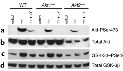
Akt1 and Akt2 are expressed in mouse platelets and activated by thrombin. It has been reported previously that human platelets express mRNA for both Akt1 and Akt2, but not Akt3 (14). Akt1 and Akt2 are 81% identical at the amino acid level. Because Ab’s raised to Akt1 versus Akt2 have not proved to be very selective, immunoblots were made with platelets from WT, Akt1–/–, and Akt2–/–
mice using an Ab that recognizes both isoforms, as well as with a second Ab that recognizes the activation-dependent phosphorylation of Akt at serine 473 (ser-ine 474 in Akt2) (22). The immunoblot in Figure 1a shows that Akt is phosphorylated at serine 473 in thrombin-treated WT platelets. It also shows that the extent of phosphorylation is slightly reduced in Akt1–/–
platelets and dramatically reduced in Akt2–/–platelets.
As reported previously (14, 23), Akt phosphorylation is inhibited in the presence of PI3K inhibitors. Immunoblots of platelet lysates reprobed with an Ab that recognizes all three Akt isoforms reveal that platelets from Akt2–/–mice express less total Akt than
either WT or Akt1–/–platelets (Figure 1b).
Phosphory-lation of endogenous GSK-3βwas used to determine the relative amounts of Akt activity in platelets from each genotype. The experiment illustrated in Figure 1c shows that in platelets from Akt1–/–mice, serine 9 of
GSK-3βis phosphorylated in response to thrombin to the same extent as in WT platelets, but that this increase in phosphorylation is reduced in Akt2–/–
platelets. The same results were found when platelets were stimulated with the PAR4 agonist peptide, AYPGQV (not shown). Thus, the results show that platelets express more Akt2 than Akt1 and that loss of Akt2 has a greater effect on phosphorylation of an Akt substrate than does loss of Akt1.
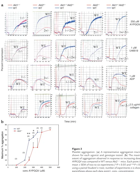
Platelets lacking Akt2 or multiple alleles of Akt1 and Akt2 have defects in platelet aggregation. Since it is clear that Akt is expressed in platelets and activated by platelet ago-nists, we next asked whether Akt plays a role in sup-porting platelet aggregation. To determine whether Akt1 or Akt2 is important in maintaining stable platelet aggregates, PRP was isolated from Akt-defi-cient mice and their WT littermates, and aggregation was observed in response to a panel of platelet agonists. Platelets from Akt2–/–mice showed a substantial defect
in aggregation in response to low concentrations of PAR4 thrombin receptor agonist, AYPGQV (250 µM), and the thromboxane A2 analogue, U46619 (1 µM) (Figure 2a). Raising the agonist concentration of either agonist overcame the defect (the results with AYPGQV are shown in Figure 2b). In contrast, platelets from
Akt1–/–mice responded normally to all concentrations
of these agonists. Notably, both Akt1–/–and Akt2–/–
platelets aggregated normally in response to ADP and collagen at all concentrations tested, including very low concentrations of these agonists (0.25 µM ADP and 2.5
[image:4.585.71.282.51.174.2]µg/ml collagen) that resulted in only 10–20% aggrega-tion (Figure 2a and addiaggrega-tional data not shown). Platelets from Akt1–/– mice and Akt2–/– mice also
Figure 1
Phosphorylation of Akt and GSK-3βin WT, Akt1–/–, and Akt2–/–mice.
Washed platelets (2 ×10⁷platelets in 100 µl) were stimulated for 10
showed no defects in the ability of epinephrine to potentiate aggregation in the presence of subthreshold concentrations of ADP (data not shown). There were no apparent defects in platelet shape change in response to any agonist tested.
Mice that are null for both Akt1and Akt2die imme-diately after birth (24). Therefore, to determine the extent to which each isoform may compensate for the other, aggregation was tested in platelets from
Akt1–/–Akt2+/–mice and Akt1+/–Akt2–/–mice. In contrast
to Akt1–/–, platelets from Akt1–/–Akt2+/–mice had mild
aggregation defects that approached those of the
Akt2–/–platelets when stimulated with low
concentra-tions of AYPGQV and U46619. This mild defect is probably largely due to the loss of the single allele of Akt2, since Akt2+/–platelets have an aggregation profile
very similar to that of Akt1–/–Akt2+/–mice (Figure 2a). Akt1+/–platelets responded normally to all agonists
test-Figure 2
[image:5.585.65.518.44.600.2]ed (data not shown). Platelets from Akt1+/–Akt2–/–mice
displayed the most obvious defects, showing no aggre-gation at 250 µM AYPGQV or 1 µM U46619, concen-trations that induce 50–80% aggregation in platelets from WT control animals. Even in Akt1+/–Akt2–/–
platelets, however, the aggregation defect was overcome at 500 µM AYPGQV and 5 µM U46619, at which con-centrations aggregation occurred normally (not shown). Akt1+/–Akt2–/–platelets also formed unstable
aggregates in response to concentrations of ADP (1
µM) that induced normal aggregation in their WT counterparts. Thus, the loss of Akt2 alone causes a shift in the dose-response curve of AYPGQV (shown in Fig-ure 2b) and U46619 required for maximal aggregation. The loss of one of the two Akt1 alleles in addition to Akt2 caused a further reduction in responses to AYPGQV and U46619 and uncovered a role for Akt in ADP-induced aggregation.
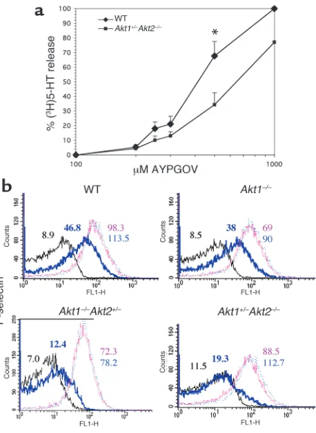
Platelets with reduced levels of Akt have defects in secretion and fibrinogen binding. When platelets are activated by thromboxane A2or low concentrations of thrombin,
secretion of ADP from platelet-dense granules is required for maximal aggregation. This is accompa-nied by secretion of adhesion proteins from α -gran-ules, which also contributes to platelet aggregation. Since loss of Akt affected platelet aggregation in response to U46619 and AYPGQV far more than did ADP, we reasoned that Akt may be involved in platelet secretion. To study dense-granule secretion, washed platelets labeled with (3H)5-HT were stimulated with
increasing concentrations of AYPGQV under nonag-gregating conditions (without fibrinogen or stirring), and release of (3H)5-HT was measured. Figure 3a
shows that in platelets from Akt1+/–Akt2–/– mice,
AYPGQV-stimulated (3H)5-HT release was reduced
nearly 50% compared with WT platelets at concentra-tions up to 500 µM. These differences, however, were reduced at higher agonist concentrations.
To determine whether Akt-deficient platelets also exhibited defects in α-granule secretion, platelets were stimulated with increasing concentrations of AYPGQV, and the binding of a FITC-conjugated Ab that recog-nizes the α-granule protein, P-selectin, was monitored by flow cytometry. A shift in the dose-response curve relative to WT platelets was seen in platelets from
Akt1+/–Akt2–/–and, to a lesser extent, Akt1–/–Akt2+/–mice.
This difference was most evident when responses to 250 µM AYPGQV were compared (Figure 3b). The responses of Akt2–/– platelets looked like those of Akt1+/–Akt2–/–(not shown). P-selectin binding to Akt1–/–
[image:6.585.57.286.52.366.2]platelets is similar to WT at all agonist concentrations tested. Therefore, platelet α-granule and dense-granule secretion are significantly impaired in platelets lacking Akt2, but not in platelets lacking Akt1 alone.
Figure 3
Secretion in WT versus Akt-deficient platelets. (a) Dense-granule secretion. Release of (3H)5-HT was determined after incubation with
the indicated concentrations of AYPGQV. The results shown are the mean ± SEM of three experiments, *P<0.05 (Student’s ttest). (b) Platelet α-granule secretion. Washed human platelets were stimu-lated with buffer alone (black line), 250 µM AYPGQV (bold blue line), 500 µM AYPGQV (pink line), or 1M AYPGQV (light blue line), stained with FITC-labeled anti–P selectin Ab, and analyzed by flow cytometry. Similar results were obtained in an additional two exper-iments. FL1-H, fluorescence intensity.
Figure 4
[image:6.585.302.523.491.670.2]To determine whether deficiencies in Akt signaling might contribute to differences in fibrinogen binding, the binding of FITC-labeled fibrinogen to the surface of WT versus Akt-deficient platelets was tested. Stimu-lation with 250 µM AYPGQV induced less fibrinogen-binding to platelets from Akt1+/–Akt2–/–, Akt2–/–
(equiv-alent to Akt1+/–Akt2–/–mice; not shown), and Akt1–/–Akt2+/–mice than to WT or Akt1–/–platelets
(Fig-ure 4). As with P-selectin expression, this difference was overcome at higher agonist concentrations, indicating that reducing the amount of platelet Akt2 expression shifts the dose-response curve for both P-selectin expression and fibrinogen binding to the right.
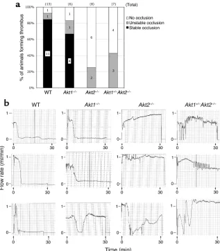
Mice lacking Akt2 are resistant to thrombosis after FeCl3 injury of the carotid artery, but have normal tail-bleeding times. To determine whether the defects in platelet func-tion observed in vitro might affect thrombus formafunc-tion in vivo, we compared responses of WT mice to those lacking one or more isoform of Akt in a carotid artery injury model. In these experiments, a filter paper soaked in 10% ferric chloride was applied to the surface of the carotid artery of the mice for 2 minutes, 30 sec-onds, after which the flow rate through the artery was recorded for 30 minutes with a Doppler flow probe. A histogram of the accumulated results is shown in
Fig-ure 5, along with representative flow traces of each genotype tested. In each of the animals tested, the flow rate before application of FeCl3was 0.8–1.1 ml/min.
After the injury, a reduction in flow rate that remained at 0–0.1 ml/min until the assay was terminated at 30 minutes was scored as a stable occlusive thrombus. Ani-mals in which blood flow stopped, but then resumed, were scored as having an unstable thrombus. Male and female mice of each genotype were tested, but no dif-ferences in results due to mouse gender were observed. In WT and Akt1–/– mice, stable occlusive thrombi
formed in 85% and 67% of the mice tested, respectively. In contrast, none of the Akt2–/–mice or Akt1+/–Akt2–/–
mice formed a stable thrombus in response to injury. In fact, the majority of the Akt2–/–mice and Akt1+/–Akt2–/–
mice never formed a thrombus in response to injury, whereas only 8% of WT and 17% of Akt1–/–mice failed
to form a thrombus. Unstable thrombi were also seen in a greater proportion of Akt2–/–and Akt1+/–Akt2–/–
mice than WT or Akt1–/–mice. Thus, mice lacking Akt2
are resistant to thrombosis after ferric chloride injury to the carotid artery. In contrast, tail bleeding times in
Akt1–/–, Akt2–/–, and Akt1+/–Akt2–/–mice were normal
[image:7.585.54.371.53.415.2]rel-ative to WT mice (Figure 6). These data suggest that Akt (Akt2 in particular) is critical for stabilizing thrombus
Figure 5
Thrombotic response of mice to ferric chloride injury of the carotid artery. Flow rates were measured in the carotid artery following exposure to FeCl3. (a) For each
formation after arterial injury, but is not required for hemostatic plug formation after transection of the veins and arterioles in the tail. The difference in the role of Akt in these two models of in vivo platelet plug for-mation may reflect differences in Akt activation under the divergent flow rates encountered in the carotid artery versus the tail veins.
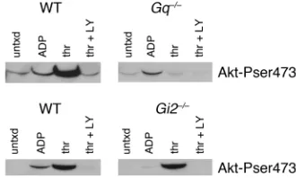
Akt is phosphorylated in a Gi2-dependent manner by ADP and in a Gq-dependent fashion by thrombin. Our data, along
with those published previously, show that phosphory-lation of Akt is detected after platelets are stimulated with thrombin, ADP, or U46619 and that Akt is phos-phorylated in a PI3K-dependent fashion. Because platelets treated with PI3K inhibitors fail to form stable aggregates, as do platelets treated with scavengers of ADP (1), it is tempting to speculate that the role of Akt in stabilizing platelet aggregates is either mediated by release of ADP or is a consequence of ADP release. In other words, either activation of Akt enhances release of ADP, thereby stabilizing platelet aggregates, or release of ADP is first required for Akt activation. Our results suggest that the former, rather than the latter, is the case, since deletion of Akt2reduces responses to PAR4 agonist more than responses to ADP. Nevertheless, to more easily distinguish between these possibilities, phosphorylation of Akt by thrombin and ADP was test-ed in platelets lacking Gαq or Gαi2. The results are
shown in Figure 7. The dominant thrombin receptor on mouse platelets, PAR4, couples directly to Gq, but
acti-vates Giby way of secreted ADP (2). ADP binds to two
receptors on platelets: P2Y1, coupled to Gq(19), and
P2Y12, coupled to Gi2(4). As previously reported,
incu-bating WT platelets with ADP (15) or thrombin (14) resulted in Akt phosphorylation. Loss of Gqabolished
Akt phosphorylation in response to thrombin, but had
little effect on phosphorylation in response to ADP. Conversely, loss of Gi2abolished Akt phosphorylation
by ADP, but only marginally reduced Akt phosphoryla-tion by thrombin (a small reducphosphoryla-tion is evident at very low exposures). These results suggest that there are both Gq- and Gi-dependent pathways that are capable of
acti-vating Akt independently. The results also show that thrombin can stimulate Akt phosphorylation inde-pendently of ADP secretion. Taken together with our data showing that removal of platelet Akt impairs secre-tion, these results suggest that the role of Akt in stabi-lizing platelet aggregates is at least partially due to a role in enhancing release of ADP, rather than due to a requirement of ADP secretion for activation of Akt.
Discussion
It has been recognized for some time that PI3Ks play a role in promoting and maintaining platelet aggrega-tion. The molecular targets of the products of PI3K in platelets and their respective functions have not been completely defined, however. One target of PI3K prod-ucts in many cells, including platelets, is the serine/thre-onine kinase Akt (25). Akt has been implicated in a diverse array of functions in different cells, including suppression of apoptosis, glucose metabolism, and cell proliferation (16). It appears from heterologous expres-sion studies and the phenotypes of the individual knockouts that the three known isoforms of Akt (Akt1, 2, and 3) serve distinct, but overlapping, functions. Akt1 is ubiquitously expressed, and Akt1–/–mice have a
gen-eral defect in organismal growth but little or no defect in glucose metabolism (17). Akt2 is highly expressed in pancreatic βcells, skeletal muscle, and brown fat (26, 27). Akt2–/–mice produced by two different groups are
hyperglycemic, hyperinsulinemic, and have defects in insulin responses (18, 28). Those produced by Cho et al. exhibit a compensatory increase in pancreatic βcell mass, while those studied by Garofalo et al. display an age-dependent loss of adipose tissue, and some Akt2–/–
[image:8.585.74.255.55.258.2]males develop more severe hyperglycemia due to βcell failure. The differences in phenotype observed between
Figure 7
Akt phosphorylation in platelets from Gq–/–and Gi2–/–mice. Washed platelets were obtained and incubated with buffer alone, ADP (10
[image:8.585.340.505.551.650.2]µM), thrombin (1 U/ml), or thrombin + LY294002 (50 µM) using the protocol described in Figure 1, then immunoblotted with an Ab specific for phosphorylated Akt serine 473. The results shown are representative of those obtained four times each.
Figure 6
these two groups is probably attributable to modifier genes present to varying extents in the 129/C57BL/6 background of Akt2–/–mice from Cho et al. versus the
inbred DBA/1lacJ background of Akt2–/– mice from
Garofalo et al. The Akt1–/–Akt2–/–mice have been
suc-cessfully generated by one group, but die immediately after birth with documented defects in bone develop-ment, adipogenesis, and growth (24). Akt3 is expressed predominantly in the brain and testes and is upregulat-ed in some cancer cells (29). The effects of genetic dele-tion of Akt3are yet to be reported.
The mRNA for Akt1 and Akt2, but not Akt3, has been detected in human platelets (14). We show here that mouse platelets express both Akt1 and Akt2 at the protein level, leading us to ask the following questions: What is the role of Akt in platelet signaling and aggre-gation? Do Akt1 and Akt2 have distinct roles? Is the role of Akt in platelet function important for throm-bus formation in vivo? To answer these questions, we have examined platelet function in mice with reduced expression of Akt1, Akt2, or a combination of both. We have also evaluated the role of Akt in the ferric chloride injury model of arterial thrombosis. The results show a definitive role for Akt in stabilizing platelet aggrega-tion in vitro and in platelet plug formaaggrega-tion in vivo.
To better understand how Akt becomes activated in platelets, agonist-mediated Akt phosphorylation was evaluated. Although thrombin, collagen, and throm-boxane A2clearly stimulate Akt phosphorylation in
platelets (5, 14, 30, 31), there are conflicting data on whether ADP is capable of doing so. The frequent fail-ure to detect ADP-mediated Akt phosphorylation is likely due to the rapid desensitization of the Gq
-cou-pled ADP receptor, P2Y1 (32). This was avoided by
iso-lating platelets in the presence of ADP scavengers, in which case Akt phosphorylation was evident, although less robust than that caused by thrombin. A second receptor for ADP on platelets, P2Y12, is coupled pri-marily to the Gifamily member, Gi2(4, 33). Studies
showing that platelets lacking Gi2or the P2Y12
recep-tor have reduced responses to thrombin (2, 4, 34) and that blockade of P2Y12 destabilized platelet aggregates (35) raised the possibility that thrombin-mediated Akt phosphorylation might be mediated by ADP release. Our studies of Akt phosphorylation in Gαi2- versus
Gαq-null platelets showed that thrombin can stimulate
Akt phosphorylation under conditions where ADP cannot, suggesting that platelets stimulated with thrombin (or U46619) do not require secreted ADP to activate Akt, although ADP can amplify Akt activation by stimulating Gi2-coupled P2Y12 receptors. This
con-clusion is consistent with two previous studies: (a) Hirsch et al. found that Akt phosphorylation by ADP in platelets is at least partially dependent on PI3Kγ, while thrombin-mediated Akt phosphorylation is largely normal in the absence of PI3Kγ(15); and (b) Li et al. recently showed that U46619-mediated phos-phorylation of Akt is only partially inhibited by antag-onists of P2Y12 (5).
What, then, is the role of Akt in platelets? The ago-nist-selective nature of the aggregation defects in platelets lacking alleles of Akt2 would suggest that Akt is more important for promoting responses to low con-centrations of agonists that stimulate Gq-coupled
receptors (such as the thromboxane prostanoid [TP] receptor for thromboxane A2 and PAR4 thrombin
receptor) than those coupled to Gi(such as the P2Y12
or α2aadrenergic receptors). Because platelets
stimu-lated with low concentrations of U46619 or AYPGQV require secretion of ADP to achieve maximum aggre-gation (2, 36), we suspected that a defect in secretion may underlie the aggregation defect. Our data showing that platelets from mice that lack Akt2alleles exhibit impaired secretion from both dense and α-granules and have a parallel reduction in fibrinogen binding rel-ative to their WT counterparts confirm that this may be the case. It is notable that the secretion measure-ments were done under nonaggregating conditions, that is, in the absence of fibrinogen and stirring. Thus, it appears that Akt-deficient platelets have a defect in secretion that results in reduced fibrinogen binding and, consequently, impaired aggregation.
[image:9.585.68.269.54.251.2]It is interesting and unexpected that collagen-medi-ated aggregation was not noticeably impaired in the Akt-deficient mice. Collagen-induced Akt phosphory-lation has been detected under nonaggregating condi-tions, although its phosphorylation is delayed (up to 5 minutes) relative to thrombin (31). It is possible that Akt activation by collagen does not take effect quickly enough to contribute to aggregation. Other possibili-ties are that loss of Akt2 is more easily compensated by Akt1 when platelets are activated by collagen than other agonists or that collagen signaling requires high
Figure 8
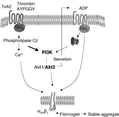
Role of Akt in platelet aggregation. Akt is activated in a PI3K-depend-ent fashion by thromboxane A2 (TxA2) or thrombin. Akt activation promotes secretion of ADP from platelet-dense granules, which stim-ulates Gi-coupled pathways by binding to the P2Y12 receptor,
shear rates to sustain Akt activation. It is interesting that mice lacking the p85αsubunit of PI3K have a defect in collagen-mediated platelet aggregation, but no defect in bleeding time, perhaps indicating that defects in collagen signaling require conditions of higher shear to affect platelet plug formation in vivo (37). Mice lacking PI3Kγalso have normal tail bleeding times but are resistant to ADP-induced thromboem-bolism (15). We show here that mice lacking Akt2, a known effector of PI3K, have an impaired ability to form thrombi following arterial injury, but, like mice deficient in PI3K isoforms, have normal bleeding times. It is tempting to speculate that the loss of Akt2 expres-sion has a greater effect on thrombus formation in the carotid because of the higher flow rates that are pres-ent in that relatively large artery compared with the small arteries and veins in the distal 1 mm of the tail. Although PI3K may regulate pathways other than those downstream of Akt, our studies, as well as others (14, 23), show that thrombin-stimulated Akt phos-phorylation is predominantly PI3K dependent. Mouse platelets treated with PI3K inhibitors, such as
Akt1+/–Akt2–/–platelets, are unresponsive to low
con-centrations of AYPGQV or U46619, indicating that removal of Akt reproduces the effects of PI3K inhibitors on aggregation. Defects in platelet function may be more severe in platelets that completely lack both Akt1 and Akt2. It appears that Akt3 is not expressed in mouse platelets, since immunoblots with an Ab selective for Akt3 were negative (not shown). Defects in platelet function were clearly more apparent in Akt2–/–than Akt1–/–platelets, demonstrating that
Akt2 plays a more important role in platelets than Akt1. This seems a simple reflection of the relative expression levels of the two isoforms, since immunore-activity of a nonsubtype selective Akt Ab and phos-phorylation of the Akt substrate GSK-3βwere clearly reduced in Akt2–/–platelets relative to both Akt1–/–and
WT platelets. Because loss of a single Akt2allele also results in mild defects in platelet aggregation, but
Akt2+/–mice are metabolically normal, the platelet
func-tion defect does not appear to be due to changes in plasma glucose or insulin concentrations.
Returning to the role of Akt in platelet function in vivo, the effect of loss of Akt2 on thrombus formation in vivo is particularly interesting given that the platelet function defects observed in vitro were agonist selective and overcome at higher agonist concentrations. Although it is possible that reduced expression of Akt2 by other cells in the vasculature may contribute to the observed effects of Akt2deletion on thrombosis in vivo, the effects correlate nicely with the observed platelet phenotype ex vivo. Attempts to transplant Akt2–/–
blood into lethally irradiated WT recipients (and vice versa) are underway to determine the contribution of components of blood versus the vascular bed to the reduced thrombosis seen in Akt2–/–mice. The ferric
chloride thrombosis model involves a substantial transmural insult and potentially indicates that even
relatively small defects in platelet function ex vivo (such as those that shift the dose-response curve for agonist activation) can translate into important defects in thrombus formation in vivo. This observation has implications as investigators try to identify new molec-ular targets for drug development to treat thrombosis. That is, molecular targets that amplify or sustain platelet responses to agonist may make particularly good candidates for development of novel therapeutics. How do we view the role of Akt in platelet function? Akt seems particularly important for supporting platelet responses to low concentrations of agonists that require secretion to amplify aggregation, such as agonists that bind to the PAR4 thrombin receptor or TP receptor for thromboxane A2. This observation is in line with the secretion defect observed in Akt2–/–and Akt1+/–Akt2–/–platelets. Akt appears to be activated by a
Gq- and PI3K-dependent mechanism and plays a role in
secretion of ADP. ADP subsequently stimulates Gi
-mediated pathways, which enhance the activation of
αIIbβ3, promote fibrinogen binding, and increase
aggre-gation (Figure 8).
Acknowledgments
Many thanks to Charles Abrams for helpful discus-sions and Donald Eslin for help in establishing the carotid artery injury model in our lab. Parts of this work were supported by grants 0365530U from the American Heart Association (to D. Woulfe) and NIH HL-40387 and NIH HL-45181 (to L.F. Brass).
1. Trumel, C., et al. 1999. A key role of adenosine diphosphate in the irre-versible platelet aggregation induced by the PAR1-activating peptide through the late activation of phosphoinositide 3-kinase. Blood.
94:4156–4165.
2. Kim, S., et al. 2002. Protease-activated receptors 1 and 4 do not stimu-late G(i) signaling pathways in the absence of secreted ADP and cause human platelet aggregation independently of G(i) signaling. Blood.
99:3629–3636.
3. Dangelmaier, C., Jin, J., Smith, J.B., and Kunapuli, S.P. 2001. Potentia-tion of thromboxane A2-induced platelet secrePotentia-tion by Gi signaling through the phosphoinositide-3 kinase pathway. Thromb. Haemost.
85:341–348.
4. Jantzen, H.-M., Milstone, D.S., Gousset, L., Conley, P.B., and Mortensen, R.M. 2001. Impaired activation of murine platelets lacking G alpha(i2).
J. Clin. Invest.108:477–483. doi:10.1172/JCI200112818.
5. Li, Z., et al. 2003. Two waves of platelet secretion induced by thrombox-ane A2 receptor and a critical role for phosphoinositide 3-kinases. J. Biol.
Chem.278:30725–30731.
6. Kovacsovics, T.J., et al. 1995. Phosphoinositide 3-kinase inhibition spares actin assembly in activating platelets but reverses platelet aggregation.
J. Biol. Chem.270:11358–11366.
7. Kucera, G.L., and Rittenhouse, S.E. 1990. Human platelets form 3-phos-phorylated phosphoinositides in response to α-thrombin, U46619, or
GTP gamma S. J. Biol. Chem.265:5345–5348.
8. Zhang, J., Shattil, S.J., Cunningham, M.C., and Rittenhouse, S.E. 1996. Phosphoinositide 3-kinase gamma and p85/phosphoinositide 3-kinase in platelets. Relative activation by thrombin receptor or beta-phorbol myristate acetate and roles in promoting the ligand-binding function of
αIIbβ3 integrin. J. Biol. Chem.271:6265–6272.
9. Sultan, C., et al. 1991. Involvement of platelet glycoprotein IIb-IIIa (α IIb-β3 integrin) in thrombin-induced synthesis of phosphatidylinositol
3′,4′-bisphosphate. J. Biol. Chem.266:23554–23557.
10. Sorisky, A., King, W.G., and Rittenhouse, S.E. 1992. Accumulation of PtdIns(3,4)P2 and PtdIns(3,4,5)P3 in thrombin-stimulated platelets. Different sensitivities to Ca2+ or functional integrin. Biochem. J.
286:581–584.
11. Banfic, H., et al. 1998. A novel integrin-activated pathway forms PKB/Akt-stimulatory phosphatidylinositol 3,4-bisphosphate via
12. Cantley, L.C. 2002. The phosphoinositide 3-kinase pathway. Science.
296:1655–1657.
13. Brazil, D.P., Park, J., and Hemmings, B.A. 2002. PKB binding proteins.
Getting in on the Akt. Cell.111:293–303.
14. Kroner, C., Eybrechts, K., and Akkerman, J.W. 2000. Dual regulation of
platelet protein kinase B. J. Biol. Chem.275:27790–27798.
15. Hirsch, E., et al. 2001. Resistance to thromboembolism in
PI3Kγ-defi-cient mice. FASEB J.15:2019–2021.
16. Lawlor, MA. 2001. PKB/Akt: a key mediator of cell proliferation, survival
and insulin responses? J. Cell Sci.114:2903–2910.
17. Cho, H., Thorvaldsen, J.L., Chu, Q., Feng, F., and Birnbaum, M.J. 2001. Akt1/PKBalpha is required for normal growth but dispensable for
main-tenance of glucose homeostasis in mice. J. Biol. Chem.276:38349–38352.
18. Cho, H., et al. 2001. Insulin resistance and a diabetes mellitus-like syn-drome in mice lacking the protein kinase Akt2 (PKB beta). Science.
292:1728–1731.
19. Offermanns, S., Toombs, C.F., Hu, Y.H., and Simon, M.I. 1997. Defective
platelet activation in G α(q)-deficient mice. Nature.389:183–186.
20. Woulfe, D., Jiang, H., Mortensen, R., Yang, J., and Brass, L.F. 2002. Acti-vation of Rap1B by G(i) family members in platelets. J. Biol. Chem.
277:23382–23390.
21. Kufrin, D., et al. 2003. Antithrombotic thrombocytes: ectopic expression
of urokinase-type plasminogen activator in platelets. Blood.102:926–933.
22. Bellacosa, A., et al. 1998. Akt activation by growth factors is a
multiple-step process: the role of the PH domain. Oncogene.17:313–325.
23. Lova, P., et al. 2003. A selective role for phosphatidylinositol 3,4,5-trisphosphate in the Gi-dependent activation of platelet Rap1B. J. Biol.
Chem.278:131–138.
24. Peng, X.D., et al. 2003. Dwarfism, impaired skin development, skeletal muscle atrophy, delayed bone development, and impeded adipogenesis
in mice lacking Akt1 and Akt2. Genes Dev.17:1352–1365.
25. Vivanco, I., and Sawyers, C.L. 2002. The phosphatidylinositol 3-kinase
AKT pathway in human cancer. Nat. Rev. Cancer.2:489–501.
26. Altomare, D.A., Lyons, G.E., Mitsuuchi, Y., Cheng, J.Q., and Testa, J.R. 1998. Akt2 mRNA is highly expressed in embryonic brown fat and the
AKT2 kinase is activated by insulin. Oncogene.16:2407–2411.
27. Altomare, D.A., et al. 1995. Cloning, chromosomal localization and expression analysis of the mouse Akt2 oncogene. Oncogene.
11:1055–1060.
28. Garofalo, R.S., et al. 2003. Severe diabetes, age-dependent loss of adipose tissue, and mild growth deficiency in mice lacking Akt2/PKBβ. J. Clin.
Invest.112:197–208. doi:10.1172/JCI200316885.
29. Nakatani, K., et al. 1999. Up-regulation of Akt3 in estrogen receptor-defi-cient breast cancers and androgen-independent prostate cancer lines.
J. Biol. Chem.274:21528–21532.
30. Banfic, H., Downes, C.P., and Rittenhouse, S.E. 1998. Biphasic activa-tion of PKBalpha/Akt in platelets. Evidence for stimulaactiva-tion both by phosphatidylinositol 3,4-bisphosphate, produced via a novel pathway, and by phosphatidylinositol 3,4,5-trisphosphate. J. Biol. Chem.
273:11630–11637.
31. Barry, F.A., and Gibbins, J.M. 2002. Protein kinase B is regulated in platelets by the collagen receptor glycoprotein VI. J. Biol. Chem.
277:12874–12878.
32. Baurand, A., et al. 2000. Desensitization of the platelet aggregation response to ADP: differential down-regulation of the P2Y1 and P2cyc
receptors. Thromb. Haemost.84:484–491.
33. Hollopeter, G., et al. 2001. Identification of the platelet ADP receptor
targeted by antithrombotic drugs. Nature.409:202–207.
34. Andre, P., et al. 2003. P2Y12 regulates platelet adhesion/activation, thrombus growth, and thrombus stability in injured arteries. J. Clin.
Invest.112:398–406. doi:10.1172/JCI200317864.
35. Storey, R.F., et al. 2000. The central role of the P(2T) receptor in ampli-fication of human platelet activation, aggregation, secretion and
proco-agulant activity. Br. J. Haematol.110:925–934.
36. Paul, B.Z., Jin, J., and Kunapuli, S.P. 1999. Molecular mechanism of thromboxane A(2)-induced platelet aggregation. Essential role for
p2t(ac) and alpha(2a) receptors. J. Biol. Chem.274:29108–29114.
37. Watanabe, N., et al. 2003. Functional phenotype of phosphoinositide 3-kinase p85α-null platelets characterized by an impaired response to