Biomechanical activation: an emerging
paradigm in endothelial adhesion biology.
M A Gimbrone Jr, … , T Nagel, J N Topper
J Clin Invest.
1997;
99(8)
:1809-1813.
https://doi.org/10.1172/JCI119346
.
Perspective
Find the latest version:
Perspectives Series:
Cell Adhesion in Vascular Biology
J. Clin. Invest.
© The American Society for Clinical Investigation, Inc. 0021-9738/97/04/1809/05 $2.00
Volume 99, Number 8, April 1997, 1809–1813
Biomechanical Activation: an Emerging Paradigm in Endothelial Adhesion Biology
Michael A. Gimbrone, Jr., Tobi Nagel, and James N. Topper
Vascular Research Division, Departments of Pathology, Brigham and Women’s Hospital, and Harvard Medical School, Boston, Massachusetts 02115
Introduction
The adhesive properties of the endothelium, the single-cell-thick lining of the cardiovascular system, are central to its biology and pathobiology. In health, the luminal endothelial surface provides a relatively nonadhesive, nonthrombogenic container for the cellular and macromolecular constituents of the blood. Specialized adhesive molecules localized at the lateral cell–cell junctions control transendothelial permeability and the move-ment of leukocytes from the blood into the tissue spaces of the body. Along its basal aspect, focal adhesion complexes, con-sisting of transmembrane integrins and associated intracellular proteins, physically link the extracellular matrix to cytoskeletal elements, providing both stability and plasticity to the vascular lining. In disease, these various adhesive interactions can un-dergo dramatic changes. As is highlighted by the articles in this Perspectives Series, the molecular biological analysis of endo-thelial adhesion pathobiology has led to the discovery of novel families of molecules (e.g., the selectins), as well as a more dy-namic appreciation of their mutual interactions (e.g., the leu-kocyte–endothelial adhesion cascade). This knowledge has added much to our basic understanding of vascular biology, as well as the pathophysiology of clinically important processes, such as acute and chronic inflammation, atherosclerosis, angio-genesis, vascular injury and repair, and developmental malfor-mations. In certain instances, these insights have provided the basis for the rational design of promising new therapeutics for cardiovascular disease.
A central premise of modern vascular biology is that the endothelial lining is a dynamically mutable interface, locally responsive to various stimuli originating from the circulating blood and/or neighboring cells and tissues, and thus can ac-tively participate in the physiological adaptation or pathophys-iological dysfunction of a given region of the vasculature (1). From a teleological standpoint, the endothelium appears ide-ally suited to function in this capacity, given its unique anatom-ical position between blood and tissues, and its ability to gen-erate an impressive repertoire of biological effectors, (e.g.,
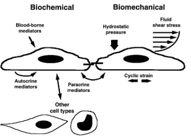
nitric oxide, eicosanoids, cytokines, growth stimulators and in-hibitors, vasoactive peptides, pro- and anticoagulants, and fi-brinolytic factors). Early studies of the mechanisms underlying this plasticity of endothelial phenotype identified certain pro-inflammatory substances, such as cytokines and bacterial prod-ucts, as important stimuli regulating the activity and expres-sion of many of these effectors. The reproducibility of this humoral mode of stimulation, and its wide-reaching patho-physiological implications, has lead to its extensive study, in both in vitro and in vivo experimental models, as a biochemi-cal paradigm of endothelial activation (2, 3). In addition to these humoral stimuli, endothelial cells also are constantly ex-posed to a spectrum of hemodynamic forces generated by pul-satile blood flow. These forces include hydrostatic pressures, cyclic strains, and wall shear stresses. There is increasing evi-dence that these biomechanical stimuli can directly influence endothelial structure and function, acutely and chronically, thus constituting a novel paradigm of endothelial activation (Fig. 1). This brief editorial review will provide a perspective on the role of biomechanical forces, alone and in conjunction with humoral stimuli, in modulating the adhesive interactions of vascular endothelium in health and disease.
Activation of endothelium by humoral factors: an established paradigm
In the early 1980s, inflammatory cytokines such as IL-1 or TNF, and bacterial products such as gram-negative endotoxins were shown to act directly on cultured human endothelial cells to alter their adhesive properties for blood leukocytes (4). By comparison to previously studied leukocyte-directed activa-tors, such as leukotrienes, activated complement components, or chemotactic peptides, the stimulatory effect of these agents was dramatic in amplitude and resulted in enhanced firm at-tachment and transmigration of leukocytes in various in vitro model systems. These adhesive changes in the cytokine-treated endothelial cell required de novo protein synthesis, which was manifested, in part, by the expression of activation antigens at the cell surface (5). Monoclonal antibodies to cer-tain of these neoantigens were effective in blocking leukocyte adhesion and thus enabled the purification and molecular cloning of novel endothelial-leukocyte adhesion molecules (ELAMs),1 such as ELAM-1 (now designated E-selectin) (6).
Address correspondence to Michael A. Gimbrone, Jr., M.D., Vascu-lar Research Division, Brigham & Women’s Hospital, 221 Longwood Avenue, LMRC-401, Boston, MA 02115-5817. Phone: 617-732-5901; FAX: 617-732-5933; E-mail: gimbrone@bustoff.bwh.harvard.edu
Received for publication 6 March 1997 and accepted in revised
form 11 March 1997. 1.
These cytokine-activated endothelial cells also expressed in-creased amounts of other adhesion molecules such as intercel-lular adhesion molecule-1 (ICAM-1), as well as chemoattrac-tant cytokines such as IL-8 and monocyte chemoattracchemoattrac-tant protein-1 (MCP-1), involved in leukocyte recruitment (7). Con-comitant changes in the expression of cell-associated procoag-ulant proteins (e.g., tissue factor), and fibrinolytic activators and inhibitors, indicated further implications of endothelial ac-tivation for hemostasis and thrombosis. Taken together, these in vitro studies thus provided a dynamic working concept of the modulation of endothelial phenotype by cytokines and other humoral factors. Immunohistochemical demonstration of endothelial activation antigens in human and animal tissues in vivo, in various acute and chronic inflammatory processes, provided further validation of this paradigm (8). In addition to recombinant cytokines and purified bacterial lipopolysaccha-rides, certain other substances, including polar phospholipids (e.g., lysophosphatidylcholine and related compounds), ho-mocyst(e)ine, and advanced glycosylation end products associ-ated with atherosclerotic and diabetic vascular disease, also have been defined as stimuli of endothelial activation in vitro (9). The induction of endothelial-leukocyte adhesion mole-cules, such as vascular cell adhesion molecule-1 (VCAM-1), has been identified as a very early event in the development of atherosclerotic lesions in experimental animal models (10). Measurement of soluble ELAMs in circulating blood samples may provide a useful indirect index of endothelial activation in clinical and epidemiological studies (11).
In summary, phenotypic modulation of vascular endothe-lium by humoral factors, such as cytokines and bacterial endo-toxin, has been dissected extensively at the cellular, molecular, and genetic regulatory levels, and at present constitutes the best studied paradigm of endothelial activation. While intri-cately related to the process of leukocyte recruitment in acute
and chronic inflammation, the biological implications of this process clearly extend beyond cell adhesion.
Activation of endothelium by biomechanical forces: an emerging paradigm
When cultured endothelial cells are exposed to quasiphysio-logical levels of fluid mechanical forces in vitro, in specially de-signed flow chambers, a spectrum of structural and functional changes is observed (12, 13). These include striking morpho-logical changes in cell shape, cell alignment, and cytoskeletal architecture, as well as more subtle changes in membrane de-formability and cell division rate. At a basic cell biological level, many of these changes can be viewed as the in vitro (re)adaptation of the cultured endothelial cell to certain of the biomechanical stimuli that are normally present in its in vivo environment within the vessel wall. This adaptation process in-volves various adhesive mechanisms. For example, real-time visualization of a cultured endothelial monolayer exposed to a unidirectional laminar shear stress (LSS) stimulus, using tan-dem scanning confocal microscopy, reveals a dynamic remod-eling of the focal contact sites along its basal aspect (12). Changes also are observed in the phosphorylation state of cy-toskeletal proteins associated with these focal adhesion com-plexes, as well as adhesion molecules such as platelet-endothe-lial adhesion molecule-1 that are localized to lateral cell–cell junctions (12, 14). These changes presumably are part of a gen-eralized cellular adaptation to applied mechanical stresses, re-flected internally in cytoskeletal architecture and externally in cell surface topography (15, 16). The net result of these struc-tural adaptations is an endothelial cell which is in dynamic equilibrium with its ambient fluid mechanical environment.
[image:3.612.58.438.57.334.2]In addition to these structural adaptations, biomechanical forces such as fluid shear stresses also stimulate the production in endothelium of a large and diverse array of potent biological
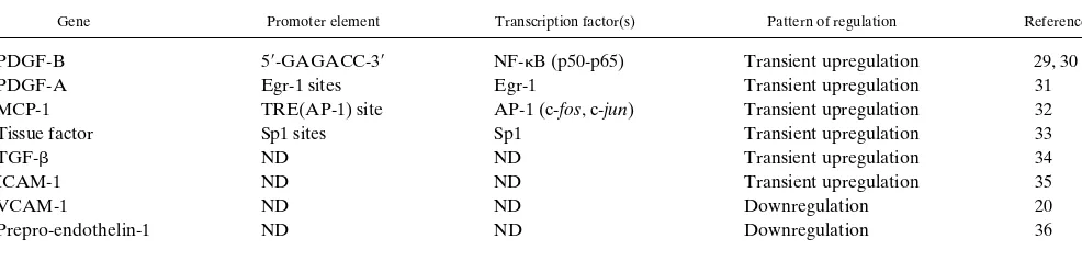
mediators (12, 13). Certain of these effects involve gene regu-lation at the transcriptional level and thus are analogous to en-dothelial activation by humoral factors. In this biomechanical paradigm of activation, the endothelial cell appears capable of responding not only to the magnitude of the applied forces but also their temporal and spatial fluctuations (e.g., steady versus pulsatile flow; uniform laminar, disturbed laminar, or turbu-lent flow regimens), thus suggesting the existence of primary flow sensors (receptors) that are coupled via distinct signaling pathways to nuclear events (12, 13). Considerable progress has been made recently in defining certain of the molecular mech-anisms involved, including the identification of positive and negative shear stress responsive elements (SSREs) in the pro-moters of biomechanically responsive genes, and transcription factors that regulate their activation (9, 13) (Table 1).
In the context of vascular adhesion biology, fluid shear stress thus far has been the best studied biomechanical stimulus of endothelial gene regulation. When cultured human umbilical vein endothelial cells (HUVEC) are exposed to a physiologi-cally relevant range (2.5–46 dyn/cm2) of steady, unidirectional LSS, there is a time-dependent induction of ICAM-1 expres-sion that is evident at the mRNA level by 2 h, and is detectable as functional cell surface protein, by immunobinding and leu-kocyte adhesion assays, for as long as 48 h of continuous flow exposure (17). This LSS-induced upregulation, in part, reflects enhanced transcriptional activity as determined by nuclear run-off analysis (18). In the same endothelial monolayers, E-selec-tin (which is normally a silent gene in this cultured cell system) and VCAM-1 (which shows a relatively low level of constitutive expression) remained unchanged in response to LSS. This se-lective upregulation of ICAM-1, but not E-selectin or VCAM-1, by LSS is in contrast to the coordinated induction of these three ELAMs that is typically observed with humoral (cyto-kine and bacterial endotoxin) stimulation in the same cultured HUVEC system.
These experimental observations with ICAM-1 suggest that certain biomechanical stimuli might act as differential reg-ulators of endothelial adhesion molecule expression, exerting additive, synergistic, or even antagonistic actions in conjunc-tion with humoral stimuli. Further evidence supporting this working concept derives from the study of VCAM-1, another member of the immunoglobulin family of adhesion molecules, in cultured endothelial cells. This mononuclear leukocyte-selective endothelial adhesion molecule, which has been impli-cated in atherogenesis (10), can be induced in vitro by
cyto-kines and also components of oxidized lipoproteins such as lysophosphatidylcholine. In cultured murine endothelial cells which have a constitutively high level of VCAM-1, the applica-tion of steady LSS suppresses its expression (19). This effect, which is induced by relatively low shear stresses (0.7–7.1 dyn/ cm2), is manifested at the level of steady state mRNA and cell surface protein and results in markedly decreased lymphocyte adhesion. Deletional analyses of the VCAM-1 promoter point to the presence of a negative SSRE that mediates this down-regulation at the transcriptional level (20). In studies with cul-tured HUVEC, LSS preconditioning exerts an inhibitory ef-fect on cytokine-induced VCAM-1 expression (21), mimicking the effects of intracellular antioxidants (22). In yet other stud-ies with cultured human saphenous vein endothelial cells, ni-tric oxide, itself an LSS-regulated endothelial product (23), acts to decrease cytokine-induced VCAM-1 expression (24, 25). These observations serve to illustrate the potential com-plexity of the interplay of biomechanical and humoral stimuli in the induction and modulation of adhesion molecule expres-sion in vascular endothelium.
To date, the majority of experimental studies of the effects of flow on adhesion molecule expression have used relatively simple in vitro fluid mechanical systems to generate uniform laminar shear stresses on cultured endothelial monolayers. By varying the viscosity of the perfusion medium, the effects of wall shear stresses, per se, can be distinguished from the influ-ence of bulk flow on boundary layer diffusion in the vicinity of the endothelial surface (12). In most cases, however, the shear-ing force is beshear-ing applied to a static culture and thus represents an abrupt transition in biomechanical loading of the system. In attempts to better model in vivo hemodynamics, investigators have devised unsteady flow systems that generate temporal and/or spatial fluctuations of fluid shear stresses (12, 13), and have even combined shear stress, pressure, and circumferential stretch in the same in vitro system to generate a quasiphysio-logical biomechanical environment for the endothelial cell (26).
(Patho)physiological relevance of biomechanical activation of vascular endothelium
As illustrated above, our current knowledge concerning the ef-fects of biomechanical stimulation on endothelial gene expres-sion has been derived largely from experiments in simplified in vitro model systems. This is attributable, in part, to practical limitations on experimental interventions in the in vivo setting. When surgical manipulations have been used to acutely modu-Table I. SSREs and Interacting Transcription Factors in Endothelial Expressed Genes*
Gene Promoter element Transcription factor(s) Pattern of regulation Reference
PDGF-B 59-GAGACC-39 NF-kB (p50-p65) Transient upregulation 29, 30
PDGF-A Egr-1 sites Egr-1 Transient upregulation 31
MCP-1 TRE(AP-1) site AP-1 (c-fos, c-jun) Transient upregulation 32
Tissue factor Sp1 sites Sp1 Transient upregulation 33
TGF-b ND ND Transient upregulation 34
ICAM-1 ND ND Transient upregulation 35
VCAM-1 ND ND Downregulation 20
Prepro-endothelin-1 ND ND Downregulation 36
[image:4.612.59.552.73.192.2]late wall shear stresses in the rabbit carotid artery, significant flow-dependent changes in endothelial VCAM-1 and ICAM-1 expression were observed (27). However, the most striking example of flow-related changes in vessel wall biology is pro-vided by an experiment of Nature — the nonrandom distribu-tion of the early lesions of atherosclerosis in humans and ex-perimental animals. Arterial bifurcations and curvatures, where disturbed flow patterns (flow separation, flow reversal, low amplitude, and fluctuating wall shear stresses) occur, typically are lesion-prone areas, whereas geometries associated with uni-form laminar flow (pulsatile without flow reversal) and rela-tively constant (time-averaged) wall shear stresses, such as the straight tubular portions of the aorta and its primary tributar-ies, tend to be lesion-protected areas (9, 18). These patterns also are retained in genetically modified mouse models of atherogenesis, in which systemic risk factors such as markedly elevated levels of atherogenic plasma lipoproteins have been deliberately induced (28). A hallmark of these early lesions in the animal models is the localized upregulation of endothelial VCAM-1, an event that precedes mononuclear leukocyte re-cruitment (9, 10). These observations suggest that the endothe-lial cells in these lesion-prone areas are responding differen-tially to their fluid mechanical environment. Experimental evidence supporting this hypothesis comes from in vitro mo-lecular biological experiments, using RT-PCR–based differen-tial display technology, to examine the patterns of endothelial genes that are (up- or down-) regulated by various biomechan-ical and cytokine stimuli (18). Uniform laminar shear stress stimulation, characteristically associated with lesion-protected areas, selectively induces the sustained upregulation of a set of genes, including manganese superoxide dismutase, cyclooxy-genase-2, and nitric oxide synthase (ecNOS), the activities of which (antioxidant, antithrombotic, antiadhesive) are poten-tially atheroprotective. In contrast, turbulent shear stress, a nonlaminar fluid mechanical stimulus, does not induce these genes. Other flow-regulated endothelial genes with potential proinflammatory, proatherogenic activities, such as E-selectin, MCP-1, and ICAM-1, do not exhibit sustained LSS-selective upregulation. Interestingly, certain novel endothelial genes ap-pear to be selectively induced by laminar shear stress but not by cytokine stimulation (18), thus further illustrating the dif-ferential responsiveness of the endothelial cell to biochemical and biomechanical activation (Fig. 1).
In the in vivo setting, a given endothelial cell is constantly being subjected to combinations of various biochemical and biomechanical stimuli, as well as information transduced via integrin-receptors from extracellular matrix components. The phenotype of a given endothelial cell thus represents an inte-grated response to its local (patho)physiological milieu. Cyto-kines, growth factors, and other mediators secreted by emi-grating leukocytes or adjacent cells within the vessel wall can acutely modify this local environment as part of a response-to-injury program. Biomechanical stimuli, in a manner analo-gous to extracellular matrix components, appear to contribute in a more sustained way to the regulation of endothelial phe-notype. This chronic mode of endothelial activation likely in-fluences the vascular remodeling that occurs in diseases such as atherosclerosis and hypertension, as well as after interven-tions such as coronary artery bypass grafting and percutaneous angioplasty. Biomechanical modulation of endothelial gene expression, in particular the genes encoding adhesion mole-cules involved in cell–cell and cell–matrix interactions, may
also play an active role during embryonic development of blood vessels, and at times of hemodynamic transitions (e.g., in the neonatal period). Clearly, biomechanical forces have im-portant implications for endothelial adhesion biology beyond their direct rheologic effects on leukocyte–endothelial interac-tions (37). The emerging paradigm of biomechanical activation of endothelial cells promises to be a conceptually rich and pathophysiologically relevant area for future investigation.
Acknowledgments
Regrettably, the space limitations of this editorial format preclude ci-tation of many primary publications in this rapidly evolving area. We wish to acknowledge the contributions of past and current members of the Vascular Research Division, in particular, K. Anderson, W. Atkinson, T. Collins, L. Khachigian, N. Resnick, S. Wasserman, and the long-standing collaboration of C.F. Dewey and colleagues in the Fluid Mechanics Laboratory at the Massachusetts Institute of Tech-nology.
Original studies in Dr. Gimbrone’s laboratory were supported by grants from the National Institutes of Health (PO1-HL30628, R37-HL51150) and Millennium Pharmaceuticals, Inc., and an unrestricted cardiovascular research award from the Bristol-Myers Squibb Re-search Institute. Dr. Topper is a recipient of a Howard Hughes Medi-cal Institute Postdoctoral Research Fellowship for Physicians. Ms. Nagel is a recipient of a Graduate Fellowship from the National Sci-ence Foundation.
References
1. Gimbrone, M.A., Jr. 1995. Vascular endothelium in health and disease.
In Molecular Cardiovascular Medicine. E. Haber, editor. Scientific American Medicine, New York. 49–61.
2. Pober, J.S., and R.C. Cotran. 1990. Cytokines and endothelial cell biol-ogy. Physiol. Rev. 70:427–451.
3. Mantovani, A., F. Bussolino, and E. Dejana. 1992. Cytokine regulation of endothelial cell function. FASEB (Fed. Am. Soc. Exp. Biol.) J. 6:2591–2599.
4. Bevilacqua, M.P., J.S. Pober, M.E. Wheeler, R.S. Cotran, and M.A. Gim-brone, Jr. 1985. Interleukin-1 acts on cultured vascular endothelium to increase the adhesion of polymorphonuclear leukocytes, monocytes and related leuko-cyte cell lines. J. Clin. Invest. 76:2003–2011.
5. Pober, J.S., M.P. Bevilacqua, D.L. Mendrick, L.A. Lapierre, W. Fiers, and M.A. Gimbrone, Jr. 1986. Two distinct monokines, interleukin 1 and tumor necrosis factor, each independently induce biosynthesis and transient expres-sion of the same antigen on the surface of cultured human vascular endothelial cells. J. Immunol. 136:1680–1687.
6. Bevilacqua, M.P., S. Stengelin, M.A. Gimbrone, Jr., and B. Seed. 1989. Endothelial-leukocyte adhesion molecule 1: an inducible receptor for neutro-phils related to complement regulatory proteins and lectins. Science (Wash. DC). 243:1160–1165.
7. Luscinskas, F.W., and M.A. Gimbrone, Jr. 1996. Endothelial-dependent mechanisms of mononuclear leukocyte recruitment. Annu. Rev. Med. 47:413–421. 8. Cotran, R.S., M.A. Gimbrone, Jr., M.P. Bevilacqua, D. Mendrick, and J.S. Pober. 1986. Induction and detection of a human endothelial activation an-tigen in vivo. J. Exp. Med. 164:661–666.
9. Gimbrone, M.A., Jr., and J.N. Topper. 1997. Biology of the vessel wall: endothelium. In Molecular Basis of Heart Diseases. K.R. Chien, J.R. Breslow, J.M. Leiden, R.D. Rosenberg, and C. Seidman, editors. W.B. Saunders, Phila-delphia. In press.
10. Cybulsky, M.I., and M.A. Gimbrone, Jr. 1991. Endothelial expression of a mononuclear leukocyte adhesion molecule during atherogenesis. Science (Wash. DC). 251:788–791.
11. Gearing, A.J.H., and W. Newman. 1993. Circulating adhesion molecules in disease. Immunol. Today. 14:506–512.
12. Davies, P.F. 1995. Flow-mediated endothelial mechanotransduction.
Physiol. Rev. 75:519–560.
13. Resnick, N., and M.A. Gimbrone, Jr. 1995. Hemodynamic forces are complex regulators of endothelial gene expression. FASEB (Fed. Am. Soc. Exp. Biol.) J. 9:874–882.
14. Osawa, M., M. Masuda, N. Harada, R.B. Lopes, Y. Kano, and K. Fuji-wara. 1997. Tyrosine phosphorylation of platelet endothelial cell adhesion mol-ecule-1 (PECAM-1, CD31) in mechanically stimulated vascular endothelial cells.
Eur. J. Cell Biol. In press.
Jr. 1992. The distribution of fluid forces on model arterial endothelium using computational fluid dynamics. J. Biochem. Eng. 114:309–316.
16. Barbee, K.A., T. Mundel, R. Lal, and P.F. Davies. 1995. Subcellular dis-tribution of shear stress at the surface of flow-aligned and nonaligned endothe-lial monolayers. Am. J. Physiol. 268:H1765–H1772.
17. Nagel, T., N. Resnick, W.J. Atkinson, C.F. Dewey, Jr., and M.A. Gim-brone, Jr. 1994. Shear stress selectively upregulates intercellular adhesion mole-cule-1 expression in cultured human vascular endothelial cells. J. Clin. Invest.
94:885–891.
18. Topper, J.N., J. Cai, D. Falb, and M.A. Gimbrone, Jr. 1996. Identifica-tion of vascular endothelial genes differentially responsive to fluid mechanical stimuli: cyclooxygenase-2, manganese superoxide dismutase, and endothelial cell nitric oxide synthase are selectively up-regulated by steady laminar shear stress. Proc. Natl. Acad. Sci. USA. 93:10417–10422.
19. Ando, J., H. Tsuboi, R. Korenaga, Y. Takada, N. Toyama-Sorimachi, M. Miyasaka, and A. Kamiya. 1994. Shear stress inhibits adhesion of cultured endothelial cells to lymphocytes by downregulating VCAM-1 expression. Am. J. Physiol. 267:C679–C687.
20. Ando, J., R. Korenaga, Y. Takada, and A. Kamiya. 1996. Down-regula-tion of VCAM-1 gene transcripDown-regula-tion by shear stress in cultured murine endothe-lial cells. J. Vasc. Res. 33(Suppl. 1):4a. (Abstr.)
21. Varner, S.E., R.M. Nerem, R.M. Medford, and R.W. Alexander. 1994. Laminar shear stress regulates VCAM-1 gene expression. Ann. Biomed. Eng.
22:40a. (Abstr.)
22. Marui, N., M. Offerman, R. Swerlick, C. Kunsch, C.A. Roxen, M. Ah-mad, R.W. Alexander, and R.M. Medford. 1993. Vascular cell-adhesion mole-cule-1 (VCAM-1) gene transcription and expression are regulated through an antioxidant sensitive mechanism in human vascular endothelial cells. J. Clin. In-vest. 92:1866–1874.
23. Corson, M.A., N.L. James, S.E. Latta, R.M. Nerem, B.C. Berk, and D.G. Harrison. 1996. Phosphorylation of endothelial nitric oxide synthase in re-sponse to fluid shear stress. Circ. Res. 79:984–991.
24. DeCaterina, R., P. Libby, H.-B. Peng, V.J. Thannickal, T.B. Rajavash-isth, M.A. Gimbrone, Jr., W.-S. Shin, and J.K. Liao. 1995. Nitric oxide decreases cytokine-induced endothelial activation. J. Clin. Invest. 96:60–68.
25. Khan, B.V., D.G. Harrison, M.T. Olbrych, R.W. Alexander, and R.M. Medford. 1996. Nitric oxide regulated vascular cell adhesion molecule 1 gene expression and redox-sensitive transcriptional events in human vascular endo-thelial cells. Proc. Natl. Acad. Sci. USA. 93:9114–9119.
26. Ziegler, T., V. Harrison, and D. Hayoz. 1996. Synergistic effects of shear
stress, cyclic stretch and pressure on ecNOS and ET-1 mRNA in endothelial cells. J. Vasc. Res. 33(Suppl. 31):113a. (Abstr.)
27. Walpola, P.L., A.I. Gotlieb, M.I. Cybulsky, and B.L. Langille. 1995. Ex-pression of ICAM-1 and VCAM-1 and monocyte adherence in arteries exposed to altered shear stress. Arterioscler. Thromb. Vasc. Biol. 15:2–10.
28. Nakashima, Y., A.S. Plump, E.W. Raines, J.L. Breslow, and R. Ross. 1994. ApoE-deficient mice develop lesions of all phases of atherosclerosis throughout the arterial tree. Arterioscler. Thromb. Vasc. Biol. 14:133–140.
29. Resnick, N., T. Collins, W. Atkinson, D.T. Bonthron, C.F. Dewey, Jr., and M.A. Gimbrone, Jr. 1993. Platelet-derived growth factor B chain promoter contains a cis-acting fluid shear-stress-responsive element. Proc. Natl. Acad. Sci. USA. 90:4591–4595.
30. Khachigian, L.M., N. Resnick, M.A. Gimbrone, Jr., and T. Collins. 1995. Nuclear factor-kB interacts functionally with the platelet-derived growth factor B-chain shear-stress response element in vascular endothelial cells exposed to fluid shear stress. J. Clin. Invest. 96:1169–1175.
31. Khachigian, L.M., K.R. Anderson, N.J. Halnon, M.A. Gimbrone, Jr., N. Resnick, and T. Collins. 1997. Egr-1 is activated in endothelial cells exposed to fluid shear-stress and interacts with a novel shear-stress response element in the PDGF A-chain promoter. Arterioscler. Thromb. Vasc. Biol. In press.
32. Shyy, J.Y.J, M.C. Lin, J.H. Han, Y.J. Lu, M. Petrime, and S. Chien. 1995. The cis-acting phorbol ester 12-O -tetradecanoylphorbol-13-acetate-responsive element is involved in shear stress-induced monocyte chemotactic protein 1 gene expression. Proc. Natl. Acad. Sci. USA. 92:8069–8073.
33. Lin, M.-C., F. Almus-Jacobs, H.-H. Chen, G.C.N. Parry, N. Mackman, J.Y.-J. Shyy, and S. Chien. 1997. Shear stress induction of the tissue factor gene.
J. Clin. Invest. 99:737–744.
34. Ohno, M., J.P. Cooke, V.J. Dzau, and G.H. Gibbons. 1995. Fluid shear stress induces endothelial transforming growth factor b-1 transcription and pro-duction. Modulation by potassium channel blockade. J. Clin. Invest. 95:1363– 1369.
35. Nagel, T., S.M. Wasserman, N. Resnick, J.N. Topper, M.E. Gerritsen, C.F. Dewey, Jr., and M.A. Gimbrone, Jr. 1996. Transcriptional upregulation of endothelial ICAM-1 by laminar shear stress. J. Vasc. Res. 33(Suppl. 1):71a. (Abstr.)
36. Malek, A.M., and S. Izumo. 1994. Molecular aspects of signal transduc-tion of shear stress in the endothelial cell (editorial review). J. Hypertension. 12: 989–999.
37. Konstantopoulos, K., and L.V. McIntire. 1996. Effects of fluid dynamic forces on vascular cell adhesion. J. Clin. Invest. 98:2661–2665.
“Cell Adhesion In Vascular Biology”
Series Editors, Mark H. Ginsberg, Zaverio M. Ruggeri, and Ajit P. Varki
October 15, 1996 Adhesion and signaling in vascular cell–cell interactions ... Guy Zimmerman, Tom McIntyre, and
... Stephen Prescott
November 1, 1996 Endothelial adherens junctions: implications in the control of vascular
permeability and angiogenesis ... Elisabetta Dejana
November 15, 1996 Genetic manipulation of vascular adhesion molecules in mice...Richard O. Hynes and Denisa D. Wagner
December 1, 1996 The extracellular matrix as a cell cycle control element in
atherosclerosis and restenosis ... Richard K. Assoian and ... Eugene E. Marcantonio
December 15, 1996 Effects of fluid dynamic forces on vascular cell adhesion... Konstantinos Konstantopoulos and
... Larry V. McIntire January 1, 1997 The biology of PECAM-1 ... Peter J. Newman January 15, 1997 Selectin ligands: Will the real ones please stand up?... Ajit Varki February 1, 1997 Cell adhesion and angiogenesis ... Joyce Bischoff February 15, 1997 von Willebrand Factor ... Zaverio Ruggeri
March 1, 1997 Therapeutic inhibition of carbohydrate–protein interactions in vivo ... John B. Lowe and Peter A. Ward
March 15, 1997 Integrins and vascular matrix assembly... Erkki Ruoslahti
April 1, 1997 Platelet GPIIb/IIIa antagonists: The first anti-integrin receptor therapeutics... Barry S. Coller
April 15, 1997 Biomechanical activation: An emerging paradigm in endothelial
adhesion biology ... Michael A. Gimbrone, Jr., Tobi Nagel, ... and James N. Topper
May 1, 1997 Proteoglycans and proteoglycan-binding proteins in vascular biology ... Robert Rosenberg
May 15, 1997 New insights into integrin-ligand interaction... Robert Liddington and Joseph Loftus
June 1, 1997 Adhesive interactions of Sickle erythrocytes with endothelium... Robert Hebbel
June 15, 1997 Cell migration in vascular biology ... Stephen Schwartz
July 1, 1997 Integrin signaling in vascular biology ... Sanford Shattil and Mark Ginsberg
July 15, 1997 Multi-step mechanisms of leukocyte homing ... Eugene Butcher