i
‘Does the human skin microbiome adapt to
antibiotic exposure?’
Ridha Khan
MRes degree
University of Salford
School of Environment and Life Sciences
ii
Table of Contents
Pages
Title page - - - i
Table of contents - - - ii
List of tables & illustrations - - - iv
Acknowledgements - - - - - vii
Acronyms & abbreviations - - - viii
Abstract - - - ix
CHAPTER 1
INTRODUCTION
1.1 Skin microbiome and Staphylococcus epidermidis - - - - 11.2 Commensal S. epidermidis preventing skin cancer - - - - 7
1.3 The influence of gut microbiome on skin microbiome - - - - 8
1.4 S. epidermidis – An opportunistic pathogen - - - - 9
1.5 Agr quorum-sensing system - - - - 11
1.6 Sensing of antimicrobial peptides (AMPs) - - - - 13
1.7 Biofilm - - - - 14
1.7.1 Biofilm formation - - - - 16
1.7.2 MSCRAMMS - - - - 18
1.7.3 PNAG/PIA - - - - 19
1.7.4 Biofilm in host evasion - - - - 20
1.8 Biofilm detachment - - - - 22
1.9 Protective exopolymers - - - - 23
1.10 Pathogen-associated molecular patterns - - - - 23
1.11 Evasion of host defences - - - - 24
1.11.1 AMP Resistance - - - - 25
1.11.2 PSMs - - - - 26
iii
1.13 Clinical background - - - - 27
1.14 Treatment - - - - 28
1.15 Prevention - - - - 30
1.16 Antibiotics - - - - 30
1.17 Antibiotic resistance - - - - 33
1.18 The importance of antibiotic research - - - - 37
1.19 Adaptive resistance (AdR) - - - - 38
1.20 Unidirectional horizontal gene transfer - - - - 43
Aims of the study - - - - 44
CHAPTER 2
MATERIALS AND METHODS
2.1 Selection of parent strains - - - 452.2 Antibiotic susceptibility testing (disc diffusion assay) - - - - 45
2.3 Selection of antibiotic-adapted strains - - - 45
2.4 Determination of cross-resistance - - - 46
2.5 Determination of biofilm formation - - - 46
2.6 Determination of relative fitness - - - - - 46
2.7 Waxworm infection assay - - - 47
2.8 Metabolite extraction for MSE - - - - - 47
2.9 Statistical analysis - - - 50
CHAPTER 3
3.0 Results-
-
-
-
-
51
3.1 Antibiotic susceptibility pattern -
-
-
-
-
51
3.2 MIC
-
-
-
-
-
64
3.3 Cross resistance - - - 65
3.4 Biofilms - - - - - 68
3.5 Relative fitness: P0 vs P9 strains - - - - - 71
iv
3.7 MSE Results - - - - - 76
CHAPTER 4
4.0 Discussion-
-
-
-
-
-
-
77
4.1 Antibiotic resistant strains - - - 77
4.2 Persister cells in biofilm - - - 79
4.3 Cross resistance - - - 81
4.4 Virulence strains in G. mellonella - - - 82
4.5 MSE analysis - - - - - - - 83
4.6 Conclusion - - - 86
4.7 Recommendations - - - 86
References
- - - 87Appendices
- - - - 113 [image:4.595.69.524.73.442.2]
LIST OF TABLES & ILLUSTRATIONS
List of figures
Page
Fig. 1. Human skin sites with bacteria - - - - 2Fig. 2A. Skin anatomy illustration with skin commensals and appendages - - 3
Fig. 2B. Factors contributing to variation in the skin microbiome - - - - 4
Fig. 3. Stimulation of innate immunity - - - - 5
Fig. 4. S. epidermidis stimulate wound recovery and tumour deterioration in the skin - 8
Fig. 5 A & B. Schematic diagram of Staphylococcus Agr quorum-sensing system - - 12
v
Fig. 7. S. epidermidis biofilm structure and function in host immune evasion - - 21
Fig. 8. A general demonstration of Staphylococcus becoming resistant to AMPs - - 25
Fig. 9. Photographic images of G. mellonella - - 42
Fig. 10. Extraction method - - - 49
Fig. 11. Mean relative biofilm units of S. epidermidis with error bars - - - 69
Fig. 12. Mean colony forming units with standard deviation error bars (p <0.05) - - 72
Fig. 13. Survival curve of the daily number of surviving larvae - - - 75
Fig. 14. MSE Results - - - 76
Fig. 15. Schematic representation of biofilm resistance - - - 81
Fig. 16. Microbial cross resistance mechanisms to antibiotics and biocide - - - 82
List of tables
Page
Table 1. Antibiotics used in this research - - - 32Table 2. Antibiotic susceptibility pattern of SEDOX - - - 51
Table 3. Antibiotic susceptibility pattern SECIPRO - - - 54
Table 4. Antibiotic susceptibility pattern SEERYTH - - - 56
Table 5. Antibiotic susceptibility pattern SECEFAL - - - 59
Table 6. Antibiotic susceptibility pattern SEAMOXI - - - 61
Table 7. Difference in MIC between Parent and P9 strains (SE1 –SE5) - - - 64
Table 8. Cross resistance based on MIC between P9 strains (SE1 –SE5) - - - 65
Table 9. Cross resistance of antibiotic-adapted strains to antibiotics based on MIC - 67
vi
vii
ACKNOWLEDGEMENTS
This research work is dedicated to God Almighty in whom all things exist, and without Him we are nothing.
I sincerely appreciate my supervisor Dr. Joe Latimer for the provided support and guidance all through the course of this research work.
viii
ACRONYMS AND ABBREVIATIONS
CoNS Coagulase-Negative Staphylococci AdR Adaptive Resistance
AMR Antimicrobial Resistance PGA Poly-γ-glutamic Acid PNAG Poly-N-acetylglucosamine
PIA Polysaccharide Intercellular Adhesin PAMPs Pathogen Associated Molecular Pattern TLRs Toll-like Receptors
CFU Colony-Forming Units
EUCAST European Committee on Antimicrobial Sensitivity Testing MHA Muller-Hinton Agar
MHB Muller-Hinton Broth
MIC Minimum Inhibitory Concentration
ix
Abstract
Staphylococcus epidermidis is a substantial element of the healthy human skin microflora and can become pathogenic to cause infections, for example, septicaemia and endocarditis. The effects of long-term sub-lethal exposure to antibiotics on the skin microbiota are unknown. How does antibiotic adaptation affect the ability of S. epidermidis to interact with host immune systems, inhibit pathogens and keep a healthy microflora? This project aims to characterise five S. epidermidis bacteria samples isolated from the skin, before and after exposure to antibiotics thus comparing their antibiotic susceptibility pattern (disc diffusion assay), biofilm formation (crystal violet microtitre plate assay) and microbial virulence (G. mellonella infection assay). Serially passaged antibiotic-adapted strains were also assessed for the following characteristics: colony and cell morphology, cross-resistance (to other antibiotics), growth rate and competitive fitness ability. S. epidermidis parent strains were firstly susceptible to antibiotics such as erythromycin and doxycycline, and later formed adaptive resistance. Erythromycin adapted strains were found to be resistant to erythromycin and cross- resistant to doxycycline based on both disk diffusion assay and MIC results. Doxycycline adapted strains formed resistance to doxycycline based on both disk diffusion assay and MIC results. Significant differences (p
1
Chapter 1
1.0
Introduction
1.1 Skin microbiome and Staphylococcus epidermidis
The human skin microflora has a vital role to play in health and disease. The growing arsenal of experimental methods are rapidly reaching new horizons in the research of human to microbe and microbe to microbe interactions. The skin covers around two square metres of the human body while maintaining the body integrity, it is the biggest organ in regard to surface area. The skin allows the body to interact with its surroundings and provide protection from several physical, mechanical, biological and chemical assaults (Leonel et al. 2019). In addition to this, the skin allows the human body to remain “healthy” by inhibiting external pathogenic microbes from entering and preventing the body from losing critical internal fluids. The skin also keeps individuals healthy by transmitting sensations and balancing body temperature (Leonel et al. 2019). The skin constantly regenerate cells, hence, it is a self-renewing organ. The skin involves an outer layer, epidermidis, a subjacent connective tissue, dermis, and subcutaneous tissue (Grice & Segre, 2011). Every skin site is composed of several components, including melanocytes, keratinocytes and Langerhans cells in the epidermis, fibroblasts, pericytes, immune cells, nerve endings and endothelial cells in the dermis, and adipocytes in the subcutaneous tissue (Grice & Segre, 2011).
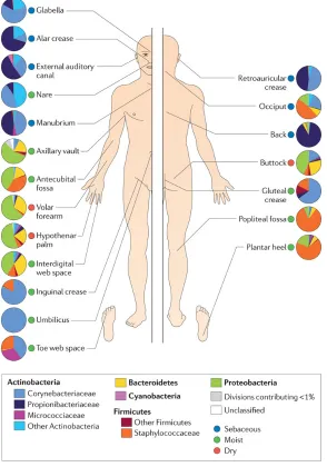
An ecogenomic study discovered that Staphylococcus and Corynebacterium species are the prevailing microbes colonising moist skin sites, culture data also suggests that these micro-organisms favour high humidity skin sites (Grice & Segre, 2011) (Figure 1). These high humidity areas involve the axillary vault, umbilicus (navel), the gluteal crease (topmost part of the fold between the buttocks), the inguinal crease (side of the groin), the popliteal fossa (behind the knee), the antecubital fossa (inner elbow) and the sole of the foot (Figure 1). Staphylococci inhabit aerobic sites on the skin and may utilise the urea present in sweat for nitrogen. Processing of apocrine sweat by Staphylococci (and other axillary vault microbes) causes the malodour characteristic connected to humans sweat (Figure 1) (Grice & Segre, 2011). One of the major Staphylococci species identified on human skin is Staphylococcus epidermidis, commonly inhabitingnares, axillae and the head (Dreno et al., 2016). Staphylococcus epidermidis is the most dominant Gram-positive, coagulase-negative cocci commensal on the healthy human skin microflora. S. epidermidis is responsible for about 60-70% of all coagulase-negative Staphylococcal species on the stratum corneum (Dreno et al., 2016).
2
[image:11.595.138.432.232.652.2]including Demodex folliculorum and Demodex brevis are microscopic arthropods and are a part of the normal skin microflora (Figure 2A) (Grice & Segre, 2011). Sebum provide Domodex mites nourishment, hence, mites are abundantly found after puberty, choosing to colonise sebaceous regions of the face. Demodex mites can similarly feed off epithelial cell lining the pilosebaceous unit and feed on microorganisms that inhabit the same area e.g. Propionibacterium acnes. The function of viruses as a skin commensal has not yet been researched, research is restricted by the current microbiological and molecular means to isolate and classify viruses (Figure 2A) (Grice & Segre, 2011).
3
glabella (amongst the eyebrows); auditory canal (within the ear); occiput (behind the scalp); retroauricular crease (behind the ear); manubrium (top chest area); and back. Moist sites are: axillary vault (armpit); nare (within the nostril); antecubital fossa (inner elbow); interdigital web space (area between the middle and ring fingers); inguinal crease (side of the groin); gluteal crease (upper area of the crease between the buttocks); popliteal fossa (back of the knee); plantar heel (underneath the heel of the foot); toe web space; and umbilicus (navel). Dry sites are: hypothenar palm (palm of the hand proximal to the little finger); volar forearm (inner area of the mid-forearm); and buttock (Grice & Segre, 2011).
[image:12.595.148.425.243.530.2]
4
[image:13.595.101.506.288.463.2]glands and sebaceous glands are few examples of skin appendages illustrated (Grice & Segre, 2011). The skin is usually cool, acidic and dry, variation of the skin microflora is reliant on the cutaneous site thickness, folds, density of hair follicles and glands. The outermost layer of the epidermis is the stratum corneum, of which 90% epidermis cells are made up of terminally differentiated keratinocytes (corneocytes). Squames (enucleated keratinocytes) comprises of keratin fibrils that are crosslinked. The keratin can retain large volumes of water between the fibers. Cornified envelopes are situated in the lipid bilayers, producing the ‘bricks and mortar’ of the epidermidis. Squames migrate from the basal layer of the skin to the outer skin surface and shed, this process could take four weeks (Grice & Segre, 2011).
5
The human skin is an organ which functions as a highly complex barrier while symbiotically interacting with bacterial populations via adaptive and innate immune system signals. Bacteria provide important nutrients, affecting cellular metabolism and strengthen the immune system (Figure 3) (Grice & Segre, 2011; Baldwin et al., 2017). Loss of protective bacteria intensifies inflammatory skin diseases (Williams & Gallo, 2017). The skin is always exposed to countless exogenous and endogenous elements which influence symbiosis, possibly leading to inflamed skin, skin allergies, infections, cutaneous tumour formation or autoimmune illnesses (Bukhari, 2015; Leonel et al., 2019). The gastrointestinal tract and faeces microflora have been researched and documented for several years while the skin or scalp microflora has not been investigated till recently (Bukhari, 2015).
[image:14.595.96.492.111.428.2]In a recent study, it has been reported that S. epidermidis strains obtained from healthy patients’ skin could induce a gathering of CD8+ T lymphocytes in the skin via dendritic cells presentation of antigens (Linehan et al., 2018). However, S. epidermidis strains that stimulate accumulation of CD8+ T
6
lymphocytes in the skin were absent from adult humans with skin disease (Linehan et al., 2018). Linehan et al. (2018) analysed S. epidermidis genome and carried out in vitro experiments to find major histocompatibility complex Ib (MHCIb) on dendritic cells, the cells displayed N-formyl methionine peptides, which are secreted by S. epidermidis, to CD8+ T lymphocytes. The researchers completed RNA sequencing of CD8+ T lymphocytes which were induced by or linked to S. epidermidis. After evaluating the global transcriptome, results revealed that CD8+ T lymphocytes can up-regulate various genes linked with immune regulation and tissue repair (Grice & Segre, 2011; Linehan et al., 2018). In addition to this, Linehan and colleagues also investigated the effects of S. epidermidis on skin injury via punch biopsy method. Interestingly, S. epidermidis stimulated CD8+ T cells, which facilitated re-epithelisation of the injured skin and promoted wound healing.
One of the functions of the skin microbiota is to avoid unwanted pathogenic bacteria from colonising the skin, this maintains an environmental equilibrium in each skin niche. The configuration of the skin microflora may largely influence medical progression for treating cancer, as cancer that use procarcinogenic or anticarcinogenic actions mainly rely on a balanced skin microflora (Garrett, 2015; Nakatsuji, et al. 2018). Skin microbiota can add to carcinogenesis, by augmenting or reducing a host’s risk, this can be categorised into three general groups: (i) modification of the host cell proliferation to death ratio (ii) influencing host immune system function and (iii) manipulation of host metabolism (Garrett, 2015). Thus, it is vital to keep a balanced skin inhabitant populace to also preserve a healthy gut microbiota (Dreno et al., 2016).
7
Nevertheless, no distinct evidence has been found to verify that S. epidermidis is capable of secreting factors that may influence colonisation of other microbes in vivo (Mirzaei et al., 2017). Certain variants of S. epidermidis yield differing lantibiotics including, Pep5, 15X, epidermin and epilancins K7, these lantibiotics are highly toxic and deadly for several Gram-positive bacteria (Espadinha et al, 2019).
Healthy skin microflora may influence the host cells leading to a stronger host defence system against other pathogenic microbes. S. epidermidis can induce endogenous antimicrobial peptides (AMPs) e.g. β defensins, hence, reinforcing the host defence system against S. aureus (Noreen et al., 2015). S. epidermidis is capable of activating mast cell-induced anti-viral host defence mechanism, for it to control inflammatory responses throughout the wound healing process, while encouraging the production of stratum corneum AMPs and inducing cutaneous T-cell development (Noreen et al., 2015). S. epidermidis hence work to protect the skin by communicating with the host immune system. Furthermore, the skin microbiota may symbolise as a filter for the environmental agents interacting with or breeching the skin. There is also evidence available on how the biological determined host immune system has a strong hold on the assembly of the microflora (Noreen et al., 2015).
Alternatively, once the existence of the skin microbiome is sensed via Toll like receptors (TLRs), epidermal Langerhans cells can induce naïve T cells to pedestal a Th17 reaction, this allows keratinocytes to control the AMPs emission (Noreen et al., 2015). Consequently, other than human innate immune system and dendritic cells in the epidermis, TLRs also appear to instruct the adaptive immune response, thus encouraging the complicated regulation of the bacteria growing in the skin (Noreen et al., 2015).
1.2 Commensal S. epidermidis preventing skin cancer
8
Figure 4. S. epidermidis stimulate wound recovery and tumour deterioration in the skin (Leonel et al., 2019). Recent studies suggest novel roles for S. epidermidis in the skin microenvironment (Linehan et al., 2018; Nakatsuji et al., 2018). S. epidermidis support wound healing through an assemblage of CD8+ T lymphocytes in the skin and inhibit cutaneous tumour development via 6-N-hydroxyaminopurine secretion. Future research will demonstrate detailed cellular and molecular mechanisms entailed in the relations between skin microbiota and numerous factors of the skin microenvironment (Grice & Segre, 2011; Leonel et al., 2019).
1.3 The influence of gut microbiome on skin microbiome
Several lines of evidence have indicated that there is a bidirectional communication between the gut microbiome and the skin microflora as various studies connect healthy gastrointestinal to skin homeostasis and allostasis (Levkovich et al., 2013). Gastrointestinal illnesses and differences in the gastrointestinal system usually show signs and symptoms on the skin (O’Neill et al., 2016). The gut microflora commonly indicate change in the pathophysiology of various inflammatory diseases (Shah et al., 2013; Thrash et al., 2013; Gloster et al., 2016).The gut microbiome has a great impact on the gastrointestinal system and can be used for healing purposes by supplementing or prescribing probiotics. Furthermore, the gut microbiota has been observed to have an effect on the skin microbiota (Schwarz et al., 2017).
9
prevalence of specific skin microflora description, which later have an effect on the skin immune defence processes (Shu et al., 2013). Propionic acid may display extreme levels of antibacterial activity against the most prevailing community-acquired methicillin-resistant Staphylococcus aureus (Samuelson et al., 2015). Certain bacteria strains which are symbiotic skin inhabitants namely S. epidermidis and P. Acnes are identified as the most tolerant species compared to the other skin microflora microbes. Overall, the study found encouraging evidence to support the theory of an active interactive system between the human gastrointestinal tract and the skin.
The gut to skin co-operation theory formulated by Arck et al. (2010) mentioned a possible gut to brain to skin co-operation, hence, the advantages of oral prebiotics and skin probiotics is explored. Additionally, as well as the oral probiotic created for the human skin, a recent group of emollients and moisturisers involving lysates of bacteria e.g. Vitreoscilla filiformis or Lactobacillus have been created. The current probiotic invention has been constructed to help manage diseases of the human skin for example, acne or atopic dermatitis. It does this by re-establishing the epidermal barrier, the microbiota of the human skin and regulating the activation of innate immune defence system.
1.4 S. epidermidis – An opportunistic pathogen
Nosocomial infections are 41% of the time caused by coagulase-negative Staphylococci. Little is known about S. epidermidis clonality, pathogenic capabilities including its virulence factors and the prevalence of antibiotic resistance genes such as, mecA, IS256, qacA/B, and icaAB (Mirzaei et al., 2017). Moreover, it is an opportunistic pathogen, as it can only become pathogenic by breaching the host innate immune system (Brown et al., 2012; Yen & Papin, 2017). Staphylococci are commonly found on human and other mammals’ skin and mucous membranes. Although S. epidermidis is not likely to cause terminal disease, treatment for S. epidermidis infections have a very low successful rate, hence, a heavy burden on the public health system (Duell et al., 2012; Otto, 2012).
S. epidermidis species show a high level of diversity with seventy-four identified sequence types (ST). The clonal complex entails many of the identified isolates, this includes the most commonly found ST2 isolate (Espadinha et al, 2019). Every ST2 isolate comprises of IS256 insertion sequences and ica genes which aid ST2 to spread successfully and S. epidermidis to be invasive (Mirzaei et al., 2017). Many of the ST2 isolates have the in vitro ability to allow S. epidermidis to form biofilms. There is information available on S. epidermidis genome, this includes negative ATCC122288 and the biofilm-positive clinical isolate RP62A9 (Grice & Segre, 2011; Espadinha et al., 2019).
10
(Brown et al., 2012). Coagulase-negative Staphylococci (CoNS) group differentiates Staphylococcus aureus with other less or non-infective Staphylococci species (Zmantar et al., 2017). S. haemolyticus and S. epidermidis are a large part of the CoNS group amongst other nosocomial pathogens. Nevertheless, previous research on species identification for CoNS infections found that most of them were caused by S. epidermidis (Otto, 2012). S. epidermidis commonly causes infections through medical devices like central intravenous or peripheral catheters. S. epidermidis infections usually transfer from the skin of a patient, individual, practitioner or employee of health institutions through the inserting process of medical devices (Zmantar et al., 2017). High medical device usage directly correlates to the increasing number of S. epidermidis infections. Which are now known to report a minimum of 22% of bacteraemia in USA critical care patients, occurring in 4–5/1000 central intravenous catheter insertions (Espadinha et al, 2019; Sandoval-Motta & Aldana, 2016).
There is a large amount of S. epidermidis on human skin with similar amounts colonising catheter surfaces (Mirzaei et al., 2017). Also, S. epidermidis are involved in vascular graft, ventriculoperitoneal medical device, surgical site, cardiac implant and prosthetic joint infection (Mansson et al., 2018). Finally, second to S. aureus, S. epidermidis has an increased percentage of intracardiac abscesses (38%), with 13% of prosthetic valve endocarditis (PVE) infections, and 24% mortality. Yet, PVE and other deadly infections are infrequent amongst S. epidermidis infections, overall S. epidermidis infections are characterised as mainly chronic and subacute (Mansson et al., 2018).
11
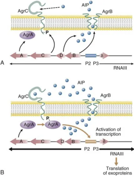
Quorum sensing is a regulation of gene expression where the bacterial cell density influences bacterial gene expression (Figure 3 & 5) (Yan & Bassler, 2019). The bacterium must secrete an autoinducer and sense a signal via a regulatory receptor protein to communicate between cells. When there is a high level of microbial population density, autoinducers amass to a vital threshold value and induce the activation of certain genes and operons (Mirzaei et al., 2017). Quorum-sensing occurs in most bacteria, Staphylococci secrete post-translational modified protein named accessory gene regulator (Agr) (Espadinha et al, 2019; Hellmark et al., 2013) (Figure 5). There is a possibility of specific genetic influence on bacterial communication and colonisation (Figure 2) including quorum-sensing signals that cross-inhibit other microbes (Figure 2). Even though quorum-sensing system is advantageous to S. epidermidis species compared to S. aureus in vitro, there is no proof that it plays a part in vivo (Espadinha et al, 2019; Sandoval-Motta & Aldana, 2016).
1.5 Agr quorum-sensing system
12
translation by blocking mRNA from binding to its target site. Agr can also express exoproteins during stationary phase of growth (high cell density) (Cheung et al., 2014).
Figure 5. A & B. Schematic diagram of Staphylococcus Agr quorum-sensing system (Mazhar & McAuliffe, 2014).
The quorum-sensing system Agr is not very active however the system controls and expresses numerous hostile virulence factors, including the proinflammatory phenol-soluble modulins (PSM), psm genes are regulated by direct binding of AgrA to their promoters (Figure 5B) (Cheung et al., 2014). S. epidermidis are known to produce six PSMs that are all genome-encoded; PSMα, PSMβ1, PSMβ2,
PSMδ, PSM𝜀, and δ-toxin. After δ-toxin, β-PSMs (43–45 amino acid length) are the main types of PSMs
found in S. epidermidis, theyare the key players in biofilm formation (1.4) and detachment (see 1.5)
(Cheung et al., 2010; Cheung et al., 2014). Enhanced production of β-type PSMs over α-type PSMs are
observed in S. epidermidis during biofilm formation (Wang et al., 2011), but how differential
production of α- versus β-type PSMs is attained systematically is unknown.
[image:21.595.184.399.143.432.2]13
being hostile and switch it to a harmless state, this reduces inflammation and stops the host immune system from identifying the pathogen. Hence, the host defence system is completely evaded by S. epidermidis, the S. epidermidis biofilm appear to be dependent on several physiological alterations. This underlines the significance of S. epidermidis biofilm being able to evade host defence system during cutaneous colonisation and biofilm related infections (Siddhiqui et al., 2018).
The acquired immune system comprises of highly specific systemic cells and processes to eliminate S. epidermidis colonisation. Nevertheless, there is not much information on how the acquired immune system can achieve this specifically for pathogenic S. epidermidis (Espadinha et al, 2019; Mirzaei et al., 2017). One way how S. epidermidis may go unidentified and remain protected from antibodies is by using exopolymers (Cheung et al., 2010). Some studies suggest that the host immune system may have evolved to react in a symbiotic way to colonising microbes (Hooper et al., 2012; Belkaid & Hand, 2014; Le et al., 2018).
1.6 Sensing of antimicrobial peptides (AMPs)
The most important biotic function antimicrobial peptides (AMPs) play in vivo is killing pathogens, including Gram-negative and -positive bacteria, viruses and fungi. Gene expression can occur in various host cells such as mucosal epithelial cells and phagocytic cells to encode AMPs (Marquette &
Bechinger, 2018). Cytokines and pathogenic microbes induce gene expression to closely regulate host
defense response. S. epidermidis cause bacterial lipid membrane leak and collaborate with human AMPs production to decrease the number of these microbes. These AMPs are vital cooperating signals between the host defence system and the microbiome. Almost thirty percent of the transcriptome of general epithelial cells use this process to communicate.
There are two main groups that AMPs belong to, cathelicidins and defensins. The AMPs from these groups with an overall positive charge are called cationic antimicrobial peptides (CAMPs). However, an anionic AMP with a negative net charge has been detected from human sweat called dermcidin.
(Espadinha et al., 2019). S. epidermidis, the Gram-positive bacteria has been found to have an AMP
sensor. The recently identified AMP sensor is made up of three crucial factors, ApsS with a sensory function, ApsR the regulator and ApsX the unique final constituent of unidentified purpose. (Vasilchenko et al., 2019)
14
important for Staphylococci to have biofilm-related proteins and PIA (Mirzaei et al., 2017).PIA is a glycan of beta-1,6-linked 2-acetamido-2-deoxy-D-glucopyranosyl residues of which 15 % are non-N-acetylated.(Spiliopoulou et al., 2012; Hofferek, 2019). These proteins are vital for colonisation and infecting the host as both commensal and pathogenic S. epidermidis strains, both type of strains have the same MSCRAMMs (microbial surface components recognising adhesive matrix molecules) (See 1.7.2) (Geoghegan et al., 2010). This indicates that S. epidermidis can be viewed as an accidental pathogen and it is clinically important to researchadherence and evasion of host immune system as this can be valuable for killing pathogenic bacteria and treating long-term infections (Espadinha et al., 2019).
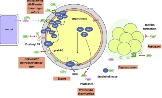
Just the way human innate immune system recognisesS. epidermidis pathogen associated molecular pattern (PAMPs), S. epidermidis also identifiesdangerous foreign molecules produced by the human host (Joo et al., 2015). There is an AMP-sensing system within S. epidermidis named Aps. Diverse number of AMPs activate Aps system and cause an up-regulation ofdefensive systems. AMP-defensive systems may includeVraFG proteins, lysylation of phospholipids by the MprF enzyme and D-alanylation of teichoic acids (Joo, & Otto, 2015). This affects the exterior of the bacteria as it diminishes the negatively charged surface and halts the attraction of positively charged AMPs, efficiently exporting AMPs from the cytoplasm membrane (Figure 8) (Lin et al., 2018). Hence, Gram-positive bacteriaAps system plays a similar role to Gram-negative AMP sensor named PhoP/PhoQ, however, the bacteria is not genetically related (Lin et al., 2018; Joo & Otto, 2015). The bacteria S. epidermidis Aps system is activated and limited to cationic AMPs (Lin et al., 2018). Aps system is a three-component regulator, it has an important component of unidentified function namely, ApsX (Joo et al., 2015). The response regulator protein ApsR (GraR) and histidine kinase ApsS (GraS) make up the other two components of the Aps system (Figure 8) (Joo et al., 2015).
1.7 Biofilm
15
It is important that ica operon express polysaccharides to form biofilms (Chua et al., 2014; Mirzaei et al., 2017). Some researchers have proposed that polysaccharide adhesin is efficient for adherence and cell to cell communication during the assembly process of biofilm production (Franca et al., 2016). Other researchers have suggested that adhesion is induced by microbial cell surface proteins and polysaccharides can only control the process of accumulating cells (Guo et al., 2019).
Many microorganism cells produce and emit extracellular substances in the form of a slime layer, thus creating a hydrophobic biofilm. Biofilms are complicated multicellular assemblage of microbes adhered to a surface (Chua et al., 2014). This allows the microbe to adhere to different kind of surfaces including metals, plastics and the human body. Adherence to medical devices including catheters, mostly occur due to hydrophobic external layer of bacteria (Figure 6). Certain proteins promote S. epidermidis adhering to surfaces, this largely involves surface protein AtlE, which has two functions; to adhere to cells or surfaces and breakdown the cell-wall peptidoglycan of S. epidermidis (Mirzaei et al., 2017; Chua et al., 2014). Another protein named Bap/Bhp protein probably promote the hydrophobicity level of the bacteria cell outer layer. S. epidermidis mostly cause prosthetic joint infections, when the bacteria accumulate on an implant surface it creates a biofilm and transitions to a stationary phase in bacterial growth due to increased microbial populace densities (Mansson et al., 2018). During the stationary phase the bacteria grows slowly compared to planktonic bacteria, the bacteria has decreased metabolic activity and, thus, high level of resistance to elimination by growth-dependent antibiotics, e.g. cell-wall-active antibiotics (Figure 6) (Spiliopoulou et al., 2012; Hellmark et al., 2013). Additionally, the biofilm decreases the penetrability of antibiotics and host immune components such as complement and antibodies (Figure 6) (Stipetic et al., 2016).
16
1.7.1 Biofilm formation
17
[image:26.595.93.502.71.387.2]Adherence to medical devices including catheters, mostly occurs due to hydrophobic external layer of bacteria (Figure 6 & 7). Certain proteins promote S. epidermidis adhering to surfaces, this largely involves surface protein AtlE, which has two functions; to adhere to cells or surfaces and breakdown the cell-wall peptidoglycan of S. epidermidis (Chua et al., 2014; Mirzaei et al., 2017). Another protein named Bap/Bhp protein probably promote the hydrophobicity level of the bacteria cell outer layer. S. epidermidis mostly cause prosthetic joint infections, when the bacteria accumulate on an implant surface it creates a biofilm and transitions to a stationary phase in bacterial growth due to increased
Figure 6. Steps of Biofilm Formation (Monroe, 2007; Aguinaldo, 2015): 1. Bacteria adhere to a surface
2. Bacteria cells replicate and form a single thick polysaccharide matrix layer. Biofilm is not visible at this stage.
3. A 3D biofilm structure is formed by large bacterial colonies secreting exopolysaccharide and quorum-sensing.
4. The exopolysaccharide slime protects the bacteria from harsh environment, antibiotics, toxic chemicals etc. Nutrients are diffused through the matrix. A secondary bacterial species joins the biofilm. The small proximity between both species allows genetic exchange.
18
microbial populace densities (Mansson et al., 2018). During the stationary phase the bacteria grows slowly compared to planktonic bacteria, the bacteria has decreased metabolic activity and, thus, high level of resistance to killing by growth-dependent antibiotics, e.g. cell-wall-active antibiotics (Figure 6 & 7) (Spiliopoulou et al., 2012; Hellmark et al., 2013). Additionally, the biofilm decreases the penetrability of antibiotics and host immune components such as complement and antibodies (Figure 6 & 7) (Stipetic et al., 2016).
1.7.2 MSCRAMMS
19
2013). This process is meant to cause a highly stable MSCRAMM-substance communication (Geoghegan et al., 2010). This emphasises the significance of SdrG assisting S. epidermidis in causing infections, also, there is an increase in the expression of SdrG in an in vivo condition with antibodies to SdrG are identified in human blood (Faustino et al., 2013). Not long ago, SdrF has been found promoting infection by encouraging S. epidermidis to adhere to ventricular medical devices. Additionally, various other S. epidermidis MSCRAMMs are yet to be identified and undergo characterisation to understand the function they play in matrix protein binding process and the pathogenicity of S. epidermidis (Geoghegan et al., 2010).
1.7.3 PNAG/PIA
Several strains of S. epidermidis create a PNAG homopolymer also known as PIA, which surround and adheres S. epidermidis cells within a biofilm (Mirzaei et al., 2016; Spiliopoulou et al., 2012). This protein molecule is different to poly-N-acetylglucosamine polymers observed in flora, for example chitin, this is due to its β 1–6 connection. Recently, PIA/PNAG has also been found in several other microbes such as Yersinia pestis and Escherichia coli. Biosynthesis of PIA/PNAG is vital for formation of biofilm in vitro and it has a substantial influence on S. epidermidis infection in animal models (Spiliopoulou et al., 2012). The process of PIA/PNAG production is initiated by a product of a gene namely intercellular adhesion (ica) locus (Mirzaei et al., 2016). A chain of reaction from activated N-acetylglucosamine monomers, is formed by IcaD and IcaA where the prolongation relies on the IcaC protein, this is possibly due to the exporting role it is known to play. Once exportation occurs, partial de-acetylation of the N-acetylglucosamine residues is completed by the enzyme IcaB, which is situated on the surface of the cell. Due to de-acetylation the neutral polymer becomes positively charged, this allows PIA/PNAG to bind to surfaces and for it to play a part in numerous biotic interactions, such as S. epidermidis evading the host immune defence system (Spiliopoulou et al., 2012). Several universal virulence regulators influence the formation of PIA/PNAG excluding the quorum-sensing regulator agr (Figure 5). Although there is limited understanding on what environmental signals regulate the expression of PIA/PNAG, especially in vivo, the intricacy of regulation reinforces the significance of PIA/PNAG for S. epidermidis physiopathology (Mirzaei et al., 2016).
20
association of G5 tandem repeats, this underlines that Aap forms fibril-type structures on the microbial outer layer. These domains are seen interacting with N-acetylglucosamine and may possibly bind to PIA/PNAG to form a polysaccharide/protein biofilm system (Mirzaei et al., 2016). A chelating agent was used to prevent the production of biofilm in vitro, the result highlighted that strong biofilm forming strains such as S. epidermidis RP62A strain are completely reliant on Aap (Otto, 2009). Additionally, monoclonal antibodies produced against Aap stopped S. epidermidis RP62A from forming biofilm. Nonetheless, the findings from this report contradicted another report which did not find any protein that induced biofilm formation in the identical strain. Hence, certain proteins may contribute to S. epidermidis forming biofilms, however this requires further investigation. Finally, the concept of biofilms being produced only by proteins is not robust as the one on PIA/PNAG that demonstrates both exopolysaccharide and proteins are required to promote S. epidermidis biofilm production. The overall information on molecular processes of S. epidermidis regulation and production of biofilm is nearly completely reliant on in vitro study (Franca et al., 2016). Animal models have been used to contribute to the pathogenesis of S. epidermidis by demonstrating few factors including PIA/PNAG, Fbe, AtlE, SdrF, SdrG and regulators Agr, sigB and luxS (Mirzaei et al., 2017; Pereira et al., 2018). Additionally, there is evidence signifying the importance of expressed biofilm elements, in vivo. However, there is an imperative requirement for more comprehensive in vivo studies to provide complete understanding of S. epidermidis biofilm related infections (Franca et al., 2016).
1.7.4 Biofilm in host evasion
Biofilm has a distinctive composition and construction for the development of biofilm resistance to numerous antibiotics and evasion of host defence systems (Franca et al., 2016). S. epidermidis show a large variety of genetic variations throughout the biofilm growing process, this includes down-regulation of basic cell mechanisms, for example, nucleic acid, protein biosynthesis and cell wall (Franca et al., 2016). The regulation of gene expression may alter and lead to the inactivity of several antibiotics which target against S. epidermidis developing biofilm cells, these involve antibiotics such as, quinolones, aminoglycosides and penicillin (Hellmark et al., 2013).
21
Furthermore, the bacteria must survive host immune defence attacks, especially antibacterial peptides. β-defensin is one of the key antibacterial peptides found mostly at epithelial surfaces. Gram-positive bacteria can act as a chemo-attractant between innate and acquired immune systems (Siddhiqui et al., 2018).
[image:30.595.75.529.224.514.2]
Figure 7. S. epidermidis biofilm structure and function in host immune evasion. The biofilm matrix is made up of PIA/PNAG, Aap, Bap/Bhp, extracellular matrix binding protein (Embp), extracellular DNA (eDNA) and teichoic acids. Biofilm channels are produced by PSMs, which eventually cause cell cluster detachment and overall dissemination of the biofilm. PSMs also play a role in bacteremia and sepsis in immunocompromised individuals. Both biofilm structure and matrix provide protection from host defences such as opsonising immunoglobulins binding, complement components and AMPs. Furthermore, there is a drastic reduction in leukocytes attacks (Le et al., 2018).
Brandwein et al. (2016)isolated Staphylococcal strains from hospital settings and patient body sites,
22
transcription and translation as well as a switch from the aerobic system to fermentation to produce energy (Allen et al. 2014). These alterations are possibly due to the reduced level of oxygen within the biofilm and the limited amount of nutrition available. Notably, this causes the biofilm to become dormant and enter the stationary phase of growth, thereby, the antibiotic used against the biofilm becomes ineffective as the biofilm now has low sensitivity for the antibiotic treatment (Brandwein et al., 2016). Likewise, the biofilm is capable of evading the host defence mechanisms as cytokines and antibiotics mainly rely on highly functional cell metabolism and cell-development mechanisms.
1.8 Biofilm detachment
Unlike the intercellular accumulation phase, there is little understanding of S. epidermidis biofilm structure and the process of detachment. It is commonly known that S. epidermidis biofilm detachment is regulated by the Agr quorum sensing mechanism (Pereira et al., 2018). Biofilms that are not controlled by Agr due to dysfunction are a lot denser and display clear deficiencies during detachment (Boles & Horswill, 2011). A model that had Agr expressed in the outer surface of biofilm was known to encourage cluster of cells detaching from the biofilm surface, hence, regulating biofilm dissemination (Pereira et al., 2018). Similarly, the function of Agr in S. epidermidis is restricted to the exposed surfaces of a biofilm, this suggests that both closely related Staphylococci species share the same quorum-sensing system to control the detachment of biofilm (Pereira et al., 2018). The mutation of both Agr system or psm genes, can support the formation of compact biofilms (Wang et al., 2011), however, the biofilm can no longer detach and form biofilm elsewhere (Joo and Otto, 2012).
23
are suggested to have this detachment mechanism (Boles & Horswill, 2011). S. epidermidis exoproteases and PSMs are strongly controlled by Agr, thus, supporting the concept of exoproteases and PSMs having a function in organising the structure of the biofilm (Cheung et al., 2014; Boles & Horswill, 2011).
1.9 Protective exopolymers
S. epidermidis and Bacillus anthracis are the only species known to date where PGA plays a role in the spread of disease. Additionally, PGA encourages S. epidermidis to grow in high salt concentrations (Chua et al., 2014). Numerous halophilic microorganisms PGA have similar function that contribute to osmotolerance and PGA also promote S. epidermidis colonisation. Also, cap genes are increasingly expressed during biofilm growth. Remarkably, PGA exist in several CoNS, but is missing in S. aureus (Cave et al, 2019).
Furthermore, exopolysaccharide PIA/PNAG can also protect S. epidermidis from human host defence mechanisms such as AMPs, neutrophil phagocytosis, immunoglobulins and complement deposition. Positively charged PIA/PNAG protect S. epidermidis from AMPs with either negative or positive charge, this indicates that the activity of PIA/PNAG may not be restricted to electrostatic repulsion of AMPs with identical charge (Yan & Bassler, 2019).
1.10 Pathogen-associated molecular patterns
24
It is imperative to know that lipoproteins were the foundation of the study, which is a strong pro-inflammatory contaminant that may result in incorrect identification of TLR2 stimulants (Noreen et al., 2015). Likewise, pro-inflammatory capabilities of S. epidermidis PSMs are yet to be verified using gene deletion mutation or synthetic peptides (Otto, 2014). Nonetheless, the similitude of the confirmed function of S. epidermidis and S. aureus PSMs show that the detailed pro-inflammatory influence of S. epidermidis PSMs is authentic; however, the activation of TLR2 by PSMs require confirmation (Cheung et al., 2014; Noreen et al., 2015). Lastly, an abnormal short-chain pro-inflammatory lipoteichoic acid has been observed in S. epidermidis. On the other hand, the chemical characterisation of the purified element has no indication of teichoic acid-associated polymer, and hence the character of this substance and the reported pro-inflammatory function require confirmation. Thereby, further research is needed for characterisation of S. epidermidis substances that stimulate the host immune defence system.
1.11 Evasion of host defences
A pathogen must evade the human body host defence in order to withstand and cause disease. Few host defence systems exist, for example, AMPs on the skin. S. epidermidis must survive other host defence responses once it breeches the epithelial barrier. Invading microbes as well as S. epidermidis interact with the innate immune system in an unspecified way. The colonising S. epidermidis bacteria have numerous mechanisms to avoid being ingested and killed by neutrophils while also evading AMPs.
A pathogen must evade multiple human body natural defences for survival (Otto, 2009). There is limited subset of mechanisms of host defence mechanisms existing on the human skin. S. epidermidis must avoid AMPs and survive several other defence mechanisms after penetrating the epithelial barrier (Whitney et al., 2010). The innate immune system reacts without being specific to any microbe entering, including S. epidermidis (Cheung, et al., 2010). The innate immune system mainly reacts by ingesting microbes using neutrophils, eradicating them with AMPs and reactive oxygen species (Otto, 2009). S. epidermidis can avoid being ingested and killed by neutrophils via AMPs resistance mechanism (Figure 6) (Le et al., 2018).
25
PGA, the gene product of cap locus is imperative for S. epidermidis protection from AMPs and neutrophil phagocytosis. Furthermore, the host defence system may have adapted overtime to not have a strong response against predominant infectious pathogens.
[image:34.595.37.554.215.530.2]1.11.1 AMP Resistance
26
by the host from the cell membrane (CM). The exported proteases that have little substrate specificity tend to destroy both anionic AMPs and CAMPs, regulated by global regulators such as Agr and Staphylococcal accessory regulator (SarA) (Figure 8). PSMs and other toxins are degraded by aureolysin, a protease which is also regulated by multiple global regulators, including Agr and SarA. CAMPs are inactivated by staphylokinase, through a sequestration process. This method is observed in surface polymers that have an opposite charge to AMPs e.g. positively charged PIA exopolysaccharide sequestrating negatively charged AMPs. The production of a biofilms adds to AMP resistance through various methods such as reduced penetration. Additionally, the cationic biofilm PIA may repulse CAMPs (Figure 8) (Joo & Otto, 2015).
1.11.2 PSMs
S. epidermidis as well as all the other Staphylococci species including the pathogenic S. aureus contain genes encoding PSMs and other related peptides (Cheung et al., 2014). These are both hydrophilic and hydrophobic helix shaped small peptides. PSMs are a group of virulence toxins that are soluble in phenols and may contribute to the pathogenicity of S. epidermidis (Otto, 2014). Although, PSMs commonly lack bacteriostatic characteristics (Cheung et al., 2014). AMPs as well as PSMs of alpha-type (20–25 amino acids) are cationic, whereas beta-alpha-type PSMs (43–45 amino acids) have a net anionic charge (Cheung et al., 2010). The amphipathic α-helix nature of PSMs allow it to have an affinity for lipids, hence, α-type PSMs are more active against eukaryotic cells compared to prokaryotic cells. PSMs with big hydrophobic side chains prevent antibacterial activity by avoiding microbial membrane disruption. Cheung et al., (2014) findings showed that PSMs can have a different effect on prokaryotic cell membrane compared to eukaryotic cells. The α-type PSMs can lyse human cells such as erythrocytes and leukocytes while promote inflammatory responses (Cheung et al., 2014).
1.12 Pathogenicity
27
the major toxin it does produce are namely PSMs. All S. epidermidis produce PSMs except S. epidermidis strains with natural agr dysfunction (Pereira et al., 2018). PSMs are typically α-helical, short and amphipathic (Cheung et al., 2014) and sometimes have cytolytic function but mainly pro- inflammatory function.S. epidermidis produce PSMγ/δ-toxin (a 24-amino acid peptide) and S. aureus produces a similar peptide but the positioning of one amino acid varies from the S. epidermidis homologue (Cheung et al., 2014). Several reports have found that S. epidermidis PSMγ assist in necrotising enterocolitis in new-borns. Few S. aureus PSMs have similar function as S. epidermidis PSMs of lysing host neutrophils (Cheung et al., 2014). Nevertheless, S. epidermidis PSM production mostly constitutes of noncytolytic β-type and mild cytolytic PSMs (Otto, 2014). In conclusion, although S. epidermidis may lack highly virulence toxic molecules compared to S. aureus, due to the S. epidermidis PSM production, S. epidermidis has an evolutionary advantage over more virulence Staphylococci strains (Otto, 2014). Although S. epidermidis toxicity factor has been researched the process of toxicity is not well understood.
Furthermore, other possible virulence substances produced by S. epidermidis are now being studied as well as the fibrinogen binding process (Becker et al., 2014). A gene called fbe consists of an open reading frame of 3,276 nucleotides which encode a protein named Fbe, which has a molecular weight of ~119 kDa (Salgueiro et al., 2017). Biomaterial implants are quickly inhabited by plasma substances and fibrinogen aid bacterial cells adhere to the biomaterial surface. A problem may occur when adhesion occurs on the identical regions of medical implant by pathogenic microorganisms, hence, causing infection (Salgueiro et al., 2017; Becker et al., 2014).
1.13 Clinical background
28
Septicemia or bacteraemia is the poisoning of the blood caused by infectious bacteria (Kleinschmidt et al., 2015). Septicemia is a microbial infection in the bloodstream where the bacteria may enter through the urinary tract, skin or lungs. Septicemia tend to affect the weak immunocompromised neonatal, older individuals and patients that had surgery (Kleinschmidt et al., 2015). Septicemia is a life-threatening disease as the bacteria toxins spread through the bloodstream to the entire body, septicemia must be cured quickly in a healthcare facility, if it is left unchecked septicemia can develop into sepsis (Kleinschmidt et al., 2015). Septicemia symptoms include chills, fever, rapid respiration and fast heart rate. If septicemia is not treated properly or left untouched, the symptoms become more severe. These symptoms may include nausea, red marks on the skin, confusion, shock, lack of concentration and pass low volumes of urine.
Endocarditis is the microbial colonisation of the inside the lining of heart muscles and heart valves (endocardium). Without proper early treatment, endocarditis may impair or destroy the heart valves that may lead to fatal complications. Endocarditis treatment mainly include the usage of antibiotics and sometimes surgery. People with artificial heart valves, damaged heart valves, or other heart defects are at a higher risk of developing endocarditis. Symptoms of endocarditis include, fatigue, aching muscles and joints chills, fever and night sweats, shortness of breath, swelling of feet, legs or abdomen and changed heart murmur (the sound of blood rushing through the heart).
When Sahal & Bilkay (2014) examined blood samples from patients that had cardiovascular surgery as well as wound samples, S. epidermidis strains displayed a strong capacity to form biofilms (surgery samples 35% and wound samples 40%), 80% of them were resistant to β-lactam and 100% of them were multi antibiotic resistant (Sahal & Bilkay, 2014). The spread and quick growth of antibiotic resistant S. epidermidis microbe in healthcare settings are associated with certain factors for example, increased selective pressure, that occur due to improper and widespread use of antimicrobials mainly in hospitals. Moreover, there are other possible agents that cause dissemination of microbes, which may involve cross transmission amongst patients via inadequate infection and control strategies, bacterial mutation and horizontal resistance genes transfer along with an intricate relationship among nominated antimicrobial substances and microbial resistance.
1.14 Treatment
29
speeds up. 80% of catheters infected with S. epidermidis are treated with antibiotics including vancomycin without removing the catheter. Two treatments that are frequently favoured in treating other microbes including S. aureus is decolonisation and vaccination, however these options are ineffective for S. epidermidis. Firstly, there is no vaccine available for S. epidermidis infectionsand lots of studies show that the use of traditional immunisation is problematic for Staphylococci infection. Secondly, S. epidermidis is a major part of the human microbiota, hence it is difficult for it to be eradicated, if S. epidermidis were to be destroyed, quick re-colonisation from other individuals can occur. By excluding S. epidermidis from the human microflora it may hypothetically lead to more aggressive pathogens replacing S. epidermidis. Therefore, it is generally agreed upon that the most effective way of reducing the number of S. epidermidis infections is by preventing the spread of S. epidermidis (Szemraj et al., 2019). This consists of medical equipment sterilisation, health care personnel in contact with indwelling medical devices during surgery and patient body parts sterilisation.
It is a challenging burden on the healthcare system to treat infections linked to S. epidermidis biofilms. This is due to the bacteria being resistant to antibacterial molecules and the human immune defence mechanism (Hoiby et al., 2010). In addition to this, currently the occurrence of virulence microbial strains becoming resistant to several antibiotics is drastically rising. Thus, making it difficult to treat microbial infections which has quickly become the deadliest health threat to all humans (Guridi et al., 2015). This difficulty has mostly occurred due to the phenotypic resistance of microorganisms as they act as a ‘reservoir’ and harbour resistance genes in the bacteria chromosome and/or plasmid DNA.
30
1.15 Prevention
Health care professionals should focus on carrying out preventive measures to reduce the incidence of S. epidermidis infections or to completely eliminate the bacteria (Perez & Patel, 2018). These practices involve controlling risk factors, for example reintubation, nasogastric and endotracheal tubes, tracheotomy, poor infection control and hand washing procedures among hospital employees, old antibiotic treatments, contaminated respiratory aids, water or drugs are effective means of preventing S. epidermidis infections (Perez & Patel, 2018).
A small synthetic antimicrobial peptide called bactericidal peptide 2 (BP2) is shown to reduce 80% risk of acquiring S. epidermidis infection in the hospital via implants in a research carried out by (Perez & Patel, 2018). BP2 has strong bactericidal activity at micromolar concentrations for a broad spectrum of microorganisms, involving antibiotic-resistant bacteria. The staphylocidal activity of BP2 is not affected by physiological salt concentrations and is only slightly affected by the presence of human plasma (Simonetti et al., 2013). BP2 is more active than HNP1-3 and as active as LL-37, a highly potent human antimicrobial peptide. Unlike LL-37 (Mahlapuu et al., 2016) and HNP1-3 (Papot et al., 2017), the microbicidal activity of BP2 is not inhibited by physiological salt concentrations, stressing the potential of BP2.
1.16 Antibiotics
31
Antibiotics have different molecular ways to eliminate bacteria, for example, inhibiting growth, protein synthesis, nucleic acid synthesis, cell wall synthesis, enzyme activity, specific biochemical processes and altering cell membrane permeability (Kapoor, Saigal & Elongavan, 2017). Some examples are briefly explained in Table 1. Traditionally, antibiotics have been classified as bacteriostats which stop cell growth or bactericides that kill bacteria. Bacteriostats rely mostly on the host immune system to eliminate the bacterial infection. However, bacteriostatic antibiotics can become bactericidal when used at a high concentration, hence, this method of classification is not enough (Kapoor, Saigal & Elongavan, 2017). Another more recent method to classify antibiotics is to observe antibiotic activity, if it is reliant on time, concentration or is it co-dependent (Table 1). Information on how the antibiotic operates is important, however, more data is required on the spectrum of activity (which microbes are susceptible to the antibiotics), level of resistance detected (due to repetitive use) and if the concentrated antibiotic is effective in vivo with therapeutic dose rates (Kapoor, Saigal & Elongavan, 2017).
32 Tabl e 1 . An tib io tic s us ed in th is res ea rc h Spe ct rum ac tiv Na rr
Broad Broad Broad Broad Broad Narr Broad Broad
Revi sed tab le from Bou ch ard (2017) Bac te ric idal or Bac te rios tat ic ? Bact eri ci dal Bact eri ci dal Bact eri ci dal Bact eri ci dal Bact eri os tat ic Bact eri os tat ic Bot h Bact eri os tat ic Bact eri os tat ic Si te of Ac tion Nu cl ei c ac id sy nthe si s Nu cl ei c ac id sy nthe si s Ce ll w al l Ce ll w al l Pr ot ei n Sy nthe si s Fol ate sy nthe si s Pr ot ei n sy nthe si s Pr ot ei n sy nthe si s Pr ot ei n sy nthe si s Ant ibi ot ic s Me tr on id az ol e Ci prof loxaci n Am ox ic ill in , Co -Am ox ic la v Ce fal exi n Ery throm yci n Tri m ethopri m Li ne zo lid Dox ycycl in e Cl ari throm yci n, Az ith ro m yc in Ch em ic al gr ou ps Ni tr oi mi da zo le Fl uoroqui nol one Pen ic ill in Ce phal ospori n Ma cr ol id e Di am in op yri m id in e Ox az ol id in on e Te tracy cl ine Ket ol id e Mo de o f a ct io n El im inati on is re liant on conce ntrati on, w ith a strong post -anti bi oti c ef fec t. El im inati on is re liant on tim e w ith littl e or no post -anti bi oti c ef fe ct. El im inati on is re liant on the le ve l of conce ntrati on and le ngth of e xposure . Ye ar of in tr od uctio n
1960 1987 2000 1985 1964 1951 1972 1985 1967 1990 1981
Ye ar of di sc ov er y
33
1.17 Antibiotic resistance
At present, there is a universal concern for the era of antibiotics as it is coming to an end (Santiago & Maximino, 2016). It is predicted that medicine in the near future may return to 19th century medicine where one slight injury may cause deadly infection and death (WHO, 2014). For decades antibiotics have been used globally and repeatedly. Since 1935, all infections caused by bacteria have been cured quickly, even if the last generation of bacteria showed resistance to antibiotics (Aminov, 2010; Domínguez & Meza-Rodriguez, 2019). Resistance to antibiotics is inevitable. All animals including humans live with a large variety of bacteria in their gut and skin microflora, most of these microbes namely commensals are highly valuable or completely innocuous (Conlan et al., 2012). Only a small portion of these commensals are capable of becoming harmful based on what niche they live in or what area of the host they occupy (Conlan et al., 2012).
The discovery of antibiotics was and still is a breakthrough in treating bacterial infection, however, due to the overuse of antibiotics there are now antibiotic-resistant strains of pathogenic microbes (Aminov, 2010; Domínguez & Meza-Rodriguez, 2019). This method of treatment lead to non-reversible mutation in new resistant strains of pathogens or commensal bacteria (Conlan et al., 2012; Santiago & Maximino, 2016). This is a global health problem which requires an urgent solution as it may lead to a permanent unbalance skin and gut microflora. This is why there are laws and regulations put in place to limit the use of antibiotics for minor infections (Dreno et al., 2016).
34
Substantial number of mutants are bound to exist in the estimated world population of bacteria (5 x10$%) (Namkeleja et al., 2016).
Strong and speedy antibiotic prescriptions work remarkably well when the bacterial population is destroyed completely (Aminov, 2010; Domínguez & Meza-Rodriguez, 2019). However, when this does not occur, the outcome of antibiotic usage becomes disadvantageous. As S. epidermidis is a part of the normal microbiota for healthy humans, it has developed resistance to numerous antibiotics that are used frequently, for example, methicillin, novobiocin, clindamycin, and benzyl penicillin (Saffari et al., 2016). Consequently, other antibiotics namely vancomycin or rifampin are now used to treat infections.
S. epidermidis contain integrated plasmids that carry genes encoding species-specific LPXTG surface proteins and resistance to cadmium. S. epidermidis Genome Island may encode possible virulence factors including numerous phenol-soluble modulins (Xue et al., 2017). S. epidermidis contain cap operon (capABC) and gamma-glutamyl transpeptidase gene as found on the B. anthracis pX02 plasmid, which encodes polyglutamate capsule, a key virulence factor of Bacillus anthracis (Xue et al.,
2017). Any other phenotypic variances are possibly caused by single nucleotide polymorphisms which can be seen in most cell envelope proteins. Phylogenetic study of cap genes in the operon specify that the attainment of this locus may be created by a plasmid-mediated transfer event from the ancestor of bacilli to S. epidermidis. Nevertheless, several species-specific metabolic functions including polyphosphate synthesis and acetoin dehydrogenase are encoded by complete operons in S. epidermidis, this may be due to gene loss by a common ancestor (Xue et al., 2017).
Five key mechanisms have been identified to cause antibiotic resistance, including, the formation of deactivating enzymes, alteration in target sites, initiation of alternative pathway e.g. bypass pathway, the inability to activate antibiotics and antibiotic prohibition from active efflux of target site (MacGowan & Macnaughton, 2017). The main mechanism that cause antibiotic resistance is the chemically modified antibiotic molecule as well as the destroyed antibiotic molecule. Secondly, mutated target sites, altered enzymatic function of the target sites and full substitution or bypass of target sites cause the most antibiotic resistance. Furthermore, majority of antibiotic resistance occur when there is low porosity and deceased amount of efflux pumps. (Munita & Arias, 2016).
35
During the late 1960s, semisynthetic penicillin and methicillin were extensively used and led to the rise of methicillin-resistant S. aureus (MRSA) and S. epidermidis (MRSE), which persists today in both community environments and health care settings (Stamatiou et al., 2013). S. epidermidis are known to contain specific antibiotic resistant genes, especially for methicillin, which is one of the first antibiotic used to treat S. epidermidis infections, approximately 75–90% of S. epidermidis isolates found in a hospital setting had methicillin resistant gene, oppose to S. aureus isolates with only 40– 60% harbouring methicillin resistant gene (Stamatiou et al., 2013). S. epidermidis carry mobile genetic elements (MGEs) called Staphylococcal cassette chromosome mec (SCCmec) comprising of mecA gene which encode a penicillin-binding protein (PBP) with low methicillin affinity (Rolo et al., 2012; Mirzaei et al., 2017). PSM-mec (20–25 amino acids), a short α-type PSM is also encoded by SCCmec (Cheung
et al., 2014). PSM-mec has the ability to activate and stimulate neutrophils (Qin et al., 2016) via human
formyl peptide receptor 2 (FPR2) interaction, the G protein-coupled receptor is expressed on different
types of host immune cells. PSM-mec canimmediately up-regulate the expression of both CD11b and
gp91phox, induce calcium flux, chemotaxis, and IL-8 release in neutrophils (Qin et al., 2016). Not only
does S. epidermidis mecA gene encode for methicillin resistance but also broad β-lactam resistance (Saffari et al., 2016).
In addition to this, ten various SCCmec elements have been found in S. epidermidis. The most commonly identifiedSCCmec element in S. epidermidis is the smallSCCmec type IV structure which may spread without damaging the fitness of its host bacteria and evade specific antibiotic pressures (Xue et al., 2017). Moreover, Staphylococcal variants carry various type ofSCCmec structures and S. epidermidis frequently uptakes and emits SCCmec elements.
Furthermore, S. epidermidis is resistant to lots of other antibiotics besides methicillin such as erythromycin chloramphenicol, gentamicin, flouroquinolones, rifamycin,sulfonamides,tetracycline and clindamycin (Saffari et al., 2016). Also, S. epidermidis is less likely to be resistant totigecycline, streptogramins, and linezolid and is found to be intermediate resistant to vancomycin. The capability of S. epidermidis to form biofilm greatly reduces the likelihood of vancomycin and other antibiotics to have an effect (Saffari et al., 2016). Bacteria plasmids mostly encode antibiotic resistant genes and are frequently found in methicillin-resistant instead of methicillin-susceptible variants. Endemic nosocomial strains of S. epidermidis are usually resistant to both methicillin and other antibiotics (Stamatiou et al., 2013).