International Journal of Emerging Technology and Advanced Engineering
Website: www.ijetae.com (ISSN 2250-2459,ISO 9001:2008 Certified Journal, Volume 5, Issue 1, January 2015)
133
Detection of Diabetic Retinopathy with Feature Extraction
using Image Processing
Meera Walvekar
1, Geeta Salunke
21
G.S M C.O.E., Pune, India
2AISSMS IOIT, Pune, India
Abstract - Diabetes has become a new global challenge. If not diagnosed and treated in time, diabetes can encourage other illnesses in the body of patients. One such illness is related to the retina of human eyes that affects the retina and retinal structure in certain ways. The screening for detection of such abnormalities in the retina is called Diabetic Retinopathy (DR).
Latest technological advances in the image processing helps auto detection of diabetic retinopathy based on the analysis of feature extractions. This analysis not only helps diagnose the disease but also helps detecting the severity of the disease.
In this paper, we will look at the extraction and outcome of important features, using image processing, and the severity of Diabetic Retinopathy. The datasets used for this study are DRIVE and STARE.
Keywords - features extraction, optic disc, microaneurysm, blood vessels, exudates, retinal image, diabetic retinopathy
I. INTRODUCTION
The World Health Organization (WHO) recently indicated that there are about 135 million people in the world having diabetes mellitus and this number may go up to 300 million by 2025. There has been focus on methods and algorithms that would help experts in the medical fields to process digital inputs, analyse results and diagnose different illnesses.
This approach is consisting of digital image studies with an aim of providing ways in diagnosing the diabetic retinopathy and identifying the severity of the disease. It typically includes application of image processing on digital images of the retinal structures. Progress in this area has been achieved in recent times and improved medical care is available for the patients.
According to a recent survey, diabetes has been recognized as the main cause of blindness. If not diagnosed early and treated in time, it can lead to severe damage to retinal structure leading to partial or even complete blindness.
The constant check up and screening activities like medical digital image processing for detection and diagnosis of diabetes related disease, like diabetic retinopathy, is very important.
The automated way of such screening of digital images will help medical experts screen and diagnose a large population of patients. Over a period of last decade, there is a lot of research conducted by different experts and authors over automatic detection of diabetic retinopathy based on extraction of features of retinal images.
In [3], the author has analysed the performance of three template matching algorithms to detect blood vessels in the retinal images for both gray level and coloured images. The complete and continuous map of the blood vessels blood vessels is detected using the proposed 2D Gaussian matched filtering. [4], the author proposed to remove from the eye funds images and then retinal vessels bifurcations and crossover points to locate their facilities. [5], the author suggested to remove vascular network, using akariki operators proposed. [6], the authors proposed that retinal blood vessels were automatically segmented digital images using Gabor Wavelet transform.
In [7], the detection of blood vessels is improved by using better filter parameters.
In [8], a method has been proposed to identify mild NPDR and severe NPDR using a procedure involving global image feature extraction. Using the scale and orientation selective Gabor filter banks, the abnormalities are detected.
In [9], the classification of the four stages of Diabetic Retinopathy using back propagation algorithm has been used by author.
In this paper we are presenting the overview of diabetic retinopathy methods and techniques. In addition to this we are presenting the outcome of feature extraction and the severity identification in diabetic retinopathy.
II. HUMAN EYE STRUCTURE
International Journal of Emerging Technology and Advanced Engineering
Website: www.ijetae.com (ISSN 2250-2459,ISO 9001:2008 Certified Journal, Volume 5, Issue 1, January 2015)
[image:2.612.59.279.141.327.2]134
Figure I: Human Eye Structure
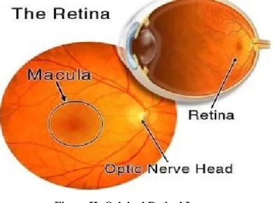
[image:2.612.72.268.490.635.2]Outside light enters the eye through the pupil and is focused on the retina. The lens helps in focusing images from different distance. The quantity of light entering the eye is controlled by iris. The culinary body controls and automatically focuses the lens of the eye structures. Optic disc image is brighter than other parts of the eye and is normally round in shape. It is also an entry to enter and leave the retina and brain from nerves to a point near the center of the retina oval shape object macula. Macula is lies near the center of the Fovea. Cells, due to the high volume of fovea are responsible for the most perfect vision.
Figure II: Original Retinal Image
(Reference: http://www.stlukeseye.com/Anatomy/retina.html)
The retina of the eye sensory tissue that lines a multilayered is behind millions of photoreceptors that capture light rays and convert electrical impulses to travel with these impulses from the optic nerve to the brain where they are turned into images of photoreceptors in the retina.
The two types of photoreceptors in the human retina are rods and cones. Around 120 million rods are more sensitive and responsible for vision at low light levels and cones are capable of colour vision.
A. Diabetic Retinopathy
In diabetic retinopathy, the sensitive inner area of the eye is damaged. The two types of diabetic retinopathy are non-proliferative (NPDR) and proliferative (PDR) diabetic retinopathy.
The early stage of the disease that affects fewer blood vessels in the eye leading blurred vision, due to fluid leaks, is known as non proliferative diabetic retinopathy. In majority of the cases it remains like this and may not affect vision. However, in some cases, may involve macula and that may lead to more advanced stage such as proliferative retinopathy.
The fluid leaks are more serious in the proliferative diabetic retinopathy. The pressure in the blood vessels may rupture causing the bleeding. This bleeding is called haemorrhage and this may cause vision loss and scarring of the retina.
Typically, everyone who is above 30 years and has diabetes shows symptoms of diabetic retinopathy. So everyone with diabetes should have regular eye checkups.
III. OVERALL APPROACH
In this paper we are looking at the automated detection of diabetic retinopathy using feature extraction from digital fundus images. This feature extraction is done using MATLAB. The features studied are micro-aneurysms, optic disc, exudates and blood vessels.
This system consists of different modules and the overall output depends upon the success rate of every step. The figure III gives overall approach to feature extraction for detection of disease severity.
[image:2.612.325.564.558.690.2]International Journal of Emerging Technology and Advanced Engineering
Website: www.ijetae.com (ISSN 2250-2459,ISO 9001:2008 Certified Journal, Volume 5, Issue 1, January 2015)
135 A. Image Pre-processing
Image pre-processing is the initial step in automated retinal pathology diagnosis. It includes techniques such as contrast enhancement, gray/green component, image de-noising, etc. In a binary image, white pixels are normally taken to represent foreground regions, while black pixels denote background. In case of Gray scale image, the intensity value represents height above a base plane. Thus, the Gray scale image represents a surface in three-dimensional Euclidean space.
Intensity conversion: In the RGB images the green channel exhibits the best contrast between the vessels and background while the red and blue ones tends to be more noisy. Since the retinal blood vessels appear darker in gray image, the green channel is used to convert the intensity of the image.
Filtering: Filtering is used to remove the noise which gets added into the fundus image. Here median filtering is quite useful as it is very robust and has the capability to filter any outliers. It is also the preferred choice for removal of salt and pepper noise. Median filtering effectively suppresses isolated noise without fading sharp edges. It replaces a pixel by the median of all pixels in the neighbourhood of small sliding window.
Adaptive Histogram Equalization: One of the challenges associated with fundus images is uneven illumination. Some areas of the fundus images appear to be brighter than the other. The quality of an image can be improved using image enhancement techniques. Adaptive Histogram equalization is a constant enhancement technique which provides an enhanced method for modifying the dynamic range and contrast of an image by altering the image. It is finding of cumulative distribution function for a given probability density function. The small area of pixels, considered to be noise, is removed after applying morphological operations. Post the transformation, the probability density function of the output will be uniform and the image will have high contrast.
B. Feature Extraction
The features such as blood vessels, exudates, micro-aneurysms and optic discs are extracted for further analysis. In this extraction process the morphological operations such as opening, closing, erode and dilate are used [13, 14]. This image is converted into a binary image. The logical operations („AND‟, „OR‟) and filters like „colpit‟ are applied and the segmentation is done for exudates, micro-aneurysms and blood vessels [15, 16].
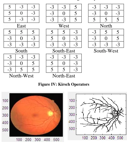
Blood Vessel: Kirsch‟s non-linear edge detector is used to
search the maximum edge in a few determined directions. Taking a single mask and rotating it to 8 major compass orientations (East, West, North, South, East, South-West, North-West and North-East) helps find the edge direction based on the maximum magnitude produced.
5 -3 -3 -3 -3 5 -3 -3 -3
5 0 -3 -3 0 5 -3 0 -3
5 -3 -3 -3 -3 5 5 5 5
East West North
5 5 5 5 5 -3 -3 5 5
-3 0 -3 5 0 -3 -3 0 5
-3 -3 -3 -3 -3 -3 -3 -3 -3
South South-East South-West
-3 -3 -3 -3 -3 -3
-3 0 5 5 0 -3
-3 5 5 5 5 -3
[image:3.612.321.566.203.480.2]North-West North-East
Figure IV: Kirsch Operators
Figure V: Actual result of Blood vessels extraction
Exudates: Small yellow white patches with sharp margins and different shapes. Exudates are one of the early occurring lesions.
The method attempts to detect hard exudates using two features of this lesion: its color and its sharp edges. The coloured fundus image is split into number of non-overlapping blocks. For each block of the image, the coloured histogram is calculated. The threshold value, based on the colour histogram, is used to detect exudates.
Hard and soft exudates are separated based on the chosen threshold value.
International Journal of Emerging Technology and Advanced Engineering
Website: www.ijetae.com (ISSN 2250-2459,ISO 9001:2008 Certified Journal, Volume 5, Issue 1, January 2015)
[image:4.612.49.294.388.486.2]136
Figure VI: Actual result of Exudates extraction
Microaneurysms: These are the first clinical abnormality to be noticed in the eye. They may appear in isolation or in clusters as tiny, dark red spots or looking like tiny haemorrhages within the light sensitive retina. Their sizes range from 10-100 microns i.e. less than 1/12th the diameter of an average optics disc and are circular in shape [7].
From analysis and experiment, the pixel count for candidate microaneurysm ranges from 30 to 5000 pixels for a (1320x1024) image. An area less than the range of 30 to 5000 pixels is regarded as a background noise.
Figure VII: Actual result of Microaneurysms extraction
Optic disc: The image is filtered in order to eliminate large gray level variations within the papillary region. The vessels are filled applying a simple Closing operation. Classical Watershed transformation is applied to the gradient to detect contours of the optic disc.
Figure VIII: Actual result of Optic disc extraction
C. Disease severity:
The severity of the disease is measured depending on the area calculated from the pre-processing and feature extraction. Depending on the severity, there are three categories such as mild, moderate and severe stage. A treatment can also be based on the severity. Certain known treatments are Vitrectomy, Scatter laser treatment, Focal laser treatment and Laser photocoagulation.
IV. RETINAL IMAGES -PUBLIC DATABASES
We have referred to primarily two image databases here.
A. DRIVE Database
The DRIVE database has been established to enable comparative studies on segmentation of blood vessels in retinal images. Test the database research community on their algorithms and results through this Web site [10] other researchers invited to share with the drive of the diabetic retinopathy screening photos database program was obtained in the Netherlands. The tests were carried out on four hundred diabetic patients between age 25 to 90 and images were captured in JPEG format [10]. Canon CR5 3CCD camera with a 45 degree field of view was used to capture the images.
B. STARE Database
Gaze (retinal structural analysis) project conceived by Michael Goldbaum, MD, University of California, San Diego in 1975. It was launched in U.S. and was funded by the national institutes of health. More than thirty people with backgrounds ranging from engineering to medical science contributed to the project. Clinical images were provided by University of California and the Veterans Administration Medical Center, San Diego [12].
REFERENCES
[1] Saiprasad Ravishankar, Arpit Jain, Anurag Mittal “Automated feature extraction for early detection of diabetic retinopathy in funds images”, IEEE 2009.
[2] D. Welfer and D. R Marinho “A course to fine strategy for automatically detecting exudates in color eye funds images”, computerized medical imaging and graphics, vol 34, 2010. [3] Banumathi A, Karthika, R., Kumar.A (2003), “Performance analysis
of matched filter techniques for automated detection of blood vessels in retinal images”, Conference on Convergent Technologies for Asia Pacific Region, 2, pp 543–546.
[image:4.612.47.296.575.672.2]International Journal of Emerging Technology and Advanced Engineering
Website: www.ijetae.com (ISSN 2250-2459,ISO 9001:2008 Certified Journal, Volume 5, Issue 1, January 2015)
137 [5] Chaudhury S, Chatterjee S, Katz N., Nelson M, Goldbaum, M,
(1989), “Detection of blood vessels in retinal images using two dimensional matched filters”, IEEE Transactions on medical imaging, 8, pp 3.
[6] Herbert F. Jelinek, Michael J. Cree, Jorge J. G. Leandro, João V. B. Soares and Roberto M. Cesar, Jr. A. Luckie, May (2007), “Automated segmentation of retinal blood vessels and identification of proliferative diabetic retinopathy”, Optical society of America, 24, pp 14481456.
[7] Mohammed AlRawi, Munib Qutaishat, Mohammed Arrar, (2006), “An improved matched filter for blood vessel detection of digital retinal images”, Computers in Biology and Medicine, pp 262 – 267. [8] Vallabha, D., Dorairaj, R., Namuduri, K., Thompson, H., (2004),
“Automated Detection and Classification of Vascular Abnormalities in Diabetic Retinopathy” in: Proceedings of 13th IEEE Signals, Systems and Computers, 2, pp 1625–1629.
[9] Wong Li Yun , U. Rajendra Acharya, Y.V. Venkatesh , Caroline Cheec,Lim Choo Min, (2008), “Identification of different stages of diabetic retinopathy using retinal optical images”, E.Y.K. Ng / Information Sciences 178 , pp 106–121.
[10] http://www.isi.uu.nl/Research/Databases/DRIVE/
[11] J.J. Staal, M.D. Abramoff, M. Niemeijer, M.A. Viergever, B. van Ginneken, “Ridge based vessel segmentation in color images of the retina”, IEEE Transactions on Medical Imaging, 2004, vol. 23, pp. 501-509.
[12] http://www.parl.clemson.edu/~ahoover/stare/index.html
[13] Dietrich Paulus and Serge Chastel and Tobias Feldmann (2005), “Vessel segmentation in retinal images”, Proceedings of SPIE, Vol. 5746, No.696.
[14] Ana Maria Mendonca and Aurelio Campilho (2006), “Segmentation of Retinal Blood Vessels by Combining the Detection of centerlines and Morphological Reconstruction”, IEEE Transaction on Medical Imaging, Vol. 25, No. 9, pp. 1200-1213.
[15] Jagadish Nayak, Subbanna Bhat (2008), “Automated identification of diabetic retinopathy stages using digital fundus images”, Journal of medical systems, Vol.32, pp. 107-115.
[16] Akara Sopharak, Bunyarit Uyyanonvara, Sarah Barman, Thomas H.Williamson (2008), “Automatic detection of diabetic retinopathy exudates from non-dilated retinal images using mathematical morphology methods”, Computerized Medical Imaging and Graphics, Vol. 32, pp. 720-727.
[17] http://www.stlukeseye.com/Anatomy/retina.html
[18] http://www.ukessays.com/essays/psychology/multimodal-retinal-vessel-segmentation-psychology-essay.php