R E S E A R C H A R T I C L E
Open Access
Three-dimensional postoperative accuracy
of extra-articular forearm osteotomies using
CT-scan based patient-specific surgical
guides
Lazaros Vlachopoulos
1*, Andreas Schweizer
2, Matthias Graf
1, Ladislav Nagy
2and Philipp Fürnstahl
1Abstract
Background:Computer assisted corrective osteotomy of the diaphyseal forearm and the distal radius based on computer simulation and patient-specific guides has been described as a promising technique for accurate reconstruction of forearm deformities. Thereby, the intraoperative use of patient-specific drill and cutting guides facilitate the transfer of the preoperative plan to the surgery. However, the difference between planned and performed reduction is difficult to assess with conventional radiographs. The aim of this study was to evaluate the accuracy of this surgical technique based on postoperative three-dimensional (3D) computed tomography (CT) data. Methods:Fourteen patients (mean age 23.2 (range, 12-58) years) with an extra-articular deformity of the forearm had undergone computer assisted corrective osteotomy with the healthy anatomy of the contralateral uninjured side as a reconstruction template. 3D bone surface models of the pathological and contralateral side were created from CT data for the computer simulation. Patient-specific drill and cutting guides including the
preoperative planned screw direction of the angular-stable locking plates and the osteotomy planes were used for the intraoperative realization of the preoperative plan. There were seven opening wedge osteotomies and nine closing wedge (or single-cut) osteotomies performed.
Eight-ten weeks postoperatively CT scans were obtained to assess bony consolidation and additionally used to generate a 3D model of the forearm. The simulated osteotomies- preoperative bone models with simulated correction - and the performed osteotomies - postoperative bone models – were analyzed for residual differences in 3D alignment.
Results: On average, a significant higher residual rotational deformity was observed in opening wedge osteotomies (8.30° ± 5.35°) compared to closing wedge osteotomies (3.47° ± 1.09°). The average residual translation was comparable small in both groups, i.e., below 1.5 mm and 1.1 mm for opening and closing wedge osteotomies, respectively.
Conclusions: The technique demonstrated high accuracy in performing closing wedge (or single-cut) osteotomies. However, for opening wedge osteotomies with extensive lengthening, probably due to the fact that precise reduction was difficult to achieve or maintain, the final corrections were less accurate.
Keywords:Computer-assisted planning, Patient-specific, Drill guides, Cutting guides, Forearm, Osteotomies, Additive manufacturing, Angular-stable locking plate
* Correspondence:[email protected] 1
Computer Assisted Research and Development Group, Balgrist University Hospital, University of Zurich, Zurich, Switzerland
Full list of author information is available at the end of the article
Background
Posttraumatic deformities of the forearm bones (radius shaft, ulna shaft or distal radius) may cause impairment of the forearm function, i.e. limited range of motion (ROM), especially decreased pro- and supination and a painful or instable distal radioulnar joint [1, 2]. Correct-ive osteotomies with anatomical reduction and restor-ation of the normal anatomy are regularly considered in symptomatic cases in order to relieve pain and/or to improve the ROM. Dependent on the deformity, a bone wedge has to be either removed (i.e., closing wedge osteotomy) or a gap has to be created (i.e., opening wedge osteotomy) to achieve the desired reduction [3].
Computer assisted corrective osteotomy of the diaphy-seal forearm and the distal radius based on computer simulation has been described as a promising technique for accurate reconstruction of forearm deformities. Three-dimensional (3D) bone models are therefore fre-quently extracted from computed tomography (CT) scans [4–11]. The planned correction is simulated by manipulating the bone models in virtual 3D space. How-ever, the transfer of the final planned correction into the intraoperative realization remains technically challen-ging. With the increasing use of patient- specific guides several methods have been described suitable for this purpose. Croitoru et al. [12] first introduced the idea of integrating a digitized model of an osteosynthesis plate into the preoperative plan [12] in order to predrill the screw holes intraoperatively according to the preopera-tive plan previous to the osteotomy. After osteotomy, by inserting the screw through the plate into the predrilled
holes an indirect – automatic - reduction can be achieved. More recently, Miyake et al. [13] and Kunz et al. [8] further developed this technique by using patient-specific drill and cutting guides in order to avoid a navi-gation system used by Croitoru et al. [12].
As exact restoration of the normal anatomy is essential for a good clinical outcome [14], the reproducibility and accuracy of the technique needs to be assessed. How-ever, most studies have relied on 2D based postoperative evaluation, disregarding the 3D characteristic of the residual deformities.
The purpose of the study was to assess the 3D accuracy of the performed corrective osteotomies of the forearm with patient-specific guides and integrated digitized model of an angular- stable locking plate into the pre-operative plan compared to the planned correction.
Methods
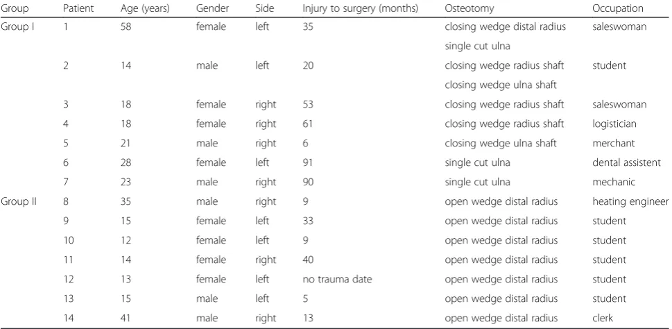
[image:2.595.56.541.496.733.2]Between January and September 2013, a total of 14 pa-tients (mean age 23.2 (range 12-58) years, 8 female) with malunited fractures of the radius or ulna were treated by corrective osteotomy at our institution. Demographic data are summarized in Table 1. Inclusion criteria were patients with a symptomatic extra-articular malunion, according to the criteria of Leong et al. [9] and McQueen et al. [15], with restriction in flexion and pro-/supination, radial shortening due to the malunion or premature close of the epiphyseal growth plates as well as pain due to dis-tal radioulnar instability more than three months after ini-tial trauma. Exclusion criteria were an intra-articular malunion and pathology of the contralateral forearm.
Table 1Demographic and clinical data are given for each patient
Group Patient Age (years) Gender Side Injury to surgery (months) Osteotomy Occupation
Group I 1 58 female left 35 closing wedge distal radius saleswoman
single cut ulna
2 14 male left 20 closing wedge radius shaft student
closing wedge ulna shaft
3 18 female right 53 closing wedge radius shaft saleswoman
4 18 female right 61 closing wedge radius shaft logistician
5 21 male right 6 closing wedge ulna shaft merchant
6 28 female left 91 single cut ulna dental assistent
7 23 male right 90 single cut ulna mechanic
Group II 8 35 male right 9 open wedge distal radius heating engineer
9 15 female left 33 open wedge distal radius student
10 12 female left 9 open wedge distal radius student
11 14 female right 40 open wedge distal radius student
12 13 female left no trauma date open wedge distal radius student
13 15 male left 5 open wedge distal radius student
Informed consent was obtained from all patients pre-operatively (for patients under 18 years in addition paren-tal consent was obtained) regarding the permission to report on their medical history, demographics, character-istics and postoperative results and to publish personal in-formation (as contained in Table 1). Ethical approval was obtained from the ethical committee of the Canton of Zurich for retrospective data analysis. The patients were assigned to one of two groups according to the type of osteotomy. Group I represented corrective osteotomies with the bone fragments being in direct contact at the osteotomy site after reduction, i.e., closing wedge and single-cut [16] osteotomies. Contrary, opening wedge osteotomies were assigned to Group II. All patients with an osteotomy of the radius shaft or ulna shaft were assigned to Group I, since the surgeons avoid due to con-cerns about the healing of the osteotomies an open wedge in this region. All patients except one with an osteotomy of the distal radius were assigned to Group II, since most of the patients with a distal radius malunion had a various degree of radial shortening. Osteotomies were preopera-tively planned in 3D and patient-specific drill and cutting guides were applied intraoperatively to perform the correction.
Preoperative planning and surgical technique
3D triangular surface models of the radius and ulna of the pathological and contralateral uninjured side were generated by segmenting the bones from CT data (slice thickness 1 mm; 120 kV; Philips Brilliance 40 CT, Philips Healthcare, The Netherlands) using commercial software (Mimics, Materialise, Belgium). The CT protocol for corrective osteotomies of the forearm at our institution includes a contiguously scan of the whole forearm from the elbow to the radiocarpal joint. For segmentation, manual thresholding and region growing was applied. A senior surgeon performed the computer planning on a standard personal computer using the custom-made software application CASPA (Balgrist CARD AG, Zur-ich, Switzerland). To quantify the malunion in 3D, the model of the contralateral bone was mirrored and aligned with the pathological bone using the Iterative Closest Point (ICP) surface registration algorithm as pre-viously described [5, 6] (Fig. 1a). After simulated
osteotomy, the distal part was reduced to the contralat-eral bone using ICP. Next, a 3D model of the angular-stable locking plate was positioned on the bone surface. Dependent on the malunion, one of the following im-plants was chosen by the surgeon: A 2.7 mm LCP plate (Synthes, Solothurn, Switzerland), a 2.7 mm LCP ulnar shortening osteotomy plate (Synthes), a 2.4 mm LCP distal radius plate (Synthes), or an Elegantus distal radius plate (Intercus Schweiz GmbH, Aarau, Switzerland). The plate models included cylinders representing the exact position and direction of the angular-stable locking screws (Fig. 1b) in relation to the plate. The screw models placed in the distal fragment were transformed with the previously registered distal part of the bone back to the pathological bone position by applying the inverse transformation. Thereby, during surgery the final direction of the screws can be predrilled previous to the osteotomy. Additionally, when a closing wedge osteot-omy was planned, we used a metallic inlay that contains a cutting slit to guide the 0.4mm thick saw blade. The inlay was inserted into a dedicated frame in the guide body for alignment according to the planned osteotomy planes. In case of an open wedge osteotomy, since all osteotomies were complete with a resulting distraction gap, the exact position of the plane is not as important as for closing wedge or single cut osteotomies. Lastly, a surgical guide with drill sleeves and the dedicated frames for the metallic inlay was designed (Fig. 1c). To achieve a unique fit, the shape of the guide body was designed to contain irregular convex and concave parts covering the bone from different directions and the undersurface of the guide to be placed on the bone as an exact replica-tion of the surface of the bone model. The guides were manufactured by Medacta International S.A. (Castel San Pietro, Switzerland) with a selective laser sinter-ing device.
During surgery, periosteum and other soft tissues were striped from the bone to permit a unique fit of the guide. After placing the guide on its intended position, it was fixed with two K-wires. Next, the screw holes were predrilled and the osteotomy was performed, resulting in a proximal and distal fragment. The osteosynthesis plate was first fixed to the distal fragment using the pre-drilled holes and thereafter the proximal fragment was
[image:3.595.50.537.636.677.2]indirectly reduced by plate fixation. On both sides of the osteotomy at least three angular stable locking screws were placed. In case of a distal radial osteotomy in the distal fragment at least four angular stable locking screws were placed. In four cases (two in each group) the plate was preoperatively prebend based on the printout of a bone in the planned correction. Au-togenous iliac cancellous bone or resected callous for-mation was used to fill the osteotomy gap in case of opening wedge osteotomies. Postoperative the patients were placed in a volar splint made of plaster for 6 weeks. Active assisted ROM exercises were initiated from the first postoperative day.
Postoperative evaluation method
During follow up plain radiographs were regularly ac-quired at 4 weeks, 8–10 weeks, 4 months and 12 months. Additionally, CT scans were performed 8–10 weeks postoperatively with the same scanning protocol as preoperatively to assess bony consolidation. Consoli-dation was defined as disappearance of the visibility of the osteotomy lines on plain radiographs, continuous bone trabeculae at least half of the diameter of the bone on CT and absence of pain or swelling at the level of the osteotomy. The CT data were used to generate a 3D model of the postoperative bone, the implant, and the screws by applying the same segmentation method as in the preoperative planning. The bone parts proximal to the osteotomy were used as a common reference for comparing the postoperative bone with the preopera-tively planned reduction. The proximal parts were regis-tered using ICP as shown in Fig. 2a. Thereafter, the difference between planned and performed reduction (i.e., residual deformity) was quantified in all six degrees of freedom by computing the difference between the dis-tal bone parts using ICP (Fig. 2b). The resulting 4×4 transformation matrix was decomposed in a rotational and translation part: The residual rotation was expressed in axis-angle representation and additionally as three constitutive rotations (i.e., Euler rotations) [17] around a standardized coordinate system as depicted in Fig. 3. Rotation around the x-, y-, and z-axis of the coordinate systems correspond to rotations in the frontal (ulnar-/ radialduction), transverse plane (pronation/supination), and sagittal (flexion/extension) plane, respectively. The
residual translation was expressed as a 3D vector de-scribing the displacement with respect to the same co-ordinate axes.
Statistical analysis
Group differences in baseline characteristics were compared using Mann–Whitney U test (for age) and unpaired t test (for the planned 3D correction angle). The residual rotational error and the clinical parame-ters (ROM and grip strength) were analyzed with an ANCOVA with the preoperative measurement as a covariable in the model. For graphical visualization Tukey boxplots were depicted with whiskers with max-imum 1.5 interquartile range (ICR). The significance level was set at P < 0.05. The software R (Version 3.0; R Foundation, Vienna, Austria) was used for statistical evaluation.
Results
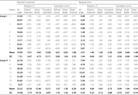
Table 1 summarizes the demographic data of the patients for group I (i.e., closing wedge and single-cut osteoto-mies) and group II (i.e., opening wedge osteotoosteoto-mies). The median age of the patients in group I was 21.5 years (range 14–58 years), the median age of the patients in group II was 15 years (range 12–41 years). The me-dian time from injury to surgery was 38 months (range 5–91 months). There was no significant differ-ence in the baseline characteristics of both groups for age (Table 1;P= 0.22) and the amount of planned correction (Table 2; 19.94° ± 7.21° vs. 23.25° ± 10.48°,P= 0.49).
Results of the preoperative planning and the postop-erative accuracy evaluation are given in Table 2 and Fig. 4. On average, a significant higher (i.e., P = 0.03) difference in rotation between planned correction and postoperative result was observed in group II (8.30° ± 5.35°) compared to group I (3.47° ± 1.09°). The highest residual deformity was observed in case 10 and case 11 (Fig. 5). In these cases, also a deviation from the planned screw direction was located. For group I, the mean residual rotational deformity in sagittal, frontal, and transverse plane was 1.4° ± 0.84°, 1.56° ± 1.01°, and 2.39° ± 1.31°, respect-ively. For Group II, a residual rotation of 3.84° ± 4.23° in the sagittal plane, 3.97° ± 4.12° in the frontal plane, and 3.73° ± 3.88° in the transverse plane was measured. The average displacement between planned and performed
[image:4.595.58.535.655.697.2]reduction was between 0.5–1.48 mm and 0.94–1.04 mm for group I and group II, respectively.
Table 3 reveals the mean ROM and grip strength pre-operative and change at the latest follow up. There was a significant difference between the preoperative and the postoperative values in flexion, pronation and in grip strength. The difference between the groups was not significant.
All osteotomies healed after a mean time of 3.6 months (±1.8 months). The closing wedge osteotomies
of group I healed after 4.4 months (±3.1 months) and the opening wedge osteotomies of group II after 3.4 (±2.0 months), respectively.
Discussion
Accuracy in the preoperative quantification of a deform-ity and, subsequently, in the surgical reconstruction are crucial, because over- and under-correction is associated with loss of ROM and grip strength [14]. Therefore, computerized 3D comparison to the contralateral bone
[image:5.595.60.538.89.167.2]Fig. 3Definition of the anatomical coordinate system for radius (a) and ulna (b). Rotation around the x-axis (red) corresponds to a correction in the frontal (ulnarduction/radialduction) plane, around the y-axis (green) to a correction in the transverse plane (pronation/supination) and around the z-axis (blue) to a correction in sagittal (flexion/extension) plane. The coordinate system was adapted in that way that a positive rotation around the defined axis defined for both sides of the radius and ulna an ulnarduction, pronation and flexion, respectively
Table 2Rotation is described by the 3D angle (axis-angle representation) and by Euler rotations around the anatomical coordinate system. Translation is given with respect to the anatomical coordinate system. Mean and standard deviation (SD) are based on absolute values
Planned Correction Residual error
Rotation (°) Translation (mm) Rotation (°) Translation (mm)
Patient 3D angle
Flexion/ Extension
Ulnar/ Radial
Pronation/ Supination
Palmar/ Dorsal
Ulnar/ Radial
Distal/ Proximal
3D angle
Flexion/ Extension
Ulnar/ Radial
Pronation/ Supination
Palmar/ Dorsal
Ulnar/ Radial
Distal/ Proximal
Group I 1 12.42 11.99 1.24 −3.14 −0.15 −0.09 −2.17 4.46 2.07 3.53 −1.82 −0.80 −0.27 0.99
29.57 −4.89 −4.63 28.61 0.75 0.95 0.69 4.56 −2.50 1.83 3.38 −1.32 −0.99 0.61
2 25.51 20.51 15.00 1.18 −2.81 −2.03 −8.43 4.92 −1.50 1.67 4.40 1.42 0.04 −5.10
14.24 −9.28 5.75 −8.70 0.38 −3.03 −1.43 3.88 −0.29 0.46 −3.84 −0.39 −2.17 −4.79
3 18.04 14.16 −0.16 11.22 −0.51 −0.47 −1.59 1.49 −0.64 −1.34 −0.14 −0.30 0.14 0.10
4 12.15 11.72 3.19 −0.60 −0.20 −0.14 −1.21 3.16 −2.56 0.12 1.85 0.28 1.06 0.37
5 13.71 −10.06 9.35 1.04 −0.45 0.37 −1.15 3.14 −0.49 1.92 2.44 0.67 0.19 0.47
6 25.61 0.86 −3.21 −25.37 0.15 0.18 −0.22 2.72 −1.29 0.98 2.20 0.10 −0.44 −0.50
7 28.20 0.27 −1.07 28.18 −0.09 0.10 0.01 2.87 1.27 2.15 −1.43 −0.09 −0.79 0.38
Mean 19.94 9.31 4.85 12.00 0.61 0.82 1.88 3.47 1.40 1.56 2.39 0.60 0.68 1.48
SD 7.21 6.47 4.73 12.11 0.85 1.04 2.55 1.09 0.84 1.01 1.31 0.50 0.68 1.98
Group II 8 22.79 19.33 10.98 −7.39 −1.66 1.16 5.31 7.94 4.90 2.33 −5.90 0.10 1.79 0.86
9 10.49 5.78 8.76 −0.71 0.55 0.36 7.66 4.76 3.63 0.56 −3.05 −1.99 −0.31 1.60
10 28.26 24.34 −9.81 12.96 −0.54 0.81 9.08 17.51 13.08 −3.74 11.50 −0.92 −1.66 1.44
11 35.39 7.42 32.61 9.84 0.09 1.15 17.93 12.47 4.06 10.86 4.25 −1.56 −1.21 2.17
12 23.58 −10.46 −19.88 5.70 0.09 0.72 5.81 9.38 −2.24 −9.11 −0.21 1.70 1.14 −0.44
13 33.65 −12.59 −16.12 −28.73 1.13 1.50 1.93 3.36 −0.36 −0.55 −3.29 −0.90 −1.08 0.08
14 8.57 −2.51 0.48 8.19 4.43 4.57 1.83 2.70 1.14 2.44 −0.18 0.14 −0.35 0.53
Mean 23.25 10.34 12.46 12.71 1.07 1.30 6.20 8.30 3.84 3.97 3.73 0.94 1.04 0.94
SD 10.48 7.73 10.18 8.87 1.53 1.42 5.49 5.35 4.23 4.12 3.88 0.75 0.57 0.74
[image:5.595.59.536.395.725.2]has been increasingly used to quantify complex malu-nions and the preoperative planning of corrective osteotomies. Different techniques, such as navigation systems [4, 12] or patient-specific guides were proposed to support the surgeon in performing the reduction exactly as preoperatively planned. Particularly, patient-specific guides are becoming popular, because they are easier to handle and more precise in vitro compared to navigation systems [18, 19]. Laboratory studies have re-ported average residual errors less than 1° and 1 mm for simulated osteotomies that used patient-specific guides [18, 19]. In these studies, the accuracy of the reduction was similar regardless whether K-wire based patient-specific reduction guides [19] or patient-patient-specific drill guides based on the implant screw directions [18] were used.
Studies have already described promising clinical re-sults of using the herein investigated technique [8, 12, 13, 20, 21]. Kunz et al. [8] reported on the radiological results of 8 patients treated by 3D planned distal radius opening wedge osteotomy using volar plating and patient-specific drill guides. The average volar tilt devi-ation after correction was 1.9° (±1.5°) and the average radial inclination deviation 1.8° (±0.8°). However, their evaluation was only based on intraoperative plane im-ages using image intensifier. Moreover, the amount of the correction and the demographic data of the patients were not mentioned. Miyake et al. [13] analyzed the sur-gical outcome of open wedge osteotomy in ten patients using the same technique as in the current study with volar plates and autogenous iliac cancellous bone graft. The average difference to the contralateral unaffected
side was 5.1° in volar tilt and 3.4° in radial inclination. Although Miyake et al. stated that 3D based methods may be more effective for complex deformities [22], the postoperative evaluation relied on 2D radiographs. The differences between 2D deformity assessment based on standard radiographs and 3D based methods was ana-lyzed by Vroemen et al. [23]. Their study retrospectively investigated the residual deformity after conventionally performed corrective osteotomies. Only the 3D deform-ity measurements showed signification correlation with the clinical outcome.
The current study reported on the accuracy of deform-ity correction using patient-specific drill guides by retro-spectively analyzing available postoperative 3D CT data. In the closing wedge group, the residual rotational de-formity observed in the three anatomical planes was between 1.4° to 2.4° on average. These results are com-parable with in-vitro findings [18, 19]. Contrary, in group II residual deformities above 10° were measured in two cases. In these cases an excessive lengthening of the bone additionally to the rotational component was required (case 10: 17 mm; case 11: 10 mm) to achieve the planned reduction, resulting in very high soft tissue tensioning. Although a surgical spreader was used to temporary stabilize the fragment, it was difficult to per-form the reduction solely based on the pre-drilled screw holes. The stress between the fragments might addition-ally cause the screws to lock into the plate slightly differently from the planned direction (see Fig. 6). Nevertheless, all residual deformities are considerable smaller compared to corrective osteotomies performed without patient-specific drill guides [23].
Fig. 4Planned Correction and Residual Error.aBoxplot illustrating the planned correction and residual deformity between group I and group II, bEach osteotomy is represented by a line. One endpoint is the planned correction, the other one is the residual deformity after surgery
[image:6.595.58.539.89.220.2] [image:6.595.61.535.670.707.2]All patients with an osteotomy of the radius shaft or the ulna shaft were allocated to group I, while all pa-tients with an open wedge distal radius osteotomy were allocated to group II. The main reason was that sur-geons avoid planning an open wedge at the level of the diaphysis of the radius and ulna due to concerns about the healing of the osteotomies in this region. Beside the type of osteotomy the region where the osteotomy was performed (i.e. shaft or distal radius) may also had in-fluenced the difference in the accuracy. In all but one case of Group I, where an incision of the interosseous membrane had to be performed, there was no additional soft tissue release. However, in two cases of Group II in-traoperatively a considerable tension was noticed. This could also have influenced the accuracy of the achieved reduction.
Surprisingly, although not significant, the time to union was in the closing wedge/single cut osteotomy
group with 4.4 months on average one month longer than in the open wedge osteotomy group. We believe there are two possible explanations for this more or less unexpected result. One the one hand the cancellous bone in the distal radius in combination with the au-togenous iliac cancellous bone graft could have a posi-tive effect on the consolidation and all open wedge osteotomies were at level of the distal radius. On the other hand in our experience in the diaphyseal region of the radius and especially of the ulna the fracture lines are regularly visible for several months or years and could have led to an overestimate of the time to union. However, we do not know the reasons therefore. Whether the necessary periosteal stripping in order to achieve a unique fitting of the guide causes a delayed union is not investigated so far.
The surgeons recognized no relevant difference in the guide fit between the groups. However, in our experience in the shaft region of the radius and ulna due to the more circular shape it can be difficult to find the exact fit. The demonstrated accuracy in performing closing wedge osteotomies at this level indicates that this was a concern in the current study.
A weakness of the study is that the CT data used in the evaluation were acquired 8–10 weeks after surgery to monitor consolidation. As a consequence the mea-sured residual deformities might also been caused by postoperative loss of correction due to soft-tissue ten-sioning, weak cortical bone, delayed union, bone-plate-constructs that failed or were not rigid enough. For opening wedge osteotomies the use of structural bone graft despite the donor side morbidity [24] or a more bending resistant plate may reduce loss of correction in these cases. Another limitation is that the proposed evaluation technique can be applied only if postopera-tive CT data is available. Disadvantage of the actual sur-gical technique is that a 3D model of the implant and all screws must be available. Moreover, an uncontoured fixed-angle locking plate must be used which may fit poorly on the bone. In the future a custom-made plate [25] may be integrated in the planning application to overcome this problem.
Conclusions
[image:7.595.57.291.133.230.2]The presented in-vivo evaluation demonstrates high accuracy of the surgical technique for closing wedge and single-cut osteotomies. Whether an additional re-duction guide based on K-wires may further improve accuracy in case of soft-tissue tension has to be eval-uated in future studies. As the approach solely relies on the used implant, any deviation from the planned implant position or screw direction will directly influ-ence the reduction.
Table 3Reveals the preoperative clinical values and the change at the latest follow up. P-values Change are given for the difference between preoperative and postoperative values, P-Values Group for the differences between the two groups
Preoperative value Chance Chance Group
mean (± SD) mean (± SD) P-value * P-value **
Flexion (°) 69 (±16) 7 (±11) 0.005 * 0.21
Extension (°) 75 (±14) −3 (±14) 0.12 0.55
Pronation (°) 62 (±22) 10 (±19) 0.04 * 0.25
Supination (°) 65 (±26) 14 (±25) 0.33 0.90
Jamar (kg) 24 (±9) 5 (±6) 0.005 * 0.58
* P < 0.05 significant change of the preoperative ROM or grip strength ** P < 0.05 significant difference between the groups
[image:7.595.56.291.501.693.2]Competing interests
Three of the authors (AS, LN, PF) are shareholders of the Balgrist CARD AG, a company developing preoperative planning software. The other authors hereby declare that they have no conflicts of interest to disclose.
Authors’contributions
LV, MG and PF implemented the technical demands regarding preoperative planning and intraoperative realization. AS and LN instructed the operative requirements and performed the surgical procedures. LV and PF were mainly accountable for analyzing the postoperative results and writing the manuscript. All authors read and approved the final manuscript.
Acknowledgements
The authors would like to thank all patients for participating in this study and their consents to report on the postoperative results.
Author details 1
Computer Assisted Research and Development Group, Balgrist University Hospital, University of Zurich, Zurich, Switzerland.2Department of Orthopaedics, Balgrist University Hospital, University of Zurich, Zurich, Switzerland.
Received: 29 October 2014 Accepted: 28 October 2015
References
1. Trousdale RT, Linscheid RL. Operative treatment of malunited fractures of the forearm. J Bone Joint Surg Am. 1995;77:894–902.
2. Colaris JW, Allema JH, Reijman M, de Vries MR, Ulas Biter L, Bloem RM, van de Ven CP, Verhaar JA: Which factors affect limitation of pronation/ supination after forearm fractures in children? A prospective multicentre study. Injury 2013
3. Jayakumar P, Jupiter JB. Reconstruction of malunited diaphyseal fractures of the forearm. Hand. 2014;9:265–73.
4. Athwal GS, Ellis RE, Small CF, Pichora DR. Computer-assisted distal radius osteotomy. J Hand Surg [Am]. 2003;28:951–8.
5. Murase T, Oka K, Moritomo H, Goto A, Yoshikawa H, Sugamoto K. Three-dimensional corrective osteotomy of malunited fractures of the upper extremity with use of a computer simulation system. J Bone Joint Surg Am. 2008;90:2375–89.
6. Schweizer A, Furnstahl P, Harders M, Szekely G, Nagy L. Complex radius shaft malunion: osteotomy with computer-assisted planning. Hand. 2009;5:171–8.
7. Dobbe JG, Strackee SD, Schreurs AW, Jonges R, Carelsen B, Vroemen JC, et al. Computer-assisted planning and navigation for corrective distal radius osteotomy, based on pre- and intraoperative imaging. IEEE Trans Biomed Eng. 2011;58:182–90.
8. Kunz M, Ma B, Rudan JF, Ellis RE, Pichora DR. Image-guided distal radius osteotomy using patient-specific instrument guides. J Hand Surg [Am]. 2013;38:1618–24.
9. Leong NL, Buijze GA, Fu EC, Stockmans F, Jupiter JB, Distal Radius Malunion collaborative g. Computer-assisted versus non-computer-assisted preoperative planning of corrective osteotomy for extra-articular distal radius malunions: a randomized controlled trial. BMC Musculoskelet Disord. 2010;11:282.
10. Miyake J, Murase T, Oka K, Moritomo H, Sugamoto K, Yoshikawa H. Computer-assisted corrective osteotomy for malunited diaphyseal forearm fractures. J Bone Joint Surg Am. 2012;94:e150.
11. Schweizer A, Furnstahl P, Nagy L. Three-dimensional correction of distal radius intra-articular malunions using patient-specific drill guides. J Hand Surg [Am]. 2013;38:2339–47.
12. Croitoru H, Ellis RE, Prihar R, Small CF, Pichora DR. Fixation-based surgery: a new technique for distal radius osteotomy. Comput Aided Surg. 2001;6:160–9.
13. Miyake J, Murase T, Moritomo H, Sugamoto K, Yoshikawa H. Distal radius osteotomy with volar locking plates based on computer simulation. Clin Orthop Relat Res. 2011;469:1766–73.
14. Schemitsch EH, Richards RR. The effect of malunion on functional outcome after plate fixation of fractures of both bones of the forearm in adults. J Bone Joint Surg Am. 1992;74:1068–78.
15. McQueen MM, Hajducka C, Court-Brown CM. Redisplaced unstable fractures of the distal radius: a prospective randomised comparison of four methods of treatment. J Bone Joint Surg (Br). 1996;78:404–9.
16. Meyer DC, Siebenrock KA, Schiele B, Gerber C. A new methodology for the planning of single-cut corrective osteotomies of mal-aligned long bones. Clin Biomech (Bristol, Avon). 2005;20:223–7.
17. Schneider P, Eberly DH: Geometric tools for computer graphics. Morgan Kaufmann; 2002
18. Ma B, Kunz M, Gammon B, Ellis RE, Pichora DR. A laboratory comparison of computer navigation and individualized guides for distal radius osteotomy. Int J Comput Assist Radiol Surg. 2014;9:713–24.
19. Oka K, Murase T, Moritomo H, Goto A, Nakao R, Sugamoto K, et al. Accuracy of corrective osteotomy using a custom-designed device based on a novel computer simulation system. J Orthop Sci. 2011;16:85–92.
20. Farshad M, Hess F, Nagy L, Schweizer A. Corrective osteotomy of distal radial deformities: a new method of guided locking fixed screw positioning. J Hand Surg Eur Vol. 2013;38:29–34.
21. Fürnstahl P, Wirth S, Nagy L, Schweizer A: Advantages and pitfalls in computer assisted orthopedic surgery planning using rapid-prototyped guides.RTejournal - Forum für Rapid Technologie, 2014
22. Miyake J, Oka K, Kataoka T, Moritomo H, Sugamoto K, Murase T. 3-dimensional deformity analysis of malunited forearm diaphyseal fractures. J Hand Surg [Am]. 2013;38:1356–65.
23. Vroemen JC, Dobbe JG, Strackee SD, Streekstra GJ. Positioning evaluation of corrective osteotomy for the malunited radius: 3-D CT versus 2-D radiographs. Orthopedics. 2013;36:e193–199.
24. Arrington ED, Smith WJ, Chambers HG, Bucknell AL, Davino NA: Complications of iliac crest bone graft harvesting. Clinical orthopaedics and related research 1996:300–309
25. Omori S, Murase T, Kataoka T, Kawanishi Y, Oura K, Miyake J, et al. Three-dimensional corrective osteotomy using a patient-specific osteotomy guide and bone plate based on a computer simulation system: accuracy analysis in a cadaver study. Int J Med Robot. 2014;10:196–202.
Submit your next manuscript to BioMed Central and take full advantage of:
• Convenient online submission
• Thorough peer review
• No space constraints or color figure charges
• Immediate publication on acceptance
• Inclusion in PubMed, CAS, Scopus and Google Scholar
• Research which is freely available for redistribution