for Cytomegalovirus Detection: Feasibility of Blood Donor Screening
T. Vollmer, C. Knabbe, J. Dreier
Institut für Laboratoriums- und Transfusionsmedizin, Herz- und Diabeteszentrum Nordrhein-Westfalen, Universitätsklinik der Ruhr-Universität Bochum, Bad Oeynhausen, Germany
Acute primary cytomegalovirus (CMV) infections, which commonly occur asymptomatically among blood donors, represent a significant risk for serious morbidity in immunocompromised patients (a major group of transfusion recipients). We imple-mented a routine CMV pool screening procedure for plasma for the identification of CMV DNA-positive donors, and we evalu-ated the sensitivities and performance of different CMV DNA amplification systems. Minipools (MPs) of samples from 18,405 individual donors (54,451 donations) were screened for CMV DNA using the RealStar CMV PCR assay (Altona Diagnostic Tech-nologies), with a minimum detection limit of 11.14 IU/ml. DNA was extracted with a high-volume protocol (4.8 ml, Chemagic Viral 5K kit; PerkinElmer) for blood donor pool screening (MP-nucleic acid testing [NAT]) and with the Nuclisens easyMAG system (0.5 ml; bioMérieux) for individual donation (ID)-NAT. In total, six CMV DNA-positive donors (0.03%) were identified by routine CMV screening, with DNA concentrations ranging from 4.35ⴛ102to 4.30ⴛ103IU/ml. Five donors already showed
seroconversion and detectable IgA, IgM, and/or IgG antibody titers (IgAⴙ/IgMⴙ/IgGⴚor IgAⴙ/IgMⴙ/IgGⴙ), and one donor showed no CMV-specific antibodies. Comparison of three commercial assays, i.e., the RealStar CMV PCR kit, the Sentosa SA CMV quantitative PCR kit (Vela Diagnostics), and the CMV R-gene PCR kit (bioMérieux), for MP-NAT and ID-NAT showed comparably good analytical sensitivities, ranging from 10.23 to 11.14 IU/ml (MP-NAT) or from 37.66 to 57.94 IU/ml (ID-NAT). The clinical relevance of transfusion-associated CMV infections requires further investigation, and the evaluated methods pres-ent powerful basic tools providing sensitive possibilities for viral testing. The application of CMV MP-NAT facilitated the idpres-enti- identi-fication of one donor with a window-phase donation during acute primary CMV infection.
H
uman cytomegalovirus (CMV) is a ubiquitous viral pathogenthat causes mostly asymptomatic disease in immunocompe-tent individuals. In immunocompromised patients, however, CMV infection represents a significant risk for serious morbidity,
e.g., due to interstitial pneumonia or hepatitis (1–3).
Immuno-compromised patients, such as patients undergoing hematopoi-etic stem cell transplantation (HSCT), solid-organ transplant re-cipients, infants with low birth weights, fetuses, pregnant woman, HIV patients, and patients being treated for hematological malig-nancies, belong to the major groups of transfusion recipients, and CMV-seronegative individuals were considered high-risk patients
for transfusion-transmitted (TT)-CMV infections (4–6). The
in-troduction of leukodepletion of blood products and provision of CMV-seronegative blood products reduced the incidence of CMV infections in at-risk populations by 92%. However, TT-CMV breakthrough infections occur in 1 to 3% of high-risk
patients who receive transfusions (4–6), possibly due to
window-phase donations during acute primary CMV infections.
The seroprevalence rates of CMV antibodies among blood do-nors show geographic differences, ranging from 45.8% in
Ger-many to 96.5% in Brazil (3,7). Primary CMV infections in blood
donors occur in all age groups, with prevalence rates between 0.2
and 1.2% (3,6,8,9). The disease presentation is mostly
asymp-tomatic, frequently with a prolonged course (10,11).
Mononucle-osis-like symptoms are rare, whereas nonspecific viral disease symptoms occur often but not at significantly increased rates,
compared to matched control groups (3,5).
Determination of the prevalence of primary CMV infections among blood donors represents an important parameter for the
effective prevention of TT-CMV infections (3,12). The aim of the
present study was implementation of routine CMV DNA
screen-ing accordscreen-ing to the setup used for our standard viral nucleic acid testing (NAT) (for HIV-1, hepatitis B virus [HBV], hepatitis C virus [HCV], hepatitis A virus [HAV], and parvovirus B19 [PVB19]). The sensitivity and performance of different amplifica-tion systems were evaluated for blood donor pool screening or testing of individuals with acute or chronic infections.
MATERIALS AND METHODS
Blood donors.A total of 54,451 allogeneic blood donations from 18,405 individual German blood donors were routinely screened for the presence of CMV DNA by the Uni.Blutspendedienst OWL between March and
June 2013. Master pools of 96 donations were set up by combining 200-l
EDTA-treated plasma samples; reactive pools were retested in duplicate. Repeatedly reactive pools were further tested by generation of subpools of
a maximum of 10 donations (200l/donor); pools were brought up to 4.8
ml with negative human plasma. Plasma specimens in positive subpools were tested individually in order to identify the individual reactive
do-Received23 April 2015Returned for modification21 May 2015 Accepted16 July 2015
Accepted manuscript posted online22 July 2015
CitationVollmer T, Knabbe C, Dreier J. 2015. Systematic evaluation of different nucleic acid amplification assays for cytomegalovirus detection: feasibility of blood donor screening. J Clin Microbiol 53:3219 –3225.
doi:10.1128/JCM.01091-15.
Editor:A. M. Caliendo
Address correspondence to T. Vollmer, [email protected].
Supplemental material for this article may be found athttp://dx.doi.org/10.1128 /JCM.01091-15.
Copyright © 2015, American Society for Microbiology. All Rights Reserved.
doi:10.1128/JCM.01091-15
on May 16, 2020 by guest
http://jcm.asm.org/
nors. Serological testing was performed only with samples from individ-ual CMV DNA-positive donors. Screening for CMV DNA was performed using a RealStar CMV PCR kit (Altona Diagnostic Technologies [ADT], Hamburg, Germany). Quantification of CMV DNA in positive plasma samples was performed using four different quantification standards of the RealStar CMV PCR kit (ADT secondary standard), which were cali-brated against the 1st WHO International Standard for human cytomeg-alovirus (HCMV) for nucleic acid amplification techniques (National In-stitute for Biological Standards and Control [NIBSC], Potters Bar, Hertfordshire, United Kingdom). Samples donated at continuous inter-vals after the initial CMV DNA-positive donation (day 0) were available for four blood donors (one male donor and three female donors). All donors underwent predonation medical examinations and denied current diseases or any known risk factors for viral infection.
Nucleic acid extraction. (i) Pool screening.DNA extraction from 4.8 ml of plasma of master pools or subpools was performed using the Chemagic viral DNA/RNA reagent kit (Viral 5k kit; PerkinElmer Chema-gen Technologie GmbH, Baesweiler, Germany) combined with the auto-mated Chemagic Magnetic Separation Module I (PerkinElmer Chemagen Technologie GmbH). Briefly, 4.8 ml of plasma was mixed with 4.8 ml of
lysis buffer, 30l of protease, and 7l of poly(A), followed by incubation
of the samples at 55°C for 10 min. Subsequently, lysates were mixed with
15 ml of binding buffer containing 100l of magnetic beads. The
Mag-netic Separation Module I automatically performed the nucleic acid ex-traction process, including binding, two washes, and elution in a final
volume of elution buffer of 100l.
(ii) Individual donation screening.Extraction of total DNA from 500
l of plasma was performed using the NucliSens easyMAG automated
RNA/DNA extraction system (bioMérieux, Nürtingen, Germany). DNA
was eluted in 55l of elution buffer.
Real-time PCR. Amplification using the RealStar CMV PCR kit (ADT) was performed with the Rotor-Gene 3000 system (Corbett Life Sciences, Sydney, Australia), whereas amplification using the Sentosa SA CMV quantitative PCR kit (Vela Diagnostics Germany GmbH, Hamburg, Germany) was carried out on the Rotor-Gene-Q platform (Vela Diagnos-tics Germany GmbH). Amplification using the CMV R-gene PCR kit (bioMérieux) was implemented with the LightCycler 480 II real-time PCR system (Roche Diagnostics, Mannheim, Germany). PCR setup and con-ditions for the three assays were as described in the manufacturer’s in-structions. All assays included a heterologous amplification system (inter-nal control) to identify possible PCR inhibition.
Analytical sensitivities and comparison of different amplification methods.The analytical sensitivity and precision of the three commer-cially available assays, i.e., the RealStar CMV PCR kit (ADT), the Sentosa SA CMV quantitative PCR kit (Vela Diagnostics Germany GmbH), and the CMV R-gene PCR kit (bioMérieux) were determined using a 2-fold dilution series of plasma inoculated with the 1st WHO International Stan-dard for human cytomegalovirus for nucleic acid amplification tech-niques. DNA was extracted using the Viral 5K kit for minipool (MP)-NAT and the Nuclisens easyMAG system for individual donation (ID)-NAT. Analysis was performed for each assay with 6 dilution steps and 24
repli-cates. The 95% limit of detection (LOD) was calculated by probit analysis using the SPSS software (IBM SPSS Statistics, version 21; IBM GmbH, Ehningen, Germany).
Serological testing.Plasma from CMV DNA-positive donors was screened for the presence of CMV-specific IgM and IgG antibodies with a Liaison XL instrument, using Liaison CMV IgG II and CMV IgM II im-munoassays (Diasorin Germany GmbH, Dietzenbach, Germany). The presence of CMV-specific IgA antibodies and IgG avidity was determined using the corresponding anti-CMV enzyme-linked immunosorbent as-says (ELISAs) (IgA and CMV IgG avidity; Euroimmun AG, Lübeck, Ger-many). The CMV-specific antigens in samples from donors were further characterized using the recomLine CMV IgG/IgM assay (Mikrogen GmbH, Neuried, Germany). Samples were analyzed according to the manufacturer’s instructions.
RESULTS
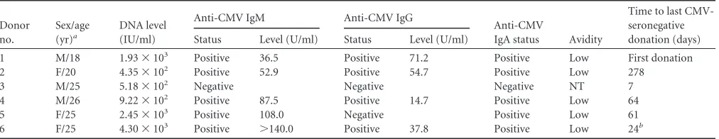
Screening of blood donors.A total of 6 of 18,405 individual blood
donors were CMV DNA positive (0.03%) (Table 1). The
serosta-tus and antibody avidity revealed that all donors had acute pri-mary CMV infections; the donors denied any current diseases during predonation medical examinations. One donor had a NAT-only-positive donation, and a second donor had a positive IgM titer but the IgG result remained negative. Confirmation of CMV infections was performed by determination of the serostatus in subsequent samples, showing complete seroconversion with IgM-negative and IgG-positive status for all donors. CMV DNA-positive donations were immediately rejected for transfusion.
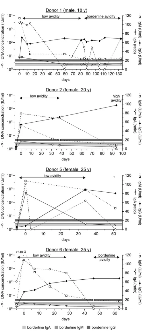
Progression of asymptomatic CMV infections.The progres-sion of CMV DNA and CMV-specific IgA, IgM, and IgG
antibod-ies in plasma is shown inFig. 1. The day of the first detection of
CMV DNA by PCR screening was defined as day 0. CMV viremia in samples from donor 1 lasted over 130 days, with alternating
positive and negative results and a maximum value of 2.98⫻103
IU/ml. IgA, IgM, and IgG antibodies were detected in parallel at the first sampling point. The maximum IgM titer was observed at that sampling point, followed by a rapid decrease until day 15 and a subsequent linear decrease toward the reference range, How-ever, IgM titers above the reference range were still detectable after 130 days. The IgG titer was still increasing after the first detection of CMV DNA, to a maximum value of 71.2 U/ml on day 3, fol-lowed by consistent titers at that level until day 130. IgA antibodies were clearly detectable at the first and second sampling points, followed by alternating positive and negative values around the reference range. Low-affinity antibodies were observed until day 88, followed by borderline results for the next sampling time points.
For donor 2, maximum viremia was also observed on day 0, followed by a continuous decrease until day 95. Similarly,
CMV-TABLE 1DNA concentrations and antibody status for CMV-positive donors at the first positive screening result
Donor no.
Sex/age
(yr)a
DNA level (IU/ml)
Anti-CMV IgM Anti-CMV IgG
Anti-CMV
IgA status Avidity
Time to last CMV-seronegative donation (days)
Status Level (U/ml) Status Level (U/ml)
1 M/18 1.93⫻103 Positive 36.5 Positive 71.2 Positive Low First donation
2 F/20 4.35⫻102 Positive 52.9 Positive 54.7 Positive Low 278
3 M/25 5.18⫻102 Negative Negative Negative NT 7
4 M/26 9.22⫻102 Positive 87.5 Positive 14.7 Positive Low 64
5 F/25 2.45⫻103 Positive 108.0 Negative Positive Low 61
6 F/25 4.30⫻103 Positive ⬎140.0 Positive 37.8 Positive Low 24b
aM, male; F, female; NT, not tested.
b
Seronegative but with an individual PCR-positive donation that had been collected outside the study period.
on May 16, 2020 by guest
http://jcm.asm.org/
[image:2.585.41.547.77.175.2]specific IgA, IgM, and IgG antibodies were present on day 0. The IgM and IgA titers continuously decreased toward borderline val-ues after 30 days, with negative valval-ues after 98 days; IgG titers continuously increased to 97.5 U/ml. Low-avidity antibodies were observed within the first 38 days, changing to high avidity after 98 days. In samples from donor 5, CMV DNA was undetectable sub-sequent to the initial positive screening, followed by intermit-tently positive and negative results. IgA and IgM antibodies were already detectable on day 0, followed by decreasing values, whereas IgG antibodies were first detected after 34 days. Only low-affinity antibodies were observed until day 34. Samples from donor 6 showed DNA-positive results in a previous donation that had been collected outside the study period. That donation showed no anti-CMV IgA, IgM, or IgG. The viremia of that donor
reached the maximum value of 3.30⫻103IU/ml on day 0 and
continuously decreased until its disappearance on day 46. IgA was
detectable on day 0, in parallel with the maximum IgM titer (⬎140
U/ml), followed by borderline results for the next 17 days and disappearance after 28 days. IgM titers continuously decreased toward negative values after 63 days, which was linked to a parallel constant increase in IgG titers. Low-affinity antibodies were ob-served on day 7, followed by borderline results between day 46 and 63.
All donors showed antibodies (IgM and IgG) against nonstruc-tural or tegument proteins in the seropositive samples (see Table S1 in the supplemental material). Antibodies (IgG) against mem-brane glycoprotein B (gB) were detected only in IgG-positive sam-ples (see Table S1 in the supplemental material). Donor 3 was not tested, since no anti-CMV IgA, IgM, or IgG antibodies were de-tected.
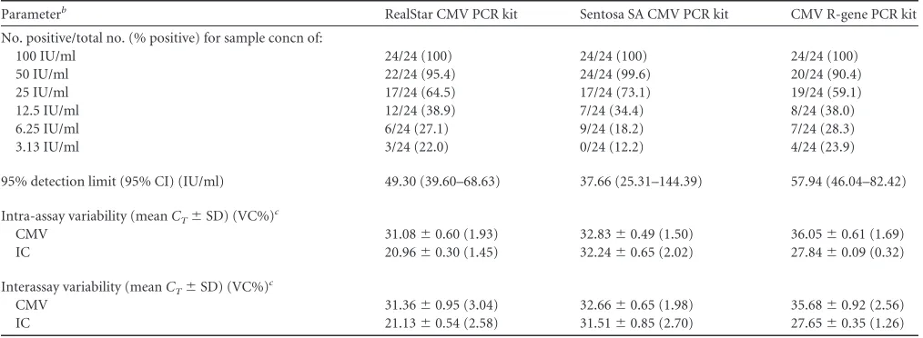
Comparison of NAT assays.The analytical sensitivities and precisions of different assays, which potentially can be imple-mented in our routine viral nucleic acid screening, are shown in
Table 2(MP-NAT) andTable 3(ID-NAT). For blood donor pool screening, all assays showed almost equally high analytical sensi-tivities, with 95% detection limits of 10.23 IU/ml (95% confidence interval [CI], 7.87 to 15.81 IU/ml) for the Sentosa SA CMV quan-titative PCR kit, 11.08 IU/ml (95% CI, 7.11 to 47.94 IU/ml) for the CMV R-gene PCR kit, and 11.14 IU/ml (95% CI, 8.49 to 17.65 IU/ml) for the RealStar CMV PCR kit. The Sentosa SA CMV quantitative PCR kit had the greatest sensitivity, i.e., 37.66 IU/ml (95% CI, 25.31 to 144.39 IU/ml), for individual donor screening using the Nuclisens easyMAG system for DNA extraction, fol-lowed by the RealStar CMV PCR kit with 49.30 IU/ml (95% CI, 39.60 to 68.63 IU/ml) and the R-gene PCR kit with 57.94 IU/ml (95% CI, 46.04 to 82.42 IU/ml). With LODs being determined with reference to the Sentosa SA CMV quantitative PCR kit as the most sensitive assay, deviations in the LODs ranged from 0.85 to 0.91 IU/ml (for MP-NAT, with extraction with the Chemagen Viral 5k kit) or from 11.61 to 20.28 IU/ml (for ID-NAT, with extraction with the Nuclisens easyMAG system) for the two other assays. The CIs for all assays overlapped over a wide range, and the mean values for the CMV R-gene PCR kit and the RealStar CMV PCR kit were located within the confidence interval for the Sen-tosa SA CMV quantitative PCR kit; therefore, there is no evidence that the assays had statistically significant differences. Wider fluc-tuations observed using the Nuclisens easyMAG extraction method might be due to the smaller input volume.
The reproducibility of all assays was demonstrated by analyz-ing the intra-assay and interassay variations for the cycle threshold
FIG 1Progression of CMV DNA and anti-CMV IgA, IgM, and IgG
antibod-ies. Displayed are the courses of CMV DNA concentrations (Œ) and immune
responses, i.e., CMV IgA (p), CMV IgM (䊐), and CMV IgG (䉬) levels. The
day of detection of CMV DNA by PCR screening was defined as day 0. Gray horizontal lines, cutoff values for the different serological assays.
on May 16, 2020 by guest
http://jcm.asm.org/
[image:3.585.39.288.54.628.2](CT) values. The intra-assay variability was calculated from eight
replicates, and the interassay variability was determined from three independent PCR runs with eight replicates per run. The standard deviations for the CMV target and the internal control
wereCTvalues of⬍1.0, and the intra-assay and interassay
varia-tions were less than 5% for all evaluated assays, independent of the
extraction method used (Tables 2and3).
DISCUSSION
It has been shown that the concentrations of plasma CMV DNA
correlate with the progression of CMV disease (13,14). Therefore,
the molecular detection of CMV DNA is essential for the diagnosis of acute CMV and the assessment of disease progression through monitoring of active CMV infections (virus clearance versus per-sistence). Early studies using different PCR assays, mainly
in-house tests, revealed considerable differences in their sensitivities and specificities, confirming the need for implementation of
val-idated CMV assays (15). Therefore, the performance of different
commercially available amplification systems was evaluated. These methods could be used for screening of individuals with acute or chronic infections as well as for blood donor pool screen-ing, since the prevention of TT-CMV infections is the subject of
recurrent debate (16). The risk of TT-CMV infections in at-risk
populations was reduced by 92% after the introduction of leu-kodepletion in 2002 and the provision of CMV-seronegative
blood products (4). However, a residual risk of TT-CMV
break-through infections persists, on the order of 1 to 3% among
high-risk patients who receive transfusions (4,5). This remaining risk is
most likely based on residual white blood cells (WBCs) despite leukodepletion. Additionally, non-cell-associated infectious
viri-TABLE 2Analytical sensitivity of pool screening (MP-NAT)a
Parameterb RealStar CMV PCR kit Sentosa SA CMV PCR kit CMV R-gene PCR kit
No. positive/total no. (% positive) for sample concn of:
25 IU/ml 24/24 (100) 24/24 (100) 24/24 (100)
12.5 IU/ml 23/24 (97.1) 23/24 (98.2) 22/24 (97.3)
6.25 IU/ml 18/24 (77.7) 21/24 (79.8) 21/24 (75.1)
3.13 IU/ml 18/24 (57.8) 14/24 (57.9) 16/24 (52.0)
1.56 IU/ml 10/24 (46.5) 10/24 (45.2) 7/24 (39.6)
0.78 IU/ml 8/24 (41.0) 9/24 (39.0) 6/24 (33.7)
95% detection limit (95% CI) (IU/ml) 11.14 (8.49–17.65) 10.23 (7.87–15.81) 11.08 (7.11–47.94)
Intra-assay variability (meanCT⫾SD) (VC%)
c
CMV 30.65⫾0.52 (1.71) 32.54⫾0.73 (2.25) 34.50⫾0.53 (1.52)
IC 22.7⫾0.18 (0.79) 29.64⫾0.85 (2.88) 28.43⫾0.35 (1.22)
Interassay variability (meanCT⫾SD) (VC%)c
CMV 30.99⫾1.19 (3.84) 32.27⫾0.97 (3.01) 34.83⫾1.49 (4.29)
IC 22.15⫾0.98 (4.42) 30.53⫾1.05 (3.44) 27.90⫾0.69 (2.47)
a
Extraction was performed with the Chemagen Viral 5K kit (4.8 ml of plasma).
bCT, cycle threshold; SD, standard deviation; VC%, variation coefficient; IC, internal control. c
[image:4.585.40.547.77.263.2]Intra-assay and interassay variabilities were calculated for the concentration of 25 IU/ml as an example.
TABLE 3Analytical sensitivity of single-donation testing (ID-NAT)a
Parameterb RealStar CMV PCR kit Sentosa SA CMV PCR kit CMV R-gene PCR kit
No. positive/total no. (% positive) for sample concn of:
100 IU/ml 24/24 (100) 24/24 (100) 24/24 (100)
50 IU/ml 22/24 (95.4) 24/24 (99.6) 20/24 (90.4)
25 IU/ml 17/24 (64.5) 17/24 (73.1) 19/24 (59.1)
12.5 IU/ml 12/24 (38.9) 7/24 (34.4) 8/24 (38.0)
6.25 IU/ml 6/24 (27.1) 9/24 (18.2) 7/24 (28.3)
3.13 IU/ml 3/24 (22.0) 0/24 (12.2) 4/24 (23.9)
95% detection limit (95% CI) (IU/ml) 49.30 (39.60–68.63) 37.66 (25.31–144.39) 57.94 (46.04–82.42)
Intra-assay variability (meanCT⫾SD) (VC%)c
CMV 31.08⫾0.60 (1.93) 32.83⫾0.49 (1.50) 36.05⫾0.61 (1.69)
IC 20.96⫾0.30 (1.45) 32.24⫾0.65 (2.02) 27.84⫾0.09 (0.32)
Interassay variability (meanCT⫾SD) (VC%)
c
CMV 31.36⫾0.95 (3.04) 32.66⫾0.65 (1.98) 35.68⫾0.92 (2.56)
IC 21.13⫾0.54 (2.58) 31.51⫾0.85 (2.70) 27.65⫾0.35 (1.26)
aExtraction was performed with the Nuclisens easyMAG system (0.5 ml of plasma).
b
CT, cycle threshold; SD, standard deviation; VC%, variation coefficient; IC, internal control.
cIntra-assay and interassay variabilities were calculated for the concentration of 100 IU/ml as an example.
on May 16, 2020 by guest
http://jcm.asm.org/
[image:4.585.40.547.512.698.2]ons related to the detection of plasma CMV DNA, as observed in
primary CMV infections or reactivation of latent infections (6,17,
18), might be a cause of breakthrough infections. Currently, three
different strategies in addition to leukodepletion, with different assets and drawbacks, have been proposed to avoid TT-CMV
in-fection, i.e., (i) provision of seronegative blood products (19), (ii)
transfusion of blood products from long-term-seropositive
do-nors (16), or (iii) transfusion of CMV DNA-negative blood
prod-ucts (20).
The transfusion of CMV-seronegative blood products is a rea-sonable approach to avoid the transfusion of blood products from donors with latent infections or late-phase primary infections, but it could involve the risk of transfusion of blood products from donors with primary CMV infections (window-period dona-tions), since IgG antibodies after primary CMV infection do not
occur until 6 to 8 weeks after infection (11). A published report of
CMV breakthrough cases after transfusion of CMV-seronegative
blood products (21) suggested a role for window-period
dona-tions, but further studies are required to assess the incidence of
window-period donations (12,22). Indeed, the infectivity of these
window-period donations needs to be elucidated, and even sero-negative donors in the window period may present a low risk
regarding TT-CMV infection (23). To our knowledge, the
char-acterization of window-period donations is limited to one study that detected four individual donors (median CMV DNA concen-trations, 40 IU/ml in plasma and 400 IU/ml in whole-blood
sam-ples) (6). We identified one window-period donation in this
study, with a plasma CMV DNA concentration of 5.18⫻102
IU/ml.
The transfusion of blood products from long-term-seroposi-tive donors is connected to the residual risk of reactivation of latent infections. To date, however, CMV DNA has not been ob-served in plasma samples from CMV-seropositive donors
(posi-tive for anti-CMV IgA, IgM, and IgG [24] or anti-CMV IgG [18]).
Recently, Ziemann et al. proposed a novel strategy that included the provision of WBC-reduced blood products from donors with
anti-CMV seropositivity for⬎1 year (6). Transfusion of CMV
DNA-negative blood products contains major requirements for blood donation screening. The most rapid and effective solution would be the implementation of CMV screening within the cur-rent routine screening procedures, which most often include minipool screening of up to 96 plasma samples. Currently avail-able MP screening procedures are restricted to screening of donor plasma for non-cell-associated viral particles, since the availability of methods for whole-blood screening is limited to single-dona-tion testing, with major cost implicasingle-dona-tions. In this study, we dem-onstrate the technical feasibility of CMV blood donor screening on a routine basis, with compatibility with the virus NAT screen-ing used in our blood transfusion service. Indeed, CMV DNA in plasma from newly seropositive donors has been detected with
different frequencies, ranging from 1.6% (24) to 44% (18), most
likely as a result of significant differences in assay sensitivities or long intervals between the last seronegative sample and the
subse-quent seropositive sample (18). The annual seroconversion rate
among German blood donors was determined to be 1% (3,5,10).
The prevalence of primary CMV infections among blood donors
was determined to be between 0.2 and 1.2% (3,8,9). Taking these
findings into account, the detection of CMV DNA in only 0.03% of 54,451 donations in this study is at least 1 order of magnitude lower than the expected rate, which is most likely due to the large
pool size of 96 samples. However, we initially selected the current pooling strategy to demonstrate the feasibility of CMV NAT blood donor screening. We have shown that the sensitivity of the meth-ods compared in this study allowed both MP screening to identify CMV DNA-positive donors and screening of individuals for the presence of CMV DNA. However, the pool size needs to be ad-justed in the future, since concerns remain regarding the required sensitivity. Unfortunately, the absence of detailed knowledge of important factors regarding transfusion-associated CMV infec-tions, including the infectious dose, the infectivity of cell-associ-ated versus free CMV DNA, the influence of neutralizing
antibod-ies (5), the role of anti-CMV antibodies in the recipient, and the
frequency of clinically apparent transfusion-transmitted CMV in-fections, limits determination of the minimum required sensitiv-ity. Furthermore, it remains to be determined whether plasma CMV DNA is infectious. The presence of CMV DNA in the blood cannot be equated inevitably with the presence of infectious virus
(16). Several studies analyzed the presence of viable CMV in the
peripheral blood of donors but, with the exception of a single
study by Diosi et al. (25), viral cultures often failed to detect viable
CMV (26), most likely due to the reduced detection limit of shell
vial assays (27). To date, the correlation of CMV DNA in the blood
with the presence of infectious virus remains unresolved. Regarding the sensitivity of 11.14 IU/ml determined for MP-NAT and the observed CMV DNA concentrations in affected blood donors, we have decided to reduce the initial pool size to 24 samples in further studies. This pool size will provide enhanced sensitivity of 268 IU/ml per single donation in our setting, cover-ing at least the majority of viremic phases observed durcover-ing the demonstrated progression of CMV viremia in this and other
stud-ies (10). Viral loads observed in primary CMV infections ranged
from 40 to 500 IU/ml (window period [6; this study]) to over
1,000 IU/ml (6), 1,600 copies/ml (24), 4,500 genomic
equiva-lents/ml (conversion factor of approximately 1:1 for units per
mil-liliter) (10), and 5,000 IU/ml (this study). It remains to be seen
whether this approach would detect all relevant viremic phases that could result in TT-CMV infections.
The natural course of CMV infections was described for 13 blood donors, revealing maximum DNA levels in plasma ranging
from⬍100 to 4,500 genomic equivalents/ml and a medium time
to DNA clearance of 137 days (10). Interestingly, comparable
courses of DNA levels in plasma and WBCs were observed, with slightly longer CMV DNA detection and considerably higher con-centrations in plasma than in WBCs. We observed a similar pro-gression, although the alternating positive and negative detection of CMV DNA in samples from donor 1 and donor 5 might be due to the analysis of plasma samples instead of whole-blood samples. Since two of the six positive samples found in this study demon-strated this pattern, the following question arose: how many of the negative samples were actually CMV positive but were not de-tected? This could be the result of lack of sensitivity for plasma samples or could be caused by the natural course of CMV infec-tion. There is an ongoing debate regarding which specimen type (whole blood or plasma) should be used for CMV detection. The frequency of detection of CMV in whole blood depends strongly on the extraction method used and the sensitivity of the down-stream amplification assay. Measuring CMV viral loads in whole-blood specimens can enhance test sensitivity, but it was demon-strated that virus levels in plasma correlated reasonably well with levels in whole blood, suggesting that both were indicative of
on May 16, 2020 by guest
http://jcm.asm.org/
tive infection (28). It remains unclear whether extremely low lev-els of CMV DNA that were detectable only in whole-blood sam-ples correlated with a relevant viremic phase or infectious dose that could result in TT-CMV infections, since blood products were further treated by leukodepletion before transfusion.
Finally, the definition of day 0 for the first detection of CMV DNA by blood donor screening limits exact conclusions regarding the duration of viremia, the occurrence and presence of CMV-specific antibodies, and calculation of the diagnostic window. Ear-lier occurrence of CMV viremia is most likely, which is supported by the presence of CMV-specific IgA, IgM, and IgG antibodies on day 0. Furthermore, the accumulation of asymptomatic CMV in-fections observed in this study represents the preselection of ap-parently healthy, immunocompetent individuals voluntarily do-nating blood and lacking detectable physical symptoms.
Identification of blood donors with acute (window-period) CMV infections could be ensured only by molecular genetic screening using a sensitive PCR method. The currently used strat-egy involving the transfusion of CMV-seronegative blood prod-ucts will invariably miss such individuals. In conclusion, we dem-onstrated sensitive, reliable, and uncomplicated implementation of a semiautomated screening method during routine analysis of blood donors for the presence of CMV DNA. The occurrence of TT-CMV infections requires further investigation, and the evalu-ated methods present powerful basic tools providing sensitive possibilities for viral testing.
ACKNOWLEDGMENT
We thank Sarah Kirkby for linguistic advice.
REFERENCES
1.de Medeiros CR, Moreira VA, Pasquini R.2000. Cytomegalovirus as a cause of very late interstitial pneumonia after bone marrow
transplanta-tion. Bone Marrow Transplant26:443– 444.http://dx.doi.org/10.1038/sj
.bmt.1702538.
2.Manez R, Kusne S, Martin M, Linden P, Irish W, Torre-Cisneros J, Kramer D, Ho M, Starzl TE.1993. The impact of blood transfusion on the occurrence of pneumonitis in primary cytomegalovirus infection after
liver transplantation. Transfusion33:594 –597.http://dx.doi.org/10.1046
/j.1537-2995.1993.33793325057.x.
3.Hecker M, Qiu D, Marquardt K, Bein G, Hackstein H.2004. Contin-uous cytomegalovirus seroconversion in a large group of healthy blood
donors. Vox Sang86:41– 44.http://dx.doi.org/10.1111/j.0042-9007.2004
.00388.x.
4.Vamvakas EC.2005. Is white blood cell reduction equivalent to antibody screening in preventing transmission of cytomegalovirus by transfusion? A review of the literature and meta-analysis. Transfus Med Rev
19:181–199.
5.Ziemann M, Hennig H. 2014. Prevention of transfusion-transmitted cytomegalovirus infections: which is the optimal strategy? Transfus Med
Hemother41:40 – 44.
6.Ziemann M, Juhl D, Gorg S, Hennig H. 2013. The impact of donor cytomegalovirus DNA on transfusion strategies for at-risk patients.
Transfusion53:2183–2189.
7.Souza MA, Passos AM, Treitinger A, Spada C.2010. Seroprevalence of cytomegalovirus antibodies in blood donors in southern Brazil. Rev
Soc Bras Med Trop 43:359 –361. http://dx.doi.org/10.1590/S0037
-86822010000400004.
8.Galea G, Urbaniak SJ.1993. Cytomegalovirus studies on blood donors in
north-east Scotland and a review of UK data. Vox Sang64:24 –30.http:
//dx.doi.org/10.1111/j.1423-0410.1993.tb02510.x.
9.Schennach H, Hessenberger G, Mayersbach P, Schonitzer D, Fuchs D.
2002. Acute cytomegalovirus infections in blood donors are indicated by
increased serum neopterin concentrations. Med Microbiol Immunol191:
115–118.http://dx.doi.org/10.1007/s00430-002-0148-8.
10. Ziemann M, Unmack A, Steppat D, Juhl D, Gorg S, Hennig H.2010.
The natural course of primary cytomegalovirus infection in blood
do-nors. Vox Sang 99:24 –33. http://dx.doi.org/10.1111/j.1423-0410.2009
.01306.x.
11. Zanghellini F, Boppana SB, Emery VC, Griffiths PD, Pass RF.1999. Asymptomatic primary cytomegalovirus infection: virologic and
im-munologic features. J Infect Dis 180:702–707. http://dx.doi.org/10
.1086/314939.
12. Preiksaitis JK.2003. Prevention of transfusion-acquired CMV infection:
is there a role for NAT? Transfusion43:302–305.http://dx.doi.org/10
.1046/j.1537-2995.2003.00373.x.
13. Emery VC, Sabin CA, Cope AV, Gor D, Hassan-Walker AF, Griffiths
PD.2000. Application of viral-load kinetics to identify patients who
de-velop cytomegalovirus disease after transplantation. Lancet355:2032–
2036.http://dx.doi.org/10.1016/S0140-6736(00)02350-3.
14. Kotton CN, Kumar D, Caliendo AM, Asberg A, Chou S, Snydman DR, Allen U, Humar A.2010. International consensus guidelines on the management of cytomegalovirus in solid organ
trans-plantation. Transplantation 89:779 –795. http://dx.doi.org/10.1097
/TP.0b013e3181cee42f.
15. Roback JD, Hillyer CD, Drew WL, Laycock ME, Luka J, Mocarski ES, Slobedman B, Smith JW, Soderberg-Naucler C, Todd DS, Woxenius S, Busch MP.2001. Multicenter evaluation of PCR methods for detecting
CMV DNA in blood donors. Transfusion41:1249 –1257.http://dx.doi.org
/10.1046/j.1537-2995.2001.41101249.x.
16. Drew WL, Roback JD.2007. Prevention of transfusion-transmitted
cy-tomegalovirus: reactivation of the debate? Transfusion47:1955–1958.
http://dx.doi.org/10.1111/j.1537-2995.2007.01494.x.
17. Dumont LJ, Luka J, VandenBroeke T, Whitley P, Ambruso DR, Elfath
MD.2001. The effect of leukocyte-reduction method on the amount of
human cytomegalovirus in blood products: a comparison of apheresis and
filtration methods. Blood97:3640 –3647.http://dx.doi.org/10.1182/blood
.V97.11.3640.
18. Ziemann M, Krueger S, Maier AB, Unmack A, Goerg S, Hennig H.
2007. High prevalence of cytomegalovirus DNA in plasma samples of
blood donors in connection with seroconversion. Transfusion47:1972–
1983.http://dx.doi.org/10.1111/j.1537-2995.2007.01420.x.
19. Yeager AS, Grumet FC, Hafleigh EB, Arvin AM, Bradley JS, Prober CG.
1981. Prevention of transfusion-acquired cytomegalovirus infections in
newborn infants. J Pediatr98:281–287.http://dx.doi.org/10.1016/S0022
-3476(81)80662-2.
20. Roback JD, Drew WL, Laycock ME, Todd D, Hillyer CD, Busch MP.
2003. CMV DNA is rarely detected in healthy blood donors using
vali-dated PCR assays. Transfusion43:314 –321.http://dx.doi.org/10.1046/j
.1537-2995.2003.00312.x.
21. Bowden RA, Slichter SJ, Sayers M, Weisdorf D, Cays M, Schoch G, Banaji M, Haake R, Welk K, Fisher L, McCullough J, Miller W.1995. A comparison of filtered leukocyte-reduced and cytomegalovirus (CMV) seronegative blood products for the prevention of
transfusion-associated CMV infection after marrow transplant. Blood86:3598 –
3603.
22. Lieberman L, Devine DV, Reesink HW, Panzer S, Wong J, Raison T, Benson S, Pink J, Leitner GC, Horvath M, Compernolle V, Prado Scuracchio PS, Wendel S, Delage G, Nahirniak S, Dongfu X, Krusius T, Juvonen E, Sainio S, Cazenave JP, Guntz P, Kientz D, Andreu G, Morel P, Seifried E, Hourfar K, Lin CK, O’Riordan J, Raspollini E, Villa S, Rebulla P, Flanagan P, Teo D, Lam S, Ang AL, Lozano M, Sauleda S, Cid J, Pereira A, Ekermo B, Niederhauser C, Waldvogel S, Fontana S, Desborough MJ, Pawson R, Li M, Kamel H, Busch M, Qu L, Triulzi D.
2014. Prevention of transfusion-transmitted cytomegalovirus (CMV)
in-fection: standards of care. Vox Sang107:276 –311.http://dx.doi.org/10
.1111/vox.12103.
23. Roback JD, Josephson CD.2013. New insights for preventing transfu-sion-transmitted cytomegalovirus and other white blood cell-associated
viral infections. Transfusion53:2112–2116.
24. Drew WL, Tegtmeier G, Alter HJ, Laycock ME, Miner RC, Busch MP.
2003. Frequency and duration of plasma CMV viremia in seroconverting
blood donors and recipients. Transfusion43:309 –313.http://dx.doi.org
/10.1046/j.1537-2995.2003.00337.x.
25. Diosi P, Moldovan E, Tomescu N.1969. Latent cytomegalovirus
infec-tion in blood donors. Br Med J4:660 – 662.http://dx.doi.org/10.1136/bmj
.4.5684.660.
26. Bayer WL, Tegtmeier GE.1976. The blood donor: detection and
on May 16, 2020 by guest
http://jcm.asm.org/
tude of cytomegalovirus carrier states and the prevalence of
cytomegalo-virus antibody. Yale J Biol Med49:5–12.
27. Boeckh M, Gallez-Hawkins GM, Myerson D, Zaia JA, Bowden RA.
1997. Plasma polymerase chain reaction for cytomegalovirus DNA af-ter allogeneic marrow transplantation: comparison with polymerase chain reaction using peripheral blood leukocytes, pp65 antigenemia,
and viral culture. Transplantation 64:108 –113.http://dx.doi.org/10
.1097/00007890-199707150-00020.
28. Tang W, Elmore SH, Fan H, Thorne LB, Gulley ML.2008. Cyto-megalovirus DNA measurement in blood and plasma using Roche
LightCycler CMV quantification reagents. Diagn Mol Pathol17:166 –
173.http://dx.doi.org/10.1097/PDM.0b013e3181599242.