0095-1137/10/$12.00 doi:10.1128/JCM.01701-09
Copyright © 2010, American Society for Microbiology. All Rights Reserved.
The Nonserotypeable Pneumococcus: Phenotypic Dynamics in the
Era of Anticapsular Vaccines
䌤
R. Marsh,
1,2†* H. Smith-Vaughan,
1,2† K. M. Hare,
1,2M. Binks,
1,2F. Kong,
3J. Warning,
3‡
G. L. Gilbert,
3P. Morris,
1,2,4and A. J. Leach
1,2Menzies School of Health Research, Darwin, Northern Territory, Australia1; Institute of Advanced Studies, Charles Darwin University,
Darwin, Northern Territory, Australia2; Centre for Infectious Diseases and Microbiology, Institute of Clinical Pathology and
Medical Research, Westmead Hospital, New South Wales, Australia3; and Northern Territory Clinical School,
Flinders University of South Australia, Darwin, Northern Territory, Australia4
Received 1 September 2009/Returned for modification 29 October 2009/Accepted 16 December 2009
Nonserotypeable pneumococci (NSP) are commonly carried by Australian Indigenous children in remote communities. The purpose of this study was to characterize carriage isolates of NSP from Indigenous children vaccinated with the seven-valent pneumococcal conjugate vaccine (PCV7) and to use these data to guide decisions on reporting of NSP. A total of 182 NSP were characterized by BOX typing, antibiogram analysis, and
multilocus sequence typing (MLST) of common BOX types. NSP positive for the wzg capsule gene were
analyzed by a multiplex PCR-based reverse line blot hybridization assay (mPCR/RLB-H) targeting capsule genes to determine the serotype. Among 182 NSP, 49 BOX types were identified. MLST of 10 representative isolates found 7 STs, including ST448 (which accounted for 11% of NSP). Non-penicillin susceptibility was
evident in 51% of the isolates. Pneumococcalwzgsequences were detected in only 23 (13%) NSP, including
10 that contained an⬃1.2-kb insert in the region. mPCR/RLB-H identified serotype 14wzysequences in
all 10 NSP, and 1 also contained a serotype 3-specificwzesequence. Among the remaining 13wzg-positive
NSP, few belonged to the serotypes represented in PCV7. It appears that most NSP identified in Australian Indigenous children are from a true nonencapsulated lineage. Few NSP represented serotypes in PCV7 that suppress capsular expression. High rates of carriage and penicillin resistance and the occasional presence of capsule genes suggest a role for NSP in the maintenance and survival of capsulated pneu-mococci. To avoid the inflation of pneumococcal carriage and antibiotic resistance rates, in clinical trials, we recommend separate reporting of rates of capsular strains and NSP and the exclusion of data for NSP from primary analyses.
Australian Indigenous children in remote communities of the Northern Territory experience dense, persistent naso-pharyngeal colonization with Streptococcus pneumoniae
(pneumococcus) from early infancy (13, 25). Cross-sectional studies report pneumococcal carriage rates of over 80% for these children (15). Pneumococcal serotype diversity con-tributed to swift serotype replacement following introduc-tion of the seven-valent pneumococcal conjugate vaccine (PCV7) in 2001. The vaccine had no significant impact on the incidence of overall pneumococcal carriage or otitis media (14).
To date, 91 pneumococcal serotypes have been described (21). In addition, a population of nonserotypeable pneumo-coccal (NSP) isolates does not react with the capsular poly-saccharide typing sera. Molecular typing has identified three categories of NSP: (i) pneumococci that lack capsule genes, (ii) pneumococci that possess capsule genes but that are phenotypically nonencapsulated, and (iii) atypical pneumo-cocci that are phenotypically NSP but that are genetically
divergent from pneumococci (8,18). Pneumococci that lack capsule genes make up a highly diverse population that includes strains that have spread globally (8). These strains have been associated with a variety of mucosal (and, occa-sionally, invasive) diseases (for a review, see reference 24). Phenotypically nonencapsulated pneumococci that possess capsule genes may represent pneumococci that have lost the ability to express the capsule or strains that have tempo-rarily ceased or downregulated capsule expression. Under-standing the distinction between these populations is par-ticularly important in the context of widespread immuniza-tion with anticapsular vaccines.
NSP frequently colonize Indigenous Australian children. In our studies with young Indigenous children, NSP were de-tected in up to 18% of nasopharyngeal swab specimens. In a recent cross-sectional carriage study, NSP were the 3rd most common pneumococcal serotype (unpublished data from ref-erence 16). It is also likely that we underestimate NSP carriage rates because of their morphological differences from their capsular counterparts; NSP tend to be smaller and dryer than capsular pneumococci, and the dimple is less conspicuous. The purpose of this study was to characterize NSP carriage isolates collected from Indigenous children after the introduction of PCV7. Our aim was to understand the potential importance of this population, particularly with regard to the presence of the capsule genes and antibiotic resistance. Importantly, we re-quire evidence to guide reporting of NSP carriage and
resis-* Corresponding author. Mailing address: Menzies School of Health Research, Building 58 (John Mathews Building), Royal Darwin Hos-pital, Darwin, Northern Territory 0810, Australia. Phone: 61 889 228196. Fax: 61 889 275187. E-mail: [email protected].
† These authors provided equal contributions to the study. ‡ Present address: Kolling Institute of Medical Research, University of Sydney, Royal North Shore Hospital, Sydney, NSW, Australia.
䌤Published ahead of print on 30 December 2009.
831
on May 16, 2020 by guest
http://jcm.asm.org/
tance in pneumococcal surveillance studies and as microbio-logical outcomes in clinical trials for otitis media.
MATERIALS AND METHODS
Ethics.Ethical approval for this study was granted by the Human Research Ethics Committee of the Northern Territory Department of Health and Families and the Menzies School of Health Research. Isolates were selected only if the parents or guardians consented at enrollment to the samples being used for future research.
Definition of NSP. NSP were grown on horse blood agar plates (Oxoid, Australia) at 37°C in 5% CO2and identified as optochin sensitive,
alpha-hemo-lytic colonies with a negative reaction to Quellung omniserum (1) and a positive autolysin (lytA) PCR result (19).
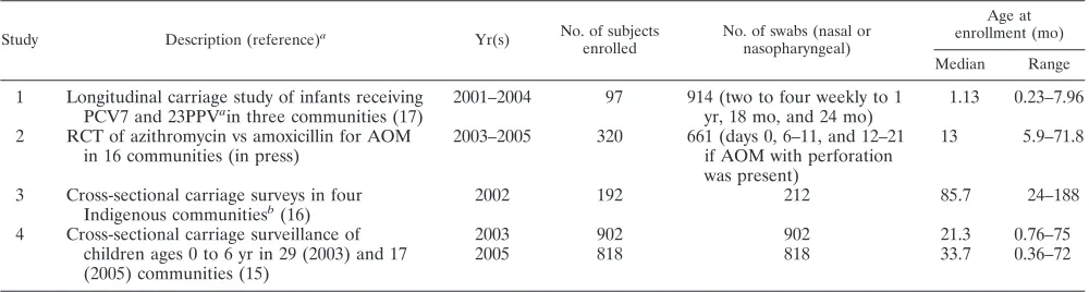
Samples.A total of 182 nasopharyngeal NSP isolates were randomly selected (Research Randomizer; http://www.randomizer.org) from four studies of otitis media (Table 1) conducted in the Northern Territory after the introduction of PCV7. The isolates were stored at⫺80°C in skim milk-tryptone-glucose-glycerol broth (20). When the specimens originated from longitudinal studies, sample selection was limited to the first NSP isolate recovered from each child.
Antibiotic susceptibility testing.Screening for penicillin and macrolide sus-ceptibility was determined by a calibrated dichotomous sussus-ceptibility (CDS) disk diffusion assay (oxacillin and erythromycin, respectively) (3). Etests (AB Biodisk, Solna, Sweden) were used to determine the penicillin and erythromycin MICs of all nonsusceptible isolates (CDS radius⬍6 mm). PCR for the detection of macrolide resistance genes (mefA/EandermBwas performed as described pre-viously (18, 26).
DNA extraction and quantification.NSP DNA for BOX typing and the de-tection of macrolide resistance genes was extracted by using the Instagene matrix (Bio-Rad), according to the manufacturer’s instructions. NSP DNA for multilo-cus sequence typing (MLST) was extracted with a QIAamp DNA minikit (Qiagen), according to the manufacturer’s instructions. The DNA concentration was determined with a Picogreen double-stranded DNA quantitation assay kit (Molecular Probes). The extraction of DNA for multiplex PCR/reverse line blot hybridization (mPCR/RLB-H) was done as described previously (12).
Capsule gene PCR.The capsule gene was detected by the method of Hanage et al. (8), with the exception that primerswzg-up (5⬘-ATCCTTGTCAGCTCTG
TGTC) and wzg-down (5⬘-TCACTTGCAACTACATGAAC) amplified a
1,797-bp product.
Molecular typing.BOX typing was done as described previously with 1 ng of DNA in each 50-l reaction mixture (27). The BOX types were defined accord-ing to the bandaccord-ing patterns between 750 bp and 3,000 bp. MLST was undertaken essentially as described previously (6), except for modification of thespiforward primer (primer spiF3, 5⬘-CAGATTTTGCAAGCCTATGG), the recPreverse primer (primer recPR2, 5⬘-GCCGTGTACAGCATTAGTTC), and theddl for-ward primer (primer ddlF2, 5⬘-GATGGCTCTGTTCAAGGATT). Molecular serotyping ofwzg-positive isolates of NSP was by done mPCR/RLB-H, as de-scribed previously (11, 28), for 90 pneumococcal serotypes. This method targets serotype-specific regions ofwzyfor all serotypes with the exception of serotype 3. In the case of serotype 3,wzeis targeted, aswzyis not present in serotype 3 strains.
RESULTS
NSP colonization of Indigenous children.Presumptive NSP
in this study were identified on the basis of colonial morphol-ogy, optochin sensitivity in 5% CO2 for distinction from S.
pseudopneumoniae(2), and a negative Quellung reaction. In
longitudinal studies 1 and 2 (Table 1), presumptive NSP were reported in 18% and 8% of the nasopharyngeal swab speci-mens, respectively. Cross-sectional studies 3 and 4 reported presumptive NSP carriage prevalence rates of 8% and 5%, respectively. These numbers are likely to underestimate the true carriage rate because NSP were not systematically sought. Variations in the rates reported between studies may also be due to subjectivity in the selection of colonies.
Characterization of NSP by BOX typing and antibiogram
analysis. Among the 182 randomly selected isolates of NSP
(which were confirmed to belytApositive), 49 BOX types were identified. The three most common BOX types (types 1, 14, and 3) represented 44% of the isolates. BOX types 1 and 14 were closely related, distinguished by a single band size differ-ence of less than 50 bp.
Intermediate resistance to penicillin (MICs, 0.12 to 1g/ml; breakpoint for oral therapy of nonmeningeal strains) was evi-dent in 49% (90/182) of the NSP isolates, penicillin resistance (MICs,ⱖ2g/ml) was detected in 2% (4/182) of the isolates, and erythromycin resistance (MICs,ⱖ4g/ml) was detected in 10% (19/182) of the isolates. Erythromycin resistance was me-diated by themefEmacrolide resistance gene in all except one of the isolates; the latter isolate was negative for themefA/E
and ermB genes. Reduced susceptibility to these commonly used antimicrobials was detected in isolates from 32 BOX types.
Characterization of NSP by MLST.Representative isolates
[image:2.585.43.543.81.215.2]of the three most common BOX types, including all antibio-grams, were analyzed by MLST (Table 2). BOX type 1 and 14 isolates were represented by six sequence types (STs), all of which were new to the MLST database. Two STs were com-mon to these BOX types. ST4485 was a double-locus variant of a single ST corresponding to a serotype 4 isolate. ST4487 and ST4488 were both double-locus variants of two STs corre-sponding to serotype 14 and 19A isolates. The BOX type 3 isolate was ST448, an NSP clone reported internationally.
TABLE 1. Carriage study details
Study Description (reference)a
Yr(s) No. of subjects enrolled
No. of swabs (nasal or nasopharyngeal)
Age at enrollment (mo)
Median Range
1 Longitudinal carriage study of infants receiving PCV7 and 23PPVain three communities (17)
2001–2004 97 914 (two to four weekly to 1 yr, 18 mo, and 24 mo)
1.13 0.23–7.96
2 RCT of azithromycin vs amoxicillin for AOM in 16 communities (in press)
2003–2005 320 661 (days 0, 6–11, and 12–21 if AOM with perforation was present)
13 5.9–71.8
3 Cross-sectional carriage surveys in four Indigenous communitiesb(16)
2002 192 212 85.7 24–188
4 Cross-sectional carriage surveillance of 2003 902 902 21.3 0.76–75
children ages 0 to 6 yr in 29 (2003) and 17 (2005) communities (15)
2005 818 818 33.7 0.36–72
a
23PPV, 23-valent pneumococcal polysaccharide vaccine; RCT, randomized controlled trial; AOM, acute otitis media. b
Children⬍16 years of age were included.
on May 16, 2020 by guest
http://jcm.asm.org/
ST448 and STs 4486, 4484, and 4565 were not closely related (less than five identical MLST loci) to STs corresponding to capsular pneumococci that are currently in the MLST data-base. All isolates were confirmed to be pneumococci by using the “is it a pneumococcus?” analysis (www.mlst.net).
Presence ofwzggene.As described previously (8), we used
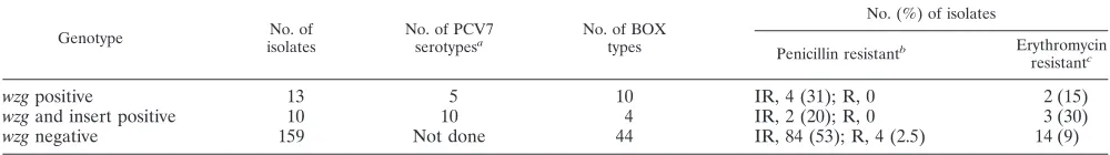
PCR for the conservedwzg(cpsA) gene to test for the presence of acpslocus (Tables 3 and 4).wzgwas detected in 23 of 182 (13%) NSP isolates and 13 BOX types. However, 10 isolates provided a product of approximately 3 kb rather than the expected 1.8-kb product. Partial sequencing of the product demonstrated a sequence homologous to that of the pneumo-coccalwzggene and an unidentified insert which was not re-lated to streptococcalaligenes, as reported previously (9). The 10 isolates represented four different BOX types, suggesting that this insertion was not a recent event.
mPCR/RLB-H analysis of the isolates with the expected 1.8-kbwzggene determined that five isolates corresponded to PCV7 serotypes 9V (or 9A) and 14, whereas eight corre-sponded to non-PCV7 serotypes 7A/F, 16F, 11A/D, 22A/F, and 13 (Table 3). Cocolonization with a capsular strain of the corresponding serotype was evident for 8 of 13 NSP with the 1.8-kbwzgproduct (Table 3). Subsequent testing of all strains with specific type antisera found that none reacted.
mPCR/RLB-H analysis determined that the 10 isolates with the 3-kbwzgPCR product corresponded to serotype 14. Two BOX type 14 isolates positive for the insertion were ST4488 (double-locus variants of STs corresponding to serotype 14 and 19A isolates).
One isolate consistently provided a serotype 14 result (based on the result forwzy) and a serotype 3 result (based on the result for wze), suggesting mosaicism of the strain and that both serotype 3 and 14 capsular genes may exist in this strain. Reduced susceptibility to antimicrobials was common in all groups (Table 4).
DISCUSSION
Australian Indigenous children living in remote communi-ties experience capsular pneumococcal carriage rates of ap-proximately 80% and NSP carriage rates of 5 to 20%. These NSP carriage rates are likely to be underestimates because NSP are morphologically distinct from capsular pneumococci and NSP were not systematically sought in the studies that determined those carriage rates. NSP detection rates in a
pop-ulation appear to be dependent on the study protocol and subjectivity in colony selection.
Cocolonization with capsular and noncapsular pneumococci occurred frequently, providing an opportunity for an interac-tion between these populainterac-tions. Previous studies of NSP report a highly diverse population with a pervasive nature, an ability to cause disease, and a likely important role in pneumococcal ecology (8, 23, 24). Although nonencapsulated forms are less likely to cause disease, their ability for gene transfer, including the transfer of antibiotic resistance and capsular/virulence genes, is predictably high due to their capacity for an increased rate of transformation (22).
The majority of NSP are likely to be of the true
nonencap-sulated lineage.Two distinct populations were detected in this
study: NSP with and without thewzg(cpsA) gene. The majority (87%) of NSP in this study werewzgnegative by PCR and were thus likely to be of the true nonencapsulated lineage. However, given that 10 of 23wzgsequences were atypical due to an inser-tion, we cannot rule out the possibility that other isolates con-tained modifiedwzgsequences not detectable by our methods.
[image:3.585.43.284.88.201.2]Our grant did not provide funding for MLST of all NSP isolates; however, MLST of representatives of the three most common BOX types identified authentic pneumococci, includ-ing six new STs and ST448 (which accounted for 11% of the NSP in this study). ST448 is a particularly interesting clone, in that it is internationally disseminated and has been associated with invasive disease (9).
TABLE 3. Investigation of serotypes associated with
wzg-positive NSPa
BOX type MLST type NSP serotype Simultaneously carried serotypes
1.8-kbwzgproduct
5 11A/11Db 11A
6 7A/7Fb 7F
8 16F 16F
8 16F 16F
14 9A/9Vb 15B
14 9A/9Vb None
14 14 19A
17 22A/22Fb 22F
25 14 19F
30 14 6B
32 13 13
52 16F 16F
57 16F 16F
3-kbwzgproduct
1 14 None
1 14 19F
1 14 None
13 14 21
14 14 19A
14 14 None
14 14 19A
14 4488c 14 16F
14 4488c 14and 3d None
15 14 15C
a
The serotype of eachwzg-positive NSP was determined by mPCR/RLB-H. Capsular pneumococcal serotypes simultaneously carried in each swab are shown. PCV7 serotypes are indicated in boldface.
b
Pneumococcal serogroups indistinguishable by mPCR/RLB-H. c
Double-locus variant of STs corresponding to serotypes 14 and 19A. d
This isolate reacted with serotypes 14 and 3 by mPCR/RLB-H. TABLE 2. MLST investigation of the three most
common NSP BOX types
BOX type No. (%) of isolates MLST type
(no. of isolates)
1 34 (19) 4485 (1)
4486 (1) 4487 (1) 4488 (1)
14 25 (14) 4484 (1)
4486 (1) 4488 (2) 4565 (1)
3 20 (11) 448 (1)
on May 16, 2020 by guest
http://jcm.asm.org/
[image:3.585.302.541.89.353.2]Identification of the majority of NSP in this study was based on colonial morphology, optochin susceptibility, and a positive
lytAPCR result. Therefore, it is likely that these strains wereS.
pneumoniaerather thanS. pseudopneumoniae(which are
gen-erally optochin resistant in the presence of increased amounts of CO2) (2) or other closely related Streptococcusspp. How-ever, aslytAis not ideal as a sole genetic criterion for pneu-mococcal identification, in due course MLST analysis may identify some strains as atypical pneumococci (divergent at the seven MLST loci by⬎5%) (7).
NSP do not represent a population of PCV7 types
suppress-ing capsule expression.Capsular genes (wzg) were detected in
a minority of the NSP isolates. Ten of these isolates had an insertion inwzgwhich may have interrupted capsule expres-sion, while 13 contained capsule genes related to both PCV7 and non-PCV7 serotypes. Additionally, among 182 isolates, only 15 were potentially downregulated PCV7 types. These data do not support the hypothesis that PCV7 types are sup-pressing capsule expression to avoid vaccine-induced immune clearance.
NSP may act as a parking lot for antibiotic resistance genes.
Transformation is a common mechanism for the development of resistance to penicillin and macrolides in pneumococci (4, 5). As NSP have transformability rates up to 1,075-fold higher than those of capsulated strains (22), NSP may act as a vector for the transmission of resistance genes between viridans group streptococci and capsulated pneumococci. Indeed, a serotype 19F clone became increasingly non-penicillin susceptible to by the uptake of pbp2x gene fragments shared with NSP (10). Despite being present in low numbers, nonencapsulated forms of PCV7 serotypes are also likely to harbor the antibiotic resistance genes of their parent capsular vaccine serotypes and thus have the potential to transfer antibiotic resistance to newly emerging nonvaccine serotypes. Our 2005 surveillance of pneumococcal carriage by Indigenous children found that 14% of the isolates were non-penicillin susceptible (MIC ⱖ 0.12
g/ml) and that 6% of the isolates were azithromycin resistant (MICⱖ 2g/ml) (15). The penicillin resistance data are in stark contrast to the 55.5% rate of non-penicillin susceptibility that we found in wzg-negative NSP and 26% in potentially revertible wzg-positive NSP. Thus, there is potential for the selection of existing capsular strains exhibiting antibiotic resis-tance and for the uptake of resisresis-tance genes by susceptible capsular pneumococci during exposure to NSP.
Reporting NSP in surveillance and clinical trials.High
car-riage rates, antibiotic resistance, and the presence of capsule genes suggest a role for NSP in the maintenance and survival
of capsular pneumococci. Until this role is better understood, we support continued reporting of NSP carriage rates. Since MLST is necessary to explicitly confirm the identity of nonse-rotypeable pneumococcal isolates, it will be necessary to ana-lyze data obtained by using less rigorous methods of identifi-cation with caution.
In populations with high NSP carriage rates, combining the data for NSP and capsular pneumococci would inflate the rates of pneumococcal carriage and antibiotic resistance. Therefore, we recommend separate reporting of rates of carriage of NSP and capsular pneumococci in surveillance studies. When the microbiological outcomes of clinical trials are reported, we recommend the inclusion of only the capsular pneumococci in primary analyses.
ACKNOWLEDGMENTS
We thank the Channel 7 Children’s Research Foundation for pro-viding financial support for this study.
We also thank the families who participated in our studies and agreed to storage and further analysis of their nasopharyngeal swab specimens. Without the support of these families, this study would not have been possible.
REFERENCES
1.American Society for Microbiology.2003. Manual of clinical microbiology, 8th ed. American Society for Microbiology, Washington, DC.
2.Arbique, J. C., C. Poyart, P. Trieu-Cuot, G. Quesne, M. G. Carvalho, A. G. Steigerwalt, R. E. Morey, D. Jackson, R. J. Davidson, and R. R. Facklam.
2004. Accuracy of phenotypic and genotypic testing for identification of Streptococcus pneumoniae and description of Streptococcus pseudopneu-moniae sp. nov. J. Clin. Microbiol.42:4686–4696.
3.Bell, S. M.1975. The CDS disc method of antibiotic sensitivity testing (calibrated dichotomous sensitivity test). Pathology7(Suppl.):48. 4.Cerda, Z. P., L. M. Laplana, C. R. Calvo, P. G. Cepero, M. C. Erazo, and R.
Gomez-Lus.2004. Molecular basis of resistance to macrolides and other antibiotics in commensal viridans group streptococci and Gemella spp. and transfer of resistance genes to Streptococcus pneumoniae. Antimicrob. Agents Chemother.48:3462–3467.
5.Coffey, T. J., C. G. Dowson, M. Daniels, and B. G. Spratt.1995. Genetics and molecular biology of beta-lactam-resistant pneumococci. Microb. Drug Re-sist.1:29–34.
6.Enright, M. C., and B. G. Spratt.1998. A multilocus sequence typing scheme for Streptococcus pneumoniae: identification of clones associated with seri-ous invasive disease. Microbiology144:3049–3060.
7.Hanage, W. P., T. Kaijalainen, E. Herva, A. Saukkoriipi, R. Syrjanen, and B. G. Spratt.2005. Using multilocus sequence data to define the pneumo-coccus. J. Bacteriol.187:6223–6230.
8.Hanage, W. P., T. Kaijalainen, A. Saukkoriipi, J. L. Rickcord, and B. G. Spratt.2006. A successful, diverse disease-associated lineage of nontypeable pneumococci that has lost the capsular biosynthesis locus. J. Clin. Microbiol.
44:743–749.
9.Hathaway, L. J., M. P. Stutzmann, P. Battig, S. Aebi, and K. Muhlemann.
2004. A homologue of aliB is found in the capsule region of nonencapsulated Streptococcus pneumoniae. J. Bacteriol.186:3721–3729.
[image:4.585.42.542.82.152.2]10.Hauser, C., S. Aebi, and K. Muhlemann.2004. An internationally spread clone of Streptococcus pneumoniae evolves from low-level to higher-level TABLE 4. Summary of typing and antimicrobial susceptibility data for NSP
Genotype No. of
isolates
No. of PCV7
serotypesa No. of BOX types
No. (%) of isolates
Penicillin resistantb Erythromycin resistantc
wzgpositive 13 5 10 IR, 4 (31); R, 0 2 (15)
wzgand insert positive 10 10 4 IR, 2 (20); R, 0 3 (30)
wzgnegative 159 Not done 44 IR, 84 (53); R, 4 (2.5) 14 (9)
aAs determined by mPCR/RLB-H.
bIntermediate resistance (IR) is a penicillin MIC of 0.12 to 1g/ml; resistance (R) is a penicillin MIC ofⱖ2g/ml. cErythromycin MIC ofⱖ4g/ml.
on May 16, 2020 by guest
http://jcm.asm.org/
penicillin resistance by uptake of penicillin-binding protein gene fragments from nonencapsulated pneumococci. Antimicrob. Agents Chemother. 48:
3563–3566.
11.Kong, F., M. Brown, A. Sabananthan, X. Zeng, and G. L. Gilbert.2006. Multiplex PCR-based reverse line blot hybridization assay to identify 23 Streptococcus pneumoniae polysaccharide vaccine serotypes. J. Clin. Micro-biol.44:1887–1891.
12.Kong, F., S. Gowan, D. Martin, G. James, and G. L. Gilbert.2002. Serotype identification of group B streptococci by PCR and sequencing. J. Clin. Microbiol.40:216–226.
13.Leach, A. J., J. B. Boswell, V. Asche, T. G. Nienhuys, and J. D. Mathews.
1994. Bacterial colonization of the nasopharynx predicts very early onset and persistence of otitis media in Australian aboriginal infants. Pediatr. Infect. Dis. J.13:983–989.
14.Leach, A. J., and P. S. Morris.2007. The burden and outcome of respiratory tract infection in Australian and aboriginal children. Pediatr. Infect. Dis. J.
26:S4–S7.
15.Leach, A. J., P. S. Morris, G. B. McCallum, C. A. Wilson, L. Stubbs, J. Beissbarth, S. Jacups, K. Hare, and H. C. Smith-Vaughan.2009. Emerging pneumococcal carriage serotypes in a high-risk population receiving 7-valent pneumococcal conjugate vaccine and 23-valent polysaccharide vaccine since 2001. BMC Infect. Dis.9:121.
16.Mackenzie, G., J. Carapetis, A. J. Leach, K. Hare, and P. Morris.2007. Indirect effects of childhood pneumococcal vaccination on pneumococcal carriage among adults and older children in Australian aboriginal commu-nities. Vaccine25:2428–2433.
17.Mackenzie, G. A., J. R. Carapetis, A. J. Leach, and P. S. Morris.2009. Pneumococcal vaccination and otitis media in Australian aboriginal infants: comparison of two birth cohorts before and after introduction of vaccination. BMC Pediatr.9:14.
18.Montanari, M. P., I. Cochetti, M. Mingoia, and P. E. Varaldo.2003. Phe-notypic and molecular characterization of tetracycline- and erythromycin-resistant strains of Streptococcus pneumoniae. Antimicrob. Agents Che-mother.47:2236–2241.
19.Nagai, K., Y. Shibasaki, K. Hasegawa, T. A. Davies, M. R. Jacobs, K.
Ubukata, and P. C. Appelbaum.2001. Evaluation of PCR primers to screen for Streptococcus pneumoniae isolates and beta-lactam resistance, and to detect common macrolide resistance determinants. J. Antimicrob. Che-mother.48:915–918.
20.O’Brien, K. L., and H. Nohynek.2003. Report from a WHO working group: standard method for detecting upper respiratory carriage ofStreptococcus pneumoniae. Pediatr. Infect. Dis. J.22:133–140.
21.Park, I. H., D. G. Pritchard, R. Cartee, A. Brandao, M. C. Brandileone, and M. H. Nahm.2007. Discovery of a new capsular serotype (6C) within sero-group 6 of Streptococcus pneumoniae. J. Clin. Microbiol.45:1225–1233. 22.Pearce, B. J., F. Iannelli, and G. Pozzi.2002. Construction of new
unencap-sulated (rough) strains of Streptococcus pneumoniae. Res. Microbiol.153:
243–247.
23.Porat, N., D. Greenberg, N. Givon-Lavi, D. S. Shuval, R. Trefler, O. Segev, W. P. Hanage, and R. Dagan.2006. The important role of nontypable Streptococcus pneumoniae international clones in acute conjunctivitis. J. In-fect. Dis.194:689–696.
24.Sa-Leao, R., A. S. Simoes, S. Nunes, N. G. Sousa, N. Frazao, and H. de Lencastre.2006. Identification, prevalence and population structure of non-typable Streptococcus pneumoniae in carriage samples isolated from pre-schoolers attending day-care centres. Microbiology152:367–376. 25.Smith-Vaughan, H., R. Byun, S. Halpin, M. A. Nadkarni, N. A. Jacques, N.
Hunter, P. S. Morris, and A. J. Leach.2008. Interventions for prevention of otitis media may be most effective if implemented in the first weeks of life. Int. J. Pediatr. Otorhinolaryngol.72:57–61.
26.Sutcliffe, J., T. Grebe, A. Tait-Kamradt, and L. Wondrack.1996. Detection of erythromycin-resistant determinants by PCR. Antimicrob. Agents Che-mother.40:2562–2566.
27.van Belkum, A., M. Sluijter, R. de Groot, H. Verbrugh, and P. W. M. Hermsn.1996. Novel BOX repeat PCR assay for high-resolution typing of
Streptococcus pneumoniaestrains. J. Clin. Microbiol.34:1176–1179. 28.Zhou, F., F. Kong, Z. Tong, and G. L. Gilbert.2007. Identification of
less-common Streptococcus pneumoniae serotypes by a multiplex PCR-based reverse line blot hybridization assay. J. Clin. Microbiol.45:3411–3415.