0095-1137/10/$12.00 doi:10.1128/JCM.00420-10
Copyright © 2010, American Society for Microbiology. All Rights Reserved.
Development of a Skin Test for Bovine Tuberculosis for
Differentiating Infected from Vaccinated Animals
䌤
Adam O. Whelan,
1* Derek Clifford,
2Bhagwati Upadhyay,
1Eleanor L. Breadon,
3James McNair,
3Glyn R. Hewinson,
1and Martin H. Vordermeier
1TB Research Group,1and Animal Services Unit,2Veterinary Laboratories Agency, New Haw, Addlestone, Surrey KT15 3NB,
United Kingdom, and Agri-Food and Biosciences Institute, Bacteriology Department, Veterinary Sciences Division, Stormont, Belfast BT4 3SD, Northern Ireland3
Received 1 March 2010/Returned for modification 15 June 2010/Accepted 20 June 2010
The tuberculin skin test has been used for the diagnosis of bovine and human tuberculosis (TB) for over a hundred years. However, the specificity of the test is compromised by vaccination with the Mycobacterium
bovis-derived vaccine strain bacille Calmette-Gue´rin (BCG). Since current promising vaccines against bovine
TB are based on heterologous prime-boost combinations that include BCG, there is a need for diagnostic tests for differentiating infected from vaccinated animals (DIVA). The application of antigens such as ESAT-6 and CFP-10 for DIVA has so far been realized largely through their application in the blood-based gamma interferon release assay. In the current study, we have reassessed the potential of such antigens as skin test reagents for DIVA in cattle. A cocktail of theMycobacterium tuberculosiscomplex recombinant protein antigens ESAT-6, CFP-10, MPB70, and MPB83 elicited delayed-type hypersensitivity (DTH) skin test responses in 78% of naturally infected tuberculin-positive cattle. Importantly, this cocktail induced no skin responses in BCG-vaccinated cattle despite them being sensitized for strong tuberculin responses. Further optimization of skin test antigen combinations identified that the inclusion of Rv3615c (Mb3645c) enhanced skin test sensitivity in naturally infected cattle without compromising specificity. In addition, we demonstrate for the first time the utility of synthetic peptides as promising skin test antigens for bovine TB for DIVA. Our data provide a promising basis for the future development of skin tests for DIVA with practical relevance for TB diagnosis in both veterinary and clinical settings.
Bovine tuberculosis (TB) is a disease of economic and zoo-notic importance caused by infection withMycobacterium bo-vis, a member of theMycobacterium tuberculosiscomplex. The primary diagnostic test used for the control and surveillance of bovine TB is the tuberculin skin test, a test that has remained in the forefront of TB diagnosis for both humans and cattle for over 100 years. The development of the test arose following the preparation of the first “tuberculin” by Robert Koch in 1890 (9). While Koch’s tuberculin failed to live up to its initial claims of having curative properties, its diagnostic potential was quickly realized. Interestingly, its potential to diagnose tuber-culous infection was first demonstrated in cattle prior to its widespread use in people (19). The two most common formats of the test used in cattle are the caudal fold test and the single intradermal comparative cervical tuberculin (SICCT) test (13). Both test formats use a purified protein derivative (PPD) tu-berculin prepared from a culture ofM. bovis (PPD-B) as the primary diagnostic antigen. Additionally, the SICCT test in-cludes the use of a Mycobacterium avium-derived PPD (PPD-A) to provide a measure of environmental sensitization. It is the more specific of the two tests (14, 20) and is therefore the adopted test format in the United Kingdom.
Bovine TB continues to be a significant and ongoing prob-lem in the United Kingdom (www.defra.gov.uk/foodfarm
/farmanimal/diseases/atoz/tb/index.htm). Cattle vaccination has been identified as one of the most promising long-term United Kingdom control strategies (10), and the development of an efficacious vaccine continues to be a research priority. Currently, promising vaccines against bovine TB are based on heterologous prime-boost combinations that include the live attenuated M. bovis vaccine strain bacille Calmette-Gue´rin (BCG) as one of their components (8). However, as for humans, the vaccination of cattle with BCG compromises the specificity of the tuberculin skin test since PPD contains cross-reactive antigens shared by both pathogenic and vaccine strains (4, 5, 26). Therefore, the development of diagnostic tests for differentiating vaccinated from infected animals (DIVA) is an essential prerequisite to allow the inclusion of BCG-based vaccination as part of bovine TB control strategies.
Current front-line reagents for DIVA for TB diagnosis are the M. tuberculosis complex antigens ESAT-6 and CFP-10. They are strongly recognized in TB-infected cattle and humans (6, 15, 22, 24, 25), yet since their genes are encoded in region of difference 1 (RD1), which has been deleted from all BCG strains (12), they do not elicit a response in BCG-vaccinated hosts in the absence of infection (2, 6, 22, 24, 25). The practical application of such reagents for DIVA has so far been realized largely through their use in blood-based gamma interferon (IFN-␥) release assays (IGRAs). Given the high level of famil-iarity and widespread application of the tuberculin skin test by veterinarians and clinicians, a skin test format for DIVA would provide a valuable additional test platform. This might espe-cially be the case where the logistics of access to laboratory-based resources are problematic. It is also notable that in
* Corresponding author. Mailing address: TB Research Group, Vet-erinary Laboratories Agency, Woodham Lane, New Haw, Addlestone, Surrey KT15 3NB, United Kingdom. Phone: 44 (0)1932 357506. Fax: 44 (0)1932 357260. E-mail: a.o.whelan@vla.defra.gsi.gov.uk.
䌤Published ahead of print on 30 June 2010.
3176
on May 16, 2020 by guest
http://jcm.asm.org/
recent years, there has also been renewed interest in a skin test-based test for DIVA for human TB, with several reports demonstrating the skin test potential of ESAT-6 (1, 3, 28).
The purpose of the current study was to assess the potential of definedM. bovisantigens as skin test reagents for diagnosing bovine TB. We have investigated and optimized combinations of defineM. bovisantigens with an emphasis on assessing their performance as sensitive and practical diagnostic tools for DIVA for use in the field.
MATERIALS AND METHODS
Animals.To investigate the skin test performance of defined antigens in cattle naturally exposed toM. bovis, SICCT reactor cattle were recruited from United Kingdom farms with a confirmed history of bovine TB (convenience sampling dependent on availability). Animals were identified during routine surveillance operations where their SICCT response (PPD-B⫺PPD-A) was⬎2 mm and were housed in secure segregated biocontainment facilities at the Veterinary Laboratories Agency (VLA) Weybridge. A total of 62 reactors were used in the study, which included 21 male and 41 female cattle of seven different breeds, including dairy and beef stock. The age at the time of skin test investigations ranged from 9 months to 11 years, with a median age of 21 months. All reactors underwent detailed postmortem examination to assess for the presence ofM. bovisinfection in accordance with previously described procedures (23). Visible lesions consistent with bovine TB and/or culture isolation ofM. boviswere confirmed for 55 of these reactors. For the specificity studies, Holstein-Friesian castrated male cattle (n⫽54) were sourced from United Kingdom TB-free farms and housed at the VLA in accommodations separate from those of the reactors. A cohort of these calves (n⫽20) was neonatally vaccinated with BCG to allow the evaluation of reagents for DIVA. A single dose of 1⫻106CFU of BCG Danish (Staten Serum Institute, Sweden) was administered subcutaneously to each of these calves at 4 to 6 weeks of age. Where a second skin test was performed for some cattle during the study, it was performed no earlier than 62 days later. All cattle experiments were cleared by local ethical review and animal procedures under a license granted by the British Home Office.
Antigens. Bovine and avian PPDs were supplied by the VLA Tuberculin Production Unit. Histidine-tagged recombinant proteins were expressed in Esch-erichia colicells and purified by nickel affinity chromatography. MPB70, MPB83, ESAT-6, and CFP-10 were supplied by Lionex Diagnostics and Therapeutics GmbH (Germany), and Rv3615c was produced at the Agri-Food and Biosciences Institute (Belfast, Northern Ireland). Rv3615c is referred to in the current paper using the M. tuberculosis genome annotation (http://genolist.pasteur.fr /TubercuList/). In theM. bovisgenome, it is annotated as Mb3645c (http: //genolist.pasteur.fr/BoviList/). All synthetic peptides used in the study were synthesized by solid-phase F-moc (9H-fluoren-9-yl-methoxycarbonyl) chemistry and supplied at a purity of⬎90% (Pepceuticals Ltd., United Kingdom). The identity of each peptide was confirmed by mass spectrometry. For ESAT-6 and CFP-10, 16-mer peptides with 8-amino-acid overlaps of the complete protein sequences were used (21 peptides in total). For MPB83, a single 20-mer peptide that had previously been shown to encode a bovine T-cell epitope (p195-214) was used (24), and similarly, for Rv3615c, three bovine T-cell epitopes encoding 20-mer peptides were used (p65-84, p73-92, and p84-103) (18).
Skin test procedures.The disclosing SICCT field test was performed and interpreted according to European Communities Commission Regulations (reg-ulation 1226/2002, amending annexes A and B of the consolidated Council Directive 64/432/EEC). For the evaluation of defined antigen combinations at VLA, up to eight intradermal injection sites were used on each animal, four on each side of the neck. PPD-B and PPD-A (2,500 IU) were always included in each test, and all antigens were administered in a 0.1-ml volume. Defined anti-gens were tested at a concentration of 10g whether used individually or as a component of a cocktail unless otherwise stated. Skin induration at administra-tion sites was measured by using calipers by the same operator for all experi-ments. Measurements were recorded prior to and at 72 h after skin test and additionally at 96 and 120 h for some experiments. Results are expressed as the difference in skin thicknesses (mm) between the pre- and post-skin test readings.
IFN-␥whole-blood ELISA.Heparinized whole-blood cultures were stimulated with either PPD-A (10g/ml), PPD-B (10g/ml), recombinant protein antigens (5g/ml), protein cocktail (5g/ml per constituent), staphylococcal enterotoxin B (1g/ml; Sigma-Aldrich, United Kingdom), or the no-antigen control. Cul-tures were set up within 8 h of blood collection and cultured at 37°C in 5% CO2. Antigen-stimulated IFN-␥was measured in whole-blood culture supernatants at
24 h using the commercially available Bovigam enzyme-linked immunosorbent assay (ELISA) (Prionics AG, Switzerland). Results are expressed as the back-ground-corrected optical density measured at 450 nm (OD450).
Statistical analysis.Differences in response magnitudes between different antigen combinations were compared by repeated-measures analysis of variance (ANOVA), with the application of Bonferroni’s multiple-comparison postanaly-sis test. Differences between antigen responder frequencies were analyzed by using the Fisher exact test. These statistical analyses and receiver operator curve (ROC) analysis were performed by using the Prism 5 software program (Graph-pad Inc.).
RESULTS
[image:2.585.335.503.69.332.2]A defined protein cocktail of MPB70, MPB83, ESAT-6, and CFP-10 induces skin test responses in naturally infected cat-tle.To assess the potential of defined antigens as diagnostic skin test reagents, we initially formulated a cocktail comprised of the purified recombinant M. bovis/M. tuberculosis protein antigens ESAT-6, CFP-10, MPB70, and MPB83. MPB70 and MPB83 were included in our cocktail since they had a previous demonstrated skin test potential for DIVA in a guinea pig model ofM. bovisinfection (24). In 37 SICCT-positive cattle, the protein cocktail induced a response in 29 of 37 (78%) of these cattle at 72 h posttest (Fig. 1). Skin reactions were ad-ditionally measured at 96 and 120 h since it was previously reported that the development of bovine skin test responses to
FIG. 1. Protein cocktail-induced skin test responses in cattle. Skin test responses were measured at 72, 96, and 120 h in cattle naturally exposed toM. bovis(n⫽37). (A) Comparative SICCT PPD responses (PPD-B⫺PPD-A). (B) Responses induced by a protein cocktail of 10
g each of ESAT-6, CFP-10, MPB70, and MPB83. The increase in skin induration for each animal is represented by open circles, and the horizontal line provides the mean (⫾ standard error of the mean [SEM]), with results expressed as the difference in skin thicknesses (mm) between the pre- and post-skin test readings. The statistical difference between responses was determined by using ANOVA (*,
P⬍0.05;**,P⬍0.01;***,P⬍0.001).
on May 16, 2020 by guest
http://jcm.asm.org/
ESAT-6 continued to increase after 72 h (16). In comparison with the 72-h reactions, the SICCT responses were significantly reduced at 96 and 120 h posttest (Fig. 1). The magnitudes of protein cocktail-induced reactions were comparable at each time point. The measurement of antigen-induced IFN-␥ in whole-blood cultures set up on the day of the skin test dem-onstrated that ESAT-6 and CFP-10 were the most strongly recognized constituent antigens in the protein cocktailin vitro
(Fig. 2). The protein cocktail was next tested at titrated dose concentrations of 10, 5, and 1g for each cocktail component. The respective responses in a cohort of 19 of the previously tested reactors are shown in Fig. 3. Skin test responses were
comparable between the 10- and 5-g doses but were signifi-cantly reduced at the 1-g dose, demonstrating a dose re-sponse. For all further experiments, a skin test concentration of 10g per defined antigen component was used.
The specificity of this protein cocktail was first determined for naïve cattle (n⫽20). The cocktail induced no measureable response in any of these animals when reactions were read at 72 h (Fig. 4). While all SICCT responses were also negative, measurable PPD-A-biased responses did indicate some sensi-tization of these animals by environmental mycobacteria. We additionally measured reactions at 96 and 120 h. While re-sponses remained negative at 96 h, at 120 h two animals pro-vided positive reactions of 2 and 12 mm to the protein cocktail and 5 mm to the SICCT test (data not shown). For all further experiments, reactions were recorded only at 72 h posttest.
The potential of the protein cocktail for DIVA was evalu-ated by using 3-month-old calves that had been neonatally vaccinated with BCG at 4 to 6 weeks of age (n⫽20). For these calves, only one animal failed to induce an SICCT PPD-B-biased skin test response, confirming the strong tuberculin skin test sensitization of BCG vaccination (Fig. 4). In con-trast, the defined protein cocktail did not induce a response in any of these vaccinates. Protein cocktail and SICCT re-sponses measured in either the naturally infected or BCG-vacci-nated cattle were used to generate ROC curves to provide esti-mates of skin test sensitivity and specificity for DIVA. Table 1 summarizes the sensitivity and specificity estimates for SICCT cutoffs of⬎2 mm and⬎4 mm since these represent operational “severe” and “standard” test cutoffs used in United Kingdom surveillance operations (www.defra.gov.uk/foodfarm/farmanimal /diseases/atoz/tb/control/tuberculin.htm). Also shown are the per-formance figures for SICCT and protein cocktail test cutoffs that provide 100% specificity for the respective tests for DIVA.
Optimization of defined skin test reagents, including the use of Rv3615c and synthetic peptides.In vitroassessment of the capacity of the constituent protein cocktail antigens to induce IFN-␥ suggested the immunodominance of ESAT-6 and CFP-10 (Fig. 2). Therefore, we investigated the additive
ben-FIG. 2.In vitrorecognition of protein antigens. Thein vitrocapacity of antigens to induce IFN-␥in blood from cattle naturally exposed to
[image:3.585.334.508.67.200.2]M. bovis(n⫽37) was determined for ESAT-6, CFP-10, MPB70, and MPB83 either individually (5g/ml) or as a combined protein cocktail (5g/ml per constituent). Responses induced by PPD-B (10g/ml), staphylococcal enterotoxin B (SEB) (1g/ml), or the no-antigen con-trol were also determined. The antigen-induced IFN-␥response for each animal is represented by open circles, and the horizontal line provides the mean (⫾SEM), with results expressed as the background-corrected OD450.
[image:3.585.77.245.67.194.2]FIG. 3. Skin test protein cocktail dose titration. A skin test dose titration was determined for the protein cocktail comprised of ESAT-6, CFP-10, MPB70, and MPB83 in cattle that were naturally exposed toM. bovis(n⫽19). The protein cocktail was administered at concentrations of 10, 5, and 1g for each antigen component. Protein cocktail and SICCT (PPD-B⫺PPD-A) responses measured at 72 h for each animal are represented by open circles, and the horizontal line provides the mean (⫾SEM), with results expressed as the difference in skin thicknesses (mm) between the pre- and post-skin test readings. The statistical difference between responses induced by the protein cocktails was determined by using ANOVA (*,P⬍0.05;***,P⬍0.001).
FIG. 4. Skin test specificity of a defined protein cocktail in naïve and BCG-vaccinated cattle. Skin test responses were measured for the protein cocktail comprised of ESAT-6, CFP-10, MPB70, and MPB83 (10 g for each component) and for the SICCT test (PPD-B ⫺ PPD-A) in naïve TB-free (n⫽ 19) and BCG-vaccinated (n ⫽20) cattle. Responses measured at 72 h for each animal are represented by open circles, and the horizontal line provides the mean (⫾SEM), with results expressed as the difference in skin thicknesses (mm) between the pre- and post-skin test readings.
on May 16, 2020 by guest
http://jcm.asm.org/
[image:3.585.75.243.473.625.2]efit of the inclusion of MPB70 and MPB83 to a cocktail already containing ESAT-6 and CFP-10. In a group of 12 previously untested naturally infected cattle, all of which induced positive SICCT responses (data not shown), 6/12 cattle induced a re-sponse to the cocktail containing all four antigens (1- to 6-mm responses), while only three of these cattle responded to the cocktail containing only ESAT-6 and CFP-10 (2-, 3-, and 6-mm responses). Although the difference in responder frequency was not significant, the result did indicate an additive benefit of including MPB70 and/or MPB83 to the cocktail.
To determine if the inclusion of both MPB83 and MPB70 was required for optimal skin responses, protein cocktails for-mulated with and without MPB70 were tested in a further 13 naturally infected cattle. Additionally, we used this experiment as an opportunity to evaluate the following two novel defined antigen formulations: first, a protein cocktail containing Rv3615c, a recently identified antigen for DIVA which can identify ESAT-6- or CFP-10-unresponsive M. bovis-infected cattle (18), and second, a synthetic peptide cocktail comprising overlapping peptides of ESAT-6 and CFP-10. The inclusion of MPB70 demonstrated no improvement in the responder fre-quency or magnitude of skin reactions in these cattle compared with those responses induced by the ESAT6-, CFP-10-, and MPB83-containing cocktail (Fig. 5). In contrast, the addition of Rv3615c increased the responder frequency from 10/13 to 13/13 and also resulted in significantly stronger responses (Fig. 5). The ESAT-6–CFP-10 peptide cocktail induced responses in 11/13 cattle, thus demonstrating their potential as skin test antigens.
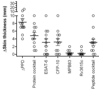
We next investigated the skin test contribution of the indi-vidual antigens present in the most promising protein cocktail comprised of ESAT-6, CFP-10, MPB83, and Rv3615c. We also extended our evaluation of synthetic peptide combinations by testing a cocktail of peptides derived from ESAT-6, CFP-10, MPB83, and Rv3615c. These reagents were tested in 12 reactor cattle that had previously demonstrated skin test responses to the protein cocktail in the previous two experiments. All nine animals that responded to any single antigen recognized CFP-10 (cutoff for positivity of ⬎1 mm), while seven, three, and two cattle responded to ESAT-6, MPB83, and Rv3615c, respectively (Fig. 6). However, the combined protein cocktail provided maximal responder frequency and reaction size, in-cluding the three animals that failed to respond to any of the individual proteins. Encouragingly, the peptide cocktail in-duced responses in 10/12 of these cattle, as shown in Fig. 6.
[image:4.585.333.502.70.250.2]Finally, the specificity of the most potent protein combina-tion of ESAT-6, CFP10, MPB83, and Rv3615c was evaluated for 14 new naïve cattle and 19 of the BCG neonatally vacci-nated calves previously tested. The protein cocktail did not
FIG. 5. Optimization of skin test response-inducing defined anti-gen combinations. Skin test responses induced by the SICCT test (PPD-B ⫺ PPD-A) and a selection of protein- and peptide-based antigen combinations were determined for cattle naturally exposed to
[image:4.585.44.283.91.151.2]M. bovis (n⫽ 13). Protein combinations of ESAT-6 (E6), CFP-10 (C10), MPB70, MPB83 (83), and/or Rv3615c were all tested at a concentration of 10 g per constituent protein. The ESAT-6 and CFP-10 (E6/C10) peptide cocktail comprised 21 peptides at a concen-tration of 10g per peptide. Responses measured at 72 h for each animal are represented by open circles, and the horizontal line pro-vides the mean (⫾SEM), with results expressed as the difference in skin thicknesses (mm) between the pre- and post-skin test readings. The statistical difference between responses induced by the indicated cocktails was determined by using ANOVA (*,P⬍0.05).
FIG. 6. Skin test response-inducing capacity of individual protein antigens. Skin test responses induced by the ESAT-6, CFP-10, MPB83, and Rv3615c proteins were determined either individually (10g) or in combination (10g per protein) for cattle naturally exposed toM. bovis(n⫽12). Additionally, responses to a cocktail of 25 peptides (10
[image:4.585.332.504.469.624.2]g per peptide) derived from ESAT-6, CFP-10, MPB83, and Rv3615c were measured in these cattle, along with SICCT skin test responses. Reactions measured at 72 h for each animal are represented by open circles, and the horizontal line provides the mean (⫾ SEM), with results expressed as the difference in skin thicknesses (mm) between the pre- and post-skin test readings.
TABLE 1. DIVA sensitivity and specificity ROC estimates for SICCT and defined protein cocktail-based skin tests
Diagnostic antigen Cutoff (mm)
Sensitivity
(%)a Specificity
(%)b Area under
the curve 95% CI d
SICCT (PPD-B-A) ⬎2 100 15.0 0.899 0.817–0.981 SICCT (PPD-B-A) ⬎4 94.1 40.0 0.899 0.817–0.981 SICCT (PPD-B-A) ⬎10 55.9 100 0.899 0.817–0.981 Protein cocktailc ⬎
1 73.6 100 0.882 0.791–0.974
aSensitivity determined by using naturally infected “reactor” cattle with dis-eased confirmed by SICCT (n⫽34).
bSpecificity determined by using disease-free BCG-vaccinated calves (n⫽20). cProtein cocktail comprised of 10g of ESAT-6, CFP-10, MPB70, and MPB83.
dCI, confidence interval.
on May 16, 2020 by guest
http://jcm.asm.org/
induce responses in any of these naïve or vaccinated cattle. While 16/19 of the BCG vaccinates still demonstrated a PPD-B-biased IFN-␥response at this time (PPD-B⫺PPD-A,⬎0.1 OD450[data not shown]), it should be noted that only 3/19 of
the BCG vaccinates still elicited a PPD-B-biased skin reaction with this test due to the extended period of nearly 12 months following neonatal vaccination at this time (data not shown).
DISCUSSION
Previous investigations on the use of defined bovine skin test reagents suggested that either immunomodulating reagents (27) or high antigen doses (ⱖ400g) (16) might be required to provide sensitive skin responses. It is therefore encouraging that in the current study, we demonstrated antigen-specific skin reactions for 78% of SICCT reactor cattle when using a protein-antigen combination of ESAT-6, CFP-10, MPB70, and MPB83. Importantly, this was achieved by using an adminis-tered concentration of 10g per protein, a dose that we con-sider to be realistic for practical field applications. Further-more, this skin test cocktail was highly specific, eliciting no response in either naïve or BCG-vaccinated calves. This is the first time that the potential of defined skin test antigens for DIVA has been demonstrated for cattle vaccinated with BCG or infected withM. bovis.
Assessment of thein vitrorecognition of the individual an-tigens in our initial protein cocktail showed that MPB70 and MPB83 had poor IFN-␥-inducing capacities in comparison with those of ESAT-6 and CFP-10. Despite their subdominant
in vitro immunogenicity in cattle, it is interesting that their inclusion in the skin test cocktail increased the skin test re-sponder frequency compared with the use of only ESAT-6 and CFP-10. In mice, it has been shown that during a delayed-type hypersensitivity (DTH) response, there must first be an early initiation phase that is required to recruit antigen-specific T cells, which then propagate the classical late-phase inflamma-tory response (21). Furthermore, the antigens that induce the initiation and effector stages can be different (17). Therefore, it may be possible that subdominant effector T-cell antigens such as MPB70 and MPB83 might still have a role in promoting reaction initiation, thereby helping to elicit a better response to dominant effector antigens such as ESAT-6 and CFP-10. Fur-ther studies investigating the nature of cellular events during the bovine DTH response could help refine and optimize fu-ture DTH-inducing reagents. However, in the absence of such knowledge, our data clearly demonstrate the importance of performing empiricalin vivostudies when evaluating skin test antigens.
Further optimization of the skin test protein cocktail dem-onstrated that the inclusion of MPB70 had no additional ben-efit over the use of MPB83. Since these antigens are closely related, sharing 61% sequence identity (7), it is likely that they share epitope specificities. Therefore, their use in combination may result in antigen redundancy, as previously observed forin vitro diagnostic assays (24). The addition of Rv3615c to a protein cocktail containing ESAT-6, CFP-10, and MPB83 did result in a significant improvement in the strength of the skin test responses and proved to be the most optimal antigen combination tested. The diagnostic potential of Rv3615c has only recently been identified, having been found to be
recog-nized by infected cattle missed by either ESAT-6 or CFP-10 (18). Our demonstration that it can also contribute to im-proved skin test responses without compromising specificity further confirms its diagnostic importance. Interestingly, eval-uation of the skin test response-inducing capacity of individual protein antigens demonstrated that Rv3615c had poor DTH-inducing potency. This observation again demonstrates the additive sensitivity benefits of using antigen combinations.
An important practical advantage of synthetic peptides com-pared to recombinant proteins as diagnostic antigens is that being chemically synthesized, quality control is more easily standardized. We have previously demonstrated the practical application of synthetic peptides as bovine TB reagents for DIVA using IFN-␥-based blood assays (25), and similarly, they have also been applied for human TB diagnosis (2, 11). How-ever, this is the first report demonstrating their potential as TB skin test antigens in cattle. Although a skin test cocktail con-taining ESAT-6, CFP-10, MPB83, and Rv3615c peptides lacked the absolute sensitivity and skin test response-inducing capacity of the equivalent protein cocktail, these important proof-of-principle data provide a basis for future optimization and improvement of a peptide-based skin test.
In considering why the results of previous ESAT-6-based cattle skin test studies looked less promising than our current data (16, 27), one explanation is likely to be our inclusion of CFP-10, which has not previously been evaluated as a bovine skin test antigen. Notably, skin test responses induced by the individual protein antigens showed CFP-10 to be the most potent defined antigen. Furthermore, the use of antigen com-binations demonstrated clear sensitivity benefits in the current study and will also have contributed to the improved responses compared with the previous responses using ESAT-6 alone (16, 27). When comparing cellular responses induced by re-combinant protein antigens, the presence of contamination endotoxin should also be considered. However, it is unlikely that immunomodulation from possible endotoxin contamina-tion can explain the potent responses to the protein antigens in the current study, since skin reactions were also induced by the synthetic peptides, which are endotoxin free.
Pollock et al. also demonstrated that PPD-induced bovine skin reactions were maximal at 72 h, while the response to ESAT-6 was often greatest at 96 h (16). Our data confirmed 72 h to be optimal for the measurement of the SICCT response (Fig. 1), and we also observed increases in the protein cocktail responder frequency when reactions were read at 96 or 120 h such that the responder frequency increased from 29/37 to 31/37 and then to 32/37 for the 72-, 96-, and 120-h time points, respectively. However, the increase in responder frequency was not significant (data not shown), and there was no increase in the magnitude of comparative responses. Furthermore, we also observed a response to both the protein cocktail and SICCT test in 2/20 naïve cattle when reactions were read at 120 h. The latter observation suggests the need for caution when considering the benefits of extending the period between administration and reading of the skin reaction for defined antigens. Additionally, since we observed highly specific reac-tions for the majority of animals at 72 h using our optimized defined cocktails, measuring responses at this time point has the practical benefit of allowing the option of testing defined
on May 16, 2020 by guest
http://jcm.asm.org/
antigens and PPD in parallel and then reading the reaction data on the same day.
The strong PPD-B-biased SICCT responses induced in our BCG-vaccinated calves confirmed that BCG sensitization com-promises the sensitivity of the test. Raising the SICCT test cutoff to⬎10 mm, similar to that in human medicine, restored test specificity but only with a significant loss of sensitivity (Table 1). This reaffirms the need for suitable reagents for DIVA such as those presented in the current study. As such, the importance of this study is that it demonstrates that com-binations of definedM. tuberculosiscomplex antigens can pro-vide practical and sensitive skin test-based diagnosis of bovine TB for DIVA. Additionally, the demonstration for the first time of the potential of synthetic peptides as skin test reagents for DIVA has practical relevance for TB diagnosis in both veterinary and clinical settings and would provide a promising basis for future test development.
ACKNOWLEDGMENTS
We are grateful for the support of the VLA Animal Support Unit for animal husbandry provision and to Animal Health (United Kingdom) officers for assisting with the recruitment of the naturally infected cattle. Recombinant ESAT-6 and CFP-10 proteins were kind gifts from Mahavir Singh, Lionex Diagnostics and Therapeutics GmbH, Germany.
This work was funded by the Department for Environment, Food and Rural Affairs (DEFRA), United Kingdom.
REFERENCES
1.Aggerbeck, H., and S. M. Madsen.2006. Safety of ESAT-6. Tuberculosis (Edinb.)86:363–373.
2.Arend, S. M., P. Andersen, K. E. van Meijgaarden, R. L. Skjot, Y. W. Subronto, J. T. van Dissel, and T. H. Ottenhoff.2000. Detection of active tuberculosis infection by T cell responses to early-secreted antigenic target 6-kDa protein and culture filtrate protein 10. J. Infect. Dis.181:1850–1854. 3.Arend, S. M., W. P. Franken, H. Aggerbeck, C. Prins, J. T. van Dissel, B. Thierry-Carstensen, P. N. Tingskov, K. Weldingh, and P. Andersen.2008. Double-blind randomized phase I study comparing rdESAT-6 to tuberculin as skin test reagent in the diagnosis of tuberculosis infection. Tuberculosis (Edinb.)88:249–261.
4.Berggren, S. A.1981. Field experiment with BCG vaccine in Malawi. Br. Vet. J.137:88–94.
5.Buddle, B. M., D. Keen, A. Thomson, G. Jowett, A. R. McCarthy, J. Heslop, G. W. De Lisle, J. L. Stanford, and F. E. Aldwell.1995. Protection of cattle from bovine tuberculosis by vaccination with BCG by the respiratory or subcutaneous route, but not by vaccination with killedMycobacterium vaccae. Res. Vet. Sci.59:10–16.
6.Buddle, B. M., N. A. Parlane, D. L. Keen, F. E. Aldwell, J. M. Pollock, K. Lightbody, and P. Andersen.1999. Differentiation betweenMycobacterium bovisBCG-vaccinated andM.bovis-infected cattle by using recombinant mycobacterial antigens. Clin. Diagn. Lab. Immunol.6:1–5.
7.Hewinson, R. G., D. P. Harris, A. Whelan, and W. P. Russell.1996. Secretion of the mycobacterial 19-kilodalton protein by Escherichia coli, a novel method for the purification of recombinant mycobacterial antigens. Clin. Diagn. Lab. Immunol.3:23–29.
8.Hogarth, P. J., R. G. Hewinson, and H. M. Vordermeier.2006. Development of vaccines against bovine tuberculosis. J. Pharm. Pharmacol.58:749–757. 9.Koch, R.1890. Weitere Mitteilungen uber ein Heilmittel gegen Tuberculose.
Dtsch. Med. Wochenschr.16:1029–1032.
10.Krebs, J. R.1997. Bovine tuberculosis in cattle and badgers. Ministry of Agriculture, Fisheries and Food, London, United Kingdom.
11.Lalvani, A., P. Nagvenkar, Z. Udwadia, A. A. Pathan, K. A. Wilkinson, J. S. Shastri, K. Ewer, A. V. Hill, A. Mehta, and C. Rodrigues.2001. Enumeration of T cells specific for RD1-encoded antigens suggests a high prevalence of latentMycobacterium tuberculosisinfection in healthy urban Indians. J. In-fect. Dis.183:469–477.
12.Mahairas, G. G., P. J. Sabo, M. J. Hickey, D. C. Singh, and C. K. Stover.
1996. Molecular analysis of genetic differences betweenMycobacterium bovis BCG and virulentM. bovis. J. Bacteriol.178:1274–1282.
13.Monaghan, M. L., M. L. Doherty, J. D. Collins, J. F. Kazda, and P. J. Quinn.
1994. The tuberculin test. Vet. Microbiol.40:111–124.
14.Plum, N.1931. A study of avian tuberculosis in cattle. Cornell Vet.21:68–76. 15.Pollock, J. M., and P. Andersen.1997. The potential of the ESAT-6 antigen secreted by virulent mycobacteria for specific diagnosis of tuberculosis. J. In-fect. Dis.175:1251–1254.
16.Pollock, J. M., J. McNair, H. Bassett, J. P. Cassidy, E. Costello, H. Agger-beck, I. Rosenkrands, and P. Andersen.2003. Specific delayed-type hyper-sensitivity responses to ESAT-6 identify tuberculosis-infected cattle. J. Clin. Microbiol.41:1856–1860.
17.Ptak, W., M. Bereta, M. Ptak, and P. W. Askenase.1986. Isotype-like sup-pression of T cell-mediated immunityin vivo. II. Suppression of the early component of contact sensitivity by a Ly-2⫹T cell-derived suppressor factor that binds to contact sensitivity-initiating, antigen-specific, Ly-1⫹T cell-derived factors that are of different antigen specificities. J. Immunol.136:
1564–1570.
18.Sidders, B., C. Pirson, P. J. Hogarth, R. G. Hewinson, N. G. Stoker, H. M. Vordermeier, and K. Ewer.2008. Screening of highly expressed mycobacte-rial genes identifies Rv3615c as a useful differential diagnostic antigen for the Mycobacterium tuberculosiscomplex. Infect. Immun.76:3932–3939. 19.Snider, D. E., Jr. 1982. The tuberculin skin test. Am. Rev. Respir. Dis.
125:108–118.
20.Stenius, R.1938. Differentiation by tuberculin testing of infection in cattle due to the human, bovine and avian types of tubercle bacilli. Vet. Rec.
50:633–637.
21.van Loveren, H., R. Meade, and P. W. Askenase.1983. An early component of delayed-type hypersensitivity mediated by T cells and mast cells. J. Exp. Med.157:1604–1617.
22.van Pinxteren, L. A., P. Ravn, E. M. Agger, J. Pollock, and P. Andersen.
2000. Diagnosis of tuberculosis based on the two specific antigens ESAT-6 and CFP10. Clin. Diagn. Lab. Immunol.7:155–160.
23.Vordermeier, H. M., M. A. Chambers, P. J. Cockle, A. O. Whelan, J. Sim-mons, and R. G. Hewinson.2002. Correlation of ESAT-6-specific gamma interferon production with pathology in cattle followingMycobacterium bovis BCG vaccination against experimental bovine tuberculosis. Infect. Immun.
70:3026–3032.
24.Vordermeier, H. M., P. C. Cockle, A. Whelan, S. Rhodes, N. Palmer, D. Bakker, and R. G. Hewinson.1999. Development of diagnostic reagents to differentiate betweenMycobacterium bovisBCG vaccination andM. bovis infection in cattle. Clin. Diagn. Lab. Immunol.6:675–682.
25.Vordermeier, H. M., A. Whelan, P. J. Cockle, L. Farrant, N. Palmer, and R. G. Hewinson.2001. Use of synthetic peptides derived from the antigens esat-6 and cfp-10 for differential diagnosis of bovine tuberculosis in cattle. Clin. Diagn. Lab. Immunol.8:571–578.
26.Waddington, F. G., and D. C. Ellwood.1972. An experiment to challenge the resistance to tuberculosis in B.C.G. vaccinated cattle in Malawi. Br. Vet. J.
128:541–552.
27.Whelan, A. O., J. C. Hope, C. J. Howard, D. Clifford, R. G. Hewinson, and H. M. Vordermeier.2003. Modulation of the bovine delayed-type hypersen-sitivity responses to defined mycobacterial antigens by a synthetic bacterial lipopeptide. Infect. Immun.71:6420–6425.
28.Wu, X., L. Zhang, J. Zhang, C. Zhang, L. Zhu, and Y. Shi.2008. Recombi-nant early secreted antigen target 6 protein as a skin test antigen for the specific detection ofMycobacterium tuberculosisinfection. Clin. Exp. Immu-nol.152:81–87.