Testing in Developing Countries
Paula Gonzalez,a,bBernal Cortes,aWim Quint,dAimée R. Kreimer,cCarolina Porras,aAna Cecilia Rodríguez,aSilvia Jimenez,a Rolando Herrero,bLinda Struijk,dAllan Hildesheim,cand Willem Melcherse
Proyecto Epidemiológico Guanacaste, Fundación INCIENSA, Guanacaste, Costa Ricaa
; Prevention and Implementation Group, International Agency for Research on Cancer, Lyon, Franceb
; Division of Cancer Epidemiology and Genetics, National Cancer Institute, Rockville, Maryland, USAc
; DDL Diagnostic Laboratory, Rijswijk, Netherlandsd
; and Department of Medical Microbiology, Radboud University Nijmegen Medical Centre, Nijmegen, Netherlandse
Liquid-based methods for the collection, transportation, and storage of cervical cells are cumbersome and expensive and involve
laborious DNA extraction. An FTA cartridge is a solid carrier device, easier to handle and allowing simple DNA elution for
hu-man papillomavirus (HPV) testing. HPV-DNA results from cervical specimens collected in PreservCyt medium (Hologic, Inc.)
and the indicating FTA elute cartridge were compared in an area where transportation and storage may affect the performance of
the test. Cervical cells from 319 young adult women enrolled in the Costa Rica Vaccine Trial were collected by a nurse using a
Cervex brush (Roberts), which was placed on the FTA cartridge and subsequently rinsed in 20 ml of PreservCyt medium. Two
0.5-ml PreservCyt aliquots were frozen for HPV-PCR testing; the FTA cartridges were kept at room temperature. HPV-DNA
de-tection and typing was performed using SPF
10PCR/DEIA (DNA enzyme immunoassay detection of amplimers)/LiPA
25system.
The percent agreement, agreement among positives, and kappas were estimated. Positivity was higher for FTA compared to
Pre-servCyt specimens (54.5% versus 45.8%,
P
<
0.001). For oncogenic types, the overall agreement was 0.92, the agreement between
positives was 0.74, and the kappa was 0.79. For individual HPV types, the overall agreement ranged from 0.97 to 1.00. We did not
observe reduced cytology adequacy when specimen collection for cytology was preceded by FTA collection for HPV testing.
HPV-DNA detection from FTA cartridges is broadly comparable to detection from PC medium. The higher HPV detection
ob-served for FTA-collected specimens should be explored further. FTA cartridges could provide a simpler and more cost-effective
method for cervical cell collection, storage, and transportation for HPV-DNA detection in research settings in developing
countries.
P
ersistent infection with one of approximately 13 carcinogenic
human papillomaviruses (HPVs) is a necessary cause for the
development of cervical cancer (
14
,
23
). Detection of some or all
of these HPV types has been shown to be useful for cervical cancer
screening (
1
,
15
,
26
–
28
).
In clinical trials (including evaluation of current and future
HPV vaccines) and epidemiological studies, cervical cells for HPV
DNA detection are usually collected and preserved in liquid-based
transport medium, which in some cases is used also for cytology
slide preparation, allowing the collection of only one sample for
both tests (
7
,
12
,
13
,
24
,
25
).
However, these medium samples can be flammable and
re-quire stable transport and storage temperatures, which are
diffi-cult and expensive to provide in developing and tropical
coun-tries. If samples are tested in a different country, as is usually the
case for large research studies conducted in developing countries,
the exportation of such samples must fulfill international
regula-tions regarding the transport of hazardous samples (i.e., according
to the 53rd edition of IATA’s DGR, specimens collected in
PreservCyt [Hologic, Inc.] or SurePath [Becton, Dickinson and
Company] best fulfill the requirements of Flammable Liquids
Class 3 and Biological Substance Category B [UN 3373]) (
31
).
Moreover, these liquid-based samples require expensive and
labo-rious DNA extraction procedures susceptible to
cross-contamina-tion, especially when manual extraction of DNA is performed.
Solid carriers for DNA transportation and storage, which
con-sist of dried fluid spots on filter paper, have been used widely for
postnatal screening of certain congenital disorders and diseases;
they have also been used in studies to detect other viruses such as
measles virus, hepatitis B virus, and HIV, as well as genetic
re-search. These devices are easy to store and transport since they dry
quickly, they are compact and lightweight, and they do not require
a controlled temperature (
2
–
4
,
6
,
8
,
11
,
16
,
19
,
21
,
22
). The FTA
cartridge (GEHC-Whatman) is a paper-based system that
immo-bilizes and staimmo-bilizes nucleic acids from fresh samples applied. The
paper is impregnated with a patented chemical formula that lyses
cells and denatures proteins upon contact, which makes samples
collected in FTA cartridges neither hazardous nor infectious as the
viruses are denaturized upon application. The indicating FTA
elute cartridge also contains an indicating dye that changes color
when a sample is applied showing the location of the sample.
Furthermore, it allows DNA elution by a simple method using
only water and heat with easy reduction of the risk of
cross-con-tamination between samples to minimal by simply applying the
puncher two to three times on clean paper and wiping it off with
95% ethanol between samples (
9
,
30
). Finally, since not all of the
material is used, the residual sample can be stored.
Four studies (two in the Netherlands and two in Sweden) have
found good agreement for HPV DNA detection between cervical
Received28 June 2012 Returned for modification10 August 2012
Accepted10 September 2012
Published ahead of print19 September 2012
Address correspondence to Paula Gonzalez, [email protected]. Copyright © 2012, American Society for Microbiology. All Rights Reserved.
doi:10.1128/JCM.01698-12
on May 16, 2020 by guest
http://jcm.asm.org/
cells collected in this FTA cartridge and liquid-based medium or
frozen dry samples. Lenselink et al. compared self-collected
sam-ples at home placed in both media from 51 women aged 18 to 29
years with an agreement of 100%. These researchers also found
93% agreement between FTA self-collected and liquid-based
phy-sician-collected samples among 45 women aged 23 to 51 years
attending a gynecology clinic (
20
). Gustavsson et al. evaluated
cervical samples from 50 women visiting a dermatology and
vene-reology clinic, from whom physician-collected samples were
ob-tained using a cytobrush; each sample was first applied to FTA and
then frozen dry, and the agreement was 94% (
9
).
More recently Gustavsson et al. compared self-collected with
physician-collected cervical samples both placed in FTA
car-tridges among 50 women attending a gynecology clinic and found
and an agreement of 88% (
10
). De Bie et al. estimated agreements
of 91% for PCR testing and 77.3% for Hybrid Capture 2 (HC2;
Qiagen) comparing two physician-collected cervical brushes
placed on the liquid-based medium and on FTA cartridges among
88 women attending a gynecology clinic (
5
).
Given that the use of this cartridge is very promising, especially
in developing countries where transportation and storage is
ex-pensive and complicated and for multicentric studies with a
cen-tralized laboratory, we conducted a study within our HPV16 and
HPV18 (HPV16/18) vaccine trial in Costa Rica to evaluate the
performance of HPV DNA detection by PCR and genotyping on
samples collected on the indicating FTA elute cartridge compared
to samples collected in PreservCyt transport medium in the
con-text of research studies. To our knowledge, this is the first study of
cervical HPV DNA detection on FTA cartridges in a developing
country and is the largest comparing FTA and liquid-based
me-dium.
MATERIALS AND METHODS
Study population.Samples included in this analysis were collected from participants in the Costa Rica Vaccine Trial (CVT), who completed a follow-up screening visit or a colposcopy visit from 24 to 27 November 2009 and from 11 January to 21 May 2010. The methods of the CVT are described in detail elsewhere (12). Briefly, it is a double blind, controlled, randomized, phase III study designed to evaluate the efficacy of the HPV16/18 vaccine (Cervarix; GlaxoSmithKline) for the prevention of HPV16/18 persistent infection and associated cervical lesions (CIN2⫹). The study enrolled 7,466 women residing in the province of Guanacaste and nearby areas of Puntarenas, Costa Rica, between 2004 and 2005. The main eligibility requirements for enrollment were as follows: age, 18 to 25 years; planned residence in the area for the 6 months following enroll-ment; good general health; and neither pregnant nor breast-feeding. The trial was approved by the U.S. National Cancer Institute IRB and the INCIENSA (Instituto Costarricense de Investigación y Enseñanza en Nu-trición y Salud) IRB in Costa Rica, and all of the women provided in-formed consent.
The women were assigned to receive three doses of Cervarix or hepa-titis A virus vaccine as a control vaccine. After the vaccination period, the women were monitored once a year for 4 years. Women with mild cervical abnormalities were monitored every 6 months, and those with high-grade cervical abnormalities or persistent low-grade abnormalities were re-ferred for colposcopy.
At each study visit, one of five trained study nurses performed a pelvic exam on sexually experienced women. Clinician-collected exfoliated cells were obtained using the Cervex brush (Roberts) by inserting the central bristles into the endocervical canal until the external bristles bent against the ectocervix; while the pressure was maintained, the brush was rotated five times in a clockwise direction, and then the brush was removed and
rinsed vigorously in a vial with 20 ml of PreservCyt medium and kept at 15 to 25°C.
Our target number of participants for this substudy was 300, and 319 were included: 218 samples were collected from women attending a reg-ular 1-year screening visit (prevalences of 41.6% for any HPV and 19.3% for any oncogenic HPV), and 101 samples were collected from women attending a colposcopy visit (prevalences of 62.4% for any HPV and 38.1% for any oncogenic HPV). For these participants, immediately fol-lowing cervical cell collection (detailed above), instead of the Cervex brush being placed directly into the PreservCyt vial, it was first applied to the indicating FTA elute cartridge, and then the Cervex brush was vigor-ously rinsed in the PreservCyt vial, as indicated above. This was done to avoid using two Cervex brushes that could induce bleeding and obscure the colposcopy evaluation and also because, for use in a clinical or re-search setting, this would be the most plausible scenario. FTA cartridges were kept at room temperature, while the PreservCyt vial was kept in a cooler at 18 to 25°C. At the end of the day, both samples were sent to a local repository.
According to routine standard trial procedures, within 1 week of col-lection, two 0.5-ml aliquots for HPV PCR testing were prepared from the PreservCyt sample and frozen in liquid nitrogen (⫺150°C). The remain-ing PreservCyt sample was used to produce a cytology slide, and the re-sidual volume was used for HPV testing by HC2 for all women attending regular screening visits and for women attending colposcopy visits only if it was considered necessary to define follow-up or treatment. FTA car-tridges were stored in a box at room temperature until they were packed in individual bags and sent to the DDL Diagnostic Laboratory (Rijswijk, Netherlands) by courier for PCR HPV testing.
DNA isolation from PreservCyt specimen.Total DNA was isolated from 200l of one of the 0.5-ml PreservCyt aliquots by using the MagNA Pure LC isolation procedure (Roche Diagnostics GmbH/Roche Applied Science, Mannheim, Germany) and a total nucleic acid isolation kit (Roche Diagnostics GmbH/Roche Molecular Biochemicals, Mannheim Germany), as described by the manufacturer. DNA was eluted in 100l. Each DNA extraction run contained positive and negative controls to monitor the DNA isolation procedure.
DNA isolation from the indicating FTA elute cartridge.The FTA cartridges were punched four times using a perforator specifically de-signed for the FTA cartridges (a 3-mm Harris Uni-Core device; Whatman). For each FTA cartridge, a new sterile perforator was used to avoid any cross-contamination. The FTA elute matrix is chemically treated with proprietary reagents that lyse cells upon contact causing the release of nucleic acids. DNA was recovered from the FTA elute matrix through a simplified elution process using heat and water. The four punches were transferred into a 1.5-ml microfuge tube, and 1,500l of sterile water was added to the punches and immediately pulse vortexed three times for a total of 5 s. After centrifugation, the water was removed with a sterile fine tip pipette. An 70-l portion of sterile water was added to the punches, and the tube was transferred to a heating block at 95°C for 30 min. At the end of the incubation period, the sample was removed from the heating block and pulse vortexed⬃6 times. It was additionally centri-fuged for 30 s, and the eluted DNA was transferred into a new microcen-trifuge tube. Inhibitory components, such as hemoglobin, are retained in the FTA elute matrix. The eluted DNA was stored at⫺20°C. Finally, 10l of the eluate was used for PCR.
HPV testing and genotyping.Broad-spectrum PCR-based HPV DNA testing of DNA isolated from FTA cartridges and PreservCyt aliquots was performed at the DDL Diagnostic Laboratory by researchers blinded from the results of the comparative sample, the HC2, and the cytology, using the SPF10PCR primer system and a DNA enzyme immunoassay detection
of amplimers (DEIA), followed by genotyping using the LiPA25(line
probe assay, version 1 [Labo Biomedical Products, Rijswijk, Nether-lands]) system as described previously (17,18). LiPA25detects 25 HPV
genotypes, including HPV16 and HPV18, but to ensure that HPV16 and HPV18 infections were not missed, all specimens that were SPF10PCR/
on May 16, 2020 by guest
http://jcm.asm.org/
DEIA positive for HPV DNA but negative for HPV16 or HPV18 were also tested for the presence of HPV16 or HPV18 by using type-specific primers by the TS16 and TS18 DEIA system (29).
Statistical analysis.The HPV DNA results obtained from samples collected using both media were compared by calculating the percent overall agreement, the percent agreement between positives, the kappa value, and the weighted kappa value when applicable. The differences were evaluated for statistical significance using the exact McNemar2test.
We compared detection of any HPV, any oncogenic HPV (HPV16, -18, -31, -33, -35, -39, -45, -51, -52, -56, -58, -66, and -68/73) and individual HPV genotypes.
We stratified the analysis by visit type (regular screening versus col-poscopy visit) to evaluate the performance of the FTA in higher- and lower-HPV-prevalence settings. To evaluate the clinical significance of discordant results, we also conducted analyses comparing PC and FTA stratified by HC2 results. Given that among the 319 samples analyzed there were only 3 high-grade squamous intraepithelial lesion (HSIL) and 36 low-grade squamous intraepithelial lesion (LSIL) cytology results, we could not make definitive conclusions from the results stratified by cytol-ogy diagnosis. We also compared the number of HPV types detected in each sample to evaluate the performance of the FTA cartridge when mul-tiple infections are present.
Finally, each sample was assigned to one of four cervical cancer risk categories according to the HPV types detected by each collection me-dium and compared. The four risk categories were defined as follows: (i) HPV16/18 positive; (ii) oncogenic HPV (oncogenic HPV types 16, 18, 31, 33, 35, 39, 45, 51, 52, 56, 58, 66, and 68/73) positive but HPV16/18 nega-tive; (iii) nononcogenic HPV (nononcogenic HPV types 6, 11, 34, 40, 42, 43, 44, 53, 54, 59, 70, 74, and uncharacterized) positive but negative for all oncogenic types, including uncharacterized types; and (iv) HPV negative. We conducted separate analyses including and excluding results from the HPV16/18 type-specific PCR with comparable results (data not shown).
To investigate whether the placement of the Cervex brush on the FTA cartridge before rinsing it in the PreservCyt medium could decrease the amount of cells placed in the PreservCyt medium, thereby affecting the cytology slide adequacy (which would have relevant implications for clin-ical management of study participants), we compared the percentages of cytologies considered inadequate for evaluation due to low cellularity among the 319 women included in this analysis to the those for the other participants from CVT whose samples were collected in the same time period and by the same group of nurses but not placed on the FTA (n⫽
262).
Analyses were performed by the programming and statistical staff at the trial’s data management center (Information Management Services,
Inc., Silver Spring, MD) under contract to the National Cancer Institute (NCI), supervised by the study investigators, and handled according to standard operating procedures.
RESULTS
Study population.
Cervical samples were collected in FTA and PC
from 319 women 22 to 31 years of age (mean, 25.0 years) and
included in the present analysis. A total of 29.5% of these women
had completed elementary school, and 11.9% had attended a
uni-versity. Most of the women were married or lived like married
(58.9%), 33.2% were single, and the remaining women were
wid-owed, separated, or divorced. A total of 47.3% had never been
pregnant, while 42.0% had been pregnant once, and 10.7% had
been pregnant two or more times.
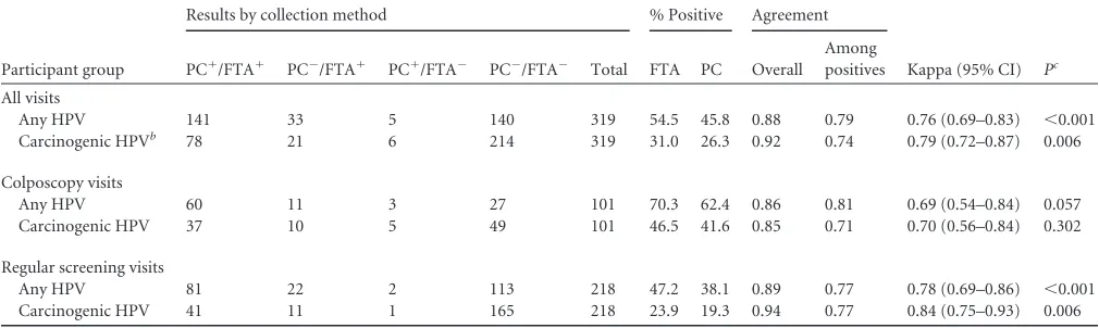
Any HPV and any oncogenic HPV detection.
Detection of any
HPV type and any oncogenic HPV type by the SPF
10LiPA
25in the
samples collected using the two different transport media among
the 319 samples were compared (
Table 1
). The overall agreements
were 0.88 for any HPV and 0.92 for any oncogenic HPV, the
agree-ments between positives were 0.79 and 0.74, respectively, and the
kappa values were 0.76 and 0.79. There was more HPV detection
of any type in the samples collected in FTA medium than in the
samples collected in PreservCyt (54.5% versus 45.8%, McNemar
P
⬍
0.001); there was also more detection of oncogenic HPV types
in FTA samples (31.0% versus 26.3%, McNemar
P
⫽
0.006).
We stratified the analysis according to the visit type (regular
screening visits versus colposcopy visits) and, although more
on-cogenic HPV was detected in FTA samples from women attending
both types of visits, this difference was significant only among
regular screening samples (McNemar
P
⫽
0.006 for screening and
McNemar
P
⫽
0.302 for colposcopy visits). (
Table 1
).
[image:3.585.40.545.78.229.2]Detection of any oncogenic HPV in samples collected in the
two media was stratified by HC2 positivity (
Table 2
). Among
HC2-positive samples, agreement between positives was 0.96
(kappa
⫽
0.93), while among HC2 negative samples agreement
between positives was 0.54 (kappa
⫽
0.66). Samples collected in
FTA were more likely to test positive for carcinogenic HPV than
PreservCyt samples when HC2 was negative (16.2% versus 10.3%,
respectively;
P
⫽
0.004) but not when HC2 was positive (69.2% in
both types of samples).
TABLE 1Comparison of HPV detection results in samples collected in liquid-based medium and solid carrier devices among CVT participantsa
Participant group
Results by collection method % Positive Agreement
Kappa (95% CI) Pc PC⫹/FTA⫹ PC⫺/FTA⫹ PC⫹/FTA⫺ PC⫺/FTA⫺ Total FTA PC Overall
Among positives
All visits
Any HPV 141 33 5 140 319 54.5 45.8 0.88 0.79 0.76 (0.69–0.83) ⬍0.001 Carcinogenic HPVb 78 21 6 214 319 31.0 26.3 0.92 0.74 0.79 (0.72–0.87) 0.006
Colposcopy visits
Any HPV 60 11 3 27 101 70.3 62.4 0.86 0.81 0.69 (0.54–0.84) 0.057 Carcinogenic HPV 37 10 5 49 101 46.5 41.6 0.85 0.71 0.70 (0.56–0.84) 0.302
Regular screening visits
Any HPV 81 22 2 113 218 47.2 38.1 0.89 0.77 0.78 (0.69–0.86) ⬍0.001 Carcinogenic HPV 41 11 1 165 218 23.9 19.3 0.94 0.77 0.84 (0.75–0.93) 0.006
aPC, PreservCyt, liquid-based medium; FTA, solid carrier device.
b
That is, HPV16, -18, -31, -33, -35, -39, -45, -51, -52, -56, -58, -66, and -68/73. cCalculated by using the exact McNemar2test.
on May 16, 2020 by guest
http://jcm.asm.org/
Among the three women with HSIL, the concordance of the
FTA and the PreservCyt results was 1.0; one woman was positive
for HPV16, one was positive for HPV52, and one was positive for
HPV58. Among the 36 women with LSIL (including ASC_US
HPV
⫹) cytology results, the agreement between positives was 0.87
(kappa
⫽
0.83) compared to 0.73 (kappa
⫽
0.80) among women
with normal cytology. FTA samples were more likely to test
onco-genic HPV positive than PC samples (26.2 versus 21.7,
respec-tively;
P
⫽
0.012) only among women with normal cytology
re-sults (
Table 2
).
Type-specific HPV detection.
The study was powered for
overall HPV detection, but we did not observe statistically
signif-icant differences between collection methods in the prevalence of
detection for any HPV type tested. The prevalence of individual
HPV types was higher in FTA samples for all HPV types except for
HPV43, HPV58, and HPV68/73, for which the prevalence was
identical, and HPV54 and HPV74, for which the prevalence in
PreservCyt samples was higher. The prevalence of
uncharacter-ized HPV types (LiPA
25positives and DEIA negatives) was
[image:4.585.40.545.88.199.2]statis-tically higher in FTA samples (
Table 3
).
TABLE 2Comparison of carcinogenic HPV detection results in samples collected in liquid-based medium and solid carrier devices among CVT participants by HPV HC2 and cytology resultsa
Analysis
No. of samples % Positive Agreement
Kappa (95% CI) Pb PC⫹/FTA⫹ PC⫺/FTA⫹ PC⫹/FTA⫺ PC⫺/FTA⫺ Total FTA PC Overall
Among positives
HPV result by HC2
Positive 44 1 1 19 65 69.2 69.2 0.97 0.96 0.93 (0.83–1.0) 1 Negative 19 14 2 169 204 16.2 10.3 0.92 0.54 0.66 (0.51–0.81) 0.004 Cytology result
HSIL 3 0 0 0 3 100.0 100.0 1.0 1.0 — —
LSIL 20 1 2 13 36 58.3 61.1 0.92 0.87 0.83 (0.64–1.0) 1 Normal 193 16 4 54 267 26.2 21.7 0.93 0.73 0.80 (0.71–0.88) 0.012
aThe carcinogenic HPV types included HPV16, -18, -31, -33, -35, -39, -45, -51, -52, -56, -58, -66, and -68/73. PC, PreservCyt, liquid-based medium; FTA, solid carrier device; HC2,
HPV Hybrid Capture 2 test.
bCalculated by using the exact McNemar2test.
TABLE 3Comparison of results of individual HPV genotype detection in samples collected in liquid-based medium and solid carrier devices among CVT participantsa
HPV type
No. of samples % Positive Agreement
Kappa (95% CI) Pb PC⫹/FTA⫹ PC⫺/FTA⫹ PC⫹/FTA⫺ PC⫺/FTA⫺ FTA PC Overall Among positives
6 4 2 0 313 1.9 1.3 0.99 0.67 0.80 (0.52–1.0) 0.500 11 2 1 0 316 0.9 0.6 1.00 0.67 0.80 (0.41–1.0) 1.000 16 17 4 1 297 6.6 5.6 0.98 0.77 0.86 (0.75–0.98) 0.375 18 4 1 1 313 1.6 1.6 0.99 0.67 0.80 (0.52–1.0) 1.000 31 5 4 2 308 2.8 2.2 0.98 0.45 0.62 (0.33–0.90) 0.686 33 0 2 1 316 0.6 0.3 0.99 0.00 0.00 (⫺0.01–0.00) 1.000 34 1 2 1 315 0.9 0.6 0.99 0.25 0.40 (⫺0.15–0.94) 1.000 35 6 2 0 311 2.5 1.9 0.99 0.75 0.85 (0.65–1.0) 0.500 39 4 3 1 311 2.2 1.6 0.99 0.50 0.66 (0.35–0.97) 0.625 40 2 2 1 314 1.3 0.9 0.99 0.40 0.57 (0.13–1.0) 1.000 42 1 1 0 317 0.6 0.3 1.00 0.50 0.67 (0.05–1.0) 1.000 43 3 1 1 314 1.3 1.3 0.99 0.60 0.75 (0.41–1.0) 1.000 44 4 4 0 311 2.5 1.3 0.99 0.50 0.66 (0.35–0.97) 0.125 45 1 2 1 315 0.9 0.6 0.99 0.25 0.40 (⫺0.15–0.94) 1.000 51 12 8 3 296 6.3 4.7 0.97 0.52 0.67 (0.48–0.85) 0.227 52 17 7 4 291 7.5 6.6 0.97 0.61 0.74 (0.59–0.86) 0.549 53 10 2 0 307 3.8 3.1 0.99 0.83 0.91 (0.78–1.0) 0.500 54 3 3 4 309 1.9 2.2 0.98 0.30 0.45 (0.11–0.79) 1.000 56 8 3 0 308 3.4 2.5 0.99 0.73 0.84 (0.66–1.0) 0.250 58 7 1 1 310 2.5 2.5 0.99 0.78 0.87 (0.70–1.0) 1.000 59 5 3 1 310 2.5 1.9 0.99 0.56 0.71 (0.44–0.98) 0.625 66 13 5 2 299 5.6 4.7 0.98 0.65 0.78 (0.62–0.94) 0.453 68/73 7 5 5 302 3.8 3.8 0.97 0.41 0.57 (0.33–0.81) 1.000 70 7 4 0 308 3.4 2.2 0.99 0.64 0.77 (0.56–0.99) 0.125 74 7 1 2 309 2.5 2.8 0.99 0.70 0.82 (0.62–1.0) 1.000 UNCc 23 18 7 271 12.9 9.4 0.92 0.48 0.61 (0.46–0.75) 0.043
aPC, PreservCyt, liquid-based medium; FTA, solid carrier device.
b
Calculated by using the exact McNemar2
test. cUNC, uncharacterized, SPF10 positive but LiPA negative.
on May 16, 2020 by guest
http://jcm.asm.org/
[image:4.585.41.548.405.697.2]Number of oncogenic HPV types detected.
The agreement
between positives between the two collection media for the
num-ber of oncogenic HPV types detected in each sample was 0.62, with
a weighted kappa of 0.75 (95% confidence interval [95% CI]
⫽
0.68 to 0.83) (
Table 4
). Detection of two or more oncogenic HPV
types in one sample was more common in samples collected in
FTA than samples collected in PreservCyt (9.1% versus 6.9%).
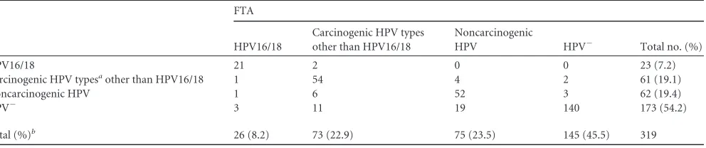
HPV detection by risk categories.
We categorized the results
according to the risk (i.e., HPV16/18, oncogenic other than
HPV16/18, nononcogenic, and HPV negative); the agreement
be-tween positives was 0.71, and the weighted kappa was 0.79 (95%
CI
⫽
0.73 to 0.85). Samples collected in FTA tended to be
catego-rized in higher-risk categories than samples collected in PreservCyt.
Thus, 8.2 and 22.9% of the FTA samples were classified as
HPV16/18 and other oncogenic HPVs, respectively, whereas 7.2
and 19.1% of the PreservCyt samples were classified in these
cat-egories (
P
⬍
0.001 exact-symmetry
2test) (
Table 5
). When we
examined only samples positive by both collection media, there
was no difference in the proportion of samples assigned to the
HPV16/18 positive or the other oncogenic HPV categories (16.3
and 44.0%, respectively, for FTA versus 16.3 and 41.8% for
liquid-based) (
P
⫽
0.82) (data not shown in the tables).
We stratified the analysis by type of visit (regular screening
visits versus colposcopy visits), and the pattern was similar, while
the agreement and the kappa were higher at regular visits (the
agreement between positives was 0.73 and the weighted kappa was
0.80 for regular visits versus 0.68 and 0.73, respectively, for
col-poscopy visits). Although more samples collected in FTA were
classified in the higher-risk categories than samples collected in
PreservCyt for both types of visits, the difference was statistically
significant only in specimens from regular screening visits (
P
⬍
0.001 versus 0.17; data not shown).
Unsatisfactory cytologies.
We evaluated the proportions of
inadequate cytologies by visit type. The difference between the two
collection media was higher for colposcopy visits than for regular
visits, but none of them was statistically significant (i.e., the
per-centage of inadequate cytologies at regular visits was 1.8% when
FTA collection preceded the liquid-based cytology (LBC) and
1.4% if no FTA collection was performed [
P
⫽
0.77], and the
percentage of inadequate cytologies at colposcopy visits was 8.9%
for FTA samples and 3.5 for liquid-based samples [
P
⫽
0.13]).
DISCUSSION
We compared HPV detection from clinician-collected cervical
cells placed in a new solid carrier device (i.e., the indicating FTA
elute cartridge) to that observed in clinician-collected cervical
cells placed in a conventional liquid-based medium (PreservCyt).
The agreement for HPV detection using FTA and PC was excellent
and was observed in both a lower-risk screening population and a
higher-risk colposcopy group. Our findings suggest that almost
no infections would be missed by using the FTA instead of the
conventional liquid-based medium and indicate that the use of
this simplified and less-expensive solid-based carrier (very
impor-tant characteristics for developing countries) for HPV testing
might be practical in the future without the need to sacrifice HPV
detection sensitivity. An added advantage of using the solid-phase
FTA device over existing liquid-based media is the simplified
DNA extraction (
20
,
30
).
Our results are consistent with those from smaller studies (
n
⬍
100) conducted in Nordic countries that observed a high overall
agreement for PCR HPV detection and genotyping between
cer-vical cells collected using a liquid-based medium and specimens
collected using the FTA cartridges both among self-collected and
clinician-collected samples (
5
,
9
,
10
,
20
).
[image:5.585.50.546.604.708.2]We observed that specimens placed on FTA were more likely
than those placed on PreservCyt to test positive for HPV (i.e.,
higher overall prevalence) and to be classified as positive for
HPV16/18 or other carcinogenic HPV types. The fact that the
differences observed were significant only among women
attend-ing regular screenattend-ing visits and among women negative by HC2
but not among higher-risk women attending a colposcopy clinic
and those with an HC2-positive result suggests that the FTA
col-lection method might have reduced specificity for the detection of
TABLE 4Comparison of number of oncogenic HPV types detected in samples collected in liquid-based medium and solid carrier devices among CVT participantsa
No. of oncogenic HPV types detected in PC samples
No. of oncogenic HPV types detected in FTA samples
0 1 2 3⫹ Total
0 214 17 4 0 235
1 4 50 7 1 62
2 1 3 9 2 15
3⫹ 1 0 0 6 7
Total 220 70 20 9 319
aThe carcinogenic HPV types included HPV16, -18, -31, -33, -35, -39, -45, -51, -52,
-56, -58, -66, and -68/73. The overall agreement was 0.87, the agreement between positives in both media was 0.62, and the weighted kappa (95% CI) was 0.75 (0.68 to 0.83).
TABLE 5Comparison of HPV test results in samples collected in liquid-based medium and solid carrier devices in CVT participants, categorized by HPV cancer risk
PC
FTA
HPV16/18
Carcinogenic HPV types other than HPV16/18
Noncarcinogenic
HPV HPV⫺ Total no. (%)
HPV16/18 21 2 0 0 23 (7.2)
Carcinogenic HPV typesaother than HPV16/18 1 54 4 2 61 (19.1)
Noncarcinogenic HPV 1 6 52 3 62 (19.4)
HPV⫺ 3 11 19 140 173 (54.2)
Total (%)b 26 (8.2) 73 (22.9) 75 (23.5) 145 (45.5) 319
a
The carcinogenic HPV types included HPV16, -18, -31, -33, -35, -39, -45, -51, -52, -56, -58, -66, and -68/73.
bOverall agreement was 0.84, the agreement between positives in both media was 0.71, and the weighted kappa (95% CI) was 0.79 (0.73 to 0.85).
on May 16, 2020 by guest
http://jcm.asm.org/
clinically relevant underlying disease. This might be due to an
increased ability to detect low-viral-load infections using the FTA
compared to the liquid-based collection method due to the fact
that FTA samples are more concentrated, leading to a greater
pro-portion of the total sample being tested, but this should be further
explored.
One theoretical concern with the use of the FTA device is the
need to place the cervical cells on the FTA cartridge prior to
pro-ducing the sample for cytological evaluation (slide for
conven-tional cytology or liquid-based medium); the possibility exists for
reduced cellularity and increased inadequacy of the specimen
col-lected for cytological evaluation. However, we did not observe
evidence of reduced cytology specimen adequacy when specimen
collection for cytology was preceded by FTA collection for HPV
testing.
Our analysis is limited by our inability to compare the FTA and
liquid-based collection methods within categories of cytological
diagnoses and, more specifically, to evaluate the performance of
FTA among women with an HSIL diagnosis. Furthermore, since
our study was conducted among young adult women who have a
high prevalence of HPV infection, it is uncertain whether our
findings can be generalized to older populations with reduced
rates of HPV infection.
In our study, the sample was applied to the FTA before being
placed in the liquid-based medium. Therefore, we were unable to
directly evaluate the possibility that the higher prevalence of HPV
detected using the FTA method is partially explained by collection
order. However, Gustavson et al. followed the same procedure
(i.e., FTA first and dry-frozen cytobrush second) and showed that
the number of copies of human single copy gene (housekeeping
gene-HMBS) was much higher for the cytobrush sample than for
the FTA cards (
9
), and in our study we did not observe evidence
for reduced cytology specimen adequacy, which is consistent with
there being sufficient material in the liquid-based sample.
There-fore, it is unlikely that the order would be the explanation for the
increased prevalence observed, but direct quantification of
the DNA present in both samples (which was not conducted in the
present study) would be needed to confirm this.
Finally, we evaluated use of the FTA specimen for HPV DNA
detection using a single HPV typing system (SPF
10/DEIA/LiPA
25),
although there is no reason to believe that this collection method
would not perform equally well for other, well-characterized
PCR-based HPV DNA detection systems.
In conclusion, our analysis demonstrates that the FTA
car-tridge is an acceptable device for the collection, storage, and
trans-port of cervical cells for HPV testing, especially in developing
countries. Use of this collection device could be considered for
future epidemiological and clinical studies that require HPV DNA
testing.
ACKNOWLEDGMENTS
We thank the women who participated in the Costa Rican Vaccine Trial. We also thank Diana Díaz, Élida Ordóñez, Gina Sánchez, Gloriana Barri-entos, and Marlen Jara for collecting these samples.
The Costa Rican Vaccine Trial is a longstanding collaboration between investigators in Costa Rica and the NCI. The trial is sponsored and funded by NCI N01-CP-11005 with support from the National Institutes of Health (NIH) Office of Research on Women’s Health and conducted in agreement with the Ministry of Health of Costa Rica. The NCI and Costa Rica investigators are responsible for the design and conduct of the study,
the collection, management, analysis, and interpretation of the data, and the preparation of the manuscript.
Vaccine was provided for our trial by GSK Biologicals under a Clinical Trials Agreement with the NCI. GSK Biologicals also provided support for aspects of the trial associated with regulatory submission needs of the company under FDA BB-IND 7920. Wim Quint and Linda Struijk are employees of DDL Diagnostic Laboratory. GEHC provided the FTA car-tridges. The authors do not have any other conflicts of interest to report.
REFERENCES
1.Arbyn M, et al.2006. Chapter 9: clinical applications of HPV testing: a summary of meta-analyses. Vaccine24(Suppl 3):78 – 89.
2.Bellini WJ, Helfand RF.2003. The challenges and strategies for labora-tory diagnosis of measles in an international setting. J. Infect. Dis.
187(Suppl 1):S283–S290.
3.Bertagnolio S, Parkin NT, Jordan M, Brooks J, Garcia-Lerma JG.2010. Dried blood spots for HIV-1 drug resistance and viral load testing: a re-view of current knowledge and WHO efforts for global HIV drug resis-tance surveillance. AIDS Rev.12:195–208.
4.Condorelli F, et al.1998. Use of a microquantity enzyme immunoassay in a large-scale study of measles, mumps and rubella immunity in Italy. Eur. J. Clin. Microbiol. Infect. Dis.17:49 –52.
5.de Bie RP, et al.2011. The indicating FTA elute cartridge a solid sample carrier to detect high-risk HPV and high-grade cervical lesions. J. Mol. Diagn.13:371–376.
6.De Swart RL, et al.2001. Combination of reverse transcriptase PCR analysis and immunoglobulin M detection on filter paper blood samples allows diagnostic and epidemiological studies of measles. J. Clin. Micro-biol.39:270 –273.
7.Franco E, et al.1999. Design and methods of the Ludwig-McGill longi-tudinal study of the natural history of human papillomavirus infection and cervical neoplasia in Brazil. Ludwig-McGill Study Group. Rev. Panam. Salud Publica6:223–233.
8.Gupta BP, Jayasuryan N, Jameel S.1992. Direct detection of hepatitis B virus from dried blood spots by polymerase chain reaction amplification. J. Clin. Microbiol.30:1913–1916.
9.Gustavsson I, Lindell M, Wilander E, Strand A, Gyllensten U.2009. Use of FTA card for dry collection, transportation and storage of cervical cell specimen to detect high-risk HPV. J. Clin. Virol.46:112–116.
10. Gustavsson I, et al.2011. Type-specific detection of high-risk human papillomavirus (HPV) in self-sampled cervicovaginal cells applied to FTA elute cartridge. J. Clin. Virol.51:255–258.
11. He H, Argiro L, Dessein H, Chevillard C.2007. Improved technique that allows the performance of large-scale SNP genotyping on DNA immobi-lized by FTA technology. Infect. Genet. Evol.7:128 –132.
12. Herrero R, et al. 2008. Rationale and design of a community-based double-blind randomized clinical trial of an HPV 16 and 18 vaccine in Guanacaste, Costa Rica. Vaccine26:4795– 4808.
13. Herrero R, et al.1997. Design and methods of a population-based natural history study of cervical neoplasia in a rural province of Costa Rica: the Guanacaste Project. Rev. Panam. Salud Publica1:362–375.
14. IARC.1995. IARC monograph on the evaluation of carcinogenic risks to humans, vol 64. Human papillomaviruses. IARC, Lyon, France. 15. Katki HA, et al.2011. Cervical cancer risk for women undergoing
con-current testing for human papillomavirus and cervical cytology: a popu-lation-based study in routine clinical practice. Lancet Oncol.12:663– 672. 16. Kerr RJ, Player G, Fiscus SA, Nelson JA. 2009. Qualitative human immunodeficiency virus RNA analysis of dried blood spots for diagnosis of infections in infants. J. Clin. Microbiol.47:220 –222.
17. Kleter B, et al.1999. Development and clinical evaluation of a highly sensitive PCR-reverse hybridization line probe assay for detection and identification of anogenital human papillomavirus. J. Clin. Microbiol.37: 2508 –2517.
18. Kleter B, et al.1998. Novel short-fragment PCR assay for highly sensitive broad-spectrum detection of anogenital human papillomaviruses. Am. J. Pathol.153:1731–1739.
19. Kraus RH, et al.2011. Avian influenza surveillance with FTA cards: field methods, biosafety, and transportation issues solved. J. Vis. Exp. e2832. 20. Lenselink CH, et al.2009. Detection and genotyping of human
papillo-mavirus in self-obtained cervicovaginal samples by using the FTA car-tridge: new possibilities for cervical cancer screening. J. Clin. Microbiol.
47:2564 –2570.
on May 16, 2020 by guest
http://jcm.asm.org/
21. Michaud V, et al.2007. Long-term storage at tropical temperature of dried-blood filter papers for detection and genotyping of RNA and DNA viruses by direct PCR. J. Virol. Methods146:257–265.
22. Milne E, et al.2006. Buccal DNA collection: comparison of buccal swabs with FTA cards. Cancer Epidemiol. Biomarkers Prev.15:816 – 819. 23. Muoz N, Castellsague X, de Gonzalez AB, Gissmann L.2006. Chapter 1:
HPV in the etiology of human cancer. Vaccine24(Suppl 3):1–10. 24. Paavonen J, et al.2009. Efficacy of human papillomavirus (HPV)-16/18
AS04-adjuvanted vaccine against cervical infection and precancer caused by oncogenic HPV types (PATRICIA): final analysis of a double-blind, randomised study in young women. Lancet374:301–314.
25. Sangwa-Lugoma G, et al.2006. Visual inspection as a cervical cancer screening method in a primary health care setting in Africa. Int. J. Cancer
119:1389 –1395.
26. Schiffman M, Castle PE, Jeronimo J, Rodriguez AC, Wacholder S.2007. Human papillomavirus and cervical cancer. Lancet370:890 –907.
27. Schiffman M, et al.2011. Human papillomavirus testing in the preven-tion of cervical cancer. J. Natl. Cancer Inst.103:368 –383.
28. Tota JE, Chevarie-Davis M, Richardson LA, Devries M, Franco EL.
2011. Epidemiology and burden of HPV infection and related diseases: implications for prevention strategies. Prev. Med.53(Suppl 1):S12– S21.
29. van Doorn LJ, Molijn A, Kleter B, Quint W, Colau B.2006. Highly effective detection of human papillomavirus 16 and 18 DNA by a testing algorithm combining broad-spectrum and type-specific PCR. J. Clin. Mi-crobiol.44:3292–3298.
30. Wolfgramm Ede V, et al.2009. Simplified buccal DNA extraction with FTA elute cards. Forensic Sci. Int. Genet.3:125–127.
31. World Health Organization.2012. Guidance on regulations for the trans-port of infectious substances 2011-2012. International Health Regulations Coordination, World Health Organization, Geneva, Switzerland.http: //www.who.int/ihr/publications/who_hse_ihr_20100801_en.pdf.