M E E T I N G A B S T R A C T S
Open Access
Lupus 2014: New Targets, New Approaches
Quebec City, Canada. 18-20 September 2014
Edited by John Esdaile, Paul R Fortin, Matthew Liang, Peter Lipsky and Daniel Wallace
Published: 18 September 2014
These abstracts are available online at http://arthritis-research.com/supplements/16/S1
M E E T I N G A B S T R A C T S
A1
Moving lupus epidemiology forward: returning to the basics
Julia F Simard
Stanford School of Medicine, Stanford, CA, USA E-mail: jsimard@stanford.edu
Arthritis Research & Therapy2014,16(Suppl 1):A1
The protracted diagnostic period and variable disease presentation not only complicate diagnosing SLE but also the epidemiologic study of it. Coupled with the remitting and relapsing nature of the disease and the challenges in managing it, clinical research in lupus requires careful attention to study design, control selection, temporality, and many often overlooked issues in the analysis phase. Between“big data”and the impressive advances in the basic sciences, it is tempting to either oversimplify methods to take advantage of“big data”or overcomplicate because the problem itself is complicated.
As we revisit the building blocks of epidemiologic research, we will uncover opportunities to move epidemiology and clinical research forward in SLE. Why do we care about effect modification and what is it? Why can we not just adjust for everything that we want to? And perhaps, most importantly, going back to the very beginning and asking ourselves: does this matter?
During this talk we will discuss issues relating to case identification methods, potential biases associated with control selection, and return to the basics of epidemiologic research. Although we shall discuss these issues in the context of environmental (nongenetic) factors, these concerns extend across the worlds of observational data analysis, can impact randomized trials, and are relevant for all types of exposures and outcomes.
A2
Is prevention of systemic lupus erythematosus a goal?
Nancy J Olsen1*, David R Karp2
1Penn State MS Hershey Medical Center, Hershey, PA, USA;2University of
Texas Southwestern Medical Center, Dallas, TX, USA E-mail: nolsen@hmc.psu.edu
Arthritis Research & Therapy2014,16(Suppl 1):A2
Background:Prevention of systemic lupus erythematosus (SLE) presents many challenges. By contrast, prevention of acquired immunodeficiency syndrome (AIDS) is relatively straightforward: an infective agent has been identified, risk behaviors are well-delineated, antiviral therapeutics are highly effective and neonates have been apparently cured. Lupus is a more complex disease, with a significant but incompletely defined genetic component, widely heterogeneous manifestations and major gaps in knowledge about pathogenesis.
Methods:The characteristic features of SLE can be exploited in the quest for preventive strategies. One of these is the presence of a latent phase
during which expressed autoantibodies are increasing in number and complexity prior to the onset of clinical symptoms. This offers a path to the development of screening blood tests that would be cost-effective and generally acceptable to subjects. ANA alone is clearly not sufficient to establish risk, as it is highly prevalent in the healthy population. Alternatively, a panel of autoantibodies, possibly combined with cytokines and gene expression levels, might be useful. The skewed demographics of SLE can also be exploited, including the higher prevalence in females, first-degree relatives and individuals < 40 years old, permitting focus on those who are most likely to be at risk.
Results:A composite index, with demographics and multiplex blood autoantibody profiles, has been proposed. This index showed statistically significant correlation with progression of disease in a small prospective cohort (P= 1 × 10⁻⁷). As the science improves, the risk definition could be augmented with targeted genetic information, as is now available for several inherited cancers; even the simple inclusion of family history as a proxy for genetic input might be of value. All such screening efforts would be for naught in the absence of an available intervention. Fortunately, several candidate treatments are available for the incomplete lupus phenotypes, and precedent for using therapeutics in individuals who are not yet ill has been established in other conditions, notably type I diabetes mellitus.
Conclusions:It is timely to propose a prevention trial in individuals at high risk for development of SLE. A placebo arm would probably be required in such a study to show that the enrollment criteria successfully identified high-risk individuals. Challenges to trial design include acceptance of“no treatment”by persons profiled as high risk and the relatively long timeline likely to be required to achieve observable clinical change. However, despite these issues, available tools and therapeutics make prevention trials in SLE a feasible, near-term prospect.
A3
Are patient ratings of interactions with providers and health plans associated with technical quality of care in systemic lupus erythematosus?
Edward Yelin*, Laura Trupin, Jinoos Yazdany
University of California, San Francisco, San Francisco, CA, USA E-mail: ed.yelin@ucsf.edu
Arthritis Research & Therapy2014,16(Suppl 1):A3
Background:Prior research has shown that the technical quality of SLE care is associated with the degree of subsequent accumulated damage. However, it is not known whether the nature of interactions between patients and providers and health systems is associated with the technical quality of care.
Methods:We analyzed data from the UCSF Lupus Outcomes Study (LOS), a national sample of persons with SLE interviewed annually using a structured telephone survey. The survey includes batteries from the Consumer Assessment of Health Plans developed by the US Agency for Healthcare Research and Quality and the Interpersonal Processes of Care
Scales to rate care along six dimensions about providers (patient-provider communication, shared decision-making, and trust) and health systems (promptness/timeliness of care, care coordination, and assessment of health plans) from 0 to 100. Due to the fact that the ratings were not normally distributed, we dichotomized the measures at the lowest versus the highest three quartiles. The survey also includes the 13 quality indicators (QIs) for SLE that can be reliably reported by patients. The QIs were aggregated into a pass rate, defined as the number of QIs received as a proportion of those for which individuals are eligible. We used generalized estimating equations to model the relationship of the QI pass rate with being in the lowest quartile of ratings of each individual dimension and with being in the lowest quartile on zero, one to three, and four to six of the dimensions. Models were adjusted for age, race/ ethnicity, education, poverty status, presence and kind of health insurance, specialty of principal SLE physician, disease duration, disease activity (SLAQ), and disease damage (BILD).
Results:A total of 640 LOS participants with≥1 visit to their principal SLE provider in the year prior to interview were eligible for analysis. Mean age was 52.8 ± 12.6 years and mean disease duration was 20.1 ± 8.8 years; 38% were nonwhites, and 14% were in poverty. Being in the lowest quartile of ratings on any one individual dimension was not associated with a statistically significant difference in QI pass rates (data not shown). Being in the lowest quartile of ratings on four to six dimensions was associated with significantly lower pass rates (0.63 vs. 0.71 for those in the lowest quartile on no dimensions,P= 0.02) (Table 1).
Conclusions:Low ratings on multiple dimensions of interactions may be a sentinel for poor technical quality of care. In the USA, ratings of providers and health plans are in the public domain and this information can help persons with SLE choose providers and health plans more likely to achieve high technical quality of care.
Acknowledgements:NIAMS Grants P60 AR05308 and R01 AR056476 and Robert Wood Johnson Investigator Award in Health Policy Research.
A4
Targeted delivery of PGE2ameliorates autoimmune nephritis
Nino Kvirkvelia, Maggie McMenaminn, Michael P Madaio* Georgia Regents University, Augusta, GA, USA E-mail: mmadaio@gru.edu
Arthritis Research & Therapy2014,16(Suppl 1):A4
Background:In SLE, pathogenic mechanisms responsible for immune activation, immune deposit formation, cell infiltration and organ damage are complex and variable. However, immune cells and their products contribute to the nature and intensity of organ involvement as well as the host’s response to therapy. In many respects, renal involvement is a microcosm of systemic disease, with variable glomerulonephritis, interstitial nephritis and vasculitis. Both systemic autoimmune and renal cellular responses influence individual disease phenotype. Nevertheless, antigenic specificity determines local events, in particular where immune deposits form. Direct binding of autoantibodies to glomerular antigens is particularly relevant, and the site of complex formation, along with the capacity of deposited Ig to engage FcR and activate complement play important roles in disease expression. Cellular infiltration (for example, T cells, B cells, macrophages) and the renal cellular response to these events are also important and influential. For the most part, therapy is systemic and directed at controlling inflammation and autoimmunity, rationalizing that limiting autoreactivity and inflammatory cell infiltration will control disease. Although this approach is somewhat efficacious, it has limitations. Thus, we reasoned that in some circumstances, targeted
therapy could provide advantage of better ongoing disease in a given organ, while sparing systemic side effects.
Methods: To approach this problem, we postulated that human monoclonal anti-a3(IV) antibodies (Ab) that localize in glomeruli could serve as vehicles for targeted drug delivery for glomerular diseases, including lupus nephritis. As a potential disease-modifying agent, we took advantage of recent observations that PGE2enhanced renal cellular recovery and
regeneration after established immunologic injury during the course of nephrotoxic nephritis (NTN). To enhance efficacy and limit undesirable systemic effects and target the kidney, PGE2was coupled to a human
monoclonal anti-a3(IV) Ab. Given enhanced glomerular expression ofa3(IV), with very limited epitope exposure in other organs, it provides an ideal target for delivery of glomerular disease modifying agents. Therefore, PGE2
was chemically linked to human anti-a3(IV) Ab. Chemical composition of conjugates was assessed by western blotting, and preserved anti-a3(IV) activity was confirmed by ELISA.
Results: Initially, glomerular localization of the anti-a3(IV) Ab-PGE2
conjugates was determined after injection in normal mice (IF). Once confirmed, the capacity of the conjugates to modify disease was determined during established NTN. Proteinuria, BUN levels and histology normalized in PGE2conjugate-treated mice, as compared with untreated mice or mice
treated with an equivalent dose of PGE2alone.
Conclusions:The results provide a novel means of targeting glomeruli during systemic disease, by providing efficient drug delivery, while limiting systemic effects.
A5
Lupus skin disease
Victoria P Werth1,2 1
University of Pennsylvania, Philadelphia, PA, USA;2Philadelphia VAMC, Philadelphia, PA, USA
E-mail: werth@mail.med.upenn.edu
Arthritis Research & Therapy2014,16(Suppl 1):A5
Skin findings in lupus erythematosus:The skin findings in lupus erythematosus include lupus-specific and lupus-nonspecific categorizations. Lupus specific includes chronic cutaneous, subacute cutaneous, and acute cutaneous lupus. Lupus-specific findings include having a skin biopsy that shows LE-specific histology. The diagnosis of cutaneous lupus can be made regardless of whether the patient meets ACR or SLICC criteria for lupus. Lupus-nonspecific skin findings refer to lesions such as vasculitis or urticaria, where the findings are not histopathologically distinct for lupus and/or may be seen as a feature of other disease processes beyond lupus erythematosus. Chronic cutaneous lupus includes localized, generalized, and hypertrophic lupus, lupus panniculitis, and papulomucinous lupus. There is currently an ongoing international Delphi approach to unify the classification of cutaneous lupus, since the ongoing proliferation of how best to group the various presentations of cutaneous lupus is confusing.
Skin and SLE criteria:Four of the SLE criteria are dermatologic, and the number of skin criteria contributes to there being many skin predominant lupus patients who meet criteria for SLE. Another approach to how best to classify SLE was recently published by the SLICC group. There is recognition of the variety of skin lesions that can be seen with the new criteria, but some criteria such as alopecia may be difficult in terms of attribution to lupus. The specificity of the new criteria will require ongoing investigation. In addition, some patients with cutaneous lupus initially do progress to SLE, but recent data suggest that, during progression to SLE, the SLE criteria are often met with skin, arthritis, hematologic, and serologic findings.
Pathophysiology and triggers of cutaneous lupus erythematosus:
The pathophysiologic findings of cutaneous lupus include interface dermatitis, with dendritic cells, CD4 and CD8 lymphocytes, and activation of innate immune proteins, including antimicrobial peptides. An interferon signature is seen in the skin and frequently in the blood with patients with cutaneous lupus, and this correlates with the activity of lupus in the skin. There is evidence that medications are a frequent trigger of subacute cutaneous lupus, with about one-third of patients having drugs as a trigger or aggravating factor. In particular, medications such as terbinafine, TNF inhibitors, and omeprazole, in addition to usual culprits such as thiazide, should be considered as risk factors.
Quality of life and cutaneous lupus erythematosus:Recent studies indicate an extremely large impact of cutaneous lupus on quality of life,
Table 1(abstract A3) Technical quality of care pass rates
by ratings of healthcare experiences in SLE, aggregate
Number of dimensions QI pass rate (95% CI)
None 0.71 (0.68, 0.74)
One to three 0.70 (0.67, 0.72)
[image:2.595.56.291.111.163.2]particularly related to activity of the skin disease. Studies with the SF-36 demonstrate that domains related to mental health, role emotion, and social function are worse in cutaneous lupus than in type II diabetes and recent myocardia infarction. Sixty percent of cutaneous lupus patients are depressed.
Measurement of disease severity in cutaneous lupus erythematosus:
The cutaneous lupus erythematosus area and severity index (CLASI) is a way to measure skin severity. The CLASI has undergone many validation studies for inter-rater and intra-rater reliability, responsiveness, correlation with QoL, and correlation with disease biomarkers. The CLASI has been studied in many different ethnic and racial groups, and has now been used in large international multicenter trials. These studies have demonstrated the importance of smoking as a risk factor for onset and severity in cutaneous lupus, as well as lack of responsiveness to current treatments.
Treatment of cutaneous lupus erythematosus:There is still a paucity of successful trials for cutaneous lupus. Hydroxychloroquine works for 50 to 60% of patients. Addition of quinacrine to hydroxychloroquine can improve response in two-thirds of patients refractory to hydroxychloroquine alone. There is a correlation of hydroxychloroquine levels with response. Lenalidomide appears to have been beneficial in two small open-label trials of refractory cutaneous lupus. Rituximab has helped some patients with refractory bullous lupus, a disease normally mediated by antibodies against type VII collagen. With the improved understanding of pathogenesis, measures of disease severity, and the understanding of the impact of lupus skin disease on patients, there is increasing interest in improving the approaches to treatment for patients.
A6
Modeling the stochastic behavior of lupus
Derry C Roopenian1*, Elisabeth B Adkins1,2, Giljun Park1, Herbert C Morse III3, Gregory W Carter1
1The Jackson Laboratory, Bar Harbor, ME, USA;2Sackler School of Graduate
Biomedical Sciences, Tufts University, Boston, MA, USA;3National Institute of Allergy and Infectious Diseases, Rockville, MD, USA
E-mail: derry.roopenian@jax.org
Arthritis Research & Therapy2014,16(Suppl 1):A6
Background: Looking forward to preclinical risk assessment and early interventions:A major aspiration of clinical medicine is early diagnosis and interventions that interrupt autoimmune diseases in its earliest stages, before they become increasingly difficult and dangerous to treat. Definition of the relative contributions of three critical components at the earliest phases of disease is required to realize this goal: 1) genes; 2) environment; and 3) chance. Lupus, like all common human disorders, results from the interplay of these components. Currently >35 allelically variant genes, many of which are shared with other autoimmune disorders, are reported to contribute to the pathogenesis of lupus. The roadmap for genetic risk assessment is relatively straightforward. Even given that present studies are confounded by the failure of defined genetic variation to fully account for disease heritability, definition of the genetic components can be generated digitally through deep genome sequencing, genome wide association (GWA) studies and family linkage studies. However, thinking forward to the best-case scenario in which all heritability is explained, there will still be a substantial gap. This gap is commonly attributed to unknowns casually referred to as environmental factors and chance events. The importance of these extragenetic unknowns is underscored by studies of monozygotic twins, which have typically revealed >70% discordance for common autoimmune syndromes, including lupus. While epigenetic mechanisms may eventually explain some of this discordance, nebulously defined environmental factors and chance will continue to cloud genome-based risk prognostications and limit the potential for identifying individuals with sufficient genetic evidence to justify early interventions.
What are contributions of environment?:The identification of relevant environmental factors is in its infancy. Lupus flares caused by sunburn and drug-induced lupus-like syndromes stand out as well described environmental triggers, but the larger question of environmental exposure history, including prior viral infections, gut microbiome composition, chemical exposures, and what have you, that promote lupus pathogenesis will only be incrementally solved. Extracting the contributions of environmental factors would be made more tractable by first understanding the extent to which chance confounds
the ability to derive cause-effect relationships between environment and disease.
What is the contribution of chance?:That which cannot be attributed to environmental factors would logically fall into the nebulous category of chance, termed more scientifically as stochastic behavior. This indeterminate parameter is increasingly recognized to be an integral component of all physical and biological systems. The prototype is Brownian motion of a particle traveling through a solution and colliding with molecules in a random manner. While random and unpredictable at the inception, stochastic behavior become biologically relevant if the“collision”initiates a chain of molecular and/or cellular processes that evolve into measurable
“deterministic”behaviors. Thus, the chance engagement of molecule A with molecule B within a critical cell type cell or the chance cognate engagement of a critical cell type (such as a naïve CD4+T cell) with an antigen presenting cell (APC) could trigger the development of lupus.
The intrinsic variability of inbred strains of laboratory mice provides a means to understand the impact of stochastic behavior on disease:
Investigation into the potential contributions of stochastic factors in complex disease processes is not practical in humans. Inbred strains of laboratory mice are the mammalian organism best suited for the task. Missing heritability as a confounding factor is negligible in highly inbred laboratory mice of the same inbred strain because each mouse is an identical twin. Environmental variation that cannot be readily controlled in humans can be minimized in mice by consistent husbandry in a stable laboratory colony.
The BXSB.Yaamodel of lupus:BXSB male mice carrying theYaamutation spontaneously develop a systemic autoimmune disease with multiple similarities to severe forms of human lupus. A duplicated copy of Toll-like receptor 7 (Tlr7) caused by theYaamutation is the primary genetic cause of this lethal lupus-like disease. The consequence of theTlr7duplication -realized only inYaamales - is the potent evocation of the IFN1 response starting at a remarkably young age and the development of follicular T cells (TFH) that secrete IL21 and drive massive germinal center B cell and
plasmablast expansions. This results in high levels of autoantibodies to RNA, DNA and other self-antigens, formation of nucleic acid containing immune complexes and lethal immune complex-mediated glomerulonephritis that lead to premature deaths.
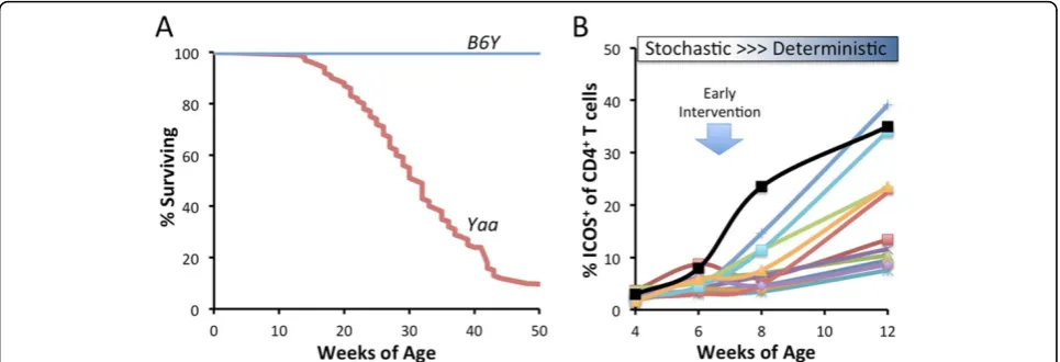
Methods and results: Robust variation in the autoimmune disease outcomes is apparent in individual BXSB.Yaawith uniform genetics and husbandry environment:BXSB.Yaamice have been inbred for at least 100 generations, 50 of which were performed in our colony. At least to the depth of genetic analysis performed to date, our BXSB.Yaacolony is genetically fixed. Moreover, continuous microbial monitoring has not revealed any changes over the last 20 years. Thus as a first approximation the environment in our BXSB.Yaaresearch colony is notably stable. However, we repeatedly observe substantial variation in the timing and severity of autoimmune disease among individual, highly inbred BXSB.Yaa
mice in well-powered studies measuring overall mouse survival (example Figure 1A). This variation is not consistent with genetic drift and/or fitness selection for healthier breeders. Substantial disease variation determined here by outcomes (timing of survival) is a durable feature of inbred BXSB.
Yaamice.
Robust variation in longitudinal expression of TFHin blood is observed
in BXSB.Yaamice at early stages of disease:Variation in the survival times of BXSB.Yaawould predict that biomarkers of mechanistically important cellular processes would vary similarly. Variation in such biomarkers at early stages of disease would be of most value because they may identify targets for early therapeutic interventions. TFHare critical drivers of the BXSB.Yaa
disease. Preliminary longitudinal studies investigating the expression of circulating ICOS+CD4+T cells show remarkable individual variation in the
frequencies of these cells by at least 8 weeks of age (Figure 1B). While such results are not yet linked to the timing of survival of the mice or correlated with other important biomarkers of disease, it is reasonable to surmise that stochastic events acting within weeks of birth bifurcate into important deterministic processes (activation and expansions of TFHin this case) that
may be have long term consequences.
However, such variation is usually disregarded. Conventional experimental studies are usually designed to override“stochastic noise”by attempts to size comparator cohorts with sufficient discriminative power to override individual variability. When significance in cohort comparisons is not achieved, the null hypothesis is invoked. The individual variability within genetically matched cohorts is an untapped source of biological information that can inform causal mechanisms of disease.
Extrapolation to disease prognosis and early interventions in humans:Variation in the severity and presentation among individuals diagnosed with a human autoimmune disease is the rule rather than the exception. While genetic risk and environment factors are certain to play important parts, the considerable phenotypic variation described above underscores the potential for stochastic behaviors to contribute significantly to disease variation. Genetically and environmentally fixed BXSB.Yaamice exhibit patterns that are consistent with stochastic events giving rise to key deterministic events at the inception of autoimmune disease. By extrapolation, therapeutic interventions designed to abrupt such early fate decisions may head off lupus at its inception and have enduring effects on overall disease pathogenesis (Figure 1B).
Acknowledgements:This work was supported by grants from the Alliance for Lupus Research and the National Institutes of Health (D.C.R. and G.W.C.), the Lupus Foundation of America (E.B.A.), and the Intramural Research Program of the National Institute of Allergy and Infectious Diseases (H.C.M.). The authors do not declare a conflict of interest.
A7
DNA repair in lupus
Jiadi Xu1, Zubin Patel1, Leah Kottyan1, Richard A Gatti2, Deborah K McCurdy2, Kenneth M Kaufman3, John B Harley3*
1
Cincinnati Children’s Hospital Medical Center, University of Cincinnati, Cincinnati, OH, USA;2University of California at Los Angeles, Los Angeles, CA, USA;3US Department of Veterans Affairs Medical Center, Cincinnati, OH, USA E-mail: john.harley@cchmc.org
Arthritis Research & Therapy2014,16(Suppl 1):A7
Background:The myriad of mouse genetic alterations that result in a lupus-like phenotype and the >100 genes now known to be involved in human lupus are all probably only a hint at the potential complexity of the many mechanisms that lead to systemic lupus erythematosus (SLE).
Methods:We have applied exome sequencing to 24 SLE patients and their parents (trios) in an effort to conquer the data analysis and to identify candidate genes that may contribute when the activity of their gene products were substantially altered.
Results:The proband of one of our SLE trios hadde novomutations in
RAD54Bthat was predicted to change ARG to GLN and to be severely
damaging to protein product activity by multiple algorithms. A second much more conservativede novomutation inDOCK8was not predicted to be consequential.RAD54Bis a component of the homologous recombination DNA repair pathway. Cells from the SLE proband were unusually sensitive to ionizing radiation by the colony survival and comet tail assays. Ionizing radiation selectively induced interferon responsive genes in cells from this patient and not from controls. Transfection of the wild-type gene into the cells from this patient led to overexpression of theRAD54Bgene product and returned ionizing radiation resistance toward normal.
Conclusion:These data in addition to the other five other genes directly or indirectly involved in altering risk of SLE by influencing DNA repair (TREX1, RAD51B, XRCC1, XRCC3, andXRCC4) implicate base excision repair, nonhomologous end joining, and homologous recombination. These results suggest that multiple DNA repair mechanisms contribute to SLE susceptibility.
A8
Impact of genetic variants on B-cell development and function in systemic lupus erythematosus
Karen Cerosaletti1, Tania Habib1, Richard James2, Janice Chen1, Melissa Pickett1, Andrew Funk1, Shan Wei1, Jane H Buckner1*
1Benaroya Research Institute at Virginia Mason, Seattle, WA, USA;2Seattle
Children’s Research Institute, Seattle, WA, USA E-mail: jbuckner@benaroyaresearch.org
Arthritis Research & Therapy2014,16(Suppl 1):A8
Background:Systemic lupus erythematosus (SLE) is an autoimmune disorder characterized by the progressive loss of tolerance to nuclear antigens and the production of pathogenic autoantibodies. Genetic polymorphisms in genes involved in B-cell signaling,PTPN22, CSK, BLKand
BANK1, are associated with susceptibility to SLE.
Methods:We have utilized a cohort of genotyped healthy subjects to better understand how these genetic variants contribute to the failure of B-cell tolerance seen in SLE. PBMC from these subjects are analyzed using multiparameter flow cytometry to assess the composition of the B-cell compartment and the response of B cells to stimulation via BCR and CD40.
Results: Our studies in healthy subjects who carry this variant have demonstrated alterations in the composition of the transitional and naïve B-cell pool. This is functionally correlated with the altered BCR response and enhanced survival of these cells in carriers of the risk variant. Three potentially functionalBANK1SNPs (including a splice branch point-site variant and two coding variants) are associated with SLE. We have shown that theBANK1risk variants are associated with homeostatic changes in the peripheral B-cell pool that include a significant expansion of the total memory and preswitch memory compartment; and a significant decrease in
Figure 1(abstract A6) A, Considerable individual variation in the survival of BXSB.Yaamale mice, n = 93. Long term survival of 26 males carrying the C57BL/6 Y chromosome (B6Y) demonstrate the dependency of the autoimmune disease onYaa.B, Longitudinal phenotyping of a cohort of BXSB.
[image:4.595.57.540.89.254.2]naïve B cells in subjects homozygous for theBANK1risk alleles. In further studies we have demonstrated that theBANK1splice variant is associated with significantly reduced expression of theΔ2 isoform and that the risk haplotype is further associated with blunted proximal BCR signaling in naïve B cells and enhanced p-AKT in memory B cells. Studies are ongoing to investigate the impact of theBANK1variant on plasma cell differentiation.
Conclusions: Studies of healthy subjects who carry SLE risk genes demonstrate alterations in B-cell function and fate. These studies can then be extended to subjects with SLE to understand how these genetic variants impact B-cell development and tolerance in the setting of disease.
A9
Differential methylation of interferon-related genes is associated with autoantibody production in systemic lupus erythematosus
Sharon A Chung1, Joanne Nititham1, Emon Elboudwarej2, Hong L Quach2, Kimberly E Taylor1, Lisa F Barcellos2, Lindsey A Criswell1*
1Rosalind Russell/Ephraim P. Engleman Rheumatology Research Center,
University of California, San Francisco, CA, USA;2School of Public Health, University of California, Berkeley, CA, USA
E-mail: Lindsey.Criswell@ucsf.edu
Arthritis Research & Therapy2014,16(Suppl 1):A9
Background:DNA methylation, an epigenetic modification, influences gene expression and has been implicated in the pathogenesis of systemic lupus erythematosus (SLE). Two recent studies suggest that interferon-regulated genes are hypomethylated in SLE patients compared with unaffected controls. However, these studies have not examined whether DNA methylation is associated with specific disease manifestations. The goal for the current study was to determine whether differential DNA methylation is associated with autoantibody production in SLE, with a focus on the anti-dsDNA autoantibody.
Methods:The methylation status of 467,314 CpG sites across the genome was characterized for 326 women with SLE. Associations between anti-dsDNA autoantibody production and methylation status was assessed using a discovery and replication study design. Multivariable regression was used to adjust for confounders including estimated leukocyte cell proportions and population substructure. In secondary analyses, we assessed differential methylation associations with anti-SSA, anti-Sm, and anti-RNP autoantibody production.
Results:Significant associations between anti-dsDNA autoantibody production and methylation status were replicated for 16 CpG sites (Pdiscovery< 1.07 × 10⁻⁷andPreplication< 0.0029) in 11 genes. The adjusted
mean difference in methylation between the two autoantibody subgroups ranged from 1 to 19%, and the adjusted odds ratio for anti-dsDNA autoantibody production comparing the lowest with the highest tertile of methylation ranged from 6.8 to 18.2. Differential methylation for these sites was also associated with anti-SSA, anti-Sm, and anti-RNP autoantibody production. All associated sites were less methylated in autoantibody-positive compared with autoantibody-negative cases. Seven of the 11 associated genes either induce interferon or regulate the interferon signaling pathway. Among the differentially methylated genes associated with the production of at least one autoantibody, cytokine and interferon signaling pathways were the most represented.
Conclusions:Hypomethylation of interferon-related and other genes is associated with autoantibody production among SLE cases. Differential methylation of these 11 genes in autoantibody-positive SLE cases may explain their recently reported associations with SLE risk, and the extent of hypomethylation may influence disease manifestations.
A10
Subphenotype mapping in systemic lupus erythematosus identifies multiple novel loci associated with circulating interferon alpha
Silvia N Kariuki1, Yogita Ghodke-Puranik2, Jessica M Dorschner2, Beverly S Chrabot3, Jennifer A Kelly4, Betty P Tsao5, Robert P Kimberly6, Marta E Alarcón-Riquelme4,7, Chaim O Jacob8, Lindsey A Criswell9, Kathy L Sivils4, Carl D Langefeld10, John B Harley11, Andrew D Skol1, Timothy B Niewold2*
1University of Chicago, Chicago, IL, USA;2Mayo Clinic, Rochester, MN, USA; 3
Gwen Knapp Center for Lupus Research, University of Chicago, Chicago, IL, USA;4Oklahoma Medical Research Foundation, Oklahoma City, OK, USA;
5University of California, Los Angeles, CA, USA;6University of Alabama,
Birmingham, AL, USA;7GENYO Centre for Genomics and Oncological Research: Pfizer/University of Granada/Andalusian Regional Government, Grenada, Spain;8University of Southern California, Los Angeles, CA, USA; 9
Rosalind Russell/Ephraim P. Engleman Rheumatology Research Center, University of California, San Francisco, CA, USA;10Wake Forest University, Winston-Salem, NC, USA;11Cincinnati Children’s Hospital Medical Center and Cincinnati VA Medical Center, Cincinnati, OH, USA, USA
E-mail: niewold.timothy@mayo.edu
Arthritis Research & Therapy2014,16(Suppl 1):A10
Background:Systemic lupus erythematosus (SLE) is a chronic autoimmune disorder characterized by inflammation of multiple organ systems, loss of tolerance to self-antigens, and dysregulated interferon responses. SLE is both genetically and phenotypically heterogeneous, and we hypothesize that this greatly reduces the power of overall case-control studies in SLE. Increased circulating level of the cytokine interferon alpha (IFNa) is a stable, heritable trait in SLE which has been implicated in primary disease pathogenesis. Forty to 50% of patients have high IFNa, and high levels correspond with clinical differences.
Methods:To study genetic heterogeneity in SLE, we performed a case-case study comparing patients with high versus low IFNain over 1,800 SLE cases. Four hundred European ancestry cases formed the discovery GWAS set, and 1,443 cases from a large independent multi-ancestral replication cohort were used to validate associations.
Results:In meta-analysis, the top associations in European ancestry were PRKG1 rs7897633 (PMeta= 2.75 × 10⁻⁸) and PNP rs1049564 (PMeta= 1.24 ×
10−7). We also found evidence for cross-ancestral background associations with theANKRD44andPLEKHF2loci. These loci have not been previously identified in case-control SLE genetics studies. Bioinformatic analyses implicate these loci functionally in dendritic cells and natural killer cells, both of which are involved in IFNaproduction in SLE.
Conclusions:As case-control studies of complex heterogeneous diseases reach a limit of feasibility with respect to subject number and detectable effect size, the study of informative pathogenic subphenotypes becomes a highly attractive and efficient strategy for genetic discovery in complex human disease.
Acknowledgements: SNK and YG-P contributed equally to the manuscript.
A11
Fine mapping and functional study of the systemic lupus erythematosus-associatedNMNAT2/SMG7locus
Jian Zhao, Yun Deng, Jennifer M Grossman, Betty P Tsao*
David Geffen School of Medicine University of California, Los Angeles, CA, USA E-mail: btsao@mednet.ucla.edu
Arthritis Research & Therapy2014,16(Suppl 1):A11
Background:NMNAT2(rs2022013 located at intron 1) was identified as a SLE risk locus in a European-derived population in a genome-wide association study (GWAS). NMNAT2 (nicotinamide mononucleotide adenylyltransferase 2) is expressed mainly in the brain, regulating energy metabolism. Proximal to the SLE-associatedNMNAT2variant isSMG7, encoding a component of the mRNA quality control pathway that regulates spliceosomal machinery such as Sm and snRNP via alternative splicing. We fine mapped theNMNAT2/SMG7region in multiple ancestries and explored functional consequences of the identified variants.
Materials and Methods:We genotyped/imputed 313 SNPs covering an ~550 kbNMNAT2/SMG7 region in 15,424 case-control subjects from European-Americans (EA), African Americans, Asians and Amerindian/ Hispanics, assessed SNPs for association with SLE using a logistic regression model adjusted for sex and ancestry, and used haplotype-based conditional testing to distinguish independent associations. Quantitative real-time PCR and luciferase reporter assays were used to examine allelic differences in
SMG7expression and transcription activity. PBMCs from SLE patients (n= 13) were cultured with or without siRNA targetingSMG7, GAPDH(positive control) or siRNA with a nontargeting sequence (NC, negative control), and culture supernatants were measured by ELISA for levels of antinuclear antibody (ANA) and cytokines/chemokines.
(P= 1.5 × 10⁻¹⁰, OR = 1.38); and multipleSMG7SNPs tagged by rs2275675 (P= 5.7 × 10⁻⁸, OR = 1.22). Expression quantitative trait locus data showed SLE-risk alleles ofNMNAT2/SMG7variants consistently associated with decreased mRNAs ofSMG7, but notNMNAT2, in cell lines, suggestingSMG7
is a more likely risk gene for SLE. The rs2275675 risk allele was associated with decreasedSMG7mRNAs dose dependently in PBMCs of 86 SLE patients and 119 controls (P= 0.001 and 6.84 × 10⁻⁸, respectively), and reduced transcription activity in two transfected cell lines (P≤0.004).SMG7
mRNA levels in PBMCs correlated inversely with ANA titers in 68 SLE patients (P= 0.0089,r=−0.31). Compared with culture supernatants of SLE PBMCs treated with NC-siRNA, those treated withSMG7-siRNA showed increased ANA (P< 0.0001) and CCL19 (P= 0.0002; a ligand for CCR7 promoting movement/ interaction of B-Th cells and antibody production).
Conclusions:We confirmed the previous GWASNMNAT2association and identified independentSMG7association with SLE in an EA population. The SLE-risk alleles are dose-dependently associated with decreased
SMG7mRNAs, andSMG7reduction increases ANA and CCL19 production in PBMC cultures of SLE patients, suggesting that dysfunction in mRNA surveillance conferred by SLE-associatedSMG7variants contributes to SLE manifestations.
A12
Impact of provider specialty on the diagnosis and management of systemic lupus erythematosus in the American Indian/Alaska Native population
John A McDougall1, Charles G Helmick2, S Sam Lim3, Caroline Gordon4, Elizabeth D Ferucci5*
1Dartmouth-Hitchcock Medical Center, Lebanon, NH, USA;2National Center
for Chronic Disease Prevention and Health Promotion, Centers for Disease Control and Prevention, Atlanta, GA, USA;3Emory University, Atlanta, GA, USA;4University of Birmingham, Edgbaston, UK;5Community Health Services, Alaska Native Tribal Health Consortium, Anchorage, AK, USA
E-mail: edferucci@anthc.org
Arthritis Research & Therapy2014,16(Suppl 1):A12
Background:Systemic lupus erythematosus (SLE) is a complex disease that is traditionally diagnosed and managed by specialists, typically rheumatologists. Higher SLE prevalence in racial/ethnic minorities such as American Indian/Alaska Native (AI/AN) people, often residing in areas with less access to rheumatologists, may necessitate diagnosis and management of SLE by primary care providers (PCP) in some cases. The purpose of this analysis was to identify areas of potential difference between PCP and specialist diagnosis and management of SLE in a population-based lupus registry of AI/AN people.
Methods:All individuals with SLE meeting our inclusion criteria were selected from the 2009 Indian Health Service lupus registry population. Inclusion in this analysis was limited to individuals with a final diagnosis of SLE made by a PCP or specialist (dermatologist, nephrologist or rheumatologist) and documented in the medical record. Based on medical record abstraction, SLE classification criteria were validated for each individual. Testing for biologic markers of SLE and medication use at any time during the course of the disease was also abstracted.
Results:Of the 320 patients identified with a documented physician diagnosis of SLE, 71 had been diagnosed by a PCP. SLE diagnosis by a specialist was associated with a higher median number of American College of Rheumatology (ACR) classification criteria (5 vs. 2), a higher percentage of patients meeting the definition of SLE by ACR criteria (79% vs. 22%), the Boston Weighted criteria (82% vs. 32%), and an abridged version of the Systemic Lupus International Collaborating Clinics criteria (83% vs. 35%) (P < 0.001 for all comparisons). Additionally, specialist diagnosis was associated with an increased proportion with any testing for anti-double-stranded DNA antibody (93% vs. 73%) and complement C3 and C4 (84% vs. 52%) documented in the medical record (P< 0.001 for all). Lastly, specialist diagnosis was associated with ever treatment with hydroxychloroquine (86% vs. 64%,
P< 0.001) as documented in the medical record at any time during their disease course.
Conclusions:Within the population studied, specialist diagnosis of SLE was associated with a higher number of SLE classification criteria met, a higher percentage of patients tested for biomarkers of disease, and a higher percentage of patients ever treated with hydroxychloroquine.
A13
Racial discrimination and disease damage among African American women with systemic lupus erythematosus
David H Chae1, Christina M Drenkard2, Tené T Lewis3, S Sam Lim2* 1
School of Public Health, University of Maryland at College Park, College Park, MD, USA;2School of Medicine, Emory University, Atlanta, GA, USA; 3School of Public Health, Emory University, Atlanta, GA, USA
E-mail: sslim@emory.edu
Arthritis Research & Therapy2014,16(Suppl 1):A13
Background: African American women with SLE experience faster progression and worse consequences of disease compared with their White counterparts. This study sought to examine whether self-reported routine experiences of discrimination, as a source of psychosocial stress, is associated with disease damage among African American women with SLE.
Methods:Participants were 578 African American women in the Georgians Organized Against Lupus study, a population-based cohort of SLE patients in Atlanta, GA, USA. Disease damage was assessed using the Self-Administered Brief Index of Lupus Damage (SA-BILD), a validated, patient-reported measure of organ damage since the onset of SLE. Discrimination was assessed using the Everyday Discrimination Scale, a widely used measure of routine experiences of unfair treatment. Ordinary least-squares regression analyses were used to examine the outcome of SA-BILD score by the primary predictors: unfair treatment, racial discrimination attribution, and their interaction, controlling for age and years since SLE diagnosis.
Results:The average SLE damage score in our sample was 2.3 (SD = 2.4), and the mean years since initial diagnosis was 13.6 years (SD = 9.3). The mean unfair treatment score was 1.92 (SD = 0.95), indicating that on average participants reported experiencing each of the forms of unfair treatment approximately once a year. A total of 159 participants (27.6%) reported not experiencing any unfair treatment. Among participants reporting any unfair treatment, most did not make an attribution of racial discrimination (n= 258 compared withn= 146). Age (r= 0.23,P< 0.001) and years since diagnosis (r= 0.25,P< 0.001) were significantly correlated with SLE damage. Reports of unfair treatment and making an attribution to racial discrimination were not significantly associated with SLE damage. In multivariable regression analyses controlling for age and years since diagnosis, we found a significant interaction between unfair treatment and attributions to racial discrimination (b= -0.52, SE = 0.24,P= 0.03). Greater unfair treatment attributed to nonracial causes was associated with higher SA-BILD score, whereas unfair treatment attributed to race showed an inverse association (Figure 1).
Conclusions:This study highlights the role that social stressors have in contributing to the progression of SLE and is the first to examine whether unfair treatment and racial discrimination are associated with disease damage among African American women with SLE. Consistent with findings from studies on discrimination and other health outcomes, these results suggest more complex, interactive rather than direct associations with SLE damage, with differential relationships being found between those who attributed unfair treatment primarily to racial discrimination versus those who did not.
[image:6.595.305.539.558.677.2]A14
The Scleroderma Patient-centered Intervention Network for conducting large-scale international trials of nonpharmacological interventions in scleroderma: a way ahead for lupus?
Brett D Thombs
McGill University and the Lady Davis Institute for Medical Research of the Jewish General Hospital, Montréal, QC, Canada
E-mail: brett.thombs@mcgill.ca
Arthritis Research & Therapy2014,16(Suppl 1):A14
Background:Psychosocial and rehabilitation interventions are increasingly used to attenuate disability and improve health-related quality of life (HRQL) in chronic diseases, but are typically not available for patients with less common diseases such as scleroderma or lupus. Conducting rigorous, adequately-powered trials of nonpharmacological interventions for patients with rare diseases is difficult. The Scleroderma Patient-centered Intervention Network (SPIN) is an international collaboration of patient organizations, clinicians, and researchers. The aim of SPIN is to develop a research infrastructure to test accessible, low-cost self-guided online interventions to reduce disability and improve HRQL for people living with scleroderma. Once tested, effective interventions will be made accessible through patient organizations partnering with SPIN.
Methods:SPIN will utilize a novel research design, the cohort multiple randomized controlled trial (cmRCT) design, to collect longitudinal data related to problems experienced by people living with scleroderma and as a framework for developing, evaluating, and delivering psychosocial and rehabilitation interventions. In the cmRCT design, patients consent to participate in a cohort for ongoing data collection. The aim is to recruit 1,500 to 2,000 patients from centers across the world within a period of 5 years (2014 to 2018). Eligible participants are persons≥18 years of age with a verified diagnosis of scleroderma. In addition to baseline medical data, participants will complete patient-reported outcome measures every 3 months. Upon enrolment in the cohort, patients will consent to be contacted in the future to participate in intervention research and to allow their data to be used for comparison purposes for interventions tested with other cohort participants. Once interventions are developed, patients from the cohort will be randomly selected and offered interventions as part of pragmatic RCTs. Outcomes from patients offered interventions will be compared with outcomes from trial-eligible patients who are not offered the interventions.
Discussion:The use of the cmRCT design, the development of self-guided online interventions, and partnerships with patient organizations will allow SPIN to develop, rigorously test, and effectively disseminate psychosocial and rehabilitation interventions for people with scleroderma. A similar approach could be used to develop and test psychosocial and rehabilitation interventions for people with lupus.
A15
Linking anti-Ro antibodies to fibrosis in congenital heart block: translation to a clinical trial
Jill P Buyon1*, Peter Izmirly1, Deborah Friedman2, Joshua Copel3 1New York University School of Medicine, New York, USA;2New York
Medical College, Valhalla, NY, USA;3Yale University School of Medicine, New Haven, Ct, USA
E-mail: jbuyonic@aol.com
Arthritis Research & Therapy2014,16(Suppl 1):A15
Background:One of the strongest clinical associations with autoantibodies (Ab) directed to components of the SSA/Ro-SSB/La ribonucleoprotein complex is the development of congenital heart block (CHB) in an offspring. Fetal disease is independent of maternal disease and often anti-Ro Ab are first sought only because CHB has been identified. The first-time risk of 2% is 10-fold higher in women who have had a previous CHB child. Tissue injury in the fetus is presumed to be dependent on the FcgR-mediated transplacental passage of maternal IgG Ab. Despite attempts of large multicenter studies to forestall disease by careful monitoring, irreversible block and extensive myocardial injury have been documented within 7 days of a normal rhythm and PR interval.In vivoandin vitrodata support that cardiac fibrosis may be consequent to macrophage Toll-like receptor (TLR) signaling following ligation of the ssRNA complexed to the Ro protein.
TLR signaling and fibrotic endpoints may be abrogated by chloroquine (CQ), which inhibits endosomal acidification. Thisin vitroobservation was initially
“translated”to patients by evaluating the use of hydroxychloroquine (HCQ) in an extensive case-control retrospective chart review of anti-Ro/La Ab exposed fetuses of mothers with SLE enrolled in three databases. This approach was followed by another study that addressed whether HCQ use reduces the expected recurrence rate of CHB. The collective results were highly encouraging.
Methods and results:Based on these studies we initiated an open-label prospective study design using Simon’s two-step approach. HCQ at 400 mg is initiated by 10 weeks following conception. Serial echocardiograms (monitor PR interval) and evaluation of maternal and cord blood biomarkers (HCQ levels, IFNasignatures, and Ab titers) are part of the protocol to address maternal compliance, pathobiology and efficacy. The first stage has been completed with 19 subjects and only one recurrence of CHB.
Future directions:Over the next 4 years, 35 subjects will be enrolled. Ultimately, HCQ will be considered efficacious for the prevention of CHB if fewer than six cases occur among a total of 54 subjects evaluated. A positive result will probably change the management of all anti-Ro-positive women who have had a previous child with CHB and illustrate the importance of translational science. Perhaps most relevant to impact, a potential prevention would justify screening of all pregnant women for anti-Ro antibodies, particularly relevant since mothers of affected children are frequently asymptomatic, a point critical to early pregnancy counseling.
A16
Targeting IRF5 inhibition in human B cells: identification of new functional roles that implicate IRF5 in systemic lupus erythematosus B-cell pathology
Saurav De1,2, Di Feng2, Alisha Valdez3, Amy Pitler1,2, Betsy J Barnes1,2* 1Rutgers Biomedical and Health Sciences, Newark, NJ, USA;2New Jersey
Medical School - Cancer Center, Rutgers Biomedical and Health Sciences, Newark, NJ, USA;3New Jersey Medical School, Rutgers Biomedical and Health Sciences, Newark, NJ, USA
E-mail: barnesbe@njms.rutgers.edu
Arthritis Research & Therapy2014,16(Suppl 1):A16
Background:The interferon regulatory factor 5 (IRF5) systemic lupus erythematosus (SLE) risk loci is considered one of the most strongly and consistently associated SLE loci identified. It has been detected using both candidate gene and genome-wide association studies. Haplotypes are associated with increased, decreased, or neutral levels of risk for SLE and have been shown to associate with functional changes in IRF5-mediated signaling, including increased expression and elevated IFNaactivity. The majority of studies, however, were performed in peripheral blood mononuclear cells and thus little is known of the function of IRF5 in specific human immune cell populations. We are interested in understanding the role of IRF5 in human B cells since previous studies in mice implicated a role for IRF5 in effector B-cell development and function and murine models of lupus lacking theIrf5gene showed reduced ANA, glomerulonephritis and pathogenic autoantibody production. Unfortunately, many of these studies were complicated by the finding of a secondary mutation in theDock2gene amongstIrf5-/-mice. Recent findings from our laboratory indicate that IRF5 is constitutively localized to the nucleus of human SLE memory B cells and that activation of healthy donor B cells results in IRF5 nuclear localization, suggesting a functional role for IRF5 in human B cells.
Methods:Human immortalized and primary B cells were utilized in this study. Primary naïve B cells were obtained by informed consent at University Hospital, Newark, NJ, USA under an approved IRB protocol, and either mock stimulated or stimulated with anti-IgM and CpG-B for activation. B-cell development, cytokine expression, autoantibody production and class switch recombination were examined in the presence or absence of siRNAs targeting IRF5 expression or peptide inhibitors targeting IRF5 nuclear localization. An IRF5-mediated B-cell signature was also examined under similar experimental conditions by ChIP-seq analysis.
Results:We find that IRF5 is indeed important for human B-cell effector functions, including, but not limited to, cytokine expression, differentiation, and autoantibody production, and have identified a pro-effector B cell gene program that is regulated by IRF5.
and thus, when overexpressed or overactivated, as seen in SLE B cells, would be expected to contribute significantly to SLE B-cell pathology. These data provide initial insight into how inhibitors of IRF5 activation may change SLE disease onset and/or progression.
A17
Platelet-derived microparticles serve as an important source of autoantigens and discriminate between levels of disease activity in systemic lupus erythematosus
Eric Boilard1*, Vincent Bissonnette1, Valérie Garceau1, Ellie Aghdassi2,
Nathalie Cloutier1, Claudia Beaudoin1, Davy Eng1, Stacey Morrison2, Paul R Fortin1,3 1
Centre de recherche du CHU de Québec, Université Laval, Quebec City, QC, Canada;2Toronto Western Research Institute, Toronto, ON, Canada;3CHU de Québec - CHUL, Quebec City, QC, Canada
E-mail: eric.boilard@crchuq.ulaval.ca
Arthritis Research & Therapy2014,16(Suppl 1):A17
Background:Immune complexes (IC) are implicated in the pathogenesis of several autoimmune diseases including systemic lupus erythematosus (SLE). In SLE, submicron extracellular vesicles, called microparticles (MP), are thought to serve as an antigenic surface promoting the deposition of immunoglobulins and the formation of MP-associated immune complexes (mpICs). However, the cellular origin of these mpICs is unknown and whether they correlate with disease activity and particular clinical features remains to establish.
Methods:The concentrations of mpICs in platelet-poor plasma from 193 women with SLE were determined using high-sensitivity flow cytometry. Considering the recently revealed role of platelets in SLE, we further scrutinized the contribution of platelets to mpICs formation. The platelet and nonplatelet MPs and mpICs were tested for association with lupus disease activity, damage, history of previous arterial disease, and the carotid intima-media thickness and plaque area on ultrasound. To assess whether disease activity and damage are associated with levels of MPs and mpICs, univariate and multivariate negative binomial models were built using the SLE disease activity index 2000 (SLEDAI-2K) and the SLICC/ACR damage index (SDI) as outcome variables. In all models, the predictor variable was the level of MPs or mpICs. When necessary, models were adjusted for covariables such as age, disease duration, menopausal status, hypertension, diabetes, anticoagulant or antiplatelet medication, antimalarial medication, prednisone use, smoking status, and ethnicity.
Results:The clinical characteristics of the 193 women studied were: age (mean (SD)) 46.3 (14.7) years; disease duration 18.5 (12.0) years; ethnicity (% Caucasian) 57%; ever-smoker 34%; menopausal in 55%; hypertensive 30%; diabetic 5%; prescribed anticoagulant or antiplatelet medication 25%; prescribed antimalarial medication 74%; prescribed prednisone 44%. Univariate analyses for activity revealed that platelet-derived mpICs, but not mpICs from other cells, were associated with SLEDAI-2K. In the multivariate model, this association remained significant (P= 0.02 for annexin V+platelet mpICs andP= 0.0006 for annexin V-platelet mpICs)
after adjusting for disease duration, hypertension and currently on prednisone. There was no association between platelet mpICs and SDI.
Conclusions:Platelet-derived MPs are a major source of autoantigens serving mpIC formation in SLE. Platelet-derived mpICs are associated with lupus disease activity level on the SLEDAI-2K but not with damage. This is the first report of an association between platelet mpICs and clinical marker of activity in SLE and in any autoimmune disease. Platelet mpICs need to be further considered as a possible biomarker of lupus disease activity.
Acknowledgements:This study was supported by the Canadian Institutes of Health Research (to PRF and EB). EB is recipient of a fellowship from the Fonds de Recherche du Quebec-santé.
A18
Vimentin is a dominant target ofin situhumoral immunity in human lupus tubulointerstitial nephritis
Marcus R Clark*, Anthony Chang, Kichul Ko, Carole J Henry Dunand, Scott Henderson, Dan Brandt, Natalya Kaverina, Vladimir Liarski, Patrick C Wilson, Andrew J Kinloch
University of Chicago, Chicago, IL, USA E-mail: mclark@medicine.bsd.uchicago.edu
Arthritis Research & Therapy2014,16(Suppl 1):A18
Background: In lupus nephritis (LuN), severe tubulointerstitial inflammation (TII) predicts progression to renal failure. Severe TII is associated with tertiary lymphoid neogenesis andin situantigen-driven clonal B-cell selection. The dominant autoantigen(s) drivingin situB-cell selection in TII are not known.
Methods:Single CD38+or Ki-67+B cells were laser captured from seven LuN biopsies. Twenty clonally expanded immunoglobulin heavy and light chain variable region pairs were cloned and expressed as antibodies. Seven more antibodies were cloned from flow-sorted CD38+cells from an eighth
biopsy. Antigen characterization was performed using a combination of confocal microscopy, ELISA, screening protoarrays, immunoprecipitation and mass spectrometry. Serum IgG titers to the dominant antigen were determined in 45 LuN and 38 non-nephritic lupus samples using purified antigen-coated arrays. Autoantigen expression was localized by immunohistochemistry and immunofluorescence on normal and LuN kidney.
Results:Thirteen of 27 antibodies reacted with cytoplasmic structures, four reacted with nuclei and none with dsDNA, Sm or RNP. Vimentin was the only autoantigen identified by both mass spectrometry and on protoarray. Eleven anticytoplasmic TII antibodies directly bound vimentin. Vimentin was highly expressed by tubulointerstitial inflammatory cells, and tested TII antibodies preferentially bound inflamed tubulointerstitium. Finally, high titers of serum anti-vimentin antibodies were associated with severe TII (P= 0.0001).
Conclusions:Vimentin, an antigenic feature of inflammation, is a dominant autoantigen targetedin situin LuN TII. This adaptive autoimmune response probably feeds forward to worsen TII and renal damage.
A19
Aggregation of MAVS antiviral protein suggests a mechanism for increased type I interferon production in SLE
Wen-Hai Shao, Brendan A Hilliard, Daniel H Shu, Stephen O Priest, Philip L Cohen*
Temple University School of Medicine, Philadelphia, PA, USA E-mail: philco@temple.edu
Arthritis Research & Therapy2014,16(Suppl 1):A19
Background:Patients with systemic lupus erythematosus (SLE) often have increased type I interferon levels (IFN-I) and activation of IFN-inducible genes (IFN signature). Because IFN-I has a key role in both the innate and adaptive immune responses, it is believed that heightened levels of this cytokine and the many genes it regulates may underlie the immune hyperreactivity and autoimmunity of SLE. The mechanism of IFN-I hyperproduction in SLE is under intense study. An important lead has emerged from our laboratory implicating the RIG-I antiviral pathway as a possible cause. In particular, the mitochondrial adaptor protein MAVS is a key intermediary in the RIG-I/MDA5 pathway, where viral RNA triggers a conformational change in RIG-I, leading to MAVS activation with subsequent IFN production. It has been reported that MAVS may form large prion-like aggregates, which might stimulate IFN-I production in a potent and prolonged fashion. We wondered whether such aggregates might be detectableex vivoin SLE patients, and whether they might play a role in the sustained increased production of IFN-I.
Methods:Peripheral blood mononuclear cells (PBMCs) were isolated from patients fulfilling ACR criteria for SLE, from healthy controls, and from patients with rheumatoid arthritis (RA). Mitochondrial lysates were prepared and MAVS aggregation was identified with a semi-denaturing agarose gel and confirmed by confocal immunofluorescent microscopy.
Results:Twenty-two of 61 SLE patients showed clear MAVS aggregation, with essentially all of their MAVS protein in a high molecular weight aggregated form. None of the RA patients and only three of 33 healthy controls had abnormal MAVS. Clinical data analysis revealed that 82.4% MAVS-aggregate-positive SLE patients (mean age 46) had anti-SSA antibodies, compared with 40% MAVS-aggregate-negative patients (mean age 44),P< 0.01 by chi-square. 64.7% aggregation-positive patients had active SLE disease (skin rash, arthritis, increased ESR, low C4, and active renal disease), while only 10% of the aggregation-negative patients had active disease.
A20
Aberrant expansion of CXCR5+memory CD4 T cells in patients with systemic lupus erythematosus
Jin-Young Choi, Sang Taek Kim, Insoo Kang, Joe Craft* Yale School of Medicine, New Haven, CT, USA E-mail: joseph.craft@yale.edu
Arthritis Research & Therapy2014,16(Suppl 1):A20
Background:Autoreactive B cells in SLE undergo autoantigen selection, suggesting a requirement for germinal center follicular helper T (Tfh) cells in their maturation. However, evidence for dysregulation of Tfh cells in SLE and their potential contribution to disease remains unclear. Recently, blood CXCR5+CD4 T cells, a heterogeneous pool consisting of functionally distinct Th1-like, Th2-like, and Th17-like subsets, have been proposed to be the circulating (blood) memory Tfh cells. We hypothesized that expanded CXCR5+memory cells in the blood of human lupus patients promote B-cell
helper function reflecting the abnormal T-B-cell responses in secondary lymphoid organs.
Methods and results:We characterized such cells in SLE patients by flow cytometry and T-B coculture studies. SLE patients had significant expansion of CXCR5+ICOShiPD-1hiCD4 T cells compared with controls. Such cells were
Bcl6-, but robustly expressed IL-21 with a portion Ki-67+, indicating their functional activity. The blood cells were capable of providing blood-born memory B cells with survival and differentiation signals to secrete isotype switched Igs, including antinuclear antibodies.
Conclusions:We speculate that therapies which alter T-B collaboration in lupus will abrogate their expansion, accompanied by reduced autoantibody titers and improved disease activity in SLE patients. Our results suggest that aberrant T-B collaboration in lupus is critical to disease pathogenesis and its blockade is likely to be important therapeutically.
Acknowledgements:This work was supported in part by the Alliance for Lupus Research and AR040072, AR44076, AR062842, and AR053495 (to JC). STK was supported by a Research Scientist Development Award from the American College of Rheumatology Research and Education Foundation and by T32 AR07107.
A21
Identification of stage-specific genes associated with lupus nephritis and response to remission induction in NZB/W and NZM2410 mice
Ramalingam Bethunaickan1, Celine C Berthier2, Weijia Zhang3, Ridvan Eksi2, Hong-Dong Li2, Yuanfang Guan2, Matthias Kretzler2, Anne Davidson1* 1
Center for Autoimmunity and Musculoskeletal Diseases, Feinstein Institute for Medical Research, Manhasset, NY, USA;2University of Michigan, Ann Arbor, MI, USA;3Mount Sinai Medical Center, New York, NY, USA E-mail: adavidson1@nshs.edu
Arthritis Research & Therapy2014,16(Suppl 1):A21
Background: Lupus nephritis affects 30 to 70% of systemic lupus erythematosus (SLE) patients and its treatment remains insufficiently effective and excessively toxic. Although biomarkers for nephritis are being identified there is still no reliable way of predicting an impending renal flare or determining which patients will respond to therapy. Because human renal tissue cannot be obtained sequentially during remission and relapse, animal models are often used to study progression of lupus nephritis. To elucidate the molecular mechanisms involved in renal inflammation during the progression, remission and relapse of nephritis we performed a transcriptome analysis of renal tissue from two murine lupus models, NZB/WF1 mice that develop proliferative glomerulonephritis and NZM2410 mice that develop glomerulosclerosis with minimal inflammation.
Methods:Kidneys from NZB/W F1 and NZM2410 mice were harvested at intervals during their disease course or after remission induction with either combination cyclophosphamide/costimulatory blockade or with BAFF inhibition. Genome-wide expression profiles were obtained from microarray analysis of perfused kidneys. Real-time PCR analysis for selected genes was used to validate the microarray data. Comparisons between groups using SAM, and unbiased analysis of the entire dataset using singular value decomposition and self-organizing map were performed.
Results:Few changes in the renal molecular profile were detected in pre-nephritic kidneys but a significant shift in gene expression, reflecting
inflammatory cell infiltration and complement activation, occurred at proteinuria onset. Subsequent changes in gene expression predominantly affected mitochondrial dysfunction and metabolic stress pathways. Remission induction reversed most, but not all, of the inflammatory changes and progression towards relapse was associated with recurrence of inflammation, mitochondrial dysfunction and metabolic stress signatures. Endothelial cell activation, tissue remodeling and tubular damage were the major pathways associated with loss of renal function.
Conclusions:Immune cell infiltration and activation is associated with proteinuria onset and reverses with immunosuppressive therapy but disease progression is associated with renal hypoxia and metabolic stress. Optimal therapy of SLE nephritis may therefore need to target both immune and nonimmune disease mechanisms. In addition, the overlap of a substantial subset of molecular markers with those expressed in human lupus kidneys suggests potential new biomarkers and therapeutic targets.
Acknowledgements:RB and CCB contributed equally.
A22
Blimp-1 and systemic lupus erythematosus
Betty Diamond1*, Sun Jung Kim1, Sebastian Schaetzle2, George Georgiou2 1
Center for Autoimmune and Musculoskeletal Diseases, Feinstein Institute for Medical Research, Manhasset, NY, USA;2The University of Texas at Austin, TX, USA
E-mail: bdiamond@nshs.edu
Arthritis Research & Therapy2014,16(Suppl 1):A22
Our laboratory has been studying the impact of low Blimp-1 expression on dendritic cell function in SLE, analyzing both a mouse model of SLE in which there is a deletion of Blimp-1 in CD11c dendritic cells (DC Blimp-1KOmice)
and healthy individuals with the SLE risk allele of Blimp-1. This allele leads to reduced Blimp-1 expression in dendritic cells. Low expression of Blimp-1 alters cytokine expression following TLR activation and antigen-processing machinery. This leads to a highly distinct TCR repertoire in T-follicular helper cells in DC Blimp-1KOmice. Much of this phenotype is under estrogen
regulation. These data therefore provide an integrated explanation for altered cytokines, autoantibodies and female predisposition in SLE.
A23
A role for cutaneous gamma delta T cells in the development of systemic lupus erythematosus
Mehran Ghoreishi, Jan P Dutz*
University of British Columbia, Vancouver, BC, Canada E-mail: dutz@interchange.ubc.ca
Arthritis Research & Therapy2014,16(Suppl 1):A23
Background:Ultraviolet (UV) light exposure promotes the development of cutaneous lupus and can induce flares of systemic lupus erythematosus. Similarly, UV light can induce lupus in genetically prone strains of mice. Nonobese diabetic (NOD) mice prone to autoimmune disease demonstrate lupus-like disease after repeated combined UV irradiation and topical application of TLR-7 agonist imiquimod (IMQ). In contrast, repeated topical application of a Toll-like receptor (TLR) 7/8 agonist promotes psoriasiform skin lesions in nonautoimmune mice such as Balb/c. The cellular and molecular determinants of these processes are still unknown.
In this study we have compared the skin lesions induced by repeated topical IMQ in Balb/CVSNOD mice. Gamma delta T cells are innate immune cells that bridge innate and adaptive immunity in part by modulating dendritic cell maturation. We have thus explored the role of gamma delta T cells in the induction of psoriasis and SLE following topical TLR 7/8 agonist application.
Methods:NOD and Balb/C mice received 70 mg topical IMQ 5% cream on shaved back skin daily for 5 days. Skin, draining lymph nodes (LNs) and sera were obtained for histological and serological analysis.