INTRODUCTION
Rodent incisors provide a remarkable example of the requirement, shared by many organs, for continuous production of progeny from stem cells; these specialized teeth grow throughout life by virtue of the existence of pools of adult stem cells that generate several differentiated cell types. This lifelong growth is counterbalanced by abrasion from material in the animal’s diet, resulting in a fixed length of the tooth. Because enamel, the hardest dental tissue, is present only on the labial (lip) but not the lingual (tongue) side of the incisor (Fig. 1A), abrasion occurs preferentially on the lingual side, causing the characteristic sharpness of the tip of the tooth. The asymmetric enamel deposition results from the presence of enamel-secreting ameloblasts exclusively on the labial side of the incisor.
Several lines of evidence indicate that a structure known as the labial cervical loop (laCL) at the proximal end of the incisor contains the stem cell niche that houses the ameloblast progenitors. Pioneering experiments using radioactive labeling of cells in the laCL and ex vivo cultures of incisors showed that epithelial
progenitors in the laCL give rise to transit-amplifying (T-A) cells that in turn differentiate into ameloblasts as they advance towards the distal end of the incisor (Fig. 1A) (Smith and Warshawsky, 1975; Harada et al., 1999). The much smaller cervical loop (CL) on the lingual side (liCL) is also presumed to contain epithelial stem cells, and the mesenchymal compartment between the CLs is thought to contain the stem cells for odontoblasts, which produce another mineralized tissue, dentin (Fig. 1A). Little is known about these latter two stem cell populations.
The development of the stem-cell-containing CLs is regulated by both transforming growth factor (TGF) and fibroblast growth factor (FGF) signaling. Increased TGFsignaling in mice carrying mutations in follistatin leads to expansion of the liCL and to the presence of ectopic stem-cell-derived ameloblasts and enamel on the lingual surface (Wang et al., 2004; Wang et al., 2007). A similar phenotype of an expanded liCL and ectopic lingual ameloblasts was observed in mice carrying mutations in sprouty genes, which encode inhibitors of FGF signaling (Klein et al., 2008). Interestingly, in both of these cases, Sonic hedgehog (Shh), the gene encoding one of the three mammalian Hedgehog (Hh) ligands, is expressed ectopically adjacent to the liCL. Shh is known to play a role in growth and morphogenesis of teeth in the embryo (Hardcastle et al., 1998; Dassule et al., 2000; Cobourne et al., 2001; Gritli-Linde et al., 2002) but the expression of Shhadjacent to the stem cell niches in these mutants pointed to a possible function for Shh in the generation of progeny from adult stem cells in the incisor.
SHH has been proposed to play a number of roles in stem cell biology, including regulation of fate decisions in embryonic stem cells (Gaspard et al., 2008) and survival and self-renewal of neural stem cells (Machold et al., 2003; Balordi and Fishell, 2007). However, the effects of SHH in adult stem cell populations remain controversial. For example, although proliferation of primitive hematopoietic stem cells has been reported to be Hh-dependent Development 137, 3753-3761 (2010) doi:10.1242/dev.056358
© 2010. Published by The Company of Biologists Ltd
1Departments of Orofacial Sciences and Pediatrics and Program in Craniofacial and Mesenchymal Biology, UCSF, 513 Parnassus Avenue, San Francisco, CA 94143-0442, USA. 2Department of Molecular Biology, Genentech Inc., 1 DNA Way, South San Francisco, CA 94080, USA. 3Developmental Biology Program, Sloan-Kettering Institute, 1275 York Avenue, New York, NY 10021, USA. 4Biomedical Imaging Group, Department of Tumor Biology and Angiogenesis, Genentech Inc., 1 DNA Way, South San Francisco, CA 94080, USA. 5Department of Pathology and Laboratory Medicine, The Joseph Stokes Jr Research Institute, The Children’s Hospital of Philadelphia, Philadelphia, PA 19104, USA. 6Howard Hughes Medical Institute, Laboratory of Mammalian Cell Biology and Development, Rockefeller University, 1230 York Avenue, New York, NY 10065, USA. 7Department of Anatomy and Program in Developmental Biology, UCSF, 1550 4th Street, San Francisco, CA 94143-2711, USA.
*These authors contributed equally to this work
†Authors for correspondence (desauvage.fred@gene.com; ophir.klein@ucsf.edu)
Accepted 13 September 2010 SUMMARY
In many organ systems such as the skin, gastrointestinal tract and hematopoietic system, homeostasis is dependent on the continuous generation of differentiated progeny from stem cells. The rodent incisor, unlike human teeth, grows throughout the life of the animal and provides a prime example of an organ that rapidly deteriorates if newly differentiated cells cease to form from adult stem cells. Hedgehog (Hh) signaling has been proposed to regulate self-renewal, survival, proliferation and/or differentiation of stem cells in several systems, but to date there is little evidence supporting a role for Hh signaling in adult stem cells. We used in vivo genetic lineage tracing to identify Hh-responsive stem cells in the mouse incisor and we show that sonic hedgehog (SHH), which is produced by the differentiating progeny of the stem cells, signals to several regions of the incisor. Using a hedgehog pathway inhibitor (HPI), we demonstrate that Hh signaling is not required for stem cell survival but is essential for the generation of ameloblasts, one of the major differentiated cell types in the tooth, from the stem cells. These results therefore reveal the existence of a positive-feedback loop in which differentiating progeny produce the signal that in turn allows them to be generated from stem cells.
KEY WORDS: Hedgehog, Stem cell, Incisor, Regeneration, Mouse
Hedgehog signaling regulates the generation of ameloblast
progenitors in the continuously growing mouse incisor
Kerstin Seidel1,*, Christina P. Ahn2,*, David Lyons1, Alexander Nee1, Kevin Ting1, Isaac Brownell3, Tim Cao4,
Richard A. D. Carano4, Tom Curran5, Markus Schober6, Elaine Fuchs6, Alexandra Joyner3, Gail R. Martin7,
Frederic J. de Sauvage2,†and Ophir D. Klein1,†
D
E
V
E
LO
P
M
E
N
3754
(Bhardwaj et al., 2001), recent work has shown that Hh signaling does not play a major role in adult hematopoietic stem cells (Gao et al., 2009; Hofmann et al., 2009).
Here, we set out to determine what role, if any, Hh signaling plays in incisor stem cell biology in adult mice. We show that SHH is the principal Hh ligand in the incisor and that it signals to three putative stem cell niches. Using genetic lineage tracing, we demonstrate that some of the Hh-responsive cells are stem cells that can give rise to differentiated progeny over extended periods of time. By blocking the pathway with a hedgehog pathway inhibitor (HPI), we demonstrate that Hh signaling is required for continued generation of enamel-producing ameloblasts but that it is dispensable for stem cell maintenance. Together, these studies provide evidence for a feedback loop in which differentiated progeny produce the signal that directs their formation from adult stem cells.
MATERIALS AND METHODS Mouse lines and drug administration
Mice carrying the Gli1lacZ(Bai et al., 2002), Ptch1lacZ(Goodrich et al., 1997), Gli1-CreERT2(Ahn and Joyner, 2004), R26R (Soriano, 1999), K5tTA(Diamond et al., 2000) or H2B-GFP (Tumbar et al., 2004) alleles or transgenes were maintained and genotyped as previously described. BrdU was injected at 40 g per gram of bodyweight. Gli1-CreERT2;R26R mice were treated by oral gavage with 10 mg of tamoxifen for lineage tracing studies or 5 mg when lineage tracing analyses were combined with the HPI treatment. HhAntag was synthesized as described (Yauch et al., 2008) and was administered orally twice daily at a dose of 100 mg/kg. CD1 females used for HhAntag studies were 6 weeks old. Mice used for lineage tracing and expression analyses were 8-10 weeks old at the start of the experiment. At least three mice were examined at each timepoint for all experiments.
Histology, gene expression analyses and detection of alkaline phosphatase activity
Jaws were dissected from perfusion-fixed animals, post-fixed in 4% PFA overnight, decalcified in RNase-free 0.5 M EDTA for 16 days and processed for paraffin embedding. Sections (7 m) were prepared and stained with Hematoxylin and Eosin using standard methods. RNA in situ hybridization was performed on tissue sections using digoxigenin-labeled probes according to standard protocols. Alkaline phosphatase activity was detected on tissue sections using BM Purple staining solution (Roche) after overnight incubation in alkaline phosphatase buffer. Brightfield images were obtained using a Leica DFC 500 camera with a Leica DM 5000B microscope.
For low-magnification fluorescence imaging (see Fig. S5 in the supplementary material), mandibles were embedded in 4% agarose, serial vibratome sections were cut and confocal microscopy was performed as below.
X-Gal staining
For X-Gal staining, jaws were dissected from freshly euthanized animals and the bone covering the proximal incisor was removed. Following fixation at 4°C in 100 mM phosphate buffer comprising 2% PFA, 5 mM EGTA, 0.2% glutaraldehyde and 2 mM MgCl2, the tissue was washed in 100 mM phosphate buffer with 0.01% Na-deoxycholate and 0.02% Nonidet-P40. Staining was performed at 37°C overnight with the solution used for the washing steps with 10 mM ferrocyanide, 10 mM K-ferricyanide and 1 mg/ml X-Gal added. The specimens were washed, fixed in 4% PFA overnight, decalcified and further processed for paraffin sectioning. Sections were counterstained with Hematoxylin and mounted in Permount (Fisher Scientific).
Quantitative RT-PCR
RNA was isolated from the incisor CL region, embryonic intestine at embryonic day (E) 16.5 and adult testis using TRIzol reagent (Invitrogen) according to the manufacturer’s recommendations for small tissue samples.
Hh pathway genes were quantitatively assessed by Taqman using an Applied Biosystems 7500 Real-Time PCR System and transcript levels were normalized to the housekeeping gene ribosomal protein L19 (RPL19). Results are expressed as normalized expression values (equal to 2–Ct).
Primer and probe sequences used for RT-PCR were as follows (FW, forward; RW; reverse; 5⬘-3⬘): mDhh-FW, CTGGCATGCATTGGTACTCT; mDhhRW, GGAAGCTCAGCCCATTAACT; mDhhProbe, CCTCCTTT -ACCGCTTGGCCG; FW, ACAAGGCATGGGACACTTG; mIhhRW, TCAGCCACAGCTGCAAAG; mIhhProbe, AGGAGGCCACCA -CA TCCTC-CA; FW, GGTG-CAAAGA-CAAGTTAAATGCC; mShhRW, CACTCGCAGCTTCACTCCAG; mShhProbe, TGGCCATCTCTG TGATGAACCAGTGG; mRPL19FW, AGAAGGTGACCTGGATGA -GAA; mRPL19-RW, TGATACATATGGCGGTCAATCT; mRPL19-Probe, CTTCTCAGGAGATACCGGGAATCCAAG.
Immunofluorescence
Paraffin sections were rehydrated, incubated in 1 mM EDTA just below boiling temperature for 30 minutes for antigen retrieval, washed in distilled H2O and treated for 20 minutes with 3% H2O2in PBS. Primary antibodies against Hh (1:500, Genentech), beta-galactosidase (1:5000, MPBiomedicals), ameloblastin (1:200, Santa Cruz) and BrdU (1:500, Abcam) were used. Washes in PBS (3⫻20 minutes) and PBS-T (1⫻5 minutes) were followed by incubation with Alexa Fluor 555 secondary antibody (1:500, Invitrogen) or biotinylated anti-rat secondary antibody (Vector) followed by signal amplification. Sections were counterstained with DAPI (Vector Laboratories) and mounted in 1% DABCO in glycerol. All images were acquired using a Leica-TCS SP5 confocal microscope except for the cell proliferation data shown in Fig. S4 in the supplementary material, which was imaged with a Leica DFC 500 camera on a Leica DM 5000B microscope.
TUNEL staining
Sections were de-paraffinized, rehydrated and incubated in a solution comprising 0.1% trypsin and 0.1% CaCl2at 37°C for 14 minutes. The In Situ Cell Death Detection Kit, TMR red (Roche) was used according to manufacturer’s instructions, and positive and negative controls were included as suggested. Sections were counterstained and mounted as described in the section above and imaged using a Leica DFC 500 camera with a Leica DM 5000B microscope.
Micro-CT analysis
The mouse jaws were imaged with a CT 40 (SCANCO Medical, Basserdorf, Switzerland) X-ray micro-CT system and maximum-intensity projections were created using standard protocols.
RESULTS
SHH is the principal hedgehog ligand in the mouse incisor and signals to three discrete regions containing label-retaining cells
Although it is widely believed that stem cells are located in the labial and lingual CLs (Smith and Warshawsky, 1975; Harada et al., 1999; Wang et al., 2007), their precise location has not been determined in vivo. Previously, cells that retain BrdU over a long period of time, and are therefore dividing infrequently (label-retaining cells, LRCs), have been identified in the laCL of cultured incisor explants from neonatal mice (Harada et al., 1999). Such label retention, although not a universal property of stem cells, is found in a number of stem cell populations and therefore can serve as an indication of the presence of stem cells (Fuchs, 2009). As a first step toward identifying the stem cells in vivo, we set out to detect such LRCs by injecting postnatal-day-2 pups with BrdU and waiting two months before assaying for label retention (Fig. 1B). This technique, which allows visualization of LRCs in all tissue compartments, is mosaic and labels only those cells that were cycling at the time of the initial labeling event. We found that LRCs were present exclusively in the proximal parts of both labial
RESEARCH ARTICLE Development 137 (22)
D
E
V
E
LO
P
M
E
N
(Fig. 1C⬙) and lingual (Fig. 1C⬘) CLs and in the mesenchyme between the two CLs (Fig. 1C), providing support to the notion that these regions contain the incisor stem cells.
Shh is known to be expressed in epithelial cells on the labial side near the CL (Fig. 1D) (Klein et al., 2008) and we detected Hh protein both in pre-secretory ameloblasts, which produce Shh mRNA, as well as in the mesenchymal cells adjacent to the laCL (Fig. 1E; see Fig. S1A,B in the supplementary material). Expression of both the transcriptional effector gene Gli1and the Hh receptor gene patched (Ptch1) is considered a reliable indicator of Hh signaling activity (Ingham and McMahon, 2001; Hooper and Scott, 2005). Using Gli1lacZandPtch1lacZreporter mice, we
sought to determine whether the LRCs that we detected are responding to Hh signaling. We used X-Gal staining, which is highly sensitive, as well as anti--galactosidase (-Gal) antibody staining, which is less sensitive but allows for higher-resolution imaging. We found that Hh signals are received by cells in the laCL near to the source of SHH. Additionally, cells in regions that are more distant from the ligand-producing cells were also found to express Gli1lacZand Ptch1lacZ(Fig. 1F-H; see Fig. S1C-E in the
supplementary material). High levels of lacZexpression in the reporter lines were observed in the proximal regions of both labial
(Fig. 1H; see Fig. S1E,H in the supplementary material) and lingual (Fig. 1G; see Fig. S1D,G in the supplementary material) CLs and in the mesenchyme between the two CLs (Fig. 1F; see Fig. S1C in the supplementary material), with much lower levels of expression in the ameloblasts, odontoblasts and pulp mesenchyme (Fig. 1F; see Fig. S1C in the supplementary material). Thus, LRCs are present within the region of presumed high-level Hh signaling. Furthermore, the presence of cells that were both BrdU-label-retaining and Gli1lacZ-positive in the CLs
and interjacent mesenchyme (Fig. 1I-I⬙) indicated that some of the putative incisor stem cells were Hh-responsive.
[image:3.612.53.515.61.323.2]To determine whether the Hh-responsive cells that are far from the cells producing SHH might be responding to other members of the Hh family, we assayed for indian hedgehog (Ihh) and desert hedgehog (Dhh) mRNA by in situ hybridization. We did not detect expression of either gene in the incisor (see Fig. S2A,B in the supplementary material). The conclusion that Shhis the principal Hh ligand gene expressed in the incisor was confirmed by a quantitative polymerase chain reaction (qPCR) analysis (see Fig. S2C in the supplementary material). Thus, all of the Hh-responding cells detected in the proximal incisor are presumably responding to SHH produced by pre-ameloblasts in the labial epithelium. Fig. 1. Hh signals to three regions of the incisor that contain label-retaining cells. (A)Schematic of the proximal region of an adult mouse incisor (sagittal plane). Ameloblasts (Am) develop from pre-ameloblasts (pre-Am), which arise from transit amplifying (T-A) cells that are derived from stem cells in the labial cervical loop (laCL). Stem cells, which have been proposed to reside in the stellate reticulum (SR) or outer enamel epithelium (OEE), also give rise to cells of the stratum intermedium (SI), which comprise a single layer that subtends the ameloblasts. BV, blood vessel; Di, distal; De, dentin; En, enamel; liCL, lingual cervical loop; Od, odontoblasts; Pr, proximal. (B)Protocol used to mark the label-retaining cells (LRCs) in C-C⬙and I-I⬙. BrdU was administered twice daily on three consecutive days (green arrows) followed by a chase period prior to sacrifice (black arrow). (C-C⬙) A section of the incisor stained with DAPI and an antibody against BrdU. LRCs are restricted to the proximal laCL (C⬙) and liCL (C⬘) and the mesenchyme in between the CLs (white arrowheads). Dotted lines outline the epithelium. (D)ShhmRNA is expressed in T-A cells, pre-ameloblasts and immature pre-ameloblasts. (E)Immunofluorescence staining detects Hh protein in both Shh-expressing epithelial cells and some adjacent mesenchymal cells (blue arrowhead). (F-H)Detection of nuclear lacZ expression in incisors from Gli1lacZmice by X-Gal staining (F) and -Gal
immunofluorescence assay (G,H). Gli1lacZis most highly expressed in the proximal laCL (H) and liCL (G) and in the proximal incisor mesenchyme.
Asterisks in F indicate the approximate domain of Shhexpression. (I-I⬙) Section of the incisor of a Gli1lacZ-expressing mouse stained with DAPI and
antibodies against -Gal (to detect Gli1-expressing cells) and BrdU. Yellow arrowheads point to double-labeled cells that are both BrdU label-retaining and Gli1lacZ-positive. Open white arrowheads indicate LRCs that do not respond to Hh signaling. Scale bars: 100m in C,D-I⬙; 50m in
C⬘,C⬙.
D
E
V
E
LO
P
M
E
N
3756
Lineage tracing studies demonstrate that
hedgehog-responsive cells in the adult incisor are stem cells
In contrast to label retention, which points to the possible presence of stem cells, inducible genetic lineage tracing permanently labels a cell and all of the progeny of that cell and is thus a method that definitively identifies adult stem cells (Joyner and Zervas, 2006; Haegebarth and Clevers, 2009). To test whether the Hh-responsive cells in the proximal mouse incisor are indeed stem cells that can give rise to differentiated cell types in the tooth over a long period of time, we genetically labeled Gli1-expressing cells and traced their descendants in vivo using Gli1-CreERT2;R26R mice (Ahn and
Joyner, 2005). Based on the knowledge that ameloblasts continuously migrate distally out of the proximal incisor, the observation that labeled progeny continue to be generated over long periods of time would demonstrate that a stem cell population has been labeled.
Upon induction with tamoxifen, Gli1-CreERT2;R26R mice
transiently express Cre recombinase in Gli1-positive cells, leading to expression of lacZin these cells and subsequently all of their progeny. Gli1-CreERT2;R26R mice not treated with
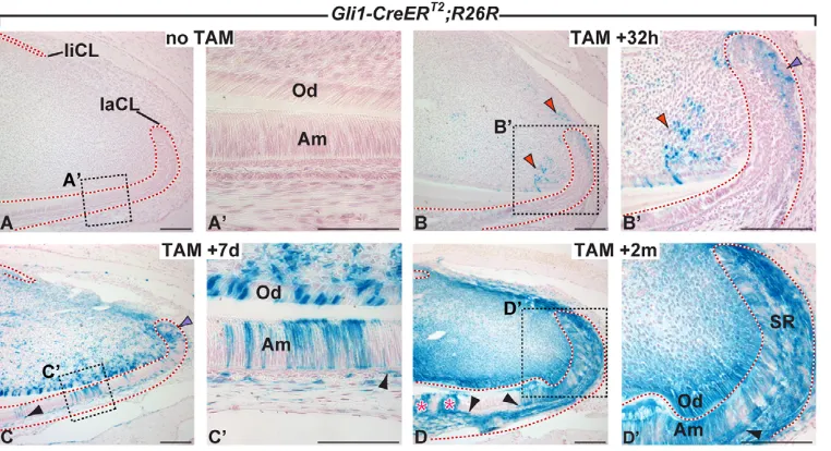
tamoxifen showed few or no lacZ-positive cells in the proximal incisor region (Fig. 2A,A⬘). By contrast, 32 hours after induction of Cre activity, lacZexpression was detected in a small number of cells of the laCL epithelium and in the proximal incisor mesenchyme (Fig. 2B,B⬘), regions that normally express Gli1 (Fig. 1F-H); because of the mosaic nature of Cre activation after a single tamoxifen injection, only a subset of the Gli1-expressing cells were lacZ-positive. Seven days after tamoxifen treatment, a higher proportion of cells in the laCL region were labeled than at the previous timepoint and we also identified clusters of labeled ameloblasts (Fig. 2C,C⬘). As ameloblasts in the mouse incisor are known to advance distally ~350 m a day (Hwang and Tonna, 1965), these X-Gal-positive ameloblasts, which emerged from the laCL one day prior to sacrifice, must have been generated from cells that were originally labeled as stem or progenitor cells in the laCL. Seven days post-induction, we also observed numerous labeled odontoblasts and other mesenchymal cells, as well as cells in the liCL (Fig. 2C,C⬘). Over time, the entire proximal incisor region filled with labeled cells. LacZ-positive ameloblasts continued to be produced two months after transient induction of
Cre activity with tamoxifen (Fig. 2D,D⬘). We also identified labeled cells in the stratum intermedium (SI), a layer of cells that underlies the ameloblasts (Fig. 2C-D⬘), as well as in the stellate reticulum (SR) in the center of the laCL (Fig. 2D⬘). We examined incisors up to 15 months after receiving a single induction and remarkably detected labeled ameloblasts at this timepoint as well (see Fig. S3 in the supplementary material). These results conclusively demonstrate that a subset of cells that receive the Hh signal (Gli1lacZ-positive cells) are stem cells in the adult mouse
incisor and that some of these stem cells are able to give rise to progeny for extended periods of time.
Inhibition of hedgehog signaling disrupts generation of ameloblasts from stem cells
To investigate the function of Hh signaling in the adult incisor, we blocked the pathway by administering HhAntag, an HPI that binds to the Hh receptor smoothened (SMO) (Yauch et al., 2008). X-ray micro-computed-tomography (micro-CT) of half-jaws of mice that received HhAntag for 28 days demonstrated that enamel is absent from the proximal portion of the incisor of these animals but is still present at the incisor tip, indicating a lack of functional proximal ameloblasts that secrete enamel (Fig. 3A,B). In situ hybridization demonstrated that expression of Gli1 and Ptch1 were downregulated in CLs of mice treated with HhAntag, confirming the inhibition of Hh signaling (see Fig. S4A-D in the supplementary material). Histological analysis showed the absence of proximal enamel in HhAntag-treated animals and revealed severe morphological alterations as well as abnormal dentin secretion (compare Fig. 3C-C⬙with 3J-J⬙).
Incisors from mice treated for 3, 5 and 14 days with HhAntag exhibited progressively more abnormal ameloblast and odontoblast morphology (Fig. 3E,G,I). At 3 days after initiation of HhAntag treatment, ameloblast and odontoblast morphology was unchanged, and enamel and dentin were still present in discrete layers (Fig. 3E). After five days of treatment, the ameloblasts appeared shorter, less polarized and more disorganized than in controls, although enamel and dentin matrices were still present (Fig. 3G). After 14 days of treatment, enamel was absent (Fig. 3I) and the ameloblasts had lost their columnar appearance (Fig. 3I). In contrast to the absence of enamel, there appeared to be an increased amount of dentin
[image:4.612.54.435.62.269.2]RESEARCH ARTICLE Development 137 (22)
Fig. 2. Hh-responsive cells produce progeny for extended periods of time. (A-D⬘) Lineage tracing of Hh-responsive cells using Gli1-CreERT2;R26Rmice.
(A,A⬘) Uninduced controls. Thirty-two hours (B,B⬘), 7 days (C,C⬘) and 2 months (D,D⬘) after tamoxifen (TAM) induction; labeled cells in the mesenchyme and proximal laCL are indicated by red and purple arrowheads, respectively. In D, pink asterisks indicate newly formed lacZ -positive ameloblasts, and black arrowheads in C-D⬘indicate labeled stratum intermedium cells. Scale bars: 100m in A,B-C,D,D⬘; 50m in A⬘,C⬘.
D
E
V
E
LO
P
M
E
N
matrix production at the 14-day timepoint. Both the odontoblasts and the secreted dentin matrix appeared disorganized, which could be due to either direct effects of SHH on the odontoblasts or to secondary effects resulting from abnormalities in the ameloblasts. Significantly, after 28 days of treatment with HhAntag, the labial epithelium appeared disorganized and was devoid of columnar cells (Fig. 3J⬙). We next examined expression of ameloblastin, a component of the enamel matrix secreted by ameloblasts, to directly assess the presence of ameloblasts (Fig. 4). After 14 days of treatment, ameloblastin levels were severely reduced (compare Fig. 4A with 4B). Interestingly, occasionally a small patch of expression remained in the central area of the incisor, whereas more distal regions lost expression. This suggested that Hh signaling might have an additional role in regulating ameloblastin expression by signaling to the ameloblasts directly. After 28 days of treatment, ameloblastin was no longer detected (Fig. 4C), indicating a complete absence of ameloblasts. These data demonstrate that Hh signaling is required for production of the ameloblasts, upon which normal incisor
growth depends. We also found that in the 28-day-treated specimens, odontoblast morphology was more perturbed and dentin secretion decreased compared with animals treated for 14 days (compare Fig. 3J⬙with 3I).
[image:5.612.54.546.56.433.2]During treatment with HhAntag, we observed changes in the shape of the laCL (Fig. 3C⬘,D,F,H,J⬘). In the region containing T-A cells, there is normally a characteristic bend of the laCL towards the liCL (Fig. 3C). Three days after initiation of HhAntag treatment, this bend was markedly less pronounced (compare blue lines in Fig. 3C⬘with those in 3D). Between 5 and 28 days of treatment, morphological changes in the laCL became gradually more severe. In the T-A region, the epithelium was markedly thinner and the central portion of the laCL, which contains the mesenchyme-like SR, was expanded (Fig. 3F,H,J⬘). Because inhibition of Hh signaling led to morphological alterations in the laCL, which houses stem cells (Smith and Warshawsky, 1975; Harada et al., 1999; Wang et al., 2007), these data support the conclusions from the lineage tracing experiments that SHH signals to the epithelial stem cell niche.
Fig. 3. Hh signaling is required for continuous incisor growth. (A,B)Micro-CT maximum-intensity projections showing normal enamel (white) in the incisor in a control animal (A) and decreased enamel after treatment with HhAntag (B). Solid yellow line indicates enamel and dotted yellow line outlines the incisor proximal to the enamel-producing region (including the cervical loop region). (C-K⬙) Hematoxylin and Eosin staining of the proximal incisor region of an untreated control (C-C⬙), after treatment with HhAntag for 3 (D,E), 5 (F,G), 14 (H,I) or 28 days (J-J⬙), and after treatment for 28 days followed by a 14-day chase (K-K⬙). Blue arrowheads delimit the T-A region. Blue line indicates bend of the T-A region. Yellow and green arrowheads point to enamel and dentin, respectively. Scale bars: 100m in C,J,K; 100m in C⬘,C⬙,D-I,J⬘,J⬙,K⬘,K⬙.
D
E
V
E
LO
P
M
E
N
3758
Survival of incisor epithelial stem cells does not require hedgehog signaling
We next tested whether the effects of HhAntag on the incisor were reversible. When HhAntag administration for 28 days was followed by a 14-day period without treatment, ameloblasts and odontoblasts with normal morphology were observed, and these cells secreted dentin and enamel matrices (Fig. 3K,K⬙). Whereas recovery occurred at the proximal end of the tooth, ameloblasts were absent in more distal regions, which consisted of tissues that had formed when Hh signaling was still being suppressed (see Fig. S4J-M in the supplementary material). Staining for ameloblastin
revealed slightly elevated levels of matrix protein production (Fig. 4D), pointing to a possible overcompensation during the recovery phase. Also, the thickness of the T-A region normalized and the shape of the laCL resembled that of the control (Fig. 3K). Thus, whereas the HhAntag studies demonstrated that Hh signaling is crucial for generation of differentiated cells in the incisor, the reversal of the effects upon withdrawal of HhAntag indicated that Hh signaling is not required for survival of the progenitors that generate these differentiated cells. These data therefore suggested that Hh signaling regulates formation of progeny from stem cells in the incisor.
[image:6.612.53.493.59.150.2]RESEARCH ARTICLE Development 137 (22)
Fig. 4. Ameloblasts do not form in the absence of Hh signaling. (A-D)Immunofluorescence staining detects ameloblastin expression in a control animal (A), after inhibition of Hh signaling for 14 (B) and 28 days (C), and after inhibition of Hh signaling for 28 days followed by a 14-day chase period (D). Yellow arrowheads demarcate the domain of ameloblastin expression. Left arrowheads point outwards, indicating that the expression domain continues distally. Open yellow arrowhead indicates reduction of ameloblastin levels. Dotted lines outline epithelium. Scale bar: 100m.
Fig. 5. Hh signaling is dispensable for survival of epithelial LRCs but is required for Hh-responsive stem cells to contribute to the ameloblast lineage.(A)Time-course for treatment of K5tTA;H2B-GFP mice with HhAntag. (B)Control animal not treated with doxycycline (DOX) or HhAntag. (C-E)H2B-GFP-marked LRCs (delimited by pink arrowheads) after continuous exposure to DOX in mice not treated with HhAntag (vehicle control; C) and after HhAntag treatment for 14 (D) and 28 (E) days. Dotted lines outline epithelium. (F)Time-course showing induction of lineage tracing followed by inhibition of Hh signaling. Vehicle or HhAntag was administered twice a day (small arrows) for 5 days after induction of Cre activity. (G,H) In vehicle-treated sample (G), labeled cells are indicated by red asterisks in the T-A region of incisors. In HhAntag-treated sample (H), green arrowheads delimit the T-A region. Dotted lines outline epithelium. (I,J)Cell proliferation in the laCL of a control animal (I) and after 14 days of treatment with HhAntag (J). Incisor sections were stained with DAPI and an antibody against BrdU (administered 1.5 hours before sacrifice for visualization of actively proliferating cells). Note that in the T-A region of control mice, proliferating cells are present in several layers, including an upper columnar layer and an underlying cuboidal layer, whereas after treatment with HhAntag for 14 days, the columnar cells are absent. Pre-Am, pre-ameloblasts. Red dotted lines outline the SI. (K,L)Detection of alkaline phosphatase staining in the incisor of a control mouse (K) and after treatment with HhAntag for 14 days (L). Yellow and black dotted lines outline the entire labial epithelium. Scale bars: 75m in B-E; 100m in G,H;
100m in J,I; 100m in K,L.
D
E
V
E
LO
P
M
E
N
[image:6.612.58.542.372.602.2]To determine the effects of Hh on stem cells, we examined how inhibition of Hh signaling affects LRCs in the laCL. We took advantage of K5tTA;H2B-GFP mice, in which expression of a doxycycline-repressible H2B-GFP transgene is controlled by the keratin 5 (K5) promoter (Fig. 5A,B; see Fig. S5 in the supplementary material). This system differs from the BrdU label retention method described above (Fig. 1) in two respects: (1) cells in the mesenchyme are not labeled because the K5 promoter is not expressed in the mesenchyme, and (2) all epithelial cells are uniformly labeled by GFP expression (which perdures for extended periods of time) before doxycycline is added (Fig. 5B). In the presence of doxycycline, expression of H2B-GFP is repressed and, subsequently, the H2B-GFP label is diluted out in proliferating cells (Tumbar et al., 2004), resulting in a population of LRCs in the proximal part of the laCL and liCL (Fig. 5A,C; see Fig. S5 in the supplementary material) similar to what was seen with BrdU label retention (Fig. 1). Blockade of Hh signaling for up to 28 days did not result in elimination of LRCs in the epithelium (Fig. 5D,E), although we cannot rule out a slight change in number or depletion of these cells. These data are consistent with our observation that the morphological changes caused by blocking Hh signaling are reversible, and support the conclusion that Hh signaling is not required for survival of the stem cell population in the laCL.
Hedgehog signaling is required for generation of ameloblasts but not stratum intermedium cells from epithelial stem cells
To directly establish whether abrogating Hh signaling affects the generation of progeny from stem cells in the laCL, Gli1-CreERT2;R26Rmice were treated with HhAntag for five days
following tamoxifen treatment to induce Cre activity (Fig. 5F). In control (vehicle-treated) mice, lacZ-positive cells were observed in the T-A regions, whose cell populations are derived from stem cells in the laCL (Fig. 5G). By contrast, very few, if any, labeled cells were detected in the T-A regions when Hh signaling was antagonized for 5 days after induction of Cre activity (Fig. 5H). As labeled cells were abundant in the proximal part of the laCL in HhAntag-treated mice (Fig. 5H), these data demonstrate that antagonizing Hh signaling prevents the generation of progeny from Hh-responsive stem cells, but not survival of Gli1-expressing cells.
[image:7.612.59.479.63.233.2]Significantly, assays for cell proliferation using short-term BrdU labeling revealed a reduction in, but not a complete absence of, proliferating cells in the T-A region after HhAntag treatment. However, whereas in control mice there were at least two layers of proliferating cells (Fig. 5I; see Fig. S4E in the supplementary material), there appeared to be only a single layer of cells in the T-A region in animals treated with HhT-Antag for 14 days (Fig. 5J). Similar findings were made in animals treated for 5 and 28 days (see Fig. S4F,G in the supplementary material) and we did not observe cell death at the 5-day timepoint (see Fig. S4H,I in the supplementary material). As cells were still being generated but ameloblasts were not formed in the HhAntag-treated incisors, we hypothesized that the differentiated progeny of the proliferating T-A cells might be the SI cells that normally underlie the ameloblasts. To test this hypothesis, we assayed for alkaline phosphatase activity, which marks SI cells within the incisor epithelium (Wise and Fan, 1989; Harada et al., 2006). In control animals, a single band of alkaline-phosphatase-positive cells was detected below the ameloblasts. In incisors from mice treated with HhAntag for 14 days, we detected a layer of cells with normal levels of alkaline Fig. 6. Model for the function of Hh signaling during stem-cell-driven incisor growth. (A)Epithelial stem cells reside in three stem cell niches (green regions) in the proximal incisor. There are two types of stem cells, those that respond to SHH signaling and those that do not (purple and yellow circles, respectively). SHH (blue) is produced by ameloblasts on the labial side of the incisor and signals to all three stem cell niches. (B) SHH-producing ameloblasts are derived from Hh-responsive stem cells in the labial cervical loop (laCL), as indicated by the purple arrow. Stratum intermedium (SI) cells are also derived from stem cells in the laCL, possibly from the same Hh-responsive stem cells as those that give rise to ameloblasts and/or from Hh-nonresponsive stem cells, as indicated by the purple and yellow arrow. (C)When Hh signaling is blocked, ameloblasts are no longer generated from the Hh-responsive stem cells. However, SI cells are still generated, possibly from Hh-nonresponsive stem cells, as indicated by the yellow arrow or from Hh-responsive stem cells in the absence of SHH signaling (not illustrated). These two possibilities are not mutually exclusive. Evidence presented in this study shows that Hh signaling is not required for stem cell survival, but instead is required for the continuous generation of the ameloblasts that produce the SHH signal from stem cells in the laCL, thus establishing a positive-feedback loop. SHH also signals to cells in the lingual cervical loop (liCL) and in the mesenchyme between the two CLs, and thus might function to ensure synchronous generation of progeny from the various stem cell niches in the proximal incisor. The paths taken by the differentiating progeny of stem cells in the liCL and mesenchyme are not known. Od, odontoblasts; pre-Am, pre-ameloblasts; T-A, transit-amplifying cells.
D
E
V
E
LO
P
M
E
N
3760
phosphatase activity adjacent to the mesenchyme (compare Fig. 5K with 5L). Thus, after 14 days of HhAntag treatment, SI cells were still being generated from T-A cells. Together, our data strongly support the hypothesis that Hh signaling is required for the generation of specific progeny – the ameloblasts – from epithelial stem cells, but that Hh signaling is not required for generation of another population – the SI cells.
DISCUSSION
Whether Hh signaling regulates adult mammalian stem cells has been an open question. Using the continuously growing rodent incisor, which provides a prime example of an adult organ that degenerates without production of progeny from stem cells, we identified a crucial role for SHH in directing the generation of progeny from adult stem cells. We initially used label retention to identify putative stem cell niches in the incisor in vivo and we found that label-retaining cells are present in three regions of the incisor: the laCL, the liCL and the mesenchyme between the two loops. These three regions contain Hh-responsive cells, as indicated by expression of the reporters Gli1and Ptch1, and many of the Hh-responsive cells are also label-retaining. We then employed genetic lineage tracing by using Gli1-CreERT2;R26R mice to indelibly
mark adult cells that are Hh-responsive and to identify the progeny that they produce. These studies showed that the Hh-responsive stem cells give rise to ameloblasts, as well as to other cell types in the incisor such as stellate reticulum cells and odontoblasts.
The principal Hh ligand in the incisor, SHH, is produced by the pre-ameloblasts on the labial surface of the incisor. Importantly, the Hh-responsive cells in the laCL are the progenitors of the cells that secrete the ligand; responsiveness of the stem cells to a ligand produced by their progeny provides a positive-feedback mechanism for regulation of the stem cells. Our studies suggest that this arrangement serves to integrate production of progeny by different stem cell populations, as the Hh signal produced by the pre-ameloblasts is received not only by cells in the laCL but also by cells in other parts of the incisor, such as the mesenchyme and the liCL (Fig. 6). We propose that the laCL stem cell population acts as the dominant stem cell population in the sense that its progeny signal to several populations of Hh-responsive cells in the incisor.
To assess how Hh signaling affects the stem cells, we specifically examined the Hh-responsive cells in the laCL, which are the progenitors of the ameloblasts. By combining lineage tracing and pharmacological pathway inhibition, we found that Hh signaling does not appear to be required for survival of the laCL stem cell population but instead that it is required for the continuous formation of ameloblasts from SHH-responsive adult stem cells. This differs from what was found in neural tissues, where Hh signaling has been shown to maintain progenitor cells (Machold et al., 2003; Balordi and Fishell, 2007). We found that Hh signaling is required in the incisor for the generation of the ameloblasts but not another lineage, the stratum intermedium (SI). When Hh signaling is inhibited, SI cells are still generated, although we do not yet know whether these cells are generated from stem cells that have the capacity to respond to Hh signaling or from an Hh-nonresponsive stem cell pool. This observation parallels the specific requirement for Hh signaling in development of certain well-defined neuronal subtypes from embryonic stem cells in vitro (Gaspard et al., 2008). This intriguing similarity suggests that Hh signaling might have a conserved function in the generation or specification of progeny from stem cells in other mammalian tissues as well.
In addition to regulating the epithelial stem cells of the laCL, Hh signaling also appears to direct the generation of progeny from mesenchymal stem cells in the incisor. Using label retention and lineage tracing, we identified a population of putative mesenchymal stem cells and showed that some of them are Hh-responsive. As odontoblasts were also affected in HhAntag-treated animals, but to a lesser degree than ameloblasts, we propose that the odontoblast progenitors are also Hh-responsive but are less sensitive to Hh signaling than the ameloblast progenitors. In both the epithelium and the mesenchyme, how responsive stem cells differ from Hh-nonresponsive stem cells remains to be determined.
Although we were unable to detect SHH protein beyond the region immediately adjacent to the Hh-producing cells, our results with theGli1and Ptch1reporter lines indicate that the ligand can traverse past many cells before reaching the target cells. The presence of Hh-responsive stem cells ~300 microns away from the source of the ligand was remarkable; Hh molecules are known to travel over many cell diameters, but these distances are typically shorter than what we have found (Gritli-Linde et al., 2001; Lewis et al., 2001; Chamberlain et al., 2008). As not all regions at a given distance from the source of the ligand were equally responsive to Hh signaling as measured by reporter activity, it appears probable that reception of the SHH signal is modulated by additional mechanisms that are yet to be identified.
A large body of work has indicated that Hh signaling is important for tooth morphogenesis and ameloblast differentiation (Hardcastle et al., 1998; Dassule et al., 2000; Cobourne et al., 2001; Gritli-Linde et al., 2002). Here, we have identified a new role for Hh signaling in the incisor and our finding that Hh signaling regulates the generation of ameloblasts from adult dental stem cells indicates that Shh has multiple functions in tooth development and stem cell biology. Understanding the role of Shh at various stages will be important future areas of research. Because HPIs are currently in clinical trials for cancer and other conditions (Von Hoff et al., 2009), our findings that HhAntag treatment affects rodent incisor stem cells, and previous work showing that HhAntag treatment affects bone growth in young animals (Kimura et al., 2008), indicate that it will be important to characterize the role for Hh signaling in adult stem cells in a context-dependent fashion.
Acknowledgements
We thank N. Strauli, D.-K. Tran and P. Mostowfi for technical assistance; B. Biehs for generously providing LRC mice; and R. Peterkova, H. Lesot, P. Sharpe and members of the Klein and de Sauvage laboratories for helpful discussions. We are grateful to Curis Inc. for initiating HhAntag studies; K. McDorman for pathology support; and A. McMahon for in situ probes. Support for this research was provided by CIRM (RN2-00933-1 to O.D.K.), NIH/NIDCR (R21-DE019958 to O.D.K. and R01-DE17744 to G.R.M.) and a Sandler Foundation grant to O.D.K. Deposited in PMC for release after 12 months.
Competing interests statement
The authors declare no competing financial interests.
Supplementary material
Supplementary material for this article is available at
http://dev.biologists.org/lookup/suppl/doi:10.1242/dev.056358/-/DC1
References
Ahn, S. and Joyner, A. L.(2004). Dynamic changes in the response of cells to positive hedgehog signaling during mouse limb patterning. Cell118, 505-516. Ahn, S. and Joyner, A. L.(2005). In vivo analysis of quiescent adult neural stem
cells responding to Sonic hedgehog. Nature437, 894-897.
Bai, C. B., Auerbach, W., Lee, J. S., Stephen, D. and Joyner, A. L.(2002). Gli2, but not Gli1, is required for initial Shh signaling and ectopic activation of the Shh pathway. Development129, 4753-4761.
RESEARCH ARTICLE Development 137 (22)
D
E
V
E
LO
P
M
E
N
Balordi, F. and Fishell, G.(2007). Mosaic removal of hedgehog signaling in the adult SVZ reveals that the residual wild-type stem cells have a limited capacity for self-renewal. J. Neurosci. 27, 14248-14259.
Bhardwaj, G., Murdoch, B., Wu, D., Baker, D. P., Williams, K. P., Chadwick, K., Ling, L. E., Karanu, F. N. and Bhatia, M.(2001). Sonic hedgehog induces the proliferation of primitive human hematopoietic cells via BMP regulation.Nat. Immunol.2, 172-180.
Chamberlain, C. E., Jeong, J., Guo, C., Allen, B. L. and McMahon, A. P.(2008). Notochord-derived Shh concentrates in close association with the apically positioned basal body in neural target cells and forms a dynamic gradient during neural patterning. Development135, 1097-1106.
Cobourne, M. T., Hardcastle, Z. and Sharpe, P. T.(2001). Sonic hedgehog regulates epithelial proliferation and cell survival in the developing tooth germ. J. Dent. Res.80, 1974-1979.
Dassule, H. R., Lewis, P., Bei, M., Maas, R. and McMahon, A. P.(2000). Sonic hedgehog regulates growth and morphogenesis of the tooth. Development 127, 4775-4785.
Diamond, I., Owolabi, T., Marco, M., Lam, C. and Glick, A.(2000). Conditional gene expression in the epidermis of transgenic mice using the tetracycline-regulated transactivators tTA and rTA linked to the keratin 5 promoter. J. Invest. Dermatol.115, 788-794.
Fuchs, E.(2009). The tortoise and the hair: slow-cycling cells in the stem cell race. Cell137, 811-819.
Gao, J., Graves, S., Koch, U., Liu, S., Jankovic, V., Buonamici, S., El Andaloussi, A., Nimer, S. D., Kee, B. L., Taichman, R. et al.(2009). Hedgehog signaling is dispensable for adult hematopoietic stem cell function. Cell Stem Cell4, 548-558.
Gaspard, N., Bouschet, T., Hourez, R., Dimidschstein, J., Naeije, G., van den Ameele, J., Espuny-Camacho, I., Herpoel, A., Passante, L., Schiffmann, S. N. et al.(2008). An intrinsic mechanism of corticogenesis from embryonic stem cells. Nature455, 351-357.
Goodrich, L. V., Milenkovic, L., Higgins, K. M. and Scott, M. P.(1997). Altered neural cell fates and medulloblastoma in mouse patched mutants. Science277, 1109-1113.
Gritli-Linde, A., Lewis, P., McMahon, A. P. and Linde, A.(2001). The whereabouts of a morphogen: direct evidence for short- and graded long-range activity of hedgehog signaling peptides. Dev. Biol. 236, 364-386.
Gritli-Linde, A., Bei, M., Maas, R., Zhang, X. M., Linde, A. and McMahon, A. P.(2002). Shh signaling within the dental epithelium is necessary for cell proliferation, growth and polarization. Development129, 5323-5337. Haegebarth, A. and Clevers, H.(2009). Wnt signaling, lgr5, and stem cells in the
intestine and skin. Am. J. Pathol.174, 715-721.
Harada, H., Kettunen, P., Jung, H. S., Mustonen, T., Wang, Y. A. and Thesleff, I.(1999). Localization of putative stem cells in dental epithelium and their association with Notch and FGF signaling.J. Cell Biol. 147, 105-120. Harada, H., Ichimori, Y., Yokohama-Tamaki, T., Ohshima, H., Kawano, S.,
Katsube, K. and Wakisaka, S.(2006). Stratum intermedium lineage diverges from ameloblast lineage via Notch signaling. Biochem. Biophys. Res. Commun. 340, 611-616.
Hardcastle, Z., Mo, R., Hui, C. C. and Sharpe, P. T.(1998). The Shh signalling pathway in tooth development: defects in Gli2 and Gli3 mutants. Development 125, 2803-2811.
Hofmann, I., Stover, E. H., Cullen, D. E., Mao, J., Morgan, K. J., Lee, B. H., Kharas, M. G., Miller, P. G., Cornejo, M. G., Okabe, R. et al.(2009).
Hedgehog signaling is dispensable for adult murine hematopoietic stem cell function and hematopoiesis. Cell Stem Cell4, 559-567.
Hooper, J. E. and Scott, M. P.(2005). Communicating with Hedgehogs.Nat. Rev. Mol. Cell Biol. 6, 306-317.
Hwang, W. S. and Tonna, E. A.(1965). Autoradiographic analysis of labeling indices and migration rates of cellular component of mouse incisors using tritiated thymidine (H3tdr). J. Dent. Res.44, 42-53.
Ingham, P. W. and McMahon, A. P.(2001). Hedgehog signaling in animal development: paradigms and principles. Genes Dev.15, 3059-3087. Joyner, A. L. and Zervas, M.(2006). Genetic inducible fate mapping in mouse:
establishing genetic lineages and defining genetic neuroanatomy in the nervous system. Dev. Dyn.235, 2376-2385.
Kimura, H., Ng, J. M. and Curran, T.(2008). Transient inhibition of the Hedgehog pathway in young mice causes permanent defects in bone structure. Cancer Cell13, 249-260.
Klein, O. D., Lyons, D. B., Balooch, G., Marshall, G. W., Basson, M. A., Peterka, M., Boran, T., Peterkova, R. and Martin, G. R.(2008). An FGF signaling loop sustains the generation of differentiated progeny from stem cells in mouse incisors. Development135, 377-385.
Lewis, P. M., Dunn, M. P., McMahon, J. A., Logan, M., Martin, J. F., St-Jacques, B. and McMahon, A. P.(2001). Cholesterol modification of sonic hedgehog is required for long-range signaling activity and effective modulation of signaling by Ptc1. Cell105, 599-612.
Machold, R., Hayashi, S., Rutlin, M., Muzumdar, M. D., Nery, S., Corbin, J. G., Gritli-Linde, A., Dellovade, T., Porter, J. A., Rubin, L. L. et al.(2003). Sonic hedgehog is required for progenitor cell maintenance in telencephalic stem cell niches. Neuron39, 937-950.
Smith, C. E. and Warshawsky, H.(1975). Cellular renewal in the enamel organ and the odontoblast layer of the rat incisor as followed by radioautography using 3H-thymidine. Anat. Rec.183, 523-561.
Soriano, P.(1999). Generalized lacZ expression with the ROSA26 Cre reporter strain. Nat. Genet. 21, 70-71.
Tumbar, T., Guasch, G., Greco, V., Blanpain, C., Lowry, W. E., Rendl, M. and Fuchs, E.(2004). Defining the epithelial stem cell niche in skin. Science303, 359-363.
Von Hoff, D. D., LoRusso, P. M., Rudin, C. M., Reddy, J. C., Yauch, R. L., Tibes, R., Weiss, G. J., Borad, M. J., Hann, C. L., Brahmer, J. R. et al.(2009). Inhibition of the hedgehog pathway in advanced basal-cell carcinoma. N. Engl. J. Med.361, 1164-1172.
Wang, X. P., Suomalainen, M., Jorgez, C. J., Matzuk, M. M., Werner, S. and Thesleff, I.(2004). Follistatin regulates enamel patterning in mouse incisors by asymmetrically inhibiting BMP signaling and ameloblast differentiation. Dev. Cell 7, 719-730.
Wang, X. P., Suomalainen, M., Felszeghy, S., Zelarayan, L. C., Alonso, M. T., Plikus, M. V., Maas, R. L., Chuong, C. M., Schimmang, T. and Thesleff, I. (2007). An integrated gene regulatory network controls stem cell proliferation in teeth. PLoS Biol. 5, e159.
Wise, G. E. and Fan, W.(1989). Changes in the tartrate-resistant acid phosphatase cell population in dental follicles and bony crypts of rat molars during tooth eruption. J. Dent. Res.68, 150-156.
Yauch, R. L., Gould, S. E., Scales, S. J., Tang, T., Tian, H., Ahn, C. P., Marshall, D., Fu, L., Januario, T., Kallop, D. et al.(2008). A paracrine requirement for hedgehog signalling in cancer. Nature455, 406-410.