ORIGINAL RESEARCH ARTICLE
COMPARISON BETWEEN PASSIVE JOINT MOBILIZATION AND MANUAL THERAPY KNEE
PROTOCOL ON PAIN, FUNCTION AND QUALITY OF LIFE IN PATIENT WITH CHRONIC
OSTEOARTHRITIS OF KNEE
*1
Dr. Manoj Kumar Mathur,
2Dr. Ajeet Kumar Saharan and
3Dr. Dhruv Taneja
1
MPT (Musculoskeletal Disorders). Assist professor, Dept of Physiotherapy, Maharaja Vinayak Global
University, Jaipur Physiotherapy College, Jaipur Rajasthan, India
2
MPT (Neuro), PhD. Associate Professor, Department of Physiotherapy, Maharaj Vinayak Global University,
Jaipur, India
3
MPT (Musculoskeletal & Sports), Assist Professor, Department of Physiotherapy, Maharaj Vinayak Global
University, Jaipur, India
ARTICLE INFO ABSTRACT
Study Objectives: To identify the better treatment protocol among MIMG knee protocol along
with kinesiotherapy and passive joint mobilization along kinesiotherapy on pain, function and quality of life in chronic osteoarthritis and to check whether the effect are maintained after 2 week of follow up.
Design: Comparative Study.
Setting: Subjects were taken from different hospitals and physiotherapy clinics in Jaipur.
Methods: A total of 30 subjects were recruited for the study on the basis of inclusion and
exclusion criteria after signing the informed consent form. The subjects were divided into two Groups (A= MIMG knee protocol & B= Passive joint mobilization).
Outcome Measure: Pain thresh hold was measured using NPRS (Numeric Pain Rating Scale),
Function was measured by WOMAC (Western Ontario and Mc Master Universities Osteoarthritis Index), Quality of Life was measured by WHOQOL(World Health Organization Quality of Life ).
Result: The result of the study shows that both passive joint mobilizations along with exercise
and MIMG knee protocol along with exercises were effective to reduce pain, improve function and improve quality of life in chronic OA. However Group B shows significant decrease in pain, improvement in function and improvement in quality of life post intervention and follow -up between the groups.
Conclusion: The finding of present study supports the application of passive joint mobilization
for the management of chronic osteoarthritis and also added to the literature about chronic OA. This study recommended that 2 weeks passive joint mobilization for chronic OA knee has got a significantly better improvement than MIMG knee protocol.
Copyright ©2018, Dr. Manoj Kumar Mathur et al. This is an open access article distributed under the Creative Commons Attribution License, which permits unrestricted use, distribution, and reproduction in any medium, provided the original work is properly cited.
INTRODUCTION
Osteoarthritis is one of the most prevalent articular disorders affecting humankind and a major cause of disability and socioeconomic burden (Henry Pollard, 2008).
*Corresponding author:Dr. Manoj Kumar Mathur,
MPT (Musculoskeletal Disorders). Assist professor, Dept of Physiotherapy, Maharaja Vinayak Global University, Jaipur Physiotherapy College, Jaipur Rajasthan, India
Osteoarthritis is a common chronic disease and a major worldwide problem for medical, psychosocial, and economic reasons. Osteoarthritis (OA) is the most common form of degenerative joint disease affecting 15 to 40% of people aged
40 and above (Abdul, 2008).For many adults OA is one of the
most important causes of long-term disability (Brosseau, 2003). The exact causes of osteoarthritis are unknown however there are a number of factors that are commonly associated
ISSN: 2230-9926
International Journal of Development Research
Vol. 08, Issue, 01, pp.18195-18202, January,2018
Article History:
Received 10th October, 2017 Received in revised form 24th November, 2017 Accepted 08th December, 2017 Published online 31st January, 2018
Key Words:
OA (Osteoarthritis), PJM (Passive Joint Mobilization), MIMG knee protocol (Macquarie Injury Management Group Knee Protocol), NPRS (Numeric Pain Rating Scale), WOMAC (Western Ontario and Mc Master Universities Osteoarthritis Index), WHOQOL (World Health Organization Quality of Life).
Citation: Dr. Manoj Kumar Mathur, Dr. Ajeet Kumar Saharan and Dr. Dhruv Taneja. 2018. “Comparison between passive joint mobilization and manual therapy knee protocol on pain, function and quality of life in patient with chronic osteoarthritis of knee”, International Journal of Development Research, 08, (01), 18195-18202.
with the onset of the disease (Ferraz, 1990). Combination of certain factors such as overweight, the aging process, joint injury, and stresses on the joints from certain jobs, genetic susceptibility, lack of exercise (sedentary lifestyle), muscle Weakness (weak quadriceps) overloading of knee joint, skewed feet and inappropriate footwear (high heels) are
considered to play major role4. In addition biomechanical,
genetic, and environmental stress has been found to contribute to this disorder too (Abdul, 2008). Osteoarthritis (OA) is one of the most common conditions affecting the quality of life of
older adults5.Ten years after ACL injury approximately half of
all patients display clinical signs of knee osteoarthritis and extrapolating these results indicates that nearly all patients will
have OA after 15–20 years (Myklebust, 2005).Occupational
stresses including prolonged kneeling and/or squatting and lifting may also increase the risk of knee osteoarthritis (McMillan, 2005). The clinical presentation of OA is characterized by joint pain, tenderness, limitation of movements, crepitus, edema, altered proprioception, and decreased muscle strength (Abdul, 2008). As the disease progresses, pain, stiffness, reduced muscle strength, and limited range of motion impact on daily activities such as walking, getting in and out of the bath and doing simple household activities, leading to difficulty in performing
functional activities (Abdul, 2008). The knee (tibio-femoral)
joint is a common site of osteoarthritis with 33% of individuals between the ages of 63- 94 having some radiographic evidence of the disease (Jordan, 2003). Knee osteoarthritis produces significant changes in health-related quality of life, particularly physical, mental and social components of health (Salaffi, 2005; Van Der Waal, 2005). Evidence of knee osteoarthritis on radiographs increases with age (Wu, 2005) and has been found in 72.1% of symptomatic participants and 41.6% of asymptomatic participants aged 40 or older (Du, 2005). However there is a low level of agreement between examiners in determining the degree of knee osteoarthritis changes on radiographs (Vilalta, 2004) and considerable variability in determining the progression of OA radiographically (Ravaud,
1998).Eleven percent of individuals 65 years old and older
report pain “on most days” due to knee OA (Jordan, 2003). This may be because it is a major weight-bearing joint, and prone to effects of obesity, trauma, as well as some metabolic diseases (Brosseau, 2003).
When knee osteoarthritis develops, the cartilage undergoes gradual changes – loosing elasticity, hardening, and cracking, becoming more easily damaged and eroded by use or injury (Symptoms of knee osteoarthritis are stiffness especially morning knee stiffness), knee pain that is aggravated by going up or down stairs, limitation in range of motion, a crunching feeling in the knee, and weakness of knee (Ferraz, 1990). The
knee may be swollen but not red and hot4. The Third National
Health and Nutrition Examination Survey showed that adults with symptomatic knee osteoarthritis used more assistive walking devices, had slower measured gait velocities, and used more nonsteroidal anti-inflammatory drugs and narcotics than those without knee OA (Dillon, 2006). Analgesic and anti inflammatory drugs are widely used in management (McColl, 2001), despite known serious adverse effects associated with
long term NSAID use (Pham, 2005). Paracetamol is the
primary oral analgesic and, if successful, the preferred long
term analgesic (Jordan, 2003).Current best evidence suggests
NSAID’s may be beneficial in the reduction of pain in the short term, but there is no support for their long term use (Bjordal, 2004). The risk of disability secondary to OA of the
knee is as great as cardiovascular disease and greater than any
other medical condition (Jordan, 2003).A recent World Health
Organisation report on the global burden of disease indicates that knee OA is likely to become the fourth most important global cause of disability in women and the eighth most important in men. The annual costs attributable to knee OA are immense (Jordan, 2003). The objectives of management of symptoms of OA of the knee are to lessen pain and stiffness, maintain or improve mobility, and minimize disability (Brosseau, 2003). Management of pain in OA knee is a
multidisciplinary approach (Wafaa, 2011). Different
physiotherapy treatments have shown to improve clinical symptoms and function of knee OA with fewer adverse effects than medical treatment (Brosseau, 2003). To address the concerns of lost function, including the ability to ambulate, several forms of physical therapy have been advocated, with various strength-based and exercise programs. Prescription of an aerobic walking and quadriceps strengthening exercise program has been used successfully, producing a reduction in both pain and disability (Roddy, 2005). The implementation of laterally wedged shoe orthotics has also been shown to provide
symptomatic relief (Marks, 2004).The application of passive
accessory movements to painful joints has long underpinned
manual therapy practice (Brosseau, 2003).Human studies have
demonstrated that joint mobilization produces rapid
hypoalgesia with concurrent sympathetic nervous system and motor system excitation, a pattern similar to that generated by direct stimulation of the periaqueductal gray matter (Brosseau,
2003).Maitland (2005), Vicenzino (2001) et al depicted that
joint mobilization which involves low-velocity passive movements within or at the limit of joint range of motion reduces pain by modulating the nervous tissues and increases joint motion (Abdul, 2008). The Macquarie Injury Management Group chiropractic knee protocol is a new technique in manual therapy developed by Dr. Henry Pollard, a practicing sports chiropractor and a clinical scientist based in Sydney. The technique involves a non-invasive myofascial mobilization procedure and an impulse thrust procedure performed on the symptomatic knee of the participants. The mobilization procedure stretches the joint capsule in the sagittal plane, gently mobilizes any restriction to normal movements within the limits of patient tolerance and likely loosens adhesions of patellofemoral articulations. Together these effects allow the greater mobility with less effort, restriction and pain. A research done by Henry Pollard supports the fact that the Macquarie Injury Management Group chiropractic knee protocol is helpful in significant reduction of pain suffered by the participants with osteoarthritic knee pain (Henry, 2008). According to the previous researches the protocol of mobilization as treatment extended to 4 weeks (Abdul, 2008). In concern with that Macquarie Injury Management Group chiropractic knee protocol was found to have positive effect in 2 weeks which attracted our attention to this technique and placed a need to evaluate its effect and to find out if it is better than conventional passive joint mobilization or equal. It also placed a need to check which one will be a better protocol for treatment for knee OA.
MATERIALS AND METHODS
An comparative study was conducted on total of 30 subjects who were included from hospitals and physiotherapy centers in Jaipur based on the inclusion and exclusion criteria and they were divided into 2 group randomly by chit method after informed consent was obtained. Group A (MIMG Knee
Protocol N=15), Group B (Passive Joint Mobilization N=15). Pre intervention measurement of pain, function and quality of life using NPRS, WOMAC, WHOQOL respectively were carried out for each patient. For both the group intervention was given 7 days per week for 2 week. Post intervention reading was taken after 2 week and a follow-up reading was taken after 4 week. Protocol for group A (MIMG Knee Protocol): All the patients in this group received MIMG Knee Protocol along with active knee exercises and static quadriceps exercises for 10 repetations/day for 7 days/per for 2 weeks. Protocol for group B (Passive Joint Mobilization): All the patients in this group received Passive Joint Mobilization along with active knee exercises and static quadriceps exercises for 10 min/day for 7 days/week for 2weeks.
MIMG Knee Protocol Part I: The patient lay supine near the homolateral edge of the couch. The practitioner sat on the homolateral side of the couch with the cephalad thigh under the leg of the patient’s involved limb and superior to the patients knee. The patients lower hamstring area rested on the practitioners thigh with their knee able to rested on 90* of flexion. A pincer contacts with the thumb and index either side of the medial and lateral superior poles of the patella. The patient was then instructed to began actively extended their knee through the pain free range of motion while the practitioner maintained contact at the patella. The patient extends the knee as far as possible in a pain free manner from the initial starting position. The practitioner maintained the contact at the patella during this movement. This was repeated up to ten times.
MIMG Knee Protocol Part II: The patient lay supine near the homolateral edge of the couch, with the involved knee overhanging the edge of the couch. The practitioner stood on the homolateral side of the couch with the patient leg gripped between the thighs to apply a distraction force to produce traction over the tibio-femoral joint. The practitioner contacts the knee with hand either side both thumb contacts on the tibial tuberosity and the fingers wrapped around the knee to the distal end of the popliteal space. An impulse type thrust was then delivered. Directed in the caudal direction to mobilized the joint in a near full extension position. Alternatively the initial contact was taken with a bias towards medial or lateral rotation of the tibio-femoral joint.
Passive Joint Mobilization: The patients were positioned comfortably in supine, knee in slight flexion, supported on a pillow. The therapist stabilized the femur with one hand whilst applying pain free large amplitude grade II and III oscillatory glides of the proximal tibia with other hand. For patellofemoral Joint, Distal Glide patient position was Supine, with knee extended. Therapist stood next to the patient’s thigh facing the patient’s feet. Therapist then placed the web space of the hand that is closer to the thigh around the superior border of the patella, use the other hand for reinforcement and glided the patella in a caudal direction, parallel to the femur. Patellofemoral Medial-Lateral Glide patient position was Supine with the knee extended. Therapist placed the heel of hand along either the medial or lateral aspect of the patella. Stand on the opposite side of the table to position hand along the medial border and on the same side of the table to position hand along the lateral border. Placed the other hand under the femur to stabilize glided the patella in a medial or lateral direction.
Data Analysis
Data Analysis was done using SPSS 16.0 version. Descriptive analysis was done to calculate the mean for age, weight and height of subjects. Repeated measure ANOVA was applied to compare the pre, post and follow up intervention reading of pain, function and quality of life with in group. Independent sample T-test was done to compare the pre, post and follow up intervention reading of pain, function and quality of life between the groups. The statistical significance was set at 95% confidence interval with p value <0.05 considered to be significant.
RESULTS
The data was analyzed for 30 subjects, the subjects
were categorized into group A and group B, descriptive statistic was used to analyze demographic data.
Mean Age, Height, Weight for group A was
(59.600-±10.315), (162.67±5.340), (73.800±4.828) respectively
for group B (62.667±8.005), (165.27±7.160),
(73.733±8.267) respectively.
Table 1. Demographic Data
Mean SD
Group A Group B Group A Group B Age 59.600 62.667 10.315 8.005 Height 16267 165.27 5.340 7.166 Weight 73.800 73.733 4.828 8.267
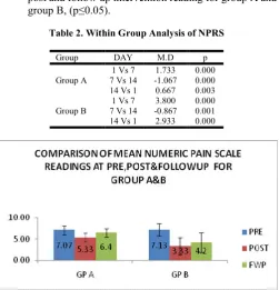
Pain was evaluated by NPRS, the score were compared
[image:3.595.310.562.446.708.2]with in the group by using repeated measure ANOVA. The result showed significant difference between pre, post and follow up intervention reading for group A and group B, (p≤0.05).
Table 2. Within Group Analysis of NPRS
Group DAY M.D p
Group A
1 Vs 7 1.733 0.000 7 Vs 14 -1.067 0.000 14 Vs 1 0.667 0.003
Group B
1 Vs 7 3.800 0.000 7 Vs 14 -0.867 0.001 14 Vs 1 2.933 0.000
Fig. 1. Within group comparison of NPRS
Independent T-test was used to compare the NPRS
Table 3. Between group analysis of NPRS
Mean SD T P
Group A
Group B
Group A
Group B
Pre 7.07 7.13 1.033 1.457 -.145 .886 Post 5.33 3.33 1.047 2.059 3.354 .002 Followup 6.4 4.2 0.986 2.336 3.361 .002
Fig. 2. Between Group Comparison of NPRS
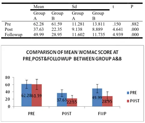
Physical function and disability was evaluated by
[image:4.595.308.560.115.329.2]WOMAC, The score was compare with in the group by using repeated measure ANOVA. The results showed significant difference between pre, post and follow up intervention reading for group A and group B, (p=0.00).
Table 4. Within Group Analysis of WOMAC
Group DAY M.D P
Group a
1 Vs 7 24.658 0.00 7 Vs 14 -12.365 0.00 14 Vs 1 12.293 0.00 Group b 1 Vs 7 39.241 0.00 7 Vs 14 -6.599 0.00 14 Vs 1 32.642 0.00
Fig. 2. Within Group Comparison of WOMAC
Independent T-test was used to compare the WOMAC
between group A and group B, the results showed non significant difference for pre intervention reading where
as for post and follow up intervention reading results showed a significant difference. (p=0.882, p=0.00 and p=0.00) respectively.
Table 5. Between Group Analysis of WOMAC
Mean Sd t P
Group A
Group B
Group A
Group B
Pre 62.28 61.59 11.281 13.811 .150 .882 Post 37.63 22.35 9.138 8.889 4.641 .000 Followup 49.99 28.95 11.602 11.735 4.939 .000
Fig. 5. Between Group Comparison of WOMAC
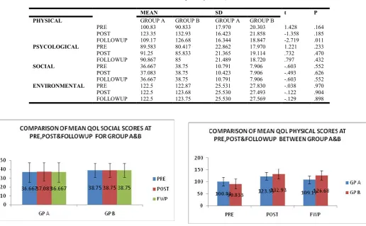
Quality of life was evaluated by WHOQOL, the score
were compared with in the group by using repeated measure ANOVA.
For the physical domain the result showed significant
difference between the pre-post, post-follow up, and pre-follow up intervention reading for group A. (p=0.00, p=0.00 and p=0.027). Same as for group B there was a significant difference between pre-post and pre- follow up where as there was non significant when post and follow up intervention readings were compared. (p=0.00, p=0.82 and p=0.00) respectively.
For the psychological domain the result showed non
significant difference between pre-post, post-follow up and pre-follow up intervention reading for group A. (p=0.334, p=0.334, p=0.378). But for the group B there was significant difference between post and pre-follow up where as there was non significant difference when post and follow intervention reading were
compared. (p=0.003, p=0.164 and p=0.006)
respectively.
For the social domain the result showed no significant
[image:4.595.43.289.284.597.2]difference for group A as well as group B for pre-post, post-follow up, pre-follow up intervention reading. Group A (p=0.334, p=0.334, p=NA) Table 6. Within Group Analysis of QOL
Mean Difference p P P
PRE-POST POST-FWP PRE-FOLLOWUP
Physical Group A -22.513 14.180 -8.333 .000 .000 .027
Group B -42.100 6.250 -35.850 .000 .082 .000
Psycological Group A -1.667 .383 -1.283 .334 .334 .378
Group B -5.417 .833 -4.583 .003 .164 .006
Social Group A -.417 -.417 0 .334 .334 N.A
Group B 0 0 0 N.A N.A N.A
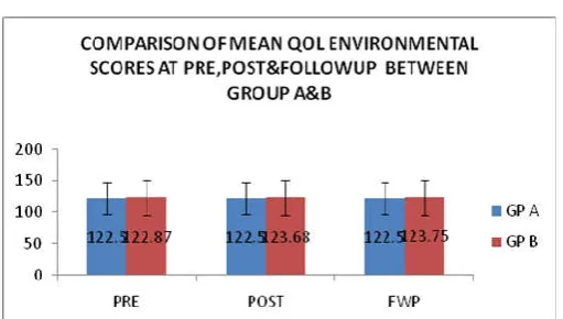
Environmental Group A -.003 0 .003 .334 .334 N.A
Group B -.817 -.067 -.883 .174 .217 .140
[image:4.595.87.508.646.744.2]respectively for group B (p=NA, p=NA, p=NA) respectively.
Fig. 5. Within Group Comparison of Physical Domain Of QOL
Fig. 6. Within Group Comparison of Psychological Domain of QOL
Fig. 7. Within Group Comparison of Social domain of QOL
For the enviromental domain the result showed no
[image:5.595.39.291.54.228.2]significant difference for group A as well as for group B for pre-post, post-follow up and pre-follow up intervention reading. Group A (p=0.334, p=0.334 and p=NA) respectively for group B (p=0.174, p=0.217 and 0.140) respectively.
Fig. 8. Within Group Comparison of enviroment Domain Of QOL
Independent T-test was used to compare the WHOQOL
between group A and group B.
For the physical domain the result showed no
significant difference for pre, post and intervention reading except for follow up intervention reading between group A and group B. (p=0.164, p=0.185, p=0.11) respectively.
For the psychological domain the result showed non
significant difference for pre, post and follow up intervention reading between group A and group B. (p=0.233, p=0.470, p=0.432)respectively.
[image:5.595.313.559.58.257.2]Fig. 9. Between Group Comparison of Physical Domain QOL Table 7. Between Group Analysis of QOL
MEAN SD t P
PHYSICAL GROUP A GROUP B GROUP A GROUP B
PRE 100.83 90.833 17.970 20.303 1.428 .164
POST 123.35 132.93 16.423 21.858 -1.358 .185
FOLLOWUP 109.17 126.68 16.344 18.847 -2.719 .011
PSYCOLOGICAL PRE 89.583 80.417 22.862 17.970 1.221 .233
POST 91.25 85.833 21.365 19.114 .732 .470
FOLLOWUP 90.867 85 21.489 18.720 .797 .432
SOCIAL PRE 36.667 38.75 10.791 7.906 -.603 .552
POST 37.083 38.75 10.423 7.906 -.493 .626
FOLLOWUP 36.667 38.75 10.791 7.906 -.603 .552
ENVIRONMENTAL PRE 122.5 122.87 25.531 27.830 -.038 .970
POST 122.5 123.68 25.530 27.493 -.122 .904
[image:5.595.43.560.448.767.2] For the psychological domain the result showed non significant difference for pre, post and follow up intervention reading between group A and group B.( p=0.233, p=0.470, p=0.432)respectively.
Fig. 10. Between Group Comparison of Psychological Domain QOL
For the social domain the result showed no significant
[image:6.595.38.293.313.460.2]difference for pre, post and follow up intervention reading between group A and group B. (p=0.552, p=0.626, p=0.552) respectively.
Fig. 11. Between Group Comparison of Social Domain QOL
For the enviromental domain the result showed no
significant difference foe pre, post and follow up intervention reading between group A and group B. (p=0.970, p=0.904, p=0.898) respectively.
Fig. 13. Between Group Comparison of Enviromental Domain QOL
INTERPRETATION OF RESULT
Both the group A and group B showed significant
decrease in pain and improvement in physical function
with intervention and showed a significant
improvement in follow up reading too.
When decrease in pain and improvement in physical
function was considered between the groups, group B showed significant improvement in post intervention and follow up.
When QOL of subjects with the group were analysed
for physical domain group A showed significant improvement in post and follow up reading where as group B showed significant improvement in post intervention reading, however the improvement was not statistically significant in follow up reading.
When QOL of subjects between the group were
analysed for physical domain group B showed significant improvement in follow up intervention.
When QOL of subjects within the group were analysed
for psychological domain group A showed non significant improvement in post and follow up reading where as group B showed a significant improvement in post intervention reading.
When QOL of subject between the groups were
analysed for enviromental and social domain group A and group B showed non significant improvement in post and follow up intervention.
DISCUSSION
Osteoarthritis (OA) is the most common form of degenerative joint disease affecting 15 to 40% of people aged 40 and above. One hundred fifty one million people worldwide experienced OA in 2004 which was ranked sixth as a leading cause of
moderate and severe disability. More severe changes are
expected in past decade. The knee is the joint most frequently affected by osteoarthritis. OA knee is two times more prevalent than OA hips in people aged over 60 years and is a significant contributor of pain and mobility impairment in
community-dwelling adults (Nor azlin, 2011).Recent researches
have proved that passive joint mobilization and manual therapy knee protocol along with kinesiotherapy have a positive effect on pain and function in patient with osteoarthritis of knee joint independently. In this context, the present study focused on to check whether both the treatment have similar effect or anyone of them proves to be better than other and which ones effect is maintained for a long term duration in rehabilitation of osteoarthritis of knee. In the present study the result depicted that both passive joint mobilization along with exercises and MIMG knee protocol along with exercises were effective to reduce pain, improve function and improve quality of life.
In support of our results Henery pollard et al, 2008 concluded in their study that the MIMG knee protocol demonstrated significant short term relief of self reported pain and
dysfunction in patient with osteoarthritis1.The result of the
present study provided that passive joint mobilization along with exercises shows better effect then MIMG knee protocol in post and follow up analysis. Pain reduction following passive joint mobilization has been already stabilized in previous studies. Mobilization may initiate local physiological mechanism, additional central mechanisms may also be involved. These central mechanisms could include activation of local segmental inhibitory pathway in the spinal cord or
descending inhibitory pathway from the brainstem (Penny
Mossa, 2007). A number of mechanism have been proposed to explain how hypoalgesic effect of passive joint mobilization may be mediated. Local mechanical disturbance may modify
[image:6.595.37.293.541.686.2]the chemical enviroment and thereby alter concentration of
inflammatory mediators. Movement may also trigger
segmental inhibitory mechanism. In addition it has been hypothesized that the mobilization may activate descending pain inhibitory systems, mediated supraspinally. Human studies have demonstrated that joint mobilization produce rapid hypoalgesia with concurrent sympathetic nervous system and motor system excitation, a pattern similar to that generated by direct stimulation of the periaqueductal gray (Penny Mossa, 2007). Sambajon et al. (2003) found a 70% reduction in levels
of cellular prostaglandin (PG) E2, a strong inflammatory
mediators causing hyperalgesia in arthritic joints, within 24 hours of mobilization in an animal study. Skyba et al. (2003) suggested that analgesic effect following knee joint mobilization was primarily due to enhancement of the descending pain inhibitory pathway in the spinal cord, which utilized serotonergic (5-HT1A) and noradrenergic receptors
(alpha-2) (Nor azlin, 2011). Serotonin and noradrenaline
releasing neurons in the spinal cord originate in supra spinal
sites in the brainstem (Penny Mossa, 2007). Activation of
supra spinal inhibitory pathways would be expected to produce a widespread analgesic response that would include areas
outside the site of injury (Nor azlin, 2011).Penny Moss et al
2006; estabilish that 9 min of accessory mobilization of the
tibio-femoral joint immediately increased knee PPT
significantly more effectively than either manual contact or no-contact control procedures, in subjects with mild to moderate knee osteoarthritis. Mobilization increased knee PPT by 27.3%, compared with 6.4% resulting from manual contact, indicating appreciably reduced sensitivity to mechanical pain (Nor azlin, 2011). A recent in vitro study of healthy animal fibroblasts by Sambajon et al. (2003) suggested that movement may alter concentrations of inflammatory mediators, known to
sensitize peripheral nociceptors (Nor azlin, 2011). MJ Jansen et
al 2011; concluded that exercise therapy plus manual mobilization showed a moderate effect size on pain compared to the small effect size for strength training or exercise therapy alone. Passive joint mobilization with exercise also proved significant effect on function in osteoarthritis patients measured by WOMAC scale. In support to our result Deyle et al 2000 demonstrate that Manual therapy technique with exercise produce a 52% improvement in self reports on
function, stiffness and pain (Gail, 2005). As previous studies
prove passive joint mobilization to have a positive impact on pain, range of motion and gait which could explain its positive effect on function. The improvement in motor activity follow passive joint mobilization has been associated with hypoalgesic and sympatho-excitatory responses produce
during the procedure (Nor azlin, 2011). The MIMG protocol
used for group A consisted of a non invasive myofascial mobilization procedure and an impulsive thrust which allows the knee for greater mobility with less effort, restriction and
pain1. However the result of present study depicted that this
protocol is less effective then passive joint mobilization the reason behind could be primary focus of MIMG protocol on patellofemoral articulation thus having a positive effect on every day activity of squatting, steps, stair climbing, kneeling and rising up from chair. Which prove that the technique
mainly focus on patellar tracking problems (Henry Pollard,
2008). The second part of MIMG protocol utilize an impulsive
thrust directed in caudal direction which leave other components of tibio femoral joint unattended in osteoarthritis patients. In addition to that MIMG procedure requires intact ligaments and capsular structure to operate successfully. One more drawback could be that the practitioner was new to the
protocol and Henery Pollard et al, 2008 suggested that for a successful implementation of procedure require practice by practitioner to acquired the motor skill necessary to perform the procedure. On the same hand the practitioner is practicing passive joint mobilization from past 5years, which could be one more reason that passive joint mobilization showed better result. When manual therapy and reinforcing exercise are utilized in a clinical sitting periodic follow up appointments helps maintain the effect of the intervention. The result of present study showed both the techniques have better follow up after 2 weeks, although passive joint mobilization proved to have a better effect, further investigation regarding motor skill and range of motion in follow up is needed. The finding from present study showed both group have positive effect on physical domain of quality of life showing their influence on physical activity. Group B showed a better impact when follow up was considered. In support of our result Vander Wall et al 2005, Salaffi et al 2005 concluded that knee osteoarthritis procedure significant changes in health related quality of life, particularly physical, mental and social components. Many other studies showed a combination or impact of osteoarthritis
on the quality of life of the patients (Ethgen, 2004).The reason
behind the result could be decrease in pain and improvement in function, Penny Moss et al; 2006 in their study added that mobilization have a positive effect on function. Gail D Deyle et al; 2000 concluded that the level of function improves when manual therapy with supervised exercises was given and results were better than conservative management. They added that with the treatment patients have improved in their walking distance which indeed would have in hance their physical activity and allow them to participate more successfully in
activity of daily life (Gail, 2005). For the psychological domain
WHOQOL group B showed better effect than group A, reason behind the result could be that in present study we used a contact procedure of 10 min of passive joint mobilization, which have positive impact on patient’s psychological level. In support of our result Penny Moss et al; 2006 stated that 9 min of accessory mobilization of the tibio-femoral joint immediately increases knee PPT significantly more effectively than non-contact procedure. Second reason behind this was feeling of independent. As there was decrease in pain and improvement in physical function the patient feels more independent in activities of daily living as compared to pre intervention. Thus we concluded that passive joint mobilization had a positive impact on physical as well as psychological aspect of quality of life.
Conclusion
The result of the present study supports the application of passive joint mobilization for the management of chronic osteoarthritis and also added to the literature about chronic osteoarthritis. This study recommended that 2 weeks passive joint mobilization for chronic osteoarthritis knee has got a significantly better improvement than MIMG Knee protocol result regarding pain, function and quality of life.
Acknowledgments
We would like to thank all our well wishers who help us in this study and give us support and courage do this.
Funding: There was no funding source for this study.
Ethical Clearance: The paper is ethically approved by the ethical commity of Dolphin (PG) institute of Biomedical and Natural Sciences, Dehradun.
REFERENCE
Abdul K. Jafar Sadiq, Self-Efficacy, Physical Function, And Quality Of Life In Individuals With Knee Osteoarthritis, Queen’s University Kingston, Ontario, Canada 2008;April. Basaran, S., Guzel, R., Seydaoglu, G. 2010. Validity,
reliability and comparison of the WOMAC osteoarthritis
index and lequesnealgofuntional index. Clin Rheumatol.
2010; july29 (7):749-56.
Bjordal JM, Ljunggren AE, Klovning A, et al. Nonsteroidal anti-inflammatory drugs, including cyclooxygenase-2 inhibitors, in osteoarthritic knee pain: meta analysis of
randomized placebo controlled trials. British Medical
J2004, 329(7478):1317.
Brosseau L, Yonge KA, Welch V, Marchand S, Judd M, Wells GA, TugwellP, Thermotherapy for treatment of
osteoarthritis (Review). Cochrane Database of Systematic
Reviews2003, Issue 4. Art. No.: CD004522.
CU Krageloh et al. validation of the WHOQOL-brief quality
of life questioner for use with medical students. Education
for healty. 2011;24(2).
Dillon CF, Rasch EK, Gu Q, et al. Prevalence of knee osteoarthritis in the United States: arthritis data from the Third National Health and Nutrition Examination Survey
1991-94. J Rheumatol 2006; 33: 2271-9.
Du H, Chen SL, Bao CD, et al. Prevalence and risk factors of
knee osteoarthritis in Huang-Pu District, Shanghai, China.
Rheumatol Int 2005; 25(8):585–590.
Ferraz MB, Quaresma MR, Aquino LR, Atra E, Tugwell P Goldsmith CH. Reliability of pain scales in the assessment
of literate and illiterate patients with rheumatoid arthritis. J
Rheumatol. 1990; Aug17 (8):1022-4.
Gail D Deyle, Stephen C Allison, Robert L Matekel Physical therapy treatment effectiveness for osteoarthritis of the knee: A randomized comparison of supervised clinical exercise and manual therapy procedures versus a home
exercise programme.. PhysTher. 2005; 85:1301-1317.
Henry Pollard, Graham Ward, Wayne Hoskins, Katie Hardy. The effect of a manual therapy knee protocol on
osteoarthritic knee pain: a randomized controlled trial. J
Can Chiropr Assoc 2008; 52(4).
Jordan KM, Arden NK, Doherty M, et al. Standing Committee for International Clinical Studies Including Therapeutic Trials ESCISIT: EULAR Recommendations2003: an evidence based approach to the management of knee osteoarthritis: Report of a Task Force of the Standing Committee for International Clinical Studies Including
Therapeutic Trials (ESCISIT). Ann Rheum Dis 2003; 62
(12):1145–1155.
K M Jordan, N K Arden, M Doherty, B Bannwarth, J W J Bijlsma. EULAR Recommendations 2003: an evidence based approach to the management of knee osteoarthritis: Report of a Task Force of the Standing Committee for International Clinical Studies Including Therapeutic Trials
(ESCISIT). Ann Rheum Dis 2003;62:1145–1155.
Kyle Gibson. Pain and function in knee osteoarthritis: Are they related to local intrinsic factors? Ethgen et al, Social support and health-related quality of life in hip and knee
osteoarthritis. Quality of Life Research March 2004,
Volume 13, Issue 2, pp 321-330
Marks R, Penton L. Are foot orthotics efficacious for treating painful medial compartment knee osteoarthritis? A review
of the literature. Int J ClinPract 2004; 58(1):49–57.
McColl GJ. Pharmacological therapies for the treatment of
osteoarthritis. Med J Aust 2001; 19; 175 Suppl: S108–111.
McMillan G, Nichols L. Osteoarthritis and Meniscus Disorders
of the knee as occupational diseases of miners. Occup
Environ Med 2005; 62(8):567–575.
MJ Jansen, W Viechthauer, A F Lenssen. Strength training alone, exercise therapy alone, and exercise therapy with passive manual mobilization each reduce pain and disability in people with knee osteoarthritis: a systematic
review. Journal of physiotherapy volume 2011; 57
(1):11-20.
Myklebust G, Bahr R. Return to play guidelines after anterior
cruciate ligament surgery. Br J Sports Med. 2005;39
(3):127–131.
Nor azlin M.N. & K. Su lyn. Effects of Passive Joint
Mobilization on Patients with Knee
Osteoarthritis.SainsMalaysiana2011;40(12):1461–1465
Penny Mossa, Kathleen Slukab, Anthony Wrighta. The initial
effects of knee joint mobilization on osteoarthritic
hyperalgesia. Manual Therapy2007;12:109-118.
Pham K, Hirschberg R. Global safety of coxibs and NSAIDs.
Curr TopMed Chem 2005; 5(5):465–473.
Ravaud P, Giraudeau B, Auleley GR, et al. Variability in knee radio graphing: implication for definition of radiological
progression in medial knee osteoarthritis. Ann Rheum Dis
1998; 57(10):624–629.
Roddy E, Zhang W, Doherty M. Aerobic walking or strengthening exercise for osteoarthritis of the knee? A
systematic review. Ann Rheum Dis 2005; 64(4):544–548.
Salaffi F, Carotti M, Stancati A, et al. Health-related quality of life in older adults with symptomatic hip and knee OA: a
comparison with matched healthy controls. Aging Clin Exp
Res 2005; 17(4):255–263.
Van Der Waal JM, Terwee CB, et al. The impact of non-traumatic hip and knee disorders on health-related quality of life as measured with the SF-36 orSF-12. A systematic
review. Qual Life Res 2005;14 (4):1141–1155.
Vilalta C, Nunez M, Segur JM, et al. Knee osteoarthritis:
interpretation variability of radiological signs. Clin
Rheumatol 2004; 23(6):501–504.
Wafaa I. Shereif and Amira A. Hassanin. Comparison between Uses of Therapeutic Exercise and Heat Application on Relieve Pain, Stiffness and improvement of Physical
Function for Patient with Knee Osteoarthritis. Life Science
Journal, 2011;8(3)
Wu CW, Kalunian KC. New developments in osteoarthritis.
Clin Geriatr Med 2005; 21(3):589–601.
![2 {[3 (2 Nitrophenyl)prop 2 enylidene]amino}phenol](data:image/gif;base64,R0lGODlhAQABAIAAAP///wAAACH5BAEAAAAALAAAAAABAAEAAAICRAEAOw==)