IJPSR (2016), Vol. 7, Issue 5 (Research Article)
Received on 09 December, 2015; received in revised form, 04 February, 2016; accepted, 03 April, 2016; published 01 May, 2016
THE EFFECT OF NANOENCAPSULATED CENTELLA ASIATICA L AND ZINGIBER
OFFICINALE ROSC. VAR. RUBRUM COMBINATION TO PROMOTECOLLAGEN SYNTHESIS AND DECREASE THE DIAMETER OF ADIPOCYTE CELLS IN FEMALE WISTAR RATS
Zullies Ikawati *1, Retno Murwanti 1, Yenny Meliana 2 and Witta Kartika 2
Depr of Pharmacology 1, Faculty of Pharmacy Universitas Gadjah Mada, Yogyakarta, Indonesia Indonesian Institute of Science 2, Jakarta, Indonesia
ABSTRACT: Cellulite is a normal condition judging from the medical aspect, but from aesthetics aspect, cellulite deserves more attention, especially for women. Centella asiatica is reported to promote both fibronectin and collagen synthesis. Meanwhile,
Zingiber officinale is reported to have lipolysis activity. Combination of the two herbals is assumed to have complementary effect as anti cellulite agents. The combination of the herbal extracts is prepared in nanoemulsion form to enhance the bioavailability. The study aimed to determine the effect of nano emulsion of C. asiatica and Z. officinale
combination (proportion of 5:1) to stimulate skin collagen synthesis and decrease the diameter of adipocyte cells using histological parameters as an indicator of lipolysis activity. Twenty female Wistar rats weighing 120-140 g were fed a high-fat diet for 30 days. The animals were divided into 4 groups, with the 3 groups received the nanoemulsion of herbal combination with the dose of 50, 100, and 200 mg/kg BW for 30 days, and 1 group received CMC Na 0, 5% as negative control. On the day 31, rats were sacrified and skin samples of 1.0 cm2, including fatty tissue, were obtained from the subjects. One part of the tissue sample was used for collagen assay, while another was used for hematoxylin-eosin staining. The amount of collagen in skin tissue was assayed using Sirius Red Collagen Detection Kit (Chondrex). The collagen thickness was also measured histologically using Sirius red staining. The diameter of adipocyte cells were measured under light microscope to represent lipolysis activity. Results showed that the amount of skin collagen was increased with the increase of extract doses, however the lowest dose showed no significant different with the normal animal. The diameter of adipocyte cells was also decreased in dose-dependent manner. Results indicate that the combination of C. asiatica and Z. officinale can be developed as herbal medicine for anti cellulite agent.
INTRODUCTION: Cellulite is an appearance changing of the skin that resembles an orange peel. Women aged 20 years old or more, 90% of them have cellulite experience with varying degree of severity, meanwhile men had a smaller number (2%) 6.
QUICK RESPONSE CODE
DOI:
10.13040/IJPSR.0975-8232.7(5).1909-14
Article can be accessed online on: www.ijpsr.com DOI link: http://dx.doi.org/10.13040/IJPSR.0975-8232.7 (5).1909-14
Cellulite is a normal condition judging from the medical aspect, but from aesthetics aspect, cellulite deserves more attention, especially for women. It is not specific to overweight women although increased adipogenicity will exacerbate the condition.
It is difficult to pinpointitsaetiology and physiology/ pathophysiology of cellulite, as thereare many factors that are involved it, affect it, and many processes that contribute simultaneously and sequentially 2. Cellulite occurs due to a microcirculation diminishing, infiltration of the interstitial fluid (edema), hypertrophy on local Key words:
cellulite, collagen synthesis, adipocytes cells diameter,
C. asiatica, Z. officinale Correspondence to Author: Prof. Dr. Zullies Ikawati., Apt
Faculty of Pharmacy Gadjah Mada Universit, Yogyakarta, Indonesia
adipose tissue, oxidative stress, inflammation mild persistent, and changes in the extracellular matrix 6,
11, 16
. Several mechanisms can reduce the appearance of cellulite, such as increasing the synthesis of collagen, stimulate lipolysis activity, using PDE inhibitors, increasing blood flow to smooth microcirculation, laser, etc.
In traditional Asian medicine, the herb of Centela asiatica has been used for hundreds of years, especially in dermatological conditions, to improve small wounds, scratches, burns, hypertrophic wounds healing, and as an anti-inflammatory agent, particularly in eczema 3. C. asiatica, which containsasiatic acid, madecassic acid, and asiaticoside is reported to stimulate human collagen synthesis 4, that often used in skin care products. Besides that, it reported that C. asiatica could increase microcirculation and capillary permeability effects on the skin. Another activity shown by C. asiatica was lipolysis and antioxidant that affect in reducing cellulite 8.
Ginger, the rhizome of the perennial plant Zingiber officinale Roscoe, is used as a flavoring agent for food, mostly in a powdered and candied form. In addition, ginger is widely used as a herbal medicine for a number of conditions including those affecting the digestive tract, headaches and motion sickness5. The characteristic pungent taste of ginger is attributed to the gingerols (6-gingerol, 8-gingerol and zingerone). Ginger was reported that it could stimulate the lipolysis activity, which is the process of triglyceride hydrolysis into glycerol and free fatty acids. This was indicated that ginger could be used to reduce lipid pile, so that it could reduce cellulite appearance 7, 15.
This recent study investigates the effect of combination of C. asiatica and Z. officinale
extracts in nanoemulsion form to stimulate collagen synthesis and decrease diameter of adipocyte cells which indicate the lipolytic action of the fatty cells. This study is the first study for this combination to develop herbal product for anti cellulite agent.
MATERIALS AND METHODS:
Experimental Animal and Materials: Twenty-five non-pregnant female Wistar rats (BW: 140-160 g, aged: 8 weeks) were used in this study. The
tested extract prepared from nanoemulsion containing 5% of C. asiatica herbs extract and 1 %
Z. officinale rhizome extract which mixed with malt dextrin as the carrier. The tested extract was dissolved with 0,5% CMC-Na before administered to the animals. Sirius Red Collagen Kit Assay was purchased from Chondrex. All other reagents used were of analytical grade.
Experimental Design:
Twenty female Wistar rats weighing 120-140 g were fed a high-fat diet for 30 days. The animals were divided into 4 groups, with the 3 groups received the nanoemulsion of herbal combination with the dose of 50, 100, and 200 mg/kg BW for 30 days, and 1 group received CMC Na 0,5% as negative control. One group of rats (n=5) was fed normal diet and served as normal control. On the day 31, rats were sacrified and skin samples of 1.0 cm2, including fatty tissue, were obtained from the subjects. One part of the tissue sample was used for collagen assay, while another was used for hematoxylin-eosin staining. The amount of collagen in skin tissue was assayed using Sirius Red Collagen Detection Kit (Chondrex). The collagen thickness was also measured histologically using Sirius red staining. The diameter of adipocyte cells were measured under light microscope to represent lipolysis activity.
Procedures:
Combination extract of C. asiatica and Red ginger was dissolved with 0,5% CMC-Na. Stock solutions test were prepared every 3 days to maintain the stability of the solution.
Treatment Applied: The rats were injected with stock solutions test and lard for 30 days. The solution volume was determined based on BW and treatment groups. Testedrats weighed every 3 days to determine the BW gain. Took the rats skin to measure the concentration and thickness of collagen after 30 days of treatment.
Skin Sample Preparation and Reading of Collagen Concentration:
physiological saline added in skin slices as much as 2 mL then crushed using homogenizer. Put the skin solution into5 mL conical flask, then 1 mL NaCl physiological solution added. Skin that been destroyed was centrifuged for 90 minutes at 2000 rpm. The supernatant discarded and the sediment was being taken. Acetic acid solution 1 mL (0.05 M) mixed with sediment then homogenized using vortex. Afterthat the solution was re-centrifuge for 90 minutes, then the supernatant was taken (1 mL) using micropipette. Supernatant stored in tubes kept in refrigerator. Meanwhile, prepared the collagen kit assay, started with making collagen standard solution. The collagen standard solution made by certain level solution to make regression equation. The equation made to determine collagen concentration in the sample.
Fill the acetic acid solution (0.05 M) into 8 tubes in amount of 250 µL each. Take 250 µL of collagen standard solution and fill into the first tube, then homogenized using vortex. After that, 250 mL solution taken from the first tube and put itinto the second tube then re-homogenized using vortex. Dilution carried out up to the seventh tube. The eighth tube was used as blank. When all the solution was already prepared, took 100 µL solution from standard, sample and blank solution thenfillit into another tube. Added 500 mL Sirius Red on each tube and homogenized using vortex, then incubated for 20 minutes in room temperature. The solvent that was incubated subsequently centrifuged at a speed of 10000 rpm for 3 minutes. The supernatant removed carefully to get the sediment. After the sediment was obtained, then added 500 µL washing solution. Re-vortex and
centrifuged at the same speed and time. Supernatant re-discarded to obtain the sediment. The sediment was added by 250 µL buffer extract solution. It was re-vortex to dissolve the sediment then was analyzed using micro plate reader. The solvent taken as much as 200 µL and placed on 96-well plate then read the absorbance at OD 550 nm.
Statistical Analyses: The collagen concentration data obtained was converted into milligrams within skin-tested milligrams. All of the data were statistically analyzed using One-Way ANOVA test with 95% confidence level.
RESULTS AND DISCUSSION: Results:
Collagen Concentration:
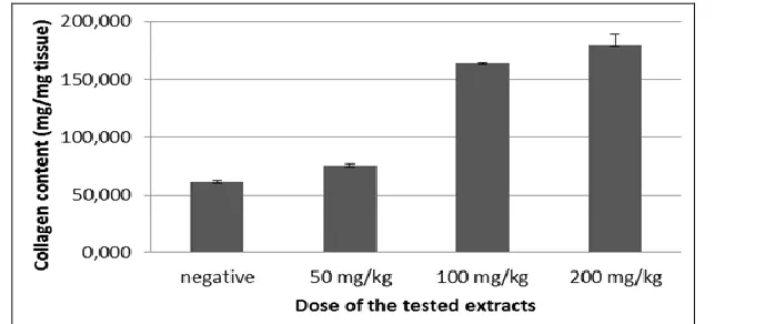
[image:3.612.147.496.562.708.2]The collagen concentration levels in C, D and E groups are 0.0752 mg/mg skin; 0.1637 mg/mg skin; 0.1785 mg/mg skin, respectively. It showed that increasing dose of solution increases collagen concentration levels in skin. Meanwhile, the collagen concentration levels in groups A and B are 0.1407 mg/mg skin and 0.0611 mg/mg skin. The result also showed that combination extract of C. asiatica and red ginger was affect collagen concentration significantly on group C towards D and E groups (p=0.045 and p=0.022). Whereas, the treatment group D and E was not different (p> 0.05). If control group was compared to the treatment group, the result showed differences between groups A to group D with a significance value of 0.023; meanwhile A to E has a significant value of 0.011. Group B had a value of p> 0.05. It means between groups B and treatment groups showed no different.
FIG. 1. COLLAGEN CONCENTRATION ON TESTED RATS FOR 30 DAYS MEASURED BY COLLAGEN KIT ASSAY
Thickness of Collagen:
FIG.2: COLLAGEN THICKNESS ON TESTED RATS FOR 30 DAYS SEEN HISTOLOGICALLY WITH SIRIUS RED STAIN
(Note: A= as a normal control, fed normal pellet and injected with CMC-Na solution; B= as a negative control, fed normal pellet and fed lard once a day then injected with CMC-Na solution; C, D, E =fed normal pellet and fed lard once a day then injected with different dose of solution i.e. 0.05 mg/g BW, 0.1 mg/g BW, 0.2 mg/g BW, respectively
Consistent results showed by collagen thickness parameter. Histologically, the thickness ofcollagen parameter also showed an increase in thickness of collagen which is directly proportional to the increase in dose of treatment groups. The average thickness of collagen in group C, D, and E was 314.33 μm; 388.80 c; 410.14 μm. On the other hand, the average in group B and A showed the results of collagen thickness was 376.28 μm and 265.99 μm. For the treatment group, C had a difference with the other treatment groups, to D and E with a value of p = 0.022 and p = 0.005,
while group D did not differ with E as the value of p> 0.05.
There were significant differences on group A as a control to treatment groups. This was found in groups D and E (p=0.001 and p=0.000). Group B did not significantly different to the treatment group, but the p value between groups B and C show the number p = 0.051, it was indicated that there was tendency to be a significant difference. Group B also had a significant difference, compared with group A (p=0.002).
[image:4.612.131.488.462.613.2]Body Weight:
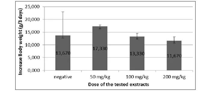
FIG. 3. BODY WEIGHT GAIN ON TESTED RATS DURING TREATMENT MEASURED EVERY 3 DAYS
Note: A= as a normal control, fed normal pellet and injected with CMC-Na solution; B= as a negative control, fed normal pellet and fed lard once a day then injected with CMC-Na solution; C, D, E =fed normal pellet and fed lard once a day then injected with different dose of solution i.e. 0.05 mg/g BW, 0.1 mg/g BW, 0.2 mg/g BW, respectively
Fig. 3. shows that the body weight gain were inversely proportional to the injection dose. The higher the dose, then the lower the body weight gain. It shown by descriptive statistic of the data that average body weight gain on group C 11,33 g/
Adipocyte Cell :
Results of Post-hoc tested analysis show a significant difference between treatment group with a dose of 50 mg/kg BB dose group BB comparedwith dose group 100 mg/kg BB and dose group of 200 mg/kg BB very minor significance 0.000. In the group of 100 mg/kg BB differ significantly with a dose of 200 mg/kg BB with significant value 0,000. Comparation between normal group and all treatment groups have the significant differences as a P value less than 0.05.The significant value of the normal group to group a dose of 50 mg/kg BB has a value of 0,001,
a dose of 100 mg/kg BB with a significance value of 0.000, while the normal group and the dose of 200 mg/kg BB have a significance value of 0.000. In the negative control group at a dose of 50 mg/kg BB did not have a significant difference because the significance value of 0.069 (p> 0.05). Whereas if compared with a dose of 100 mg/kg BB and 200 mg/kg BB, the negative control group had no significant difference, respectively 0,021 and 0,007. Normal control group with the negative control group had no significant difference due to the value of p> 0.05 is 0.200.
FIG.4: MEAN THE DIAMETER OF ADIPOCYTE CELL (µm) MEASURED ON THE 30th DAYS AFTER TESTED RATS WERE SACRIFICED
(Note : A : as a normal control, B : as a negatif control, C:a dose of 50 mg/kg BB, D: a dose of 100 mg/kg BB, E: a dose of 200 mg/kg BB). (a) showed a significant difference (p>0,05) between normal control with treatment control, (b) shows a significant between negative control with control treatment (Errors Bars: SD).
DISCUSSION: The concentration and the
thickness of collagen increased linearly by increasing the dose of combined extract C. asiatica
and red ginger. This suggests that the combination extract of C. asiatica and red ginger have dose with dependent characteristic. The significant difference was found in control and treatment groups, especially in groups D and E on both of parameters. It means that combining C. asiatica and red ginger extract can increase collagen synthesis. This is similar with the opinion of 8, that C. asiatica is a plant that can increase collagen synthesis, due to the mechanism of asiaticoside contained in stock solutions test. Asiaticoside is one of triterpene compounds and an identity compound of C. asiatica. According to the monograph conducted by WHO 1, C. asiatica contains not less than 2% triterpene glycosides in the form of asiaticoside and madecassoside. Asiaticoside was classified in saponin terpenoid. Kanzaki 9 reported that saponin could increase ability of TGF-β receptor on
fibroblast, so the ability of fibro blasts to proliferate into collagen also increased.Several possibilities regarding the mechanism of the activation of the TGF-β pathway by saponin are 1) Saponin stimulatesthe synthesis, secretion, and activating TGF-β 1 in fibroblast, 2) Saponin changes the expression of TGF-β receptors on fibroblasts so that these receptors become more sensitive to the presence of TGF-β, 3) the post-reseptor signal transduction system is mofied by saponin. So, its predicted that asiatikosida compound that plays a role in increasing the amount of collagen.
Based on control group towards treatment group, group B has higher number of collagen compared to group C. But, it was not happened in group A. This may imply that fat administration influence number of collagen in tested rats. According to previous study by Junior et al 10, shown that wistar rats treated with high-fat diet had higher body weight and collagen levels than rats fed normal diet. The differences between this study was the different sample organ taken. Junior et al 10 was used histology sample from penile organ. The mechanism of this phenomenonis still not widely known. Furthermore, it was possible that lard becomes factor that influence high levels of collagen in group B.
Lipolysis is one of anti-cellulite mechanisms beside increasing collagen level 14. On previous study was submitted that C. asiatica and red ginger has a lipolysis activity, even C. asiatica has a better lipolysis activity than caffeine 8, 12. Lipolysis is triglyceride hydrolysis process to form glycerol and free fatty acid 15. Lipolisis also associated with
Hormone Sensitive Lipase (HSL) translocation activity from cytosol to lipid droplets in a diposities7. It was possible that lard injected to treatments group becomes more controlled and had no effect on collagen levels. The results on body weight shown that treatment are able to diminishing body weight gain but not significantly, so it can’t be determined as a slimming substance.
CONCLUSION: Based on the result of this study, it can be concluded that concentration and thickness of collagen increased by increasing the dose of combination extracts of C. asiatica and red ginger.
REFERENCES:
1. Anonymous, Obesity and Overweight:http://www.who.int/
mediacentre/factsheets/fs311/en/. 1999; March, 15th 2015. 2. Almeida M, Serrano C, Roldan J, Rejano J: Cellulite's
aetiology: a review. Journal of the European Academy of Dermatology and Venereology2012; 27(3), 273–278.
3. Brinkhaus B, Lindner M, Schuppan D, Hahn EG : Chemical,
pharmacological and clinical profile of the East Asian medical plantCentella asiatica, Phytomedicine 2000; 75: 427-48. 4. Bonte F, Dumas M, Chaudagne C, Meybeck A: Influence of
asiatic acid, madecassic acid, and asiaticoside on human collagen I synthesis, Planta Med1994 Apr; 60(2):133-5. 5. Borrelli F, Capasso R, Pinto A, Izzo AA : Inhibitory effect of
ginger(Zingiber officinale) on rat ileal motility in vitro, Life Sci2004;74:2889–96
6. DupontE, Journet, M, Oula M., GomezJ, Leveille C, Loing E, Bilodeau : An integral topical gel for cellulite reduction: results from a double blind, randomized, placebo-controlled evaluation of efficacy,Clinical, Cosmetic andInvestigational Dermatology2014; 7: 73-88.
7. Han LK, Morimoto C, Zheng YN, Li W, Asami E, Okuda H,
Saito M : Effects of zingeroneon fat storage in ovariectomized rats, Yakugaku Zasshi: Journal of the Pharmaceutical Society of Japan2008 ;128(8): 1195-1201.
8. Hashim P, SidekH, Helan MH, Sabery A, Palanisamy UD, and IlhamM : Triterpene Composition and Bioactivities of Centella asiatica, Molecules, 2011; 16:1310-1322.
9. Kanzaki T, Nobuhiro M, Shiina R, SaitoY: Role of
Transforming Growth Factor-β Pathway in the Mechanism of Wound Healing by Saponin from Ginseng Radixrubra, British Journal of Pharmacology 1998 ; 125 : 255-262.
10. Junior J, Oliveira F, Silva P, Furriel A, Sampaio F, Gregorio B : Lard and/or Canola Oil-rich Diets Induce Penile Morphological Alterations in a Rat Model.Acta Cirurgica Brasileira2014; 29: 39-44.
11. KruglikovI: The Pathophysiology of Cellulite: Can the Puzzle Eventually Be Solved?, Journal of Cosmetics, Dermatological Sciences and Applications 2012 ;2: 1-7.
12. Nomura H : The Pungent Principles of Ginger. Part I. A New
Ketone, Zingerone (4-Hydroxy-3-methoxy-phenylethyl
Methyl Ketone) occuring in Ginger. Journal of the Chemical Society Transactions 1917; 769-776.
13. Novriansyah R : Perbedaan Kepadatan Kolagen di Sekitar Luka Insisi Tikus Wistar yang Dibalut Kasa Konvensional dan Penutup Oklusif Hidrokoloid Selama 2 dan 14 hari. Thesis, Program Pasca Sarjana Magister Ilmu Biomedik dan Program Dokter Spesialis I Ilmu Bedah Universitas Diponegoro, Semarang, 2008.
14. PalanisamyUD, Lin LT, ManaharaT, Sivapalan V,
SubramaniamT, Helme MH, and MasilamaniT : Standardized extract of Syzygium aqueum: a safe cosmetic ingredient, International Journal of Cosmetic Science 2011;33: 269-275.
15. Snyder PB, Esselstyn JM, Loughney K, Wolda SL, Florio VA
: The role of cyclic nucleotide phosphodiesterases in the regulation of adipocyte lipolysis, Journal Lipid Research 2005;46: 494-503.
16. TerranovaF, Berardesca E, Maibach H : Cellulite: nature and aetipathogenesis, International Journal of Cosmetic Science 2006;28: 157-167.
17. Zainol MM, Abdul-HamidA, Abu BakarF, and Pak Dek S: Effect of Different Drying Methods on the Degradation of Selected Flavonoids in Centella asiatica, International Food Research Journal 2009; 16: 531-537.
All © 2013 are reserved by International Journal of Pharmaceutical Sciences and Research. This Journal licensed under a Creative Commons Attribution-NonCommercial-ShareAlike 3.0 Unported License.
This article can be downloaded to ANDROID OS based mobile. Scan QR Code using Code/Bar Scanner from your mobile. (Scanners are available on Google Playstore)
How to cite this article: