INTRODUCTION
Although established genetic models offer unmatched resources for genetic analysis, there is strong motivation to develop genetic tools in new species. This motivation stems from the diversity that is evident in development, morphology and physiology, which means that many questions cannot be addressed in the few well-established models. The development of transgenesis in emerging animal models, such as Nematostella vectensis (Renfer et al., 2010), Parhyale hawaiensis (Pavlopoulos and Averof, 2005), Tribolium castaneum (Berghammer et al., 1999) and Ciona intestinalis (Sasakura et al., 2007), represents the first step for establishing sophisticated genetic techniques in these species. One such technique, gene trapping, captures gene expression at the site of transgene insertion. Transposon-mediated gene trapping allows the implementation of unbiased genetic screens to identify new genes and provides valuable markers for in vivo imaging and phenotypic characterisation (Bellen, 1999; Bellen et al., 1989).
In Drosophila, gene traps are also used to generate GAL4 drivers, powerful tools that exploit endogenous genes to direct gene expression with spatial and temporal specificity (Brand and Perrimon, 1993). In principle, gene trapping can be used to introduce a wide range of genetic tools, such as alternative expression drivers, recombinases, specialised markers and knockdown constructs, into a trapped locus. In practice, this is difficult to achieve because each application relies on a different transgene construct, and traps derive from unique insertions that
cannot be reproduced with each construct. Replacing one type of construct with another, at a given locus, is possible but technically challenging and restricted to highly developed genetic models (Sepp and Auld, 1999).
We present a new approach termed integrase-mediated trap conversion (iTRAC) that allows primary gene traps to be adapted for diverse applications through transgene conversion. The approach uses a primary exon-trapping vector, based on the Minos transposon, that incorporates an attPdocking site for the fC31 integrase. Once a trap has been generated and selected, a virtually unlimited range of secondary constructs carrying the cognate attB site can be integrated specifically into the docking site at the trapped locus (Fig. 1). As a proof of principle, we demonstrate iTRAC in Parhyale hawaiensis, a crustacean that has emerged as an attractive model for developmental studies (Browne et al., 2005; Extavour, 2005; Gerberding et al., 2002; Liubicich et al., 2009; Ozhan-Kizil et al., 2008; Pavlopoulos and Averof, 2005; Pavlopoulos et al., 2009; Price et al., 2010; Rehm et al., 2009; Vargas-Vila et al., 2010).
MATERIALS AND METHODS Gene-trapping constructs
The 1.3 kb PhHsp70afragment (accession FR749989) was isolated by inverse PCR from Parhyalegenomic DNA using a previously described approach (Pavlopoulos and Averof, 2005; Pavlopoulos et al., 2009) and cloned upstream of the DsRed/SV40polyA reporter cassette to obtain plasmid pSL(PhHsp70a-DsRed).
A 230 bp SpeI fragment containing the fC31attPsite from pTA-attP (Groth et al., 2000) was cloned into the SpeI site of pSL(PhHsp70a-DsRed) to generate pSL(attP;PhHsp70a-DsRed). The attP-PhHsp70a-DsRed construct was cloned as an AscI fragment into the Minos vectors pMi{3xP3-DsRed} and pMi{3xP3-EGFP} (Pavlopoulos and Averof, 2005; Pavlopoulos et al., 2004), generating pMi{3xP3-DsRed;attP;PhHsp70a-DsRed} and pMi{3xP3-EGFP;attP;PhHsp70a-pMi{3xP3-DsRed;attP;PhHsp70a-DsRed}.
The transcription initiation and splice sites of PhMS, PhHS and PhHsp70awere mapped by 5⬘RACE from transgenic animals carrying stable insertions of PhMS-DsRed(Pavlopoulos and Averof, 2005), PhHS-DsRed(Pavlopoulos et al., 2009) and PhHsp70a-DsRed(DistalDsRedtrap), Development 138, 2625-2630 (2011) doi:10.1242/dev.066324
© 2011. Published by The Company of Biologists Ltd
1Institute of Molecular Biology and Biotechnology, Foundation for Research and Technology Hellas, GR-70013 Heraklio, Crete, Greece. 2Laboratory for Development and Evolution, Department of Zoology, University of Cambridge, Cambridge CB2 3EJ, UK.
*Present address: Evolutionary Biology Group, Institute of Biology, Leiden University, 2333 BE Leiden, The Netherlands
†Author for correspondence ([email protected]) Accepted 11 April 2011
SUMMARY
Genetic model organisms such as Drosophila, C. elegansand the mouse provide formidable tools for studying mechanisms of
development, physiology and behaviour. Established models alone, however, allow us to survey only a tiny fraction of the morphological and functional diversity present in the animal kingdom. Here, we present iTRAC, a versatile gene-trapping approach that combines the implementation of unbiased genetic screens with the generation of sophisticated genetic tools both in established and emerging model organisms. The approach utilises an exon-trapping transposon vector that carries an integrase docking site, allowing the targeted integration of new constructs into trapped loci. We provide proof of principle for iTRAC in
the emerging model crustacean Parhyale hawaiensis: we generate traps that allow specific developmental and physiological
processes to be visualised in unparalleled detail, we show that trapped genes can be easily cloned from an unsequenced genome, and we demonstrate targeting of new constructs into a trapped locus. Using this approach, gene traps can serve as platforms for generating diverse reporters, drivers for tissue-specific expression, gene knockdown and other genetic tools not yet imagined.
KEY WORDS: Parhyale, Gene trapping, fC31 integrase, Regeneration, Transgenesis, iTRAC
A versatile strategy for gene trapping and trap conversion in
emerging model organisms
Zacharias Kontarakis1, Anastasios Pavlopoulos2, Alexandros Kiupakis1, Nikolaos Konstantinides1, Vassilis Douris1,* and Michalis Averof1,†
D
E
V
E
LO
P
M
E
N
respectively, using SMART-RACE (Clontech) and a reverse primer targeting the coding sequence of DsRed (5⬘CTTGGTCACCTTC -AGCTTGGCGGT-3⬘).
iTRAC constructs
The EGFP coding sequence was excised as a NcoI-NotI fragment from pMi{3xP3-EGFP} and cloned into NcoI andNotI cut pSL(PhHS-DsRed) (Pavlopoulos et al., 2009) to generate pSL(PhHS-EGFP).
Plasmid pBS(MiL;attB;PhHS-EGFP) was generated as follows: we removed the right inverted repeat of Minosfrom pMi{3xP3-DsRed} by AvrII andNheI digestion and religation; we excised DsRedwith SgrBI and NotI and replaced it with a SgrBI-NotI fragment of pSL(PhHS-EGFP) carrying PhHS-EGFP; we then excised 3xP3using SalI and replaced it with a SalI fragment containing attBfrom plasmid pTA-attB (Groth et al., 2000).
Plasmid pBS(MiL;attB;PhHsp70a-EGFP) was generated by excising PhHS from pBS(MiL;attB;PhHS-EGFP) using SmaI and NcoI and replacing it by PhHsp70aobtained by SmaI and partial NcoI digestion of pMi{3xP3-DsRed;attP;PhHsp70a-DsRed).
Parhyalegene traps
Plasmid pMi{3xP3-DsRed;attP;PhHsp70a-DsRed} was injected with Minostransposase mRNA and stable transgenic lines were isolated as described previously (Pavlopoulos and Averof, 2005). Traps were imaged on a Leica MZ16F epifluorescence stereoscope.
Trapped genes were identified by 5⬘ RACE on each line using the SMART-RACE Kit (Clontech) with reverse primers DsRed-SMART (5⬘ -CTTGGTCACCTTCAGCTTGGCGGT-3⬘) or hsp70aDsRedR (5⬘
-GAGGCCATGGTTGTGGATT-3⬘). Additional cDNA sequences from the Distal locus were obtained by 3⬘ RACE on wild-type animals using forward primers targeting the sequences already determined (accession FR821313). In situ hybridisation using the DistalcDNA probe was carried out on wild-type embryos as described previously (Rehm et al., 2009). The DistalDsRedline carries additional transgene insertions that do not give
visible traps.
Splicing to PhHsp70a-DsRedand normal splicing at the Distallocus were measured by quantitative RT-PCR using a common forward primer targeting the Distal5⬘UTR (5⬘-TGACAGTCGCTGCGAAATAG-3⬘) and two reverse primers targeting DsRed(5⬘ -GGGTGCTTCACGTACACCTT-3⬘) and a Distal3⬘exon (5⬘-GTCTGCTCGTCTTCCTTTGC-3⬘). RNA was isolated from populations of 20-30 heterozygous and homozygous DistalDsRed embryos, reverse transcribed using oligo(dT) primers and
amplified in triplicate on the MJ Research Opticon real-time PCR machine; PCR efficiency with each set of primers was 1.98 and 1.72, respectively. Trapped and normally spliced products were detected at a ratio of 0.09 (s.e.0.18) in heterozygous embryos and 0.15 (s.e.0.32) in homozygous embryos.
fC31 interplasmid assay, integration and iTRAC
In vitro synthesised fC31 integrase mRNA was prepared as described previously (Groth et al., 2004). For the interplasmid assay, 1- or 2-cell stage Parhyaleembryos were injected with plasmids carrying the attP and attBsites and fC31 integrase mRNA at 500 ng/l, 500 ng/l and 100 ng/l, respectively. Surviving embryos were collected 24 hours after injection and nucleic acids were isolated by mechanical disruption in Holmes-Bonner buffer (100 mM Tris-HCl pH 7.5, 10 mM EDTA, 300 mM NaCl, 2% SDS, 7 M urea), triple phenol/chloroform extraction and ethanol precipitation. Recombination events were detected by PCR using two forward primers (5⬘-AGGAAGGGAAGAAAGCGAAA-3⬘and 5⬘ -CCAATTTCTATCTTAGCCCAACC-3⬘) and a common reverse primer (5⬘-GGGTGCTTCACGTACACCTT-3⬘), as illustrated in Fig. 4A.
Genomic integration via fC31 integrase was tested by injecting plasmid pBS(MiL;attB;PhHS-EGFP) and integrase mRNA (300 ng/l and 100 ng/l, respectively) into 1- or 2-cell stage embryos of a line carrying multiple attPinsertions. Out of 512 injected embryos, 207 survived to late embryogenesis and 104 expressed EGFP after heat shock. Seventeen individuals with uniform expression were raised to adulthood; all produced progeny carrying the PhHS-EGFPmarker. Wild-type embryos lacking attP insertions were injected as controls; out of 619 injected embryos, 255 survived as late embryos, but none expressed EGFP after heat shock.
iTRAC was tested by injecting plasmid pBS(MiL;attB;PhHsp70a-EGFP) and fC31 integrase mRNA (300 ng/l and 100 ng/l, respectively) into 1- or 2-cell stage embryos of the DistalDsRedline. Out of 389 injected
embryos, 90 survived to late embryogenesis and 47 expressed EGFP in at least some limbs and in the characteristic pattern of Distal; 12 of these embryos showed bilateral replacement of DsRed fluorescence by EGFP. Four individuals were raised to adulthood, of which three gave rise to DistalEGFPprogeny.
RESULTS AND DISCUSSION The gene-trapping vector
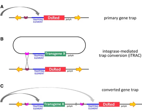
[image:2.612.50.296.53.246.2]Our Minosgene-trapping vector carries the 3xP3-DsRedor 3xP3-EGFP transformation marker (Berghammer et al., 1999; Pavlopoulos and Averof, 2005), the fC31attPsite, and a ‘trapping element’ (described below) upstream of the DsRed reporter and the early mRNA polyadenylation sequence of SV40. The two types of trapping strategies commonly employed, i.e. enhancer trapping and exon trapping, make use of a core promoter or a splice acceptor to capture the activity of cis-regulatory elements or splice donors of trapped genes, respectively. To create a trapping system that is widely applicable, we sought to identify promoters or splice acceptors capable of gene trapping in a range of species. First, we tested two core promoter elements, the Drosophila hsp70basal promoter and an artificial ‘super core promoter’ (which combines Fig. 1. Gene-trap and trap conversion strategy. (A)Primary
gene-trapping construct based on a transposon vector (yellow inverted arrows), carrying a fC31attPrecognition site (purple) and a trapping element (core promoter or splice acceptor) upstream of a reporter gene (DsRed). The vector may also include additional markers (not shown). Depending on its insertion site in the genome, the reporter might come to be expressed under the influence of nearby sequences (grey arrow). (B)Once a gene trap has been isolated, integrase-mediated trap conversion (iTRAC) utilises attPas a docking site for integrating new constructs into the trapped locus. Constructs carrying the cognate attB
site (magenta) are introduced into the locus by integrase-mediated site-specific recombination. A wide range of secondary constructs for different types of applications can be envisaged (see text); in this example, the DsRed trap is converted into one that expresses a hypothetical transgene X. (C)Integration mediated by single attPand
attBsites results in displacement of the original trapping construct by the new construct. Complete replacement is also feasible using flanking pairs of attPand attBsites.
D
E
V
E
LO
P
M
E
N
several core promoter motifs) (Juven-Gershon et al., 2006), for enhancer trapping activity in Drosophila, Triboliumand Parhyale, but neither was found to work across the species tested (Schinko et al., 2010) (data not shown). Next, we searched for core promoters and splice acceptors among sequences that lie upstream of Parhyale hsp70genes. Among the sequences tested, two elements were capable of efficient gene trapping in Parhyale: the heat-inducible element PhHS(Pavlopoulos et al., 2009) and a fragment named PhHsp70a. Both fragments could drive expression patterns specific to individual transgene insertions without any need for a heat shock (Fig. 2B-L). Using 5⬘RACE on cDNA prepared from transgenic lines, we determined that PhHS contains a core promoter upstream of the transcription start site and a large intron within the 5⬘ UTR, whereas PhHsp70ais a truncated 5⬘ UTR sequence with a splice acceptor site that becomes spliced to the exons of trapped genes (see Fig. S1 in the supplementary material). We decided to focus on exon trapping mediated by PhHsp70a.
In our first experiment using the Minos{3xP3-DsRed;PhHsp70a-DsRed}vector, we recovered at least six independent traps from ~250 injected Parhyaleembryos. Using the same vector, we also obtained four independent exon traps in Drosophilafrom ~350 injected embryos, suggesting that this construct can mediate exon trapping efficiently in diverse arthropods.
Imaging of developmental and physiological processes
A variety of exon traps have been generated to date using the PhHsp70a-DsRedtrapping construct in Parhyale(Fig. 2). These include traps with expression in the central nervous system, mesoderm, appendages, mouthparts, gills and other patterns. Most lines have been propagated through many generations over 4-5 years, demonstrating that the transgenes are stable, with continuing activity and no detrimental effects on reproduction and survival.
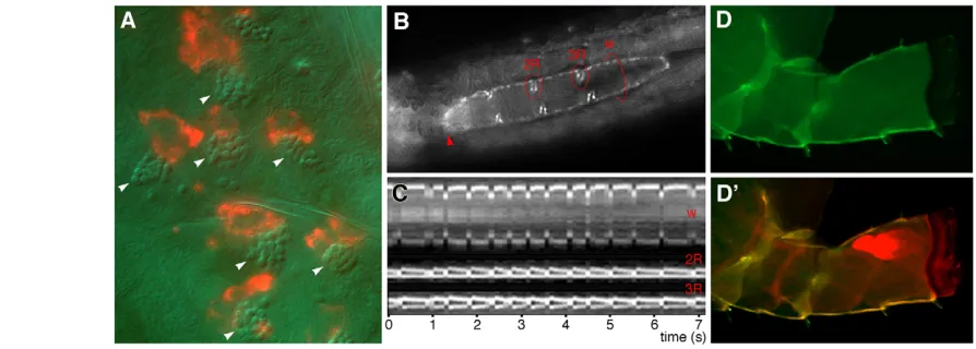
In emerging model organisms, gene traps are likely to be first used as markers for visualising specific tissues or cell types, providing a means to follow dynamic cell behaviours, to study physiological processes in vivo and to assess phenotypes following experimental manipulations. Some of our traps mark well-recognised organs, such as the nervous system, gills or paragnaths (Fig. 2D,F,H,K), whereas others mark complex populations of cells and previously undescribed cell types. For instance, one trap marks a previously uncharacterised cell type on the dorsal epidermis of late embryos, juveniles and adults (Fig. 2C) that is associated with specific sensory or structural elements in the epidermis of Parhyale (Fig. 3A).
Another trap allows us to image cardiac function. Parhyalehas a typical arthropod heart, consisting of a muscular tube with three pairs of lateral inflow valves and an anterior outflow valve (Fig. 3B). Using this trap, we were able to observe heart function and to visualise the opening and closing of valves in unprecedented detail (Fig. 3C and see Movie 1 in the supplementary material).
A third trap, which we named DistalDsRed, marks the distal part
of all Parhyale limbs in embryos, larvae and adults (Fig. 2E,L). We have used this line to monitor limb regeneration following amputation in Parhyale(Fig. 3D,D⬘).
Cloning trapped genes and mutagenic effects In emerging model organisms, transposon-based exon trapping is one of the most straightforward ways to isolate genes through unbiased genome-wide screens. The trapped gene of interest can be cloned easily by primer extension on cDNA from the trapped
line, even when the genome is unsequenced. To demonstrate this, we cloned a cDNA from the gene trapped in the DistalDsRedline of Parhyale. The cDNA contains a long open reading frame with no similarity to known proteins. A corresponding probe revealed the same expression pattern as DistalDsRedin embryos (see Fig. S2 in
[image:3.612.317.561.59.422.2]the supplementary material). Similarly, we have cloned cDNAs of several other genes trapped by PhHsp70a-DsRedin Parhyaleand Drosophila. Sequencing of these cDNAs led to our discovery of trans-splicing in Parhyale(Douris et al., 2010).
Fig. 2. Gene trapping in Parhyale hawaiensis. (A)Our primary trapping vector consists of the transformation marker 3xP3-DsRed, the fC31attPsite and the exon-trapping element PhHsp70a-DsRed, flanked by Minosinverted repeats (yellow inverted arrows). A splice acceptor in PhHsp70amediates exon trapping (dotted arrow). (B)Embryo expressing the 3xP3-DsRedmarker in the absence of gene trapping (arrowhead); the autofluorescence of yolk is also seen (red crescent). (C-J)Embryos with gene traps expressing DsRed in a variety of patterns, including specific epidermal cell types (C), brain and ventral nerve cord (D), limbs (E), gills (F), paragnaths (H), segmental stripes (I), as well as more widespread patterns (G,J). (K,L)Hatchlings with gene traps expressing DsRed in the gills (K) and limbs (L). All images show lateral views (except H,K,J, which are tilted) with anterior to the left. In H-L, DsRed fluorescence images are merged with corresponding UV autofluorescence or brightfield images (in cyan or blue) to highlight morphological features. The traps were obtained using the PhHsp70a-DsRedtrapping construct, except those shown in H and K, which were obtained using PhHS-DsRed.
D
E
V
E
LO
P
M
E
N
In exon-trapped genes, splicing of the endogenous transcript onto the gene-trap cassette generates a chimaeric mRNA that expresses, at most, the N-terminal portion of the endogenous protein. If splicing to PhHsp70a-DsRedwere 100% efficient, traps would be mutagenic and we would expect to detect a loss-of-function phenotype in animals homozygous for the trap. However, the majority of exon traps obtained using PhHsp70a-DsRed do not cause a detectable phenotype in homozygous animals. We used quantitative RT-PCR to examine splicing to PhHsp70a-DsRedrelative to normal splicing at the DistalDsRed
locus. We found that only some transcripts are spliced to the PhHsp70a-DsRedtrapping cassette, whereas a large proportion are still spliced onto the endogenous downstream exon. Thus, PhHsp70a-DsRedallows for sensitive trap detection with little disruption of endogenous gene function. This is helpful for maintaining stocks in the absence of balancer chromosomes. iTRAC, described below, provides the means to convert such traps into mutagenic insertions.
Integrase-mediated trap conversion (iTRAC)
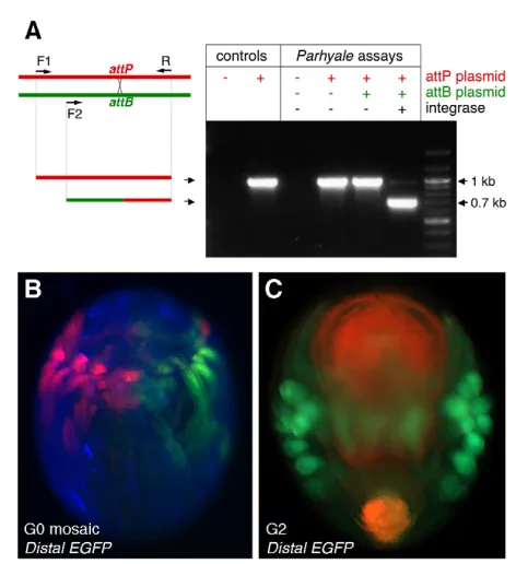
The attPsite in the trapping vector is a platform for integrating new constructs into trapped loci so as to generate new markers and tools for genetic analysis. fC31 integrase has never been used before in Parhyale. First, we devised a rapid PCR-based assay for fC31 integrase activity in vivo, which demonstrated efficient integrase-dependent recombination of attPand attBsites across two plasmids injected into early Parhyaleembryos (Fig. 4A).
Next, we tested the ability of attB-bearing plasmids to integrate at attPsites inserted in the Parhyalegenome in the presence of
fC31 integrase mRNA. A plasmid carrying attB and a heat-inducible EGFP marker (PhHS-EGFP) integrated with high efficiency into a transgenic line carrying multiple copies of the attP sequence (104 of 207 injected embryos expressed EGFP after heat shock). No integration events were recovered by injection into wild-type embryos (0 out of 255 embryos). Integration of the PhHS-EGFPtransgene also occurred in the germline, as judged from its transmission to the next generation.
Finally, we were able to demonstrate the conversion of a DsRed exon trap into one that expresses EGFP in the same pattern. A plasmid carrying attB-PhHsp70a-EGFPwas injected with fC31 integrase mRNA into early embryos of the DistalDsRedtransgenic
line. A high proportion of these embryos (47 out of 90 survivors) showed EGFP fluorescence replacing DsRed fluorescence in at least a subset of limbs (Fig. 4B). In ~20% of injected survivors, iTRAC also occurred in the germline and a stable DsRed to EGFP conversion was evident in subsequent generations (Fig. 4C). In DistalEGFPlines, DsRed expression could not be detected. These
results indicate that iTRAC can be implemented with high efficiency in Parhyale.
Conclusions
Model organisms are defined by the experimental approaches they offer to address biological questions of broad interest. Candidate gene approaches have, until now, been the main avenue for applying knowledge gained in established models to other species of interest, but these approaches are biased and incomplete. The establishment of transgenesis in new species that span the phylogenetic tree of animals, from cnidarians to protostomes and chordates, sets the stage for developing forward and reverse genetics tools and approaches in a wide range of organisms. iTRAC provides a shortcut for implementing these approaches.
[image:4.612.57.504.60.220.2]The versatility of iTRAC opens opportunities for a wide range of downstream applications: (1) the generation of markers for different types of microscopy, ranging from fluorescence-based live imaging to electron microscopy; (2) the implementation of binary systems for gene expression, such as the GAL4, LexA, tTA and Q systems; (3) the genetic marking and manipulation of clonal populations of cells using FLP or Cre recombinase; (4) the conversion of gene traps into gene knockouts, for instance by insertion of strong transcriptional terminators; (5) genetic cell ablation using cell-autonomously acting toxins; and (6) chromosome engineering. Conceivably, the same collections of traps could be used in the future to implement tools that are not yet Fig. 3. Using gene traps to visualise physiology and development in Parhyale. (A)Gene trap marking a previously undescribed cell type on the dorsal epidermis of a late embryo. DsRed-marked cells are of irregular shape and are associated with refractile structures located below the cuticle (arrowheads, visualised with Nomarski optics). (B,C)A gene trap allows us to study the function of the heart and valves by live imaging (see Movie 1 in the supplementary material). (B)Frame from live recording of a Parhyalehatchling, showing the outline of the heart tube and the three pairs of inflow valves; the outflow valve, at the anterior end of the heart (arrowhead), is out of focus. The length of the heart is 450m. Red circles mark the regions where kymograms were recorded. (C)Kymograms depicting successive contractions of the heart wall (w) and inflow valves 2 and 3 of the heart. (D,D⬘) The DistalDsRedtrap helps to visualise regenerating limbs, which are not normally visible prior to moulting. The regenerated limb (red in D⬘) can be clearly seen folded within the amputation stump (green).
D
E
V
E
LO
P
M
E
N
realised. Thus, iTRAC may serve as a genetic Swiss army knife, allowing the exploitation of gene traps in a virtually endless number of ways.
The approach and the vectors presented here are likely to be applicable in a broad range of animal models, as all the constituents are known to work in widely divergent species: the Minostransposon is an excellent vector for gene trapping not only in arthropods, but also in mammals and in Ciona (Pavlopoulos et al., 2007; Sasakura et al., 2007); the fC31 integrase system has found application in Drosophila, zebrafish, Xenopus, mouse and human (Allen and Weeks, 2005; Groth et al., 2004; Groth et al., 2000; Lister, 2010), and we have shown here that it works very efficiently in Parhyale; the PhHsp70a element can mediate exon trapping in Parhyaleand Drosophila and, given the wide conservation of splice acceptor sites, is likely to be useful more broadly.
Acknowledgements
We thank Yiannis Livadaras and Giorgos Tsoumbekos for help with Drosophila traps, Michele Calos for the fC31 integrase plasmids, Mingming Wu and Carsten Wolff for discussions on individual gene traps, and Maura Strigini for critical comments. This work was funded by the EPAN programme of the General Secretariat for Research and Technology (Greece) and by the Marie Curie RTN programme ‘ZOONET’ (European Union).
Competing interests statement
The authors declare no competing financial interests.
Author contributions
Z.K. established the fC31 integrase system and implemented iTRAC in Parhyale; A.P. cloned the hsp70sequences and established gene trapping in Parhyale; A.K. built and tested alternative trapping vectors; N.K. studied limb regeneration using gene traps; A.K. and N.K. determined promoter and splice sites in the hsp70sequences; Z.K., V.D. and A.K. cloned Distaland assessed splicing at that locus; M.A. conceived iTRAC, imaged gene traps, supervised the project and wrote the paper; all authors discussed the results and commented on the manuscript.
Supplementary material
Supplementary material for this article is available at
http://dev.biologists.org/lookup/suppl/doi:10.1242/dev.066324/-/DC1
References
Allen, B. G. and Weeks, D. L.(2005). Transgenic Xenopus laevis embryos can be generated using phiC31 integrase.Nat. Methods2, 975-979.
Bellen, H. J.(1999). Ten years of enhancer detection: lessons from the fly. Plant Cell11, 2271-2281.
Bellen, H. J., O’Kane, C. J., Wilson, C., Grossniklaus, U., Pearson, R. K. and Gehring, W. J.(1989). P-element-mediated enhancer detection: a versatile method to study development in Drosophila. Genes Dev.3, 1288-1300. Berghammer, A. J., Klingler, M. and Wimmer, E. A.(1999). A universal marker
for transgenic insects. Nature402, 370-371.
Brand, A. H. and Perrimon, N.(1993). Targeted gene expression as a means of altering cell fates and generating dominant phenotypes. Development118, 401-415.
Browne, W. E., Price, A. L., Gerberding, M. and Patel, N. H.(2005). Stages of embryonic development in the amphipod crustacean, Parhyale hawaiensis. Genesis42, 124-149.
Douris, V., Telford, M. J. and Averof, M.(2010). Evidence for multiple independent origins of trans-splicing in Metazoa. Mol. Biol. Evol.27, 684-693. Extavour, C. G.(2005). The fate of isolated blastomeres with respect to germ cell
formation in the amphipod crustacean Parhyale hawaiensis. Dev. Biol. 277, 387-402.
Gerberding, M., Browne, W. E. and Patel, N. H.(2002). Cell lineage analysis of the amphipod crustacean Parhyale hawaiensis reveals an early restriction of cell fates. Development129, 5789-5801.
Groth, A. C., Olivares, E. C., Thyagarajan, B. and Calos, M. P.(2000). A phage integrase directs efficient site-specific integration in human cells. Proc. Natl. Acad. Sci. USA97, 5995-6000.
Groth, A. C., Fish, M., Nusse, R. and Calos, M. P.(2004). Construction of transgenic Drosophila by using the site-specific integrase from phage phiC31. Genetics166, 1775-1782.
Juven-Gershon, T., Cheng, S. and Kadonaga, J. T.(2006). Rational design of a super core promoter that enhances gene expression.Nat. Methods3, 917-922. Lister, J. A.(2010). Transgene excision in zebrafish using the phiC31 integrase.
Genesis48, 137-143.
Liubicich, D. M., Serano, J. M., Pavlopoulos, A., Kontarakis, Z., Protas, M. E., Kwan, E., Chatterjee, S., Tran, K. D., Averof, M. and Patel, N. H.(2009). Knockdown of Parhyale Ultrabithorax recapitulates evolutionary changes in crustacean appendage morphology. Proc. Natl. Acad. Sci. USA106, 13892-13896.
Ozhan-Kizil, G., Havemann, J. and Gerberding, M.(2008). Germ cells in the crustacean Parhyale hawaiensis depend on Vasa protein for their maintenance but not for their formation. Dev. Biol. 218, 333-339.
Pavlopoulos, A. and Averof, M.(2005). Establishing genetic transformation for comparative developmental studies in the crustacean Parhyale hawaiensis. Proc. Natl. Acad. Sci. USA102, 7888-7893.
Pavlopoulos, A., Berghammer, A. J., Averof, M. and Klingler, M.(2004). Efficient transformation of the beetle Tribolium castaneum using the Minos transposable element: quantitative and qualitative analysis of genomic integration events. Genetics167, 737-746.
[image:5.612.54.290.64.322.2]Pavlopoulos, A., Oehler, S., Kapetanaki, M. G. and Savakis, C.(2007). The DNA transposon Minos as a tool for transgenesis and functional genomic analysis in vertebrates and invertebrates. Genome Biol. 8 Suppl. 1, S2.
Fig. 4. Integrase-mediated trap conversion (iTRAC) in Parhyale. (A)The interplasmid assay for fC31 integrase-mediated recombination involves injecting plasmids carrying an attPor an attBsite (red and green, respectively) with fC31 integrase mRNA into Parhyaleembryos, and assaying recombination by PCR using primers F1, F2 and R. Each gel lane represents a single experiment, in which different combinations of plasmids and integrase mRNA were injected. The 1 kb band results from amplification of the attPplasmid fragment (red), whereas the 0.7 kb band results from amplification of a hybrid fragment created by recombination between attPand attBsites (green-red). The 0.7 kb fragment is strictly dependent on the presence of fC31 integrase. The identity of this fragment was also verified by sequencing. (B)Ventral view of a DistalDsRedembryo (G0) injected with fC31 integrase mRNA and a plasmid carrying attBand PhHsp70a-EGFP. Integration at the 2-cell stage resulted in a mosaic in which the DistalDsRedtrap was converted to DistalEGFPon one side of the embryo; the other side retained DistalDsRedexpression. (C)Ventral view of DistalEGFPembryo, two generations after conversion (G2). DsRed fluorescence is not detectable in limbs; red autofluorescence of the yolk is shown for contrast.
D
E
V
E
LO
P
M
E
N
Pavlopoulos, A., Kontarakis, Z., Liubicich, D. M., Serano, J. M., Akam, M., Patel, N. H. and Averof, M.(2009). Probing the evolution of appendage specialization by Hox gene misexpression in an emerging model crustacean. Proc. Natl. Acad. Sci. USA106, 13897-13902.
Price, A. L., Modrell, M. S., Hannibal, R. L. and Patel, N. H.(2010). Mesoderm and ectoderm lineages in the crustacean Parhyale hawaiensis display intra-germ layer compensation. Dev. Biol. 341, 256-266.
Rehm, E. J., Hannibal, R. L., Chaw, R. C., Vargas-Vila, M. A. and Patel, N. H. (2009). The crustacean Parhyale hawaiensis: a new model for arthropod development. In Emerging Model Organisms: A Laboratory Manual, Vol. 1 (ed. R. R. Bheringer, A. D. Johnson and R. E. Krumlauf) 592 pp. Cold Spring Harbor, New York: Cold Spring Harbor Laboratory Press.
Renfer, E., Amon-Hassenzahl, A., Steinmetz, P. R. and Technau, U.(2010). A muscle-specific transgenic reporter line of the sea anemone, Nematostella vectensis. Proc. Natl. Acad. Sci. USA107, 104-108.
Sasakura, Y., Oogai, Y., Matsuoka, T., Satoh, N. and Awazu, S.(2007). Transposon mediated transgenesis in a marine invertebrate chordate: Ciona intestinalis. Genome Biol. 8 Suppl. 1, S3.
Schinko, J. B., Weber, M., Viktorinova, I., Kiupakis, A., Averof, M., Klingler, M., Wimmer, E. A. and Bucher, G.(2010). Functionality of the GAL4/UAS system in Tribolium requires the use of endogenous core promoters. BMC Dev. Biol. 10, 53.
Sepp, K. J. and Auld, V. J.(1999). Conversion of lacZ enhancer trap lines to GAL4 lines using targeted transposition in Drosophila melanogaster. Genetics151, 1093-1101.
Vargas-Vila, M. A., Hannibal, R. L., Parchem, R. J., Liu, P. Z. and Patel, N. H. (2010). A prominent requirement for single-minded and the ventral midline in patterning the dorsoventral axis of the crustacean Parhyale hawaiensis. Development137, 3469-3476.