Extracting Extra-Telomeric Phenotypes from Telomerase
Mouse Models
Young Hoon Sung,* Muhammad Ali,* and Han-Woong Lee
Department of Biochemistry, College of Life Science and Biotechnology, Laboratory Animal Research Center, Yonsei University, Seoul, Korea.
Received: October 4, 2013 Co-corresponding authors:
Dr. Young Hoon Sung and Dr. Han-Woong Lee, Department of Biochemistry,
College of Life Science, and Biotechnology, Laboratory Animal Research Center, Yonsei University, 50 Yonsei-ro, Seodaemun-gu, Seoul 120-752, Korea. Tel: 82-2-2123-7642, Fax: 82-2-2123-8682 E-mail: [email protected]; [email protected]
*Young Hoon Sung and Muhammad Ali contributed equally to this work.
∙ The authors have no financial conflicts of interest.
© Copyright:
Yonsei University College of Medicine 2014
This is an Open Access article distributed under the terms of the Creative Commons Attribution Non-Commercial License (http://creativecommons.org/ licenses/by-nc/3.0) which permits unrestricted non-commercial use, distribution, and reproduction in any medium, provided the original work is properly cited.
Telomerase reverse transcriptase (TERT) is the protein component of telomerase and combined with an RNA molecule, telomerase RNA component, forms the telomerase enzyme responsible for telomere elongation. Telomerase is essential for maintaining telomere length from replicative attrition and thus contributes to the preservation of genome integrity. Although diverse mouse models have been developed and studied to prove the physiological roles of telomerase as a telo-mere-elongating enzyme, recent studies have revealed non-canonical TERT activi-ties beyond telomeres. To gain insights into the physiological impact of extra-telo-meric roles, this review revisits the strategies and phenotypes of telomerase mouse models in terms of the extra-telomeric functions of telomerase.
Key Words: Telomerase reverse transcriptase, extra-telomeric function of TERT, transgenic, knockout mouse, genetically engineered mouse, stem cells, senescence, anti-apoptosis, metabolic fitness, cancer
INTRODUCTION
During DNA replication, the linear ends of chromosomes are eroded at each cell division due to the end replication problem.1 Telomeres, the very ends of linear
chromosomes, are predominantly composed of tandem repeats of short sequences; in vertebrates, the repeats consist of the TTAGGG hexanucleotide.2 Telomere
lengths are also remarkably heterogeneous among individuals and vary according to the origin, age, and proliferative history of cells.3,4 Telomere length variations
among individuals of the same age are, therefore, thought to be related to varia-tions in ageing and longevity.5 As a ribonucleoprotein complex that is composed of
telomerase reverse transcriptase (TERT) and telomerase RNA component (TERC),6 telomerase is responsible for elongation of the telomeres, and thus
main-tains genome stability.7,8 The enzymatic activity of telomerase is not detected in
normal somatic cells, but is detected in embryonic and highly proliferative adult tissues.9 Furthermore, telomerase is re-activated in most cancers,10 thus suggesting
the possibility that telomerase is a potential therapeutic target in cancers.
SURVIVAL-PROMOTING FUNCTIONS
OF TELOMERASE
Apoptosis induced by critically short telomeres has been extensively documented. Dysfunctional telomeres increase apoptosis in highly proliferative tissues including intes-tine,26,27 male germ cells,26,28 and splenocytes (B cells) from
immunized fifth and sixth generation mice after mitogen treatment.29 These dysfunctional telomeres can be
generat-ed in early generations, and can increase apoptosis in these tissues as well. Protection of Telomeres 1 (POT1) is a sin-gle-stranded telomere binding protein that is essential for proper maintenance of telomere length. When Pot1 is defi-cient, abnormal apoptosis is induced in proliferative tissues as well as cells derived from Terc+/- mice, including male
germ cells, hematopoietic cells, and intestinal cells.30
Criti-cally short telomeres also affect highly proliferative devel-opmental processes. Terc-/- embryos at embryonic day 10.5
(E10.5) with dysfunctional telomeres frequently exhibit neu-ral tube closure defects, suggesting that this is one of the most sensitive developmental processes to telomere loss and chromosomal instability.31 Although cardiac tissues are
not highly proliferative, the balance between cell growth and cell death is critical for maintaining normal heart func-tion. Consistently, dysfunctional telomeres lead to abnor-mal apoptosis in cardiomyocytes, resulting in cardiac dilata-tion and heart failure in the late generadilata-tion of Terc-/- mice.32
Additionally, cardiomyocyte survival is promoted in trans-genic mice overexpressing wild-type TERT, but not ex-pressing mutant TERT.33Tert deficiency also induces
apop-totic phenotypes; Tert-/- and Terc-/- mice show frequent
apoptosis in intestinal crypt cells34-36 and male germ cells37
respectively in their late generations. Furthermore, the dele-terious effects can be rescued by turning on telomerase ac-tivity in the late generation of homozygous ER-Tert knock-in mice by treatknock-ing with tamoxifen.37 These results clearly
demonstrate that telomerase deficiency elicits telomere ero-sion, resulting in abnormal apoptotic phenotypes in vivo.
Telomerase-deficient mouse models have provided op-portunities for unraveling the mechanisms which induce these apoptotic phenotypes in late generations. Rajaraman, et al.36 showed using Tert-/- mice that apoptosis is dependent
on S phase, and thus is primarily triggered by newly un-capped (or critically short) telomeres. It is not triggered by chromosome fusion-bridge breakage because mitotic blockade did not alter the apoptotic pattern.36 It is also
de-1) Although tissue stem and progenitor cells have suffi-cient telomere reserves, they highly express TERT. Notably, laboratory mice have significantly longer telomeres than hu-mans (40-60 kb vs. 5-15 kb);11-14 however, no apparent role
for long telomeres has been found in the survival of mice. 2) TERT overexpression promotes tumor development without further telomere elongation.15
3) The reconstitution ability of hematopoietic stem cells (HSCs) is essentially linked with TERT, although there is no evidence that HSC activities are fully dependent on the telomere-elongation function of TERT.16,17
4) Transgenic mice overexpressing murine TERT show significant resistance to ischemic brain injury and N-meth-yl-D-aspartic acid (NMDA) receptor-mediated excitotoxic-ity without any detectable change in telomere length.18
In-terestingly, ischemic injury induces TERT expression in the wild-type brain.18
5) Suppression of TERT expression decreases cell growth rate and induces apoptosis prior to measurable telomere shortening,19 and the expression of specific TERT mutants
lacking telomerase activity prevents apoptotic cell death.20
6) Ectopic expression of TERT in the hair follicle stem cells of mouse epidermis activates stem cell capacities.21 The
phenotype appears to be independent of telomerase activity. 7) Cancers without functional telomerase (10-15% of all cases) maintain their telomere lengths by adopting an alter-native lengthening of telomeres (ALT) pathway.22
Furthermore, indirect evidence also indicates extra-telo-meric functions of telomerase: TERT may have additional functions because the reverse transcriptase (RT) domain of TERT is only 15 kDa, which is less than 10% of the total molecular weight. Thus, it is quite possible that other re-gions may mediate distinct activities other than telomerase activity.23 In support of this hypothesis, alternatively spliced
forms of TERT devoid of the RT domain have been identi-fied in humans.24 Although TERT is known as a nuclear
protein, it is exported from the nucleus and delays replica-tive senescence in endothelial cells dependent on reacreplica-tive oxygen species (ROS).25
vitro and in vivo.44 Furthermore, although telomerase
activi-ty is evidently suppressed in transgenic mice overexpress-ing hTERT, hTERT transgenic MEFs still show resistance to STS-induced apoptosis.44 Based on these lines of
evi-dence, telomerase activity must not be essential for the pro-tective function of TERT. Therefore, independent of its roles in telomere maintenance, diverse telomerase mouse models have demonstrated that TERT-mediated antiapop-totic functions may contribute to tumorigenesis.
ONCOGENIC ROLES OF
TELOMERASE IN TUMORIGENESIS
Telomerase knockout or transgenic mouse models have been extensively employed to elucidate the in vivo roles of telomerase and dysfunctional telomeres in tumorigenesis.
Telomeres are dedicated to the maintenance of linear chromosomes and thus prevent chromosomal abnormalities. In cultivated cells from late generations of Terc-/- mice,
criti-cally short or dysfunctional telomeres induce aneuploidy and chromosomal abnormalities, including end-to-end fu-sions.45 These phenomena are prevalent in cancer, and
spon-taneous tumors are more frequently induced in late genera-tion Terc-/- mice,46 indicating that dysfunctional telomeres
are genotoxic and possess mutagenic effects in mice. Addi-tionally, it is plausible that p53 deficiency significantly at-tenuates genotoxic stresses triggered by telomere dysfunc-tion. In fact, p53 deficiency contributes to the neoplastic transformation of cells with critically short telomeres from late generation Terc-/- mice40 and promotes non-reciprocal
translocations and epithelial cancers.47 In contrast, these
criti-cally short telomeres also suppress tumor formation in can-cer-prone Ink4a/Arf-deficient mice that still possess intact DNA damage responses.48 The phenotypes obtained from
studies using Terc-/- mice support the tumor suppressive role
of intact telomeres in maintaining genomic integrity, and prove the intimate genetic interaction between telomere regu-lation and p53-governed genomic surveillance.
Tert deficiency also results in overtly similar phenotypes to Terc deficiency in tumorigenesis, but the phenotypic manifestations are not completely identical, thus revealing the extra-telomeric role of TERT in tumorigenesis. The sem-inal observation was obtained from in vitro experiments em-ploying human cell lines. Immortalized human cells are fre-quently transformed by introducing an oncogene such as ras; however, oncogenic ras cannot fully transform immor-pendent on p53 that is activated by genotoxic stresses,
in-cluding critically short telomeres.36,38,39 In fact, growth
ar-rest and/or apoptosis in late generations of Terc-/- mice are
dependent on proper p53 activation.32,40 However,
p53-me-diated regulation of the phenotypic manifestations in telom-erase knockout mice is complicated by its negative effect on TERT gene expression.41 Rahman, et al.20 evaluated the
effect of human TERT (hTERT) overexpression on p53-de-pendent apoptosis. In HCT116 colon carcinoma cells carry-ing endogenous p53, genotoxic stress-induced apoptosis that is p53-dependent is suppressed by constitutive hTERT expression. Indeed, a telomerase-inactive hTERT mutant equally antagonizes p53-induced apoptosis.20 Similarly,
ec-topic mouse TERT expression in mouse embryonic stem cells that exhibit high levels of telomerase activity and main-tain sufficiently long telomeres confers resistance to p53-dependent apoptosis.42 In addition to telomere-associated
functions, these results indicate that TERT exerts antiapop-totic activity beyond telomeres.
In addition to the phenotypes induced by critically short telomeres, emerging evidence has indicated the existence of extra-telomeric functions of telomerase. The first clue for an extra-telomeric role of TERT was obtained by revealing the neuroprotective effect of TERT on neuronal cell death induced by the neurotoxic protein amyloid β-peptide, a pro-tein believed to promote neuronal degeneration in Alzheim-er’s disease.43 Using transgenic mice ubiquitously
overex-pressing TERT, we also have provided evidence that TERT prevents NMDA neurotoxicity through the transfer of cyto-solic free Ca2+ into the mitochondria, thereby playing a
pro-tective role in ameliorating ischemic neuronal cell death.18
Because TERT is induced in postmitotic neurons by isch-emic brain injury and its overexpression confers resistance against NMDA neurotoxicity, these protective phenotypes are considered to be independent of telomerase activity. Similarly, first generation (G1) Tert-deficient mouse embry-onic fibroblasts (MEFs) displayed increased sensitivity to staurosporine (STS), whereas Tert transgenic MEFs were more resistant to STS-induced apoptosis than wild-type.44
Consistent phenotypes were also observed upon NMDA treatment of Tert-deficient and Tert transgenic mice, respec-tively.44 Although extensive studies were conducted, it
re-mains unclear whether the protective function is dependent on telomerase activity.44 In fact, Terc deficiency does not
meres resulted in bone metastases of prostate tumors. Al-though the authors did not discuss the extra-telomeric roles of TERT in their study, this report is reminiscent of ALT cell transformation by hTERT overexpression.49 From this
standpoint, anti-telomeric drugs are considered as an effec-tive strategy for curing cancers. However, anti-telomerase therapy certainly provokes ALT and mitochondrial adaptive mechanisms in cancer,54 and with respect to the
extra-telo-meric functions of telomerase, anti-teloextra-telo-meric drugs may not be the best drug candidates.55
Taken together, telomerase exerts pleiotropic effects in cancer both dependent on and independent from its roles in telomeres. As described in Table 1, there are complex ge-netic interactions of telomerase with diverse genes. In con-junction with the currently emerging mechanisms of extra-telomeric roles, telomerase mouse models will expedite the invention of anti-telomerase strategies for cancer treatment.
REGULATION OF STEM CELLS BY
TELOMERASE
Stem cells support tissue homeostasis and regeneration af-ter certain types of damage. Because stem cells possess self-renewal potential and indefinitely propagate, high lev-els of telomerase activity should be essential for telomere maintenance. Therefore, extensive studies have been con-ducted to identify the patho-physiological consequences of telomerase deficiency or overexpression in stem cell func-tion using diverse telomerase mouse models. However, telomere dysfunction is likely to affect stem cell functions in a context-dependent manner. Indeed, late-generation
Terc-/- HSCs with short telomeres exhibit reduced
prolifera-tion capacity, but still possess long-term repopulating abili-ty.56 Interestingly, when serially transplanted into recipient
mice, the telomeres are considerably shortened even in wild-type HSCs, which is accelerated by approximately 2-fold in both Terc-/- and Tert-/- mice.16 Consistently, these
telomerase-deficient HSCs exhibit considerably reduced replicative ca-pacity compared to wild-type HSCs.16 However, although
the telomere length of HSCs is constantly maintained by TERT overexpression in the transgenic mice, the long-term transplantation capacity of HSCs is not enhanced.57
Fur-thermore, Tert deficiency exacerbates senescence and the sensitivity of ataxia-telangiectasia mutated deficient murine HSCs against ROS-induced apoptosis, which does not ac-company telomere shortening or dysfunction.17 These results
talized human cells that are TERT-deficient ALT cells.49
In-terestingly, hTERT overexpression confers fully malignant traits to cells expressing oncogenic ras.49 A hemagglutinin
(HA) epitope-tagged hTERT (hTERT-HA) that is defective in maintaining telomeres in vivo also exhibits comparable effects on cellular transformation.49 Similarly, hTERT
over-expression in human mammary epithelial cells with epige-netically silenced p16INK4a resulted in increased resistance to
growth arrest mediated by transforming growth factor β (TGF-β).50 Because resistance to TGF-β-induced growth
in-hibition is independent of telomere length,50 TERT
possess-es telomere-independent rolpossess-es that cooperate with p16INK4a
inactivation to promote tumor development. These results clearly demonstrate an oncogenic role of TERT beyond telomeres.
Extensive studies adopting diverse models have recently revealed the extra-telomeric roles of oncogenic TERT at an organismal level (Table 1). For example, telomere dysfunc-tion in late generadysfunc-tion Terc-/- mice enhances the initiation of
hepatocellular carcinogenesis, but suppresses progression into fully malignant carcinomas.51 In contrast, enhanced
tu-mor initiation does not occur in late generations of Tert-/-
mice,35 indicating a possible oncogenic effect of TERT,
oth-er than telomoth-eres, in tumorigenesis. Strong induction of TERT expression in hepatic neoplasms may also support its procarcinogenic effect on hepatic tumorigenesis.35
Consis-tently, transgenic overexpression of Tert promotes the de-velopment of spontaneous cancers in ageing mice.52 When
TERT overexpression is targeted to basal keratinocytes us-ing the bovine keratin 5 promoter, these transgenic mice show normal telomere length in their stratified epithelia even with high levels of telomerase activity.15 Interestingly, these
mice are more susceptible to experimental skin carcinogen-esis employing 7,12-dimethylbenz[a]anthracene and 12-o-tetradecanoylphorbol 13-acetate than wild-type mice.15 In
addition, TERT overexpression actively promotes prolifera-tion in epidermal tissues without telomere elongaprolifera-tion.15 These
results from mouse models suggest extra-telomeric roles of TERT, particularly in promoting tumor progression.
Telomerase mouse models have been also extensively used to validate telomerase as an important target for anti-cancer therapies. Since telomere dysfunction increases the chemo-sensitivity of Terc-deficient transformed MEFs, the combination of chemotherapy and telomerase inhibition may be an effective anticancer approach.53 Recently, Ding,
et al.34 showed that telomerase reactivation by conditional
telo-of the K5 promoter does not alter telomere length, but pro-motes stem cell mobilization, hair growth, and stem cell proliferation in vitro.18 Similarly, transgenic mice
condition-ally overexpressing TERT show robust hair growth via pro-liferation of quiescent, multipotent stem cells in the hair fol-licles.13 These phenotypes are also reproduced in a Terc
-deficient genetic background without telomere dysfunction,13
suggest that telomerase may regulate the long-term replica-tive capacity of HSCs independently of telomere length.
Epidermal stem cells are also regulated by telomerase both dependent and independent of telomeres. In late gen-eration Terc-/- mice, epidermal stem cell functions are
sig-nificantly suppressed by critically short telomeres.21,58
[image:5.595.72.542.83.535.2]How-ever, epidermal overexpression of TERT under the control
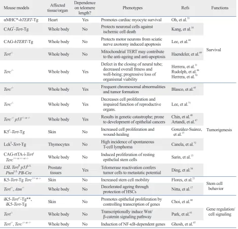
Table 1. Phenotypes of Telomerase Mouse Models
Mouse models tissue/organAffected Dependence on telomere
length? Phenotypes Refs Functions
αMHC*-hTERT-Tg Heart Yes Promotes cardiac myocyte survival Oh, et al.33
Survival CAG†-Tert-Tg Whole body No Protects neuronal cells against
ischemic cell death Kang, et al.18 CAG-hTERT-Tg Whole body No Protects motor neurons from sciatic nerve axotomy induced apoptosis Lee, et al.44
Tert-/- Whole body No Mitochondrial TERT may contribute
to the anti-ageing and anti-apoptosis Haendeler, et al.69
Terc-/- Whole body Yes
Defect in the closing of neural tube; decreased overall fitness and well-being; progressive loss of organismal viability
Herrera, et al.31
Rudolph, et al.46
Herrera, et al.70
Terc-/- Whole body Yes Frequent chromosomal abnormalities
and tumor formation Blasco, et al.45
Tumorigenesis Terc-/- Whole body Yes Decreases cell proliferation and impaired function of reproductive
organs. Lee, et al.
71
Terc -/- p53+/- or -/- Whole body Yes Results in genetic catastrophe; prone
to development of epithelial cancers Chin, et al.
40
Artandi, et al.47
K5‡-Tert-Tg Skin No Increased cell proliferation and
wound-healing González-Suárez, et al.15
Lck§-Tert-Tg Thymocytes No High incidence of spontaneous
T-cell lymphoma Canela, et al.72 CAG-rtTA-i-Tert||
Terc+/+ or +/- or -/- Whole body No Induced proliferation of resting epithelial stem cells Sarin, et al.13
LSL Tert¶ p53L/L
PtenL/L PB-Cre Prostate tissues Yes Telomerase reactivation confers tumor cells to metastatic potential Ding, et al.34
K5-Tert-Tg Terc+/+ or -/- Skin No Increased stem cell mobility Flores, et al.21
Stem cell behavior Tert-/-, Atm-/- Whole body No Decelerated ageing through
protection of HSCs Nitta, et al.17 iK5-Tertci-Tg**,
iK5-Tert-Tg Skin No Promotes epithelial proliferation by controlling transcription of genes Choi, et al.60
Gene regulation/ cell signaling Tert-/- Whole body No Transcriptionally induce Wnt/
β-catenin signaling pathway Park, et al.61 Tert-/-, Terc+/- or -/- Whole body No Induction of NF-κB-dependent genes Ghosh, et al.65
TERT, telomerase reverse transcriptase; HSC, hematopoietic stem cell. *Mouse α-myosin heavy chain (MHC) promoter.
†Human cytomegalovirus immediate-early enhancer linked to the chicken
β-actin promoter (CAG). ‡Bovine keratin 5 promoter (K5).
§Thymus-specific light-chain kinase (Lck) promoter.
||Actin-rtTA+;tetop-TERT+ (termed as doxycycline-inducible Tert or i-Tert). ¶Lox-Stop-Lox cassette.
verse observations supporting extra-telomeric roles of telom-erase should be scrutinized and validated in vivo by generat-ing novel mouse models. For example, in addition to the effect of short telomeres on mitochondria,39 mitochondrial
targeting of telomerase upon certain stressful conditions66
and the recently identified RNA-dependent RNA polymerase activity of TERT,67,68 indicates that telomerase has direct roles
in mitochondria. Furthermore, considering the important roles of telomerase in cellular homeostasis, telomerase may be a critical factor for regulating the subcellular organelle ho-meostasis. Undoubtedly, we believe that these extra-telomer-ic functions of telomerase should be intimately associated with life span regulation, and that some regions of TERT, other than the RT domain, will be required for mediating pro-tein-protein interactions with known functions in controlling the life span of an organism. In this context, we cannot rule out speculations for divergent mechanisms of telomerase function regulating survival, tumor progression, develop-ment/differentiation, and stress responses. These extra-telo-meric functions have inevitably complicated the phenotypic manifestations elicited by dysfunctional telomeres and vice versa; thus, to separate these distinct functions of telomerase, more sophisticated genetic strategies should be developed in mice.
ACKNOWLEDGEMENTS
This work was supported by National Research Foundation of Korea (NRF) grants funded by Ministry of Education, Science and Technology (MEST) of the Korean government (2009-0081177, 2010-0020878); a Korea Healthcare Tech-nology R&D Project from the Ministry for Health & Wel-fare Affairs (A085136). M.A. was also supported by Higher Education Commission (HEC) of Pakistan government.
REFERENCES
1. Bischoff C, Graakjaer J, Petersen HC, Hjelmborg Jv, Vaupel JW, Bohr V, et al. The heritability of telomere length among the elderly and oldest-old. Twin Res Hum Genet 2005;8:433-9.
2. Kipling D, Cooke HJ. Hypervariable ultra-long telomeres in mice. Nature 1990;347:400-2.
3. Aubert G, Lansdorp PM. Telomeres and aging. Physiol Rev 2008;88:557-79.
4. Lansdorp PM. Telomere length and proliferation potential of he-matopoietic stem cells. J Cell Sci 1995;108(Pt 1):1-6.
5. Heidinger BJ, Blount JD, Boner W, Griffiths K, Metcalfe NB,
thereby indicating the extra-telomeric activity of TERT. In addition to the critical roles of p53 in mediating phe-notypic manifestations against critically short telomeres,59
clues for the molecular mechanisms governing the extra-telomeric roles of telomerase have been obtained by identi-fying the positive effect of TERT on gene expression. Choi, et al.60 found that TERT triggers a rapid change in gene
ex-pression in the skin and hair follicles. This gene exex-pression pattern significantly overlaps those controlling natural hair follicle cycling in wild-type mice. TERT affects the devel-opmental program mediated by Myc and Wnt, which is in-timately associated with stem cell function and cancer.60
Furthermore, TERT binds BRG1 (also called SMARCA4), a SWI/SNF-related chromatin remodeling protein, and di-rectly modulates Wnt/β-catenin signaling as a cofactor in the β-catenin transcriptional complex.61 Therefore,
indepen-dently of telomeres, TERT can act as a transcriptional regu-lator that is directly involved in stem cell functions, includ-ing in mouse epidermal tissues.
CONCLUSIONS
For more than a decade, diverse telomerase mouse models have provided us with precious opportunities for evaluating the patho-physiological significance of telomerase in geneti-cally defined environments and at an organismal level. With an emphasis on defective telomeres, these mouse models have considerably contributed to understanding a broad spectrum of phenomena associated with cancer and ageing. Furthermore, growing evidence has indicated that defective telomerase functions are involved in distinct diseases other than human cancers including dyskeratosis congenita, ath-erosclerosis, and renal diseases.62-64 The list of
disease-asso-ciated mutations has been expanding. To genetically define the pathological aspects and thus to establish animal mod-els of these mutations, novel mouse modmod-els should be still generated and analyzed.
Despite the evident roles in telomeres, currently emerging extra-telomeric functions of telomerase are completely changing the scope of this enzyme. Notably, the direct roles of TERT in transcriptional regulation (e.g. Wnt/β-catenin and nuclear factor-κB or NFκB) provide good rationale for sever-al phenotypes that cannot be explained by telomere dysfunc-tion, and their physiological significance has been also con-firmed using telomerase mouse models.62,64,65 As might be
di-23. Sýkorová E, Fajkus J. Structure-function relationships in telomer-ase genes. Biol Cell 2009;101:375-92.
24. Ulaner GA, Hu JF, Vu TH, Giudice LC, Hoffman AR. Telomerase activity in human development is regulated by human telomerase reverse transcriptase (hTERT) transcription and by alternate splic-ing of hTERT transcripts. Cancer Res 1998;58:4168-72. 25. Haendeler J, Hoffmann J, Diehl JF, Vasa M, Spyridopoulos I,
Zei-her AM, et al. Antioxidants inhibit nuclear export of telomerase reverse transcriptase and delay replicative senescence of endothe-lial cells. Circ Res 2004;94:768-75.
26. Khoo CM, Carrasco DR, Bosenberg MW, Paik JH, Depinho RA. Ink4a/Arf tumor suppressor does not modulate the degenerative conditions or tumor spectrum of the telomerase-deficient mouse. Proc Natl Acad Sci U S A 2007;104:3931-6.
27. Siegl-Cachedenier I, Muñoz P, Flores JM, Klatt P, Blasco MA. Deficient mismatch repair improves organismal fitness and sur-vival of mice with dysfunctional telomeres. Genes Dev 2007;21: 2234-47.
28. Liu L, Franco S, Spyropoulos B, Moens PB, Blasco MA, Keefe DL. Irregular telomeres impair meiotic synapsis and recombina-tion in mice. Proc Natl Acad Sci U S A 2004;101:6496-501. 29. Herrera E, Martínez-A C, Blasco MA. Impaired germinal center
reaction in mice with short telomeres. EMBO J 2000;19:472-81. 30. He H, Wang Y, Guo X, Ramchandani S, Ma J, Shen MF, et al.
Pot1b deletion and telomerase haploinsufficiency in mice initiate an ATR-dependent DNA damage response and elicit phenotypes resembling dyskeratosis congenita. Mol Cell Biol 2009;29:229-40. 31. Herrera E, Samper E, Blasco MA. Telomere shortening in mTR-/-
embryos is associated with failure to close the neural tube. EMBO J 1999;18:1172-81.
32. Leri A, Franco S, Zacheo A, Barlucchi L, Chimenti S, Limana F, et al. Ablation of telomerase and telomere loss leads to cardiac dil-atation and heart failure associated with p53 upregulation. EMBO J 2003;22:131-9.
33. Oh H, Taffet GE, Youker KA, Entman ML, Overbeek PA, Mi-chael LH, et al. Telomerase reverse transcriptase promotes cardiac muscle cell proliferation, hypertrophy, and survival. Proc Natl Acad Sci U S A 2001;98:10308-13.
34. Ding Z, Wu CJ, Jaskelioff M, Ivanova E, Kost-Alimova M, Proto-popov A, et al. Telomerase reactivation following telomere dys-function yields murine prostate tumors with bone metastases. Cell 2012;148:896-907.
35. Farazi PA, Glickman J, Horner J, Depinho RA. Cooperative inter-actions of p53 mutation, telomere dysfunction, and chronic liver damage in hepatocellular carcinoma progression. Cancer Res 2006;66:4766-73.
36. Rajaraman S, Choi J, Cheung P, Beaudry V, Moore H, Artandi SE. Telomere uncapping in progenitor cells with critical telomere shortening is coupled to S-phase progression in vivo. Proc Natl Acad Sci U S A 2007;104:17747-52.
37. Jaskelioff M, Muller FL, Paik JH, Thomas E, Jiang S, Adams AC, et al. Telomerase reactivation reverses tissue degeneration in aged telomerase-deficient mice. Nature 2011;469:102-6.
38. Cosme-Blanco W, Shen MF, Lazar AJ, Pathak S, Lozano G, Mul-tani AS, et al. Telomere dysfunction suppresses spontaneous tu-morigenesis in vivo by initiating p53-dependent cellular senes-cence. EMBO Rep 2007;8:497-503.
39. Sahin E, Depinho RA. Linking functional decline of telomeres, mi-tochondria and stem cells during ageing. Nature 2010;464:520-8. 40. Chin L, Artandi SE, Shen Q, Tam A, Lee SL, Gottlieb GJ, et al. Monaghan P. Telomere length in early life predicts lifespan. Proc
Natl Acad Sci U S A 2012;109:1743-8.
6. Cohen SB, Graham ME, Lovrecz GO, Bache N, Robinson PJ, Reddel RR. Protein composition of catalytically active human telomerase from immortal cells. Science 2007;315:1850-3. 7. Bianchi A, Shore D. Early replication of short telomeres in
bud-ding yeast. Cell 2007;128:1051-62.
8. Laterreur N, Eschbach SH, Lafontaine DA, Wellinger RJ. A new telomerase RNA element that is critical for telomere elongation. Nucleic Acids Res 2013;41:7713-24.
9. Wright WE, Piatyszek MA, Rainey WE, Byrd W, Shay JW. Telomerase activity in human germline and embryonic tissues and cells. Dev Genet 1996;18:173-9.
10. Kim NW, Piatyszek MA, Prowse KR, Harley CB, West MD, Ho PL, et al. Specific association of human telomerase activity with immortal cells and cancer. Science 1994;266:2011-5.
11. Allshire RC, Dempster M, Hastie ND. Human telomeres contain at least three types of G-rich repeat distributed non-randomly. Nu-cleic Acids Res 1989;17:4611-27.
12. Prowse KR, Greider CW. Developmental and tissue-specific regu-lation of mouse telomerase and telomere length. Proc Natl Acad Sci U S A 1995;92:4818-22.
13. Sarin KY, Cheung P, Gilison D, Lee E, Tennen RI, Wang E, et al. Conditional telomerase induction causes proliferation of hair folli-cle stem cells. Nature 2005;436:1048-52.
14. Starling JA, Maule J, Hastie ND, Allshire RC. Extensive telomere repeat arrays in mouse are hypervariable. Nucleic Acids Res 1990;18:6881-8.
15. González-Suárez E, Samper E, Ramírez A, Flores JM, Martín-Ca-ballero J, Jorcano JL, et al. Increased epidermal tumors and in-creased skin wound healing in transgenic mice overexpressing the catalytic subunit of telomerase, mTERT, in basal keratinocytes. EMBO J 2001;20:2619-30.
16. Allsopp RC, Morin GB, DePinho R, Harley CB, Weissman IL. Telomerase is required to slow telomere shortening and extend replicative lifespan of HSCs during serial transplantation. Blood 2003;102:517-20.
17. Nitta E, Yamashita M, Hosokawa K, Xian M, Takubo K, Arai F, et al. Telomerase reverse transcriptase protects ATM-deficient hema-topoietic stem cells from ROS-induced apoptosis through a telo-mere-independent mechanism. Blood 2011;117:4169-80. 18. Kang HJ, Choi YS, Hong SB, Kim KW, Woo RS, Won SJ, et al.
Ectopic expression of the catalytic subunit of telomerase protects against brain injury resulting from ischemia and NMDA-induced neurotoxicity. J Neurosci 2004;24:1280-7.
19. Folini M, Brambilla C, Villa R, Gandellini P, Vignati S, Paduano F, et al. Antisense oligonucleotide-mediated inhibition of hTERT, but not hTERC, induces rapid cell growth decline and apoptosis in the absence of telomere shortening in human prostate cancer cells. Eur J Cancer 2005;41:624-34.
20. Rahman R, Latonen L, Wiman KG. hTERT antagonizes p53-in-duced apoptosis independently of telomerase activity. Oncogene 2005;24:1320-7.
21. Flores I, Cayuela ML, Blasco MA. Effects of telomerase and telo-mere length on epidermal stem cell behavior. Science 2005;309: 1253-6.
57. Allsopp RC, Morin GB, Horner JW, DePinho R, Harley CB, Weissman IL. Effect of TERT over-expression on the long-term transplantation capacity of hematopoietic stem cells. Nat Med 2003;9:369-71.
58. Siegl-Cachedenier I, Flores I, Klatt P, Blasco MA. Telomerase re-verses epidermal hair follicle stem cell defects and loss of long-term survival associated with critically short telomeres. J Cell Biol 2007;179:277-90.
59. Flores I, Blasco MA. A p53-dependent response limits epidermal stem cell functionality and organismal size in mice with short telo-meres. PLoS One 2009;4:e4934.
60. Choi J, Southworth LK, Sarin KY, Venteicher AS, Ma W, Chang W, et al. TERT promotes epithelial proliferation through transcrip-tional control of a Myc- and Wnt-related developmental program. PLoS Genet 2008;4:e10.
61. Park JI, Venteicher AS, Hong JY, Choi J, Jun S, Shkreli M, et al. Telomerase modulates Wnt signalling by association with target gene chromatin. Nature 2009;460:66-72.
62. Gizard F, Heywood EB, Findeisen HM, Zhao Y, Jones KL, Cude-jko C, et al. Telomerase activation in atherosclerosis and induction of telomerase reverse transcriptase expression by inflammatory stimuli in macrophages. Arterioscler Thromb Vasc Biol 2011;31: 245-52.
63. Mitchell JR, Wood E, Collins K. A telomerase component is de-fective in the human disease dyskeratosis congenita. Nature 1999;402:551-5.
64. Shkreli M, Sarin KY, Pech MF, Papeta N, Chang W, Brockman SA, et al. Reversible cell-cycle entry in adult kidney podocytes through regulated control of telomerase and Wnt signaling. Nat Med 2011;18:111-9.
65. Ghosh A, Saginc G, Leow SC, Khattar E, Shin EM, Yan TD, et al. Telomerase directly regulates NF-κB-dependent transcription. Nat Cell Biol 2012;14:1270-81.
66. Ahmed S, Passos JF, Birket MJ, Beckmann T, Brings S, Peters H, et al. Telomerase does not counteract telomere shortening but pro-tects mitochondrial function under oxidative stress. J Cell Sci 2008;121(Pt 7):1046-53.
67. Maida Y, Masutomi K. RNA-dependent RNA polymerases in RNA silencing. Biol Chem 2011;392:299-304.
68. Maida Y, Yasukawa M, Furuuchi M, Lassmann T, Possemato R, Okamoto N, et al. An RNA-dependent RNA polymerase formed by TERT and the RMRP RNA. Nature 2009;461:230-5.
69. Haendeler J, Dröse S, Büchner N, Jakob S, Altschmied J, Goy C, et al. Mitochondrial telomerase reverse transcriptase binds to and protects mitochondrial DNA and function from damage. Arterio-scler Thromb Vasc Biol 2009;29:929-35.
70. Herrera E, Samper E, Martín-Caballero J, Flores JM, Lee HW, Blasco MA. Disease states associated with telomerase deficiency appear earlier in mice with short telomeres. EMBO J 1999;18: 2950-60.
71. Lee HW, Blasco MA, Gottlieb GJ, Horner JW 2nd, Greider CW, DePinho RA. Essential role of mouse telomerase in highly prolif-erative organs. Nature 1998;392:569-74.
72. Canela A, Martín-Caballero J, Flores JM, Blasco MA. Constitu-tive expression of tert in thymocytes leads to increased incidence and dissemination of T-cell lymphoma in Lck-Tert mice. Mol Cell Biol 2004;24:4275-93.
p53 deficiency rescues the adverse effects of telomere loss and co-operates with telomere dysfunction to accelerate carcinogenesis. Cell 1999;97:527-38.
41. Xu D, Wang Q, Gruber A, Björkholm M, Chen Z, Zaid A, et al. Downregulation of telomerase reverse transcriptase mRNA ex-pression by wild type p53 in human tumor cells. Oncogene 2000;19:5123-33.
42. Lee MK, Hande MP, Sabapathy K. Ectopic mTERT expression in mouse embryonic stem cells does not affect differentiation but confers resistance to differentiation- and stress-induced p53-de-pendent apoptosis. J Cell Sci 2005;118(Pt 4):819-29.
43. Zhu H, Fu W, Mattson MP. The catalytic subunit of telomerase protects neurons against amyloid beta-peptide-induced apoptosis. J Neurochem 2000;75:117-24.
44. Lee J, Sung YH, Cheong C, Choi YS, Jeon HK, Sun W, et al. TERT promotes cellular and organismal survival independently of telomerase activity. Oncogene 2008;27:3754-60.
45. Blasco MA, Lee HW, Hande MP, Samper E, Lansdorp PM, De-Pinho RA, et al. Telomere shortening and tumor formation by mouse cells lacking telomerase RNA. Cell 1997;91:25-34. 46. Rudolph KL, Chang S, Lee HW, Blasco M, Gottlieb GJ, Greider
C, et al. Longevity, stress response, and cancer in aging telomer-ase-deficient mice. Cell 1999;96:701-12.
47. Artandi SE, Chang S, Lee SL, Alson S, Gottlieb GJ, Chin L, et al. Telomere dysfunction promotes non-reciprocal translocations and epithelial cancers in mice. Nature 2000;406:641-5.
48. Greenberg RA, Chin L, Femino A, Lee KH, Gottlieb GJ, Singer RH, et al. Short dysfunctional telomeres impair tumorigenesis in the INK4a(delta2/3) cancer-prone mouse. Cell 1999;97:515-25. 49. Stewart SA, Hahn WC, O’Connor BF, Banner EN, Lundberg AS,
Modha P, et al. Telomerase contributes to tumorigenesis by a telo-mere length-independent mechanism. Proc Natl Acad Sci U S A 2002;99:12606-11.
50. Stampfer MR, Garbe J, Levine G, Lichtsteiner S, Vasserot AP, Yaswen P. Expression of the telomerase catalytic subunit, hTERT, induces resistance to transforming growth factor beta growth inhi-bition in p16INK4A(-) human mammary epithelial cells. Proc Natl Acad Sci U S A 2001;98:4498-503.
51. Farazi PA, Glickman J, Jiang S, Yu A, Rudolph KL, DePinho RA. Differential impact of telomere dysfunction on initiation and pro-gression of hepatocellular carcinoma. Cancer Res 2003;63:5021-7. 52. Artandi SE, Alson S, Tietze MK, Sharpless NE, Ye S, Greenberg
RA, et al. Constitutive telomerase expression promotes mammary carcinomas in aging mice. Proc Natl Acad Sci U S A 2002;99: 8191-6.
53. Lee KH, Rudolph KL, Ju YJ, Greenberg RA, Cannizzaro L, Chin L, et al. Telomere dysfunction alters the chemotherapeutic profile of transformed cells. Proc Natl Acad Sci U S A 2001;98:3381-6. 54. Hu X, Li Y, Li C, Fu Y, Cai F, Chen Q, et al. Combination of
fuco-xanthin and conjugated linoleic acid attenuates body weight gain and improves lipid metabolism in high-fat diet-induced obese rats. Arch Biochem Biophys 2012;519:59-65.
55. Roh JI, Sung YH, Lee HW. Clinical implications of antitelomeric drugs with respect to the nontelomeric functions of telomerase in cancer. Onco Targets Ther 2013;6:1161-6.