Vol. 46, No. 4, pp. 579 - 583, 2005
In this study, the cytotoxicity of medical latex gloves to cultured L-929 cells was determined using various extraction conditions. According to the extraction time and temperature, three types of extraction conditions were used: 1) 24 h at 37 ; 2) 72 h at 37 ; 3) 72 h at 50 . Also, four different extraction vehicles were used, namely, distilled water (DW), 9 g/l sodium chloride (saline) in DW, and culture media with or without serum. Under the above-mentioned conditions, the samples were extracted and then 2-fold serially diluted in the concen-tration range 3.13 - 50%. When extracted with either DW or saline for 24 h or 72 h at 37 , only 50% diluted samples showed distinct cytotoxicity to L-929 cells. Moreover, no cytotoxic potentials were observed when gloves were ex-tracted with DW or saline at 50 for 72 h. Cytotoxicity was markedly greater when gloves were extracted with culture medium, irrespective of the presence of serum in the medium. These results suggest that optimal extraction conditions should be established for the cytotoxicity evaluations of biomaterials and medical devices.
Key Words:Extraction conditions, cytotoxicity, latex gloves, L-929 cells, medical devices
In recent years, the biological safety evaluation of biomaterials and medical devices has become more globally standardized, concurrent with the publication of the ISO 10993 standard for bioma-terial and medical device testing.1Although an in vitro cytotoxicity test using a mammalian cell
culture has been adopted for primary safety evaluation prior toin vivotesting in every national and international standard, the recommended methodologies and cell lines used vary, and accordingly cytotoxicity quoted results from each sample vary according to the standards.2-6
Cytotoxicity tests are recommended for all medical devices as they allow a rapid evaluation, employ standard protocols, produce quantitative and comparable data, and due to their sensitivity, allow toxic materials to be discarded prior to animal testing.6,7Generally, three types of cytotox-icity tests are used: the extract dilution method, the direct contact method, and the indirect contact (agar diffusion) method.7 The direct contact method enables weak cytotoxicity to be detected because of its high sensitivity,8,9 whereas the extract dilution method is more commonly adopted for the in vitro cytotoxicity evaluation of materials and devices used in the body, since it can be applied to a wide variety of raw materials and finished products that may release toxins from exposed surfaces.10,11 Extraction conditions (time and temperature) are dependent on the physicochemical characteristics of the material being tested and the extraction vehicle.12,13
Recommended conditions may be applied ac-cording to the device characteristics and the spe-cific conditions of use.11 These conditions are as follows: a) not less than 24 h at 37 , b) 72 h at 50 , c) 24 h at 70 , and d) 1 h at 121 .11 Extraction conditions should simulate as closely as possible the conditions under which the device will normally be used. Established cell lines are preferred and can be obtained from recognized
Evaluation of the Extraction Method for the Cytotoxicity
Testing of Latex Gloves
Hyun Sook Baek,1,4 Ja Young Yoo,1,4 Dong Kyun Rah,2,4 Dong-Wook Han,1,4 Dong Hee Lee,1,4 Oh-Hun Kwon,3,4 and Jong-Chul Park1,4
Departments of 1Medical Engineering, 2Plastic Surgery, and 3Laboratory Medicine, 4Yonsei Medical Technology and Quality Evaluation Center, Yonsei University College of Medicine, Seoul, Korea.
Received January 13, 2004 Accepted July 29, 2004
This study was supported by the Korean Ministry of Commerce, Industry and Energy (Grant No.: 00013598).
Reprint address: requests to Dr. Jong-Chul Park, Department of Medical Engineering, Yonsei University College of Medicine, 134 Shinchon-dong, Seodaemun-ku, Seoul 120-752, Korea. Tel: 82-2-2228-1917, Fax: 82-2-363-9923, E-mail: [email protected]
repositories. Where a specific sensitivity is re-quired, primary cell cultures and cell lines ob-tained directly from living tissues should only be used if reproducibility and accuracy of the re-sponse can be demonstrated.14-16 However, several problems, such as the preparation of test materials (in the case of the extract method), choice of cell type, the test procedure and method used to quantify results, etc., have been encountered in vitro cytotoxicity tests.17-19 In this study, the cyto-toxicity of latex gloves to cultured L-929 cells was determined under various extraction conditions by using the extract dilution method. Since 1980, the cytotoxicity of latex urinary catheters has been widely reported on in clinical and experimental situations using various cell lines, such as, human urothelial cells, V79 cells and L-929 cells.20,21 Therefore, latex gloves are suitable test samples for evaluating the optimal extraction conditions for cytotoxicity tests.
All reagents used for cytotoxicity testing were purchased from Sigma (St. Louis, MO., USA), unless otherwise specified. The powder free latex gloves were purchased from Woo Jin ACT Inc. Seoul, Korea. The gloves were minced and then sterilized using ethylene oxide gas prior to testing. NCTC clone 929 (L-929, mouse subcutaneous connective tissue) cells were purchased from the American Type Culture Collection (Rockville, MD, USA). The cells were initially cultured and rou-tinely maintained in Dulbecco's modified Eagle's medium (GIBCO BRL, Grand Island, NY, USA) supplemented with 10% fetal bovine serum (FBS; GIBCO BRL) and 1% penicillin-streptomycin (GIBCO BRL) at 37 and 5% CO2 in a humid environment. The collected L-929 cells were then plated in 24-well microculture plates at a density of 2×10⁵cells/well in complete culture medium. According to the extraction time and tempera-ture, the extraction conditions used for extracting latex gloves were as follows: 1) 24 h at 37 ; 2) 72 h at 37 ; 3) 72 h at 50 . In addition, the solvents used for extracting the sample were distilled water (DW), 9 g/L sodium chloride (sa-line) in DW, and culture media with or without 10% heat-inactivated FBS. The extracts were 2-fold serially diluted by adding fresh culture media (2×) containing 10% FBS. After the L-929 cells had been seeded at a density of 2.0×10⁵cells/well into
a 24-well plate in duplicate and incubated at 37 for 24 h, the medium was replaced with the diluted extracts and then the cells were incubated for a further 24 h.
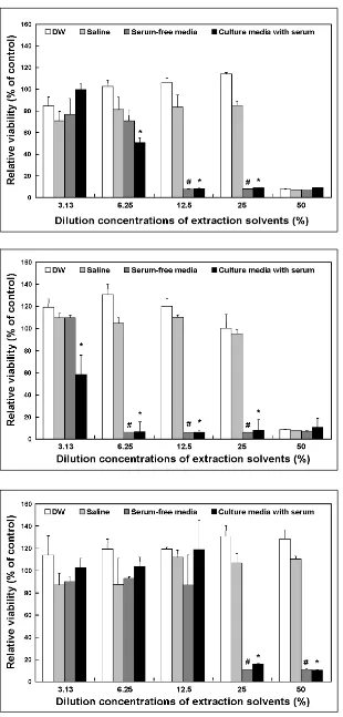
Fig. 1. The cytotoxicity of latex gloves as determined by extraction with DW, sa-line, serum-free media and culture media with serum for 24 h at 37 . The cytotoxi-city of the latex gloves to the cultured L-929 cells is expressed in terms of the relative viability. Relative cell viability was expressed as percentage of the optical density of the appropriate diluted extract versus the optical density of the corre-sponding non-treated control. All the variables were tested in three independent incubations for each experiment, and each experiment was repeated twice (n=6). The results were reported a mean ± SD and analyzed by Student t-tests. The values marked with * and # marks were signifi-cantly (p< 0.05) different from DW and saline extractions.
[image:3.595.58.368.96.743.2]Fig. 2. The cytotoxicity of latex gloves as determined by extraction with DW, sa-line, serum-free media and culture media with serum for 72 h at 37 . The cytoto-xicity of the latex gloves to the cultured L-929 cells is expressed in terms of the relative viability. Relative cell viability was expressed as percentage of the optical density of the appropriate diluted extract versus the optical density of the corre-sponding non-treated control. All the variables were tested in three inde-pendent incubations for each experiment, and each experiment was repeated twice (n= 6). The results were reported a mean ± SD and analyzed by Student t-tests. The values marked with * and # marks were significantly (p<0.05) different from DW and saline extractions.
cells was not occurred at all the extract concen-trations. In the cases of the extraction in the cul-ture medium with or without serum, the cells were non-viable due to the latex glove cytotoxicity at higher extract concentrations (25% and 50%).
Generally direct contact methods have various advantages, because they mimic physiological conditions, the zone of diffusion represents a concentration gradient of toxic chemicals, and require no extraction preparation. But the major difficulty of this assay is the risk of physical trau-ma to cultured cells from either sample move-ment or crushing due to sample weight.7,22 In vitro methods of cytotoxicity testing should quantify cell viability and growth, and be cor-related with in vivo methods or animal tests.11,23 Although an extract dilution test provides a quantitative comparison with positive and nega-tive controls, it presents difficulties in terms of preparing sample extracts. When culture medium is used as the extraction solvent, both polar and non polar components are extracted from the sample, and as was found in the present study, a culture medium containing serum had higher toxicity than normal saline.24 It has also been documented that the toxicities of polymeric biomaterials, would seem to be overwhelmingly due to their leachables.25
As shown in Figs. 1, 2 and 3, the results of cyto-toxicity tests using the extract dilution method showed dose-dependent relationships and pro-duced diverse results. Unlike extractions at 37 for 24 h or 72 h, extractions for 72 h at 50 were insufficient to evaluate sample cytotoxicity. These results seemed to be due to the degradation species released from the latex that might induce cytotoxic effects.
In conclusion, the use of culture medium with or without serum led much higher cytotoxicities than extractions performed with DW or saline. Additionally, extractions at 37 for 24 h or 72 h were found to be more sensitive and effective at evaluating latex glove cytotoxicity by the extract dilution method. Our results suggest that a combination of two or more extraction methods compatible with the physicochemical character-istics of test materials is required for in vitro
cytotoxicity testing.
REFERENCES
1. Bollen LS, Svendsen O. Regulatory guidelines for bio-compatibility safety testing. Med Plast Biomater 1997; May:16-43.
2. BS 5736, Evaluation of medical devices for biological hazards. Part 10: Method of test for toxicity to cells in culture of extracts from medical devices. 1988. 3. United States Pharmacopeia XXII: Biological reactivity
tests in vitro; 1990.
4. Hockley K, Baxter D. Use of 3T3 cell-neutral red uptake assay for irritants as an alternative to the rabbit (Draize) test. Food Chem Toxicol 1986;24:473-5. 5. Tsuchiya T. Studies on the standardization of
cytotoxi-city tests and new standard reference materials useful for evaluation the safety of biomaterials. J Biomater Appl 1994;9:138-57.
6. Shayne CG. Cytotoxicity testing. In: Shayne CG, editor. Safety evaluation of medical devices. New York: Marcel Dekker; 1997. p.75-84.
7. Ratner BD, Northup SJ. Testing biomaterials. In: Ratner BD, Hoffman AS, Schoen FJ, Lemons JE, editors. Biomaterials science: an introduction to materials in medicine. New York: Academic Press, 1996. p.215-20. 8. Ciapetti G, Granchi D, Verri E, Savarino L, Cavedagna D, Pizzoferrato A. Application of a combination of neural red and amino black staining for rapid, reliable cytotoxicity testing of biomaterials. Biomaterials 1996; 17:1259-64.
9. Tsuchiya T, Ikarashi Y, Arai T, Ohhashi J, Isama K, Nakamura A. In vitro tissue/biomaterials toxic re-sponses: correlation with cytotoxic potentials but not cell attachment. Clin Mater 1994;16:1-8.
10. International standard ISO 10993-12, Biological evalua-tion of medical devices-Part 12: Sample preparaevalua-tion and reference materials. 1996.
11. International standard ISO 10993-5, Biological evalua-tion of medical devices-Part 5: Tests for cytotoxicity:in vitro methods; 1992.
12. Lee JE, Park J-C, Park KD, Kim YH, Suh H. In vitro evaluation of PEG modified polyurethanes in cellular toxicity. Biomater Res 1998;2:65-8.
13. Lee WK, Park KD, Han DK, Suh H, Park J-C, Kim YH. Heparinized bovine pericardium as a novel cardiovas-cular bioprosthesis. Biomaterials 2000;21:2323-30. 14. Tsuchiya T, Ikarashi Y, Hata H, Toyoda K, Takahashi
M, Uchima T, et al. Comparative studies of the toxicity of standard reference materials in various cytotoxicity tests and in vivo implantation tests. J Appl Biomater 1993;4:153-6.
15. Kubota Y, Takahashi S, Takahashi I, Patrick G. Different cytotoxic response to gadolinium between mouse and rat alveolar macrophages. Toxicolin Vitro 2000;14:309-19.
17. Tsuchiya T, Ikarashi Y, Arai T, Ohhashi J, Nakamura A. Improved sensitivity and decreased sample size in a cytotoxicity test for biomaterials: a modified color microassay using a microplate and crystal violet staining. J Appl Biomater 1994;5:361-7.
18. Cenni E, Ciapetti G, Granchi D, Arciola CR, Savarino L, Stea S, et al. Established cell lines and primary cultures on testing medical devicesin vitro. Toxicolin Vitro 1999;13:801-10.
19. Liu BS, Yao CH, Chen YS, Hsu SH.In vitroevaluation of degradation and cytotoxicity of a novel composite as a bone substitute. J Biomed Mater Res 2003;67:1163-9. 20. Graham DT, Mark GE, Macarthur EB, Pomeroy AR.In vivovalidation of a cell culture test for biocompatibility testing of urinary catheters. J Biomed Mater Res 1984; 18:1125-35.
21. Ruutu M, Alfthan O, Talja M, Andersson C.
Cytotoxi-city of latex urinary catheters. Br J Urol 1985;57:82-7. 22. Pariente JL, Bordenave L, Jacob F, Bareille R, Baquey C, Le Guillou M. Cytotoxicity assessment of latex urinary catheters on cultured human urothelial cells. Eur Urol 2000;38:640-3.
23. Annual book of ASTM standards, section 13, F813-83, Standard practice for direct contact cell culture evalua-tion of materials for medical devices. 1996.
24. Ikarashi Y, Toyoda K, Ohsawa N, Uchima T, Tsuchiya T, Kaniwa M, et al. Comparative studies by cell culture andin vivoimplantation test on the toxicity of natural rubber latex materials. J Biomed Mater Res 1992;26: 339-56.