R E V I E W
Open Access
Ten things you should know about
transposable elements
Guillaume Bourque
1,2*, Kathleen H. Burns
3, Mary Gehring
4, Vera Gorbunova
5, Andrei Seluanov
5, Molly Hammell
6,
Michaël Imbeault
7, Zsuzsanna Izsvák
8, Henry L. Levin
9, Todd S. Macfarlan
9, Dixie L. Mager
10and Cédric Feschotte
11*Abstract
Transposable elements (TEs) are major components
of eukaryotic genomes. However, the extent of their
impact on genome evolution, function, and disease
remain a matter of intense interrogation. The rise of
genomics and large-scale functional assays has shed
new light on the multi-faceted activities of TEs and
implies that they should no longer be marginalized.
Here, we introduce the fundamental properties of TEs
and their complex interactions with their cellular
environment, which are crucial to understanding
their impact and manifold consequences for organismal
biology. While we draw examples primarily from
mammalian systems, the core concepts outlined
here are relevant to a broad range of organisms.
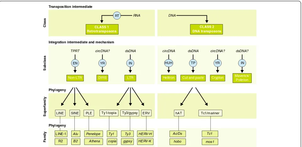
Transposable elements come in many different
forms and shapes
Transposable elements (TEs) are DNA sequences that
have the ability to change their position within a genome.
As a result of their deep evolutionary origins and
continu-ous diversification, TEs come in a bewildering variety of
forms and shapes (Fig.
1
). TEs can be divided into two
major classes based on their mechanism of transposition,
and each class can be subdivided into subclasses based on
the mechanism of chromosomal integration. Class 1
ele-ments, also known as retrotransposons, mobilize through
a
‘
copy-and-paste
’
mechanism whereby a RNA
intermedi-ate is reverse-transcribed into a cDNA copy that is
inte-grated elsewhere in the genome [
1
]. For long terminal
repeat (LTR) retrotransposons, integration occurs by
means of a cleavage and strand-transfer reaction catalyzed
* Correspondence:[email protected];[email protected]
1
Department of Human Genetics, McGill University, Montréal, Québec H3A 0G1, Canada
11Department of Molecular Biology and Genetics, Cornell University, Ithaca,
NY 14850, USA
Full list of author information is available at the end of the article
by an integrase much like retroviruses [
2
]. For non-LTR
retrotransposons, which include both long and short
in-terspersed nuclear elements (LINEs and SINEs),
chromo-somal integration is coupled to the reverse transcription
through a process referred to as target-primed reverse
transcription [
3
]. Class 2 elements, also known as DNA
transposons, are mobilized via a DNA intermediate, either
directly through a
‘
cut-and-paste
’
mechanism [
4
,
5
] or, in
the case of
Helitrons
, a
‘
peel-and-paste
’
replicative
mech-anism involving a circular DNA intermediate [
6
]. For
de-tailed reviews on individual TE types and transposition
mechanisms, we refer the reader to the monograph edited
by Craig et al. [
7
].
Each TE subclass is further divided into subgroups (or
superfamilies) that are typically found across a wide
range of organisms, but share a common genetic
organization and a monophyletic origin. For example,
Ty3/
gypsy
and Ty1/
copia
elements are two major
super-families of LTR retrotransposons that occur in virtually
all major groups of eukaryotes [
8
]. Similarly, Tc1/
mari-ner
, hAT (hobo-Ac-Tam3), and MULEs (Mutator-like
el-ements) are three superfamilies of DNA transposons
that are widespread across the eukaryotic tree [
9
]. At the
most detailed level of TE classification, elements are
grouped into families or subfamilies, which can be
de-fined as a closely related group of elements that can be
traced as descendants of a single ancestral unit [
10
]. This
ancestral copy can be inferred as a consensus sequence,
which is representative of the entire (sub)family [
11
,
12
].
Thus, in principle, every TE sequence in a genome can
be affiliated to a (sub)family, superfamily, subclass, and
class (Fig.
1
). However, much like the taxonomy of
spe-cies, the classification of TEs is in constant flux,
per-petually subject to revision due to the discovery of
completely novel TE types, the introduction of new
levels of granularity in the classification, and ongoing
de-velopment of methods and criteria to detect and classify
TEs [
13
,
14
].
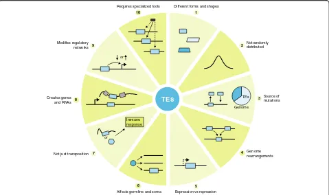
TEs are not randomly distributed in the genome
The genome may be viewed as an ecosystem inhabited
by diverse communities of TEs, which seek to propagate
and multiply through sophisticated interactions with
each other and with other components of the cell [
15
].
These interactions encompass processes familiar to
ecol-ogists, such as parasitism, cooperation, and competition
[
16
]. Thus, it is perhaps not surprising that TEs are
rarely, if ever, randomly distributed in the genome. TEs
exhibit various levels of preference for insertion within
certain features or compartments of the genome (Fig.
2
).
These are often guided by opposite selective forces, a
balancing act of facilitating future propagation while
mitigating deleterious effects on host cell function. At
the most extreme end of the site-selection spectrum,
many elements have evolved mechanisms to target
spe-cific loci where their insertions are less detrimental to
the host but favorable for their propagation [
17
]. For
in-stance, several retrotransposons in species as diverse as
slime mold and budding and fission yeast have evolved
independently, but convergently, the capacity to target
the upstream regions of genes transcribed by RNA
poly-merase III, where they do not appear to affect host gene
expression but retain the ability to be transcribed
them-selves [
17–20
].
Natural selection and genetic drift are also powerful
forces shaping the distribution and accumulation of
TEs [
21
]. Insertions that are strongly deleterious are
rapidly removed from the population. Insertions that
have little or no effects on genome function and host
fitness may reach fixation according to the efficiency of
selection and drift at purging these insertions from the
population, which vary greatly among species [
21
].
Se-lective forces can explain why some elements are more
likely to be retained in certain genomic locations than
others [
22
,
23
]. For instance, de novo insertions of the
human LINE 1 (L1) retrotransposon readily occur
within (and disrupt) gene exons [
24
], but very few if
any L1 elements have been fixed within the coding
re-gion of human genes [
25
]. Similarly, no LTR
retrotrans-poson is known to exhibit insertion preference with
regard to which DNA strand is transcribed, and yet
these elements are strongly depleted in the sense
orien-tation within human introns
—
most likely due to their
propensity to interfere with gene splicing and
polyade-nylation when inserted in sense orientation [
11
,
26
].
Perhaps because of some of these shared properties, the
evolutionary trajectories of TE accumulation in
mam-mals were found to be conserved across species in spite
of clade specific differences in TE content. [
27
]. Thus,
the success and diversity of TEs in a genome are shaped
both by properties intrinsic to the elements as well as
evolutionary forces acting at the level of the host
spe-cies. A solid comprehension of how these forces act
to-gether is paramount to understanding the impact of
TEs on organismal biology.
Transposition intermediate
RNA DNA
Integration intermediate and mechanism
Superfamily
Phylogeny
Subclass
Class
Phylogeny
Family
LTR Helitron Cut and paste Crypton Non-LTR DIRS
TPRT circDNA? dsDNA circDNA dsDNA circDNA?
Maverick/ Polinton
dsDNA?
LINE SINE PLE Ty1/copia Ty3/gypsy ERV hAT Tc1/mariner
LINE-1
R2
Alu
B2
Ty1
copia Ty3
gypsy HERV-H
HERV-K Penelope
Athena
Ac/Ds
hobo
Tc1
mos1
RT
CLASS 1 Retrotransposons
CLASS 2 DNA transposons
EN YR IN HUH TP YR IN
Fig. 1Classification of eukaryotic transposable elements. Schematic and examples showing the key features and relationships between TE classes,
[image:2.595.59.540.87.321.2]TEs are an extensive source of mutations and
genetic polymorphisms
TEs occupy a substantial portion of the genome of a
species, including a large fraction of the DNA unique to
that species. In maize, where Barbara McClintock did
her seminal work [
28
], an astonishing 60 to 70% of the
genome is comprised of LTR retrotransposons, many of
which are unique to this species or its close wild
rela-tives, but the less prevalent DNA transposons are
cur-rently the most active and mutagenic [
29–32
] (Fig.
2
).
Similarly, the vast majority of TE insertions in
Drosoph-ila melanogaster
are absent at the orthologous site in its
closest relative
D. simulans
(and vice versa), and most
are not fixed in the population [
33
,
34
]. Many TE
fam-ilies are still actively transposing and the process is
highly mutagenic; more than half of all known
pheno-typic mutants of
D. melanogaster
isolated in the
labora-tory are caused by spontaneous insertions of a wide
variety of TEs [
35
]. Transposition events are also
com-mon and mutagenic in laboratory mice, where ongoing
activity of several families of LTR elements are
respon-sible for 10
–
15% of all inherited mutant phenotypes
[
36
]. This contribution of TEs to genetic diversity may
be underestimated, as TEs can be more active when
or-ganisms are under stress, such as in their natural
envir-onment [
37
,
38
].
Because TE insertions rarely provide an immediate
fit-ness advantage to their host, those reaching fixation in
the population do so largely by genetic drift and are
sub-sequently eroded by point mutations that accumulate
neutrally [
21
]. Over time, these mutations result in TEs
that can no longer encode transposition enzymes and
produce new integration events. For instance, our
(hap-loid) genome contains ~ 500,000 L1 copies, but more
than 99.9% of these L1 copies are fixed and no longer
mobile due to various forms of mutations and
trunca-tions [
39
,
40
]. It is estimated that each person carries a
set of ~ 100 active L1 elements, and most of these are
young insertions still segregating within the human
population [
41
–
43
]. Thus, as for any other organism, the
‘
reference
’
human genome sequence does not represent
a comprehensive inventory of TEs in humans.
Thou-sands of
‘
non-reference
’
, unfixed TE insertions have been
catalogued through whole genome sequencing and other
targeted approaches [
44
]. On average, any two human
haploid genomes differ by approximately a thousand TE
insertions, primarily from the L1 or Alu families. The
number of TE insertion polymorphisms in a species with
much higher TE activity such as maize [
32
] dwarfs the
number in humans.
If TEs bring no immediate benefit to their host and
are largely decaying neutrally once inserted, how do they
Different forms and shapes
1 10
2
3
4
5 6
7 8
9 Not randomlydistributed
Source of mutations
Genome rearrangements
Expression vs repression Affects germline and soma
Not just transposition Creates genes
and RNAs Modifies regulatory
networks
Requires specialized tools
TEs
or or
Genome TEs
Immune response
Fig. 2Ten things you should know about transposable elements (TEs). Examples of how TEs can impact genomes in direct and indirect ways.
[image:3.595.59.538.88.372.2]persist in evolution? One key to this conundrum is the
ability of TEs not only to propagate vertically but also
horizontally between individuals and species. There is
now a large body of evidence supporting the idea that
horizontal transposon transfer is a common phenomenon
that affects virtually every major type of TE and all
branches of the tree of life [
45
,
46
]. While the cellular
mechanisms underlying horizontal transposon transfer
re-main murky, it is increasingly apparent that the intrinsic
mobility of TEs and ecological interactions between their
host species, including those with pathogens and parasites,
facilitate the transmission of elements between widely
di-verged taxa [
47–49
].
TEs are associated with genome rearrangements
and unique chromosome features
Transposition represents a potent mechanism of genome
expansion that over time is counteracted by the removal
of DNA via deletion. The balance between the two
pro-cesses is a major driver in the evolution of genome size
in eukaryotes [
21
,
50
,
51
]. Several studies have
demon-strated the impact and range of this shuffling and cycling
of genomic content on the evolution of plant and animal
genomes [
52–55
]. Because the insertion and removal of
TEs is often imprecise, these processes can indirectly
affect surrounding host sequences. Some of these events
occur at high enough frequency to result in vast
amounts of duplication and reshuffling of host
se-quences, including genes and regulatory sequences. For
example, a single group of DNA transposons (MULEs)
has been responsible for the capture and reshuffling of
~ 1,000 gene fragments in the rice genome [
56
]. Such
studies have led to the conclusion that the rate at which
TEs transpose, which is in part under host control, is an
important driver of genome evolution [
57
–
59
].
In addition to rearrangements induced as a byproduct of
transposition, TEs can promote genomic structural
vari-ation long after they have lost the capacity to mobilize [
60
].
In particular, recombination events can occur between the
highly homologous regions dispersed by related TEs at
dis-tant genomic positions and result in large-scale deletions,
duplications, and inversions [
59
,
61–63
] (Fig.
2
). TEs also
provide regions of microhomology that predispose to
tem-plate switching during repair of replication errors leading
to another source of structural variants [
64
]. These
non-transposition-based mechanisms for TE-induced or
TE-enabled structural variation have contributed
substan-tially to genome evolution. These processes can also make
the identification of actively transposing elements more
difficult in population studies that infer the existence of
ac-tive elements through the detection of non-reference
insertions.
TEs also contribute to specialized chromosome
fea-tures. An intriguing example is in
Drosophila
, where
LINE-like retrotransposons form and maintain the
telo-meres in replacement of the telomerase enzyme which
has been lost during dipteran evolution [
65
]. This
domes-tication event could be viewed as a replay of what might
have happened much earlier in eukaryotic evolution to
solve the
‘
end problem
’
created by the linearization of
chromosomes. Indeed, the reverse transcriptase
compo-nent of telomerase is thought to have originated from an
ancient lineage of retroelements [
66
,
67
]. TE sequences
and domesticated transposase genes also play structural
roles at centromeres [
68
–
70
].
There is an intrinsic balance between TE
expression and repression
To persist in evolution, TEs must strike a delicate
bal-ance between expression and repression (Fig.
2
).
Expres-sion should be sufficient to promote amplification, but
not so vigorous as to lead to a fitness disadvantage for
the host that would offset the benefit to the TE of
in-creased copy numbers. This balancing act may explain
why TE-encoded enzymes are naturally suboptimal for
transposition [
71
,
72
] and why some TEs have evolved
self-regulatory mechanisms controlling their own copy
numbers [
73
,
74
]. A variety of host factors are also
employed to control TE expression, which includes a
variety of small RNA, chromatin, and DNA modification
pathways [
75–78
], as well as sequence-specific
repres-sors such as the recently profiled KRAB zinc-finger
proteins [
79–82
]. However, many of these silencing
mechanisms must be at least partially released to permit
developmental regulation of host gene expression
pro-grams, particularly during early embryonic development.
For example, genome-wide loss of DNA methylation is
necessary to reset imprinted genes in primordial germ
cells [
83
]. This affords TEs an opportunity, as reduced
DNA methylation often promotes TE expression. Robust
expression of a TE in the germ lineage (but not
neces-sarily in the gametes themselves) is often its own
down-fall. In one example of a clever trick employed by the
host, TE repression is relieved in a companion cell
de-rived from the same meiotic product as flowering plant
sperm [
84
]. However, this companion cell does not
con-tribute genetic material to the next generation. Thus,
al-though TEs transpose in a meiotic product, the events
are not inherited. Instead, TE activity in the companion
cell may further dampen TE activity in sperm via the
im-port of TE-derived small RNAs [
85
].
This is indeed observed among many species, with
cili-ates representing an extreme example of this division
—
TEs are actively deleted from the somatic macronucleus
but retained in the micronucleus, or germline [
87
].
An-other example is the P-elements in
Drosophila
, which
are differentially spliced in the germline versus soma
[
88
]. Many organisms, including plants, do not
differen-tiate germ lineage cells early in development; rather, they
are specified from somatic cells shortly before meiosis
commences. Thus, TEs that transpose in somatic cells in
plants have the potential to be inherited, which suggests
that the interest of TEs and host are in conflict across
many more cells and tissues than in animals with a
seg-regated germline.
TEs are insertional mutagens in both germline
and soma
Like other species, humans contend with a contingent of
currently active TEs where the intrinsic balance between
expression and repression is still at play [
89
]. For us, this
includes L1 and other mobile elements that depend on
L1-encoded proteins for retrotransposition [
90
,
91
].
These elements are responsible for new germline
inser-tions that can cause genetic disease. More than 120
in-dependent TE insertions have been associated with
human disease [
24
]. The rate of de novo germline
trans-position in humans is approximately one in 21 births for
Alu
[
92
] and one in 95 births for L1 [
93
].
Historically, little attention has been given to
transpos-ition in somatic cells and its consequences, because
som-atic transposition may be viewed as an evolutionary
dead-end for the TE with no long-term consequences for
the host species. Yet, there is abundant evidence that TEs
are active in somatic cells in many organisms [
94
] (Fig.
2
).
In humans, L1 expression and transposition have been
de-tected in a variety of somatic contexts, including early
em-bryos and certain stem cells [
95
,
96
]. There is also a great
deal of interest in mobile element expression and activity
in the mammalian brain, where L1 transposition has been
proposed to diversify neuronal cell populations [
97–99
].
One challenge for assessing somatic activity has rested
with the development of reliable single cell insertion site
mapping strategies [
100–103
].
Somatic activity has also been observed in human
can-cers, where tumors can acquire hundreds of new L1
in-sertions [
104–109
]. Just like for human polymorphisms,
somatic activity in human cancers is caused by small
numbers of so-called
‘
hot
’
L1 loci [
41
,
107
]. The
activ-ities of these master copies varies depending on the
indi-vidual [
105
], tumor type [
105
], and timeframe in the
clonal evolution of the tumor [
106
,
110
]. Some of these
de novo L1 insertions disrupt critical tumor suppressors
and oncogenes and thus drive cancer formation [
107
],
although the vast majority appear to be
‘
passenger
’
mutations [
111
]. Host cells have evolved several
mecha-nisms to keep TEs in check. However, as the force of
natural selection begins to diminish with age and
com-pletely drops in post-reproductive life, TEs may become
more active [
112
].
TEs can be damaging in ways that do not involve
transposition
TEs are best known for their mobility, in other words their
ability to transpose to new locations. While the breakage
and insertion of DNA associated with transposition
repre-sents an obvious source of cell damage, this is not the only
or perhaps even the most common mechanism by which
TEs can be harmful to their host. Reactivated transposons
harm the host in multiple ways. First, de-repression of
transposon loci, including their own transcription, may
interfere with transcription or processing of host mRNAs
through a myriad of mechanisms [
113–115
]. Genome-wide
transcriptional de-repression of TEs has been documented
during replicative senescence of human cells [
116
] and
sev-eral mouse tissues, including liver, muscle, and brain [
117
,
118
]. De-repression of LTR and L1 promoters can also
cause oncogene activation in cancer [
119
]. Second,
TE-encoded proteins such as the endonuclease activity of L1
ORF2p can induce DNA breaks and genomic instability
[
120
]. Third, accumulation of RNA transcripts and
extra-chromosomal DNA copies derived from TEs may trigger
an innate immune response leading to autoimmune
dis-eases and sterile inflammation (Fig.
2
). Activation of
inter-feron response is now a well-documented property of
transcripts derived from endogenous retroviruses and may
give immunotherapies a boost in identifying and attacking
cancer cells [
121–123
]. The relative contribution of all the
above mechanisms in organismal pathologies remains to be
determined.
(HERV) group, HERV-K (HML-2), may play a role in
some cancers but the evidence remains circumstantial
[
129
,
130
].
A number of key coding and non-coding RNAs are
derived from TEs
Although usually detrimental, there is growing evidence
that TE insertions can provide raw material for the
emergence of protein-coding genes and non-coding
RNAs, which can take on important and, in some cases
essential, cellular function [
131–133
] (Fig.
2
). The
process of TE gene
‘
domestication
’
or exaptation over
evolutionary time contributes to both deeply conserved
functions and more recent, species-specific traits. Most
often, the ancestral or a somewhat modified role of a
TE-encoded gene is harnessed by the host and
con-served, while the rest of the TE sequence, and hence its
ability to autonomously transpose, has been lost.
Spec-tacular examples of deeply conserved TE-derived genes
are
Rag1
and
Rag2
, that catalyze V(D)J somatic
recom-bination in the vertebrate immune system. Both genes,
and probably the DNA signals they recognize, were
de-rived from an ancestral DNA transposon around 500
million years ago [
134
,
135
]. Indeed, DNA transposases
have been co-opted multiple times to form new cellular
genes [
70
,
113
].
The
gag
and
env
genes of LTR retrotransposons or
en-dogenous retroviruses (ERVs) have also been
domesti-cated numerous times to perform functions in placental
development, contribute to host defense against
exogen-ous retroviruses, act in brain development, and play
other diverse roles [
132
,
136
]. One of the most
intri-guing examples of TE domestication is the repeated,
in-dependent capture of ERV
env
genes, termed
syncytins
,
which appear to function in placentation by facilitating
cell
–
cell fusion and syncytiotrophoblast formation [
137–
139
]. Notably, one or more such
syncytin
genes have
been found in virtually every placental mammalian
lineage where they have been sought, strongly suggesting
that ERVs have played essential roles in the evolution
and extreme phenotypic variability of the mammalian
placenta. Another example of a viral-like activity
re-purposed for host cell function is provided by the
neuronal
Arc
gene, which arose from the
gag
gene from
a LTR retrotransposon domesticated in the common
an-cestor of tetrapod vertebrates [
140
]. Genetic and
bio-chemical studies of murine Arc show that it is involved
in memory and synaptic plasticity and has preserved
most of the ancestral activities of Gag, including the
packaging and intercellular trafficking of its own RNA
[
140
]. Remarkably, flies appear to have independently
evolved a similar system of trans-synaptic RNA delivery
involving a
gag
-like protein derived from a similar yet
distinct lineage of LTR retrotransposons [
141
]. Thus, the
biochemical activities of TE-derived proteins have been
repeatedly co-opted during evolution to foster the
emer-gence of convergent cellular innovations in different
organisms.
TEs can donate their own genes to the host, but they
can also add exons and rearrange and duplicate existing
host genes. In humans, intronic
Alu
elements are
par-ticularly prone to be captured as alternative exons
through cryptic splice sites residing within their
se-quences [
142
,
143
]. L1 and SVA (SINE/VNTR/Alu)
ele-ments also contribute to exon shuffling through
transduction events of adjacent host sequences during
their mobilization [
144
,
145
]. The reverse transcriptase
activity of retroelements is also responsible for the
trans-duplication of cellular mRNAs to create
‘
proc-essed
’
retrogenes in a wide range of organisms [
146
,
147
]. The L1 enzymatic machinery is thought to be
in-volved in the generation of tens of thousands of
retro-gene copies in mammalian genomes, many of which
remain transcribed and some of which have acquired
new cellular functions [
147
,
148
]. This is a process still
actively shaping our genomes; it has been estimated that
1 in every 6000 humans carries a novel retrogene
inser-tion [
93
].
TEs also make substantial contributions to non-protein
coding functions of the cell. They are major components
of thousands of long non-coding RNAs in human and
mouse genomes, often transcriptionally driven by
retro-viral LTRs [
149
]. Some of these TE-driven lncRNAs
ap-pear to play important roles in the maintenance of stem
cell pluripotency and other developmental processes
[
150
–
154
]. Many studies have demonstrated that TE
se-quences embedded within lncRNAs and mRNAs can
dir-ectly modulate RNA stability, processing, or localization
with important regulatory consequences [
114
,
155
–
158
].
Furthermore, TE-derived microRNAs [
159
] and other
small RNAs processed from TEs [
160
] can also adopt
regulatory roles serving host cell functions. The myriad of
mechanisms by which TEs contribute to coding and
non-coding RNAs illustrate the multi-faceted interactions
between these elements and their host.
TEs contribute cis-regulatory DNA elements and
modify transcriptional networks
[
176
,
177
] (reviewed in [
178
]). The varying coat colors of
agouti mice provides a striking example of a host gene
controlling coat color whose expression can be altered
by the methylation levels of a TE upstream of its
pro-moter [
179
,
180
]. In the oil palm, the methylation level
of a TE that sits within a gene important for flowering
ultimately controls whether or not the plants bear
oil-rich fruit [
181
].
As TE families typically populate a genome as a
multi-tude of related copies, it has long been postulated that
they have the potential to donate the same cis-regulatory
module to
‘
wire
’
batteries of genes dispersed throughout
the genome [
182
]. An increasing number of studies
sup-port this model and suggest that TEs have provided the
building blocks for the assembly and remodeling of
cis-regulatory networks during evolution, including
pathways underlying processes as diverse as pregnancy
[
183
,
184
], stem cell pluripotency [
150
,
151
,
171
],
neo-cortex development [
185
], innate immunity in mammals
[
163
], or the response to abiotic stress in maize [
186
].
Indeed, TE sequences harbor all the necessary features
of a
‘
classical
’
gene regulatory network [
113
,
114
]. They
are bound by diverse sets of transcription factors [
172
]
integrate multiple inputs (activation/repression), respond
to signals in both
cis
and
trans
, and are capable of
co-ordinately regulating gene expression. In this context,
TEs are highly suitable agents to modify biological
pro-cesses by creating novel cis-regulatory circuits and
fine-tuning pre-existing networks.
Analyzing TEs requires specialized tools
TEs have been historically neglected and remain
fre-quently ignored in genomic studies in part because of
their repetitive nature, which poses a number of
analyt-ical challenges and often requires the use of specialized
tools [
187
]. As genomes can harbor thousands of copies
of very similar TE sequences, uniqueness or,
alterna-tively, repetitiveness of substrings within these regions
need to be taken into consideration during both
experi-mental design and analysis. As an example, short DNA
oligos targeting a specific TE instance in the genome for
PCR, short hairpin RNA, or CRISPR-Cas9 have to be
carefully designed and validated to ensure that they are
truly specific and target unique regions of the genome.
In some scenarios, it can be acceptable or even desirable
to target many elements simultaneously [
150
] or an
en-tire TE family [
153
,
188–191
].
Similarly, uniqueness and repetitiveness are important
concepts to consider when aligning reads from next
gen-eration sequencing and analyzing TEs (Fig.
2
). Various
strategies exist to assign reads that could originate from
multiple genomic locations: 1) mapping reads to
consen-sus sequences of TE subfamilies [
172
]; 2) mapping to
the genome and keeping only uniquely-mapping reads
[
163
,
168
]; 3) assigning multiple mapping reads at
ran-dom between possible candidates [
192
]; or 4)
redistribut-ing them accordredistribut-ing to various algorithms, such as
maximum likelihood [
193
,
194
]. The choice is ultimately
guided by the technique (such as ChIP-seq and
RNA-seq) and the purpose of the analysis
—
is
informa-tion about individual TE instances needed, or is a
high-level tally of results for each subfamily sufficient?
Notably, these issues of uniqueness will differ
substan-tially depending on the species studied and the presence
or absence of recently, or currently, active TE families.
For example, mapping reads to TEs in the human
gen-ome will be less challenging than in the mouse gengen-ome
given the more recent and mobile TE landscape of
the latter species [
36
]. Finally, as sequencing
technol-ogy and bioinformatics pipelines improve, notably
with the increasing length of sequencing reads, many
of the hurdles faced by earlier studies will be
progres-sively removed [
187
].
Outlook
As potent insertional mutagens, TEs can have both
posi-tive and negaposi-tive effects on host fitness, but it is likely
that the majority of TE copies in any given species
—
and
especially those such as humans with small effective
population size
—
have reached fixation through genetic
drift alone and are now largely neutral to their host.
When can we say that TEs have been co-opted for
cellu-lar function? The publication of the initial ENCODE
paper [
195
], which asserted
‘
function for 80% of the
gen-ome
’
, was the subject of much debate and controversy.
Technically speaking, ENCODE assigned only
‘
biochem-ical
’
activity to this large fraction of the genome. Yet
critics objected to the grand proclamations in the
popu-lar press (The Washington Post Headline:
“
Junk DNA
concept debunked by new analysis of the human
gen-ome
”
) and to the ENCODE consortium
’
s failure to
pre-vent this misinterpretation [
196–198
]. To these critics,
ignoring evolutionary definitions of function was a major
misstep.
This debate can be easily extended to include TEs. TEs
make up the vast majority of what is often referred to as
The vast differences in TEs between species make
standard approaches to establish their regulatory roles
particularly challenging [
200
]. For example, intriguing
studies on the impact of HERVs, in particular HERV-H,
in stem cells and pluripotency [
150–152
] must be
inter-preted using novel paradigms that do not invoke deep
evolutionary conservation to imply function, as these
particular ERVs are absent outside of great apes.
Evolu-tionary constraint can be measured at shorter time
scales, including the population level, but this remains a
statistically challenging task especially for non-coding
se-quences. Natural loss-of-function alleles may exist in the
human population and their effect on fitness can be
studied if their impact is apparent, but these are quite
rare and do not allow systematic studies. It is possible to
engineer genetic knockouts of a particular human TE
locus to test its regulatory role but those are restricted
to in-vitro systems, especially when the orthologous TE
does not exist in the model species. In this context,
studying the impact of TEs in model species with
power-ful genome engineering tools and vast collections of
mu-tants and other genetic resources, such as plants, fungi,
and insects, will also continue to be extremely valuable.
Finally, a growing consensus is urging more rigor
when assigning cellular function to TEs, particularly for
the fitness benefit of the host [
178
]. Indeed, a TE
dis-playing biochemical activity (such as those bound by
transcription factors or lying within open chromatin
re-gions) cannot be equated to a TE that shows evidence of
purifying selection at the sequence level or, when
genet-ically altered, result in a deleterious or dysfunctional
phenotype. Recent advances in editing and manipulating
the genome and the epigenome en masse yet with
preci-sion, including repetitive elements [
153
,
154
,
189–191
],
offer the promise for a systematic assessment of the
functional significance of TEs.
Abbreviations
Env:Envelope protein; ERV: Endogenous retrovirus; HERV: Human endogenous retrovirus; L1: Long interspersed nuclear element 1; LINE: Long interspersed nuclear element; LTR: Long terminal repeat; SINE: Short interspersed nuclear element; TE: Transposable element
Acknowledgments
We apologize to our colleagues whose relevant work and original articles could not be cited owing to space constraint. We thank Patricia Goerner-Potvin and Edward Chuong for early discussions about the manuscript. We are also indebted to Ayana Moosa and Susan Mahon from the McGill Bellairs Research Institute for their help in organizing the workshop that led to the concept of this review.
Funding
This work was supported by grants from the Canadian Institute for Health Research (CIHR-MOP-115090 and CIHR-CEE-151618). GB is supported by the Fonds de Recherche Santé Québec (FRSQ-25348 and FRSQ-35279). ZI is supported by ERC-2011-AdG 294742. CF, HLL, KHB, VG, and MG acknowledge funding by the National Institutes of Health. MG is also funded by the National Science Foundation.
Authors’contributions
All authors participated in the planning and the writing of this review. All authors read and approved the final manuscript.
Competing interests
The authors declare that they have no competing interests.
Publisher
’
s Note
Springer Nature remains neutral with regard to jurisdictional claims in published maps and institutional affiliations.
Author details
1Department of Human Genetics, McGill University, Montréal, Québec H3A
0G1, Canada.2Canadian Center for Computational Genomics, McGill University, Montréal, Québec H3A 0G1, Canada.3Department of Pathology,
Johns Hopkins University School of Medicine, Baltimore, MD 21205, USA.
4Whitehead Institute for Biomedical Research and Department of Biology,
Massachusetts Institute of Technology, Cambridge, MA 02142, USA.
5Department of Biology, University of Rochester, Rochester, NY 14627, USA. 6Cold Spring Harbor Laboratory, Cold Spring Harbor, NY 11724, USA. 7Department of Genetics, University of Cambridge, Cambridge CB2 3EH, UK. 8
Max Delbrück Center for Molecular Medicine in the Helmholtz Association, 13125 Berlin, Germany.9The Eunice Kennedy Shriver National Institute of
Child Health and Human Development, The National Institutes of Health, Bethesda, Maryland, USA.10Terry Fox Laboratory, British Columbia Cancer
Agency and Department of Medical Genetics, University of BC, Vancouver, BC V5Z1L3, Canada.11Department of Molecular Biology and Genetics, Cornell
University, Ithaca, NY 14850, USA.
References
1. Boeke JD, Garfinkel DJ, Styles CA, Fink GR. Ty elements transpose through an RNA intermediate. Cell. 1985;40:491–500.
2. Brown PO, Bowerman B, Varmus HE, Bishop JM. Correct integration of retroviral DNA in vitro. Cell. 1987;49:347–56.
3. Luan DD, Korman MH, Jakubczak JL, Eickbush TH. Reverse transcription of R2Bm RNA is primed by a nick at the chromosomal target site: a mechanism for non-LTR retrotransposition. Cell. 1993;72:595–605. 4. Greenblatt IM, Brink RA. Transpositions of modulator in maize into divided
and undivided chromosome segments. Nature. 1963;197:412–3. 5. Rubin GM, Kidwell MG, Bingham PM. The molecular basis of P-M hybrid
dysgenesis: the nature of induced mutations. Cell. 1982;29:987–94. 6. Grabundzija I, Messing SA, Thomas J, Cosby RL, Bilic I, Miskey C, et al. A
Helitron transposon reconstructed from bats reveals a novel mechanism of genome shuffling in eukaryotes. Nat Commun. 2016;7:10716.
7. Craig NL, Chandler M, Gellert M, Lambowitz AM, Rice PA, Sandmeyer SB. Mobile DNA III. 3rd ed. Washington, DC: American Society for Microbiology (ASM); 2015.
8. Malik HS, Eickbush TH. Phylogenetic analysis of ribonuclease H domains suggests a late, chimeric origin of LTR retrotransposable elements and retroviruses. Genome Res. 2001;11:1187–97.
9. Feschotte C, Pritham EJ. DNA transposons and the evolution of eukaryotic genomes. Annu Rev Genet. 2007;41:331–68.
10. Britten RJ, Kohne DE. Repeated sequences in DNA. Science. 1968;161: 529–40.
11. Smit AF. Interspersed repeats and other mementos of transposable elements in mammalian genomes. Curr Opin Genet Dev. 1999;9:657–63. 12. Jurka J, Smith T. A fundamental division in the Alu family of repeated
sequences. Proc Natl Acad Sci U S A. 1988;85:4775–8.
13. Wicker T, Sabot F, Hua-Van A, Bennetzen JL, Capy P, Chalhoub B, et al. A unified classification system for eukaryotic transposable elements. Nat Rev Genet. 2007;8:973–82.
14. Arkhipova IR. Using bioinformatic and phylogenetic approaches to classify transposable elements and understand their complex evolutionary histories. Mob DNA. 2017;8:19.
16. Robillard É, Rouzic AL, Zhang Z, Capy P, Hua-Van A. Experimental evolution reveals hyperparasitic interactions among transposable elements. Proc Natl Acad Sci U S A. 2016;113:14763–8.
17. Sultana T, Zamborlini A, Cristofari G, Lesage P. Integration site selection by retroviruses and transposable elements in eukaryotes. Nat Rev Genet. 2017; 18:292–308.
18. Spaller T, Kling E, Glöckner G, Hillmann F, Winckler T. Convergent evolution of tRNA gene targeting preferences in compact genomes. Mob DNA. 2016; 7:17.
19. Cheung S, Manhas S, Measday V. Retrotransposon targeting to RNA polymerase III-transcribed genes. Mob DNA. 2018;9:14.
20. Guo Y, Singh PK, Levin HL. A long terminal repeat retrotransposon of
Schizosaccharomyces japonicusintegrates upstream of RNA pol III transcribed genes. Mob DNA. 2015;6:19.
21. Lynch M. The origins of genome architecture. 1st ed. Sunderland: Sinauer Associates; 2007.
22. Campos-Sánchez R, Cremona MA, Pini A, Chiaromonte F, Makova KD. Integration and fixation preferences of human and mouse endogenous retroviruses uncovered with functional data analysis. PLOS Comput Biol. 2016;12:e1004956.
23. Kvikstad EM, Makova KD. The (r)evolution of SINE versus LINE distributions in primate genomes: sex chromosomes are important. Genome Res. 2010; 20:600–13.
24. Hancks DC, Kazazian HH. Roles for retrotransposon insertions in human disease. Mob DNA. 2016;7:9.
25. Gotea V, Makalowski W. Do transposable elements really contribute to proteomes? Trends Genet. 2006;22:260–7.
26. Medstrand P, Van De Lagemaat LN, Mager DL. Retroelement distributions in the human genome: variations associated with age and proximity to genes. Genome Res. 2002;12:1483–95.
27. Buckley RM, Kortschak RD, Raison JM, Adelson DL. Similar evolutionary trajectories for retrotransposon accumulation in mammals. Genome Biol Evol. 2017;9:2336–53.
28. McClintock B. Controlling elements and the gene: Cold Spring Harb Symp Quant Biol. Cold Spring Harbor: Cold Spring Harbor Laboratory Press; 1956. p. 197–216.
29. Schnable PS, Ware D, Fulton RS, Stein JC, Wei F, Pasternak S, et al. The B73 maize genome: complexity, diversity, and dynamics. Science. 2009;326:1112–5. 30. Lazarow K, Doll M-L, Kunze R. Molecular Biology of Maize Ac/Ds elements:
an overview. In: Peterson T, editor. Plant transposable elements. Totowa: Humana Press; 2013. p. 59–82.
31. Liu S, Yeh C-T, Ji T, Ying K, Wu H, Tang HM, et al. Mu transposon insertion sites and meiotic recombination events co-localize with epigenetic marks for open chromatin across the maize genome. PLoS Genet. 2009;5: e1000733.
32. Springer NM, Anderson SN, Andorf CM, Ahern KR, Bai F, Barad O, et al. The maize W22 genome provides a foundation for functional genomics and transposon biology. Nat Genet. 2018;50:1282–8.
33. Kofler R, Nolte V, Schlötterer C. Tempo and mode of transposable element activity in Drosophila. PLOS Genet. 2015;11:e1005406.
34. Rahman R, Chirn G, Kanodia A, Sytnikova YA, Brembs B, Bergman CM, et al. Unique transposon landscapes are pervasive acrossDrosophila melanogaster genomes. Nucleic Acids Res. 2015;43:10655–72.
35. Eickbush TH, Furano AV. Fruit flies and humans respond differently to retrotransposons. Curr Opin Genet Dev. 2002;12:669–74.
36. Maksakova IA, Romanish MT, Gagnier L, Dunn CA, Van de Lagemaat LN, Mager DL. Retroviral elements and their hosts: insertional mutagenesis in the mouse germ line. PLoS Genet. 2006;2:e2.
37. Lanciano S, Mirouze M. Transposable elements: all mobile, all different, some stress responsive, some adaptive? Curr Opin Genet Dev. 2018;49:106–14. 38. Horváth V, Merenciano M, González J. Revisiting the relationship between
transposable elements and the eukaryotic stress response. Trends Genet. 2017;33:832–41.
39. Ostertag EM, Kazazian HH Jr. Biology of mammalian L1 retrotransposons. Annu Rev Genet. 2001;35:501–38.
40. Szak ST, Pickeral OK, Makalowski W, Boguski MS, Landsman D, Boeke JD. Molecular archeology of L1 insertions in the human genome. Genome Biol. 2002;3:research0052–1.
41. Brouha B, Schustak J, Badge RM, Lutz-Prigge S, Farley AH, Moran JV, et al. Hot L1s account for the bulk of retrotransposition in the human population. Proc Natl Acad Sci U S A. 2003;100:5280–5.
42. Sassaman DM, Dombroski BA, Moran JV, Kimberland ML, Naas TP, DeBerardinis RJ, et al. Many human L1 elements are capable of retrotransposition. Nat Genet. 1997;16:37–43.
43. Beck CR, Collier P, Macfarlane C, Malig M, Kidd JM, Eichler EE, et al. LINE-1 retrotransposition activity in human genomes. Cell. 2010;141:1159–70. 44. Sudmant PH, Rausch T, Gardner EJ, Handsaker RE, Abyzov A, Huddleston J,
et al. An integrated map of structural variation in 2,504 human genomes. Nature. 2015;526:75–81.
45. Gilbert C, Feschotte C. Horizontal acquisition of transposable elements and viral sequences: patterns and consequences. Curr Opin Genet Dev. 2018;49:15–24.
46. Wallau GL, Vieira C, Loreto ÉLS. Genetic exchange in eukaryotes through horizontal transfer: connected by the mobilome. Mob DNA. 2018;9:6. 47. Gilbert C, Cordaux R. Viruses as vectors of horizontal transfer of genetic
material in eukaryotes. Curr Opin Virol. 2017;25:16–22.
48. Metzger MJ, Paynter AN, Siddall ME, Goff SP. Horizontal transfer of retrotransposons between bivalves and other aquatic species of multiple phyla. Proc Natl Acad Sci U S A. 2018;115:E4227–35.
49. Ivancevic AM, Kortschak RD, Bertozzi T, Adelson DL. Horizontal transfer of BovB and L1 retrotransposons in eukaryotes. Genome Biol. 2018;19:85. 50. Petrov DA. Mutational equilibrium model of genome size evolution. Theor
Popul Biol. 2002;61:531–44.
51. Schubert I, Vu GTH. Genome stability and evolution: attempting a holistic view. Trends Plant Sci. 2016;21:749–57.
52. Gregory TR, Johnston JS. Genome size diversity in the family Drosophilidae. Heredity. 2008;101:228–38.
53. Kapusta A, Suh A, Feschotte C. Dynamics of genome size evolution in birds and mammals. Proc Natl Acad Sci U S A. 2017;114:E1460–9.
54. Pellicer J, Kelly LJ, Leitch IJ, Zomlefer WB, Fay MF. A universe of dwarfs and giants: genome size and chromosome evolution in the monocot family Melanthiaceae. New Phytol. 2014;201:1484–97.
55. Thybert D, Roller M, Navarro FCP, Fiddes I, Streeter I, Feig C, et al. Repeat associated mechanisms of genome evolution and function revealed by the Mus caroli and Mus pahari genomes. Genome Res. 2018;28:448–59.
56. Jiang N, Bao Z, Zhang X, Eddy SR, Wessler SR. Pack-MULE transposable elements mediate gene evolution in plants. Nature. 2004;431:569–73. 57. Cordaux R, Batzer MA. The impact of retrotransposons on human genome
evolution. Nat Rev Genet. 2009;10:691–703.
58. Freeling M, Xu J, Woodhouse M, Lisch D. A solution to the C-value paradox and the function of junk DNA: the genome balance hypothesis. Mol Plant. 2015;8:899–910.
59. Bennetzen JL, Wang H. The contributions of transposable elements to the structure, function, and evolution of plant genomes. Annu Rev Plant Biol. 2014;65:505–30.
60. Carvalho CM, Lupski JR. Mechanisms underlying structural variant formation in genomic disorders. Nat Rev Genet. 2016;17:224–38.
61. Deininger PL, Moran JV, Batzer MA, Kazazian HH. Mobile elements and mammalian genome evolution. Curr Opin Genet Dev. 2003;13:651–8. 62. Ade C, Roy-Engel AM, Deininger PL. Alu elements: an intrinsic source of
human genome instability. Curr Opin Virol. 2013;3:639–45. 63. Han K, Lee J, Meyer TJ, Remedios P, Goodwin L, Batzer MA. L1
recombination-associated deletions generate human genomic variation. Proc Natl Acad Sci U S A. 2008;105:19366–71.
64. Lee JA, Carvalho CM, Lupski JR. A DNA replication mechanism for generating nonrecurrent rearrangements associated with genomic disorders. Cell. 2007;131:1235–47.
65. Pardue M-L, DeBaryshe PG. Retrotransposons that maintain chromosome ends. Proc Natl Acad Sci U S A. 2011;108:20317–24.
66. Belfort M, Curcio MJ, Lue NF. Telomerase and retrotransposons: reverse transcriptases that shaped genomes. Proc Natl Acad Sci U S A. 2011;108: 20304–10.
67. Fulcher N, Derboven E, Valuchova S, Riha K. If the cap fits, wear it: an overview of telomeric structures over evolution. Cell Mol Life Sci. 2014; 71:847–65.
68. Casola C, Hucks D, Feschotte C. Convergent domestication of pogo-like transposases into centromere-binding proteins in fission yeast and mammals. Mol Biol Evol. 2007;25:29–41.
69. Kursel LE, Malik HS. Centromeres. Curr Biol. 2016;26:R487–90.
71. Lampe DJ, Akerley BJ, Rubin EJ, Mekalanos JJ, Robertson HM. Hyperactive transposase mutants of the Himar1 mariner transposon. Proc Natl Acad Sci U S A. 1999;96:11428–33.
72. Mátés L, Chuah MKL, Belay E, Jerchow B, Manoj N, Acosta-Sanchez A, et al. Molecular evolution of a novel hyperactiveSleeping Beautytransposase enables robust stable gene transfer in vertebrates. Nat Genet. 2009;41: 753–61.
73. Lohe AR, Hartl DL. Autoregulation of mariner transposase activity by overproduction and dominant-negative complementation. Mol Biol Evol. 1996;13:549–55.
74. Saha A, Mitchell JA, Nishida Y, Hildreth JE, Ariberre JA, Gilbert WV, et al. A trans-dominant form of gag restricts Ty1 retrotransposition and mediates copy number control. J Virol. 2015;89:3922–38.
75. Molaro A, Malik HS. Hide and seek: how chromatin-based pathways silence retroelements in the mammalian germline. Curr Opin Genet Dev. 2016;37: 51–8.
76. Liu N, Lee CH, Swigut T, Grow E, Gu B, Bassik MC, et al. Selective silencing of euchromatic L1s revealed by genome-wide screens for L1 regulators. Nature. 2017;553:228–32.
77. Goodier JL. Restricting retrotransposons: a review. Mob DNA. 2016;7:16. 78. Berrens RV, Andrews S, Spensberger D, Santos F, Dean W, Gould P, et al. An
endosiRNA-based repression mechanism counteracts transposon activation during global dna demethylation in embryonic stem cells. Cell Stem Cell. 2017;21:694–703.e7.
79. Imbeault M, Trono D. As time goes by: KRABs evolve to KAP endogenous retroelements. Dev Cell. 2014;31:257–8.
80. Imbeault M, Helleboid P-Y, Trono D. KRAB zinc-finger proteins contribute to the evolution of gene regulatory networks. Nature. 2017;543:550–4. 81. Yang P, Wang Y, Macfarlan TS. The role of KRAB-ZFPs in transposable
element repression and mammalian evolution. Trends Genet. 2017;33:871–81. 82. Ecco G, Imbeault M, Trono D. KRAB zinc finger proteins. Development. 2017;
144:2719–29.
83. Miyoshi N, Stel JM, Shioda K, Qu N, Odahima J, Mitsunaga S, et al. Erasure of DNA methylation, genomic imprints, and epimutations in a primordial germ-cell model derived from mouse pluripotent stem cells. Proc Natl Acad Sci U S A. 2016;113:9545–50.
84. Slotkin RK, Vaughn M, Borges F, TanurdžićM, Becker JD, Feijó JA, et al. Epigenetic reprogramming and small RNA silencing of transposable elements in pollen. Cell. 2009;136:461–72.
85. Martínez G, Panda K, Köhler C, Slotkin RK. Silencing in sperm cells is directed by RNA movement from the surrounding nurse cell. Nat Plants. 2016;2:16030. 86. Haig D. Transposable elements: self-seekers of the germline, team-players of
the soma. BioEssays. 2016;38:1158–66.
87. Allen SE, Nowacki M. Necessity is the mother of invention: ciliates, transposons, and transgenerational inheritance. Trends Genet. 2017;33:197–207.
88. Teixeira FK, Okuniewska M, Malone CD, Coux R-X, Rio DC, Lehmann R. piRNA-mediated regulation of transposon alternative splicing in the soma and germ line. Nature. 2017;552:268–72.
89. Huang CRL, Burns KH, Boeke JD. Active transposition in genomes. Annu Rev Genet. 2012;46:651–75.
90. Beck CR, Garcia-Perez JL, Badge RM, Moran JV. LINE-1 elements in structural variation and disease. Annu Rev Genomics Hum Genet. 2011;12:187–215. 91. Burns KH, Boeke JD. Human transposon tectonics. Cell. 2012;149:740–52. 92. Xing J, Zhang Y, Han K, Salem AH, Sen SK, Huff CD, et al. Mobile elements
create structural variation analysis of a complete human genome. Genome Res. 2009;19:1516–26.
93. Ewing AD, Kazazian HH Jr. High-throughput sequencing reveals extensive variation in human-specific L1 content in individual human genomes. Genome Res. 2010;20:1262–70.
94. Kazazian HH. Mobile DNA transposition in somatic cells. BMC Biol. 2011;9:62. 95. Garcia-Perez JL, Marchetto MC, Muotri AR, Coufal NG, Gage FH, O’shea KS,
et al. LINE-1 retrotransposition in human embryonic stem cells. Hum Mol Genet. 2007;16:1569–77.
96. Klawitter S, Fuchs NV, Upton KR, Munoz-Lopez M, Shukla R, Wang J, et al. Reprogramming triggers endogenous L1 and Alu retrotransposition in human induced pluripotent stem cells. Nat Commun. 2016;7:10286. 97. Baillie JK, Barnett MW, Upton KR, Gerhardt DJ, Richmond TA, De Sapio F,
et al. Somatic retrotransposition alters the genetic landscape of the human brain. Nature. 2011;479:534–7.
98. Erwin JA, Marchetto MC, Gage FH. Mobile DNA elements in the generation of diversity and complexity in the brain. Nat Rev Neurosci. 2014;15:497–506.
99. Muotri AR, Chu VT, Marchetto MC, Deng W, Moran JV, Gage FH. Somatic mosaicism in neuronal precursor cells mediated by L1 retrotransposition. Nature. 2005;435:903–10.
100. Evrony GD, Lee E, Park PJ, Walsh CA. Resolving rates of mutation in the brain using single-neuron genomics. Elife. 2016;5:e12966.
101. Upton KR, Gerhardt DJ, Jesuadian JS, Richardson SR, Sánchez-Luque FJ, Bodea GO, et al. Ubiquitous L1 mosaicism in hippocampal neurons. Cell. 2015;161:228–39.
102. Treiber CD, Waddell S. Resolving the prevalence of somatic transposition in
Drosophila. eLife. 2017;6:e28297.
103. Faulkner GJ, Garcia-Perez JL. L1 mosaicism in mammals: extent, effects, and evolution. Trends Genet. 2017;33:802–16.
104. Iskow RC, McCabe MT, Mills RE, Torene S, Pittard WS, Neuwald AF, et al. Natural mutagenesis of human genomes by endogenous retrotransposons. Cell. 2010;141:1253–61.
105. Lee E, Iskow R, Yang L, Gokcumen O, Haseley P, Luquette LJ, et al. Landscape of somatic retrotransposition in human cancers. Science. 2012;337:967–71.
106. Tubio JM, Li Y, Ju YS, Martincorena I, Cooke SL, Tojo M, et al. Extensive transduction of nonrepetitive DNA mediated by L1 retrotransposition in cancer genomes. Science. 2014;345:1251343.
107. Scott EC, Gardner EJ, Masood A, Chuang NT, Vertino PM, Devine SE. A hot L1 retrotransposon evades somatic repression and initiates human colorectal cancer. Genome Res. 2016;26:745–55.
108. Tang Z, Steranka JP, Ma S, Grivainis M, RodićN, Huang CRL, et al. Human transposon insertion profiling: analysis, visualization and identification of somatic LINE-1 insertions in ovarian cancer. Proc Natl Acad Sci U S A. 2017; 114:E733–40.
109. Schauer SN, Carreira PE, Shukla R, Gerhardt DJ, Gerdes P, Sanchez-Luque FJ, et al. L1 retrotransposition is a common feature of mammalian
hepatocarcinogenesis. Genome Res. 2018;28:639–53.
110. RodićN, Steranka JP, Makohon-Moore A, Moyer A, Shen P, Sharma R, et al. Retrotransposon insertions in the clonal evolution of pancreatic ductal adenocarcinoma. Nat Med. 2015;21:1060–4.
111. Burns KH. Transposable elements in cancer. Nat Rev Cancer. 2017;17:415–24. 112. Gorbunova V, Boeke JD, Helfand SL, Sedivy JM. Sleeping dogs of the
genome. Science. 2014;346:1187–8.
113. Feschotte C. Transposable elements and the evolution of regulatory networks. Nat Rev Genet. 2008;9:397–405.
114. Elbarbary RA, Lucas BA, Maquat LE. Retrotransposons as regulators of gene expression. Science. 2016;351:aac7247.
115. Daniel C, Behm M, Öhman M. The role of Alu elements in the cis-regulation of RNA processing. Cell Mol Life Sci. 2015;72:4063–76.
116. Cecco M, Criscione SW, Peckham EJ, Hillenmeyer S, Hamm EA, Manivannan J, et al. Genomes of replicatively senescent cells undergo global epigenetic changes leading to gene silencing and activation of transposable elements. Aging Cell. 2013;12:247–56.
117. De Cecco M, Criscione SW, Peterson AL, Neretti N, Sedivy JM, Kreiling JA. Transposable elements become active and mobile in the genomes of aging mammalian somatic tissues. Aging. 2013;5:867–83.
118. Van Meter M, Kashyap M, Rezazadeh S, Geneva AJ, Morello TD, Seluanov A, et al. SIRT6 represses LINE1 retrotransposons by ribosylating KAP1 but this repression fails with stress and age. Nat Commun. 2014;5:5011.
119. Babaian A, Mager DL. Endogenous retroviral promoter exaptation in human cancer. Mob DNA. 2016;7:24.
120. Hedges DJ, Deininger PL. Inviting instability: transposable elements, double-strand breaks, and the maintenance of genome integrity. Mutat Res Mol Mech Mutagen. 2007;616:46–59.
121. Chiappinelli KB, Strissel PL, Desrichard A, Li H, Henke C, Akman B, et al. Inhibiting DNA methylation causes an interferon response in cancer via dsRNA including endogenous retroviruses. Cell. 2015;162:974–86. 122. Kassiotis G, Stoye JP. Immune responses to endogenous retroelements:
taking the bad with the good. Nat Rev Immunol. 2016;16:207–19. 123. Roulois D, Yau HL, Singhania R, Wang Y, Danesh A, Shen SY, et al.
DNA-demethylating agents target colorectal cancer cells by inducing viral mimicry by endogenous transcripts. Cell. 2015;162:961–73. 124. Crow YJ, Manel N. Aicardi–Goutières syndrome and the type I
interferonopathies. Nat Rev Immunol. 2015;15:429–40.
126. Vargiu L, Rodriguez-Tomé P, Sperber GO, Cadeddu M, Grandi N, Blikstad V, et al. Classification and characterization of human endogenous retroviruses; mosaic forms are common. Retrovirology. 2016;13:7.
127. Perron H, Jouvin-Marche E, Michel M, Ounanian-Paraz A, Camelo S, Dumon A, et al. Multiple sclerosis retrovirus particles and recombinant envelope trigger an abnormal immune response in vitro, by inducing polyclonal Vβ16 T-lymphocyte activation. Virology. 2001;287:321–32.
128. Li W, Lee M-H, Henderson L, Tyagi R, Bachani M, Steiner J, et al. Human endogenous retrovirus-K contributes to motor neuron disease. Sci Transl Med. 2015;7:307ra153.
129. Downey RF, Sullivan FJ, Wang-Johanning F, Ambs S, Giles FJ, Glynn SA. Human endogenous retrovirus K and cancer: innocent bystander or tumorigenic accomplice? Int J Cancer. 2015;137:1249–57.
130. Kassiotis G, Stoye JP. Making a virtue of necessity: the pleiotropic role of human endogenous retroviruses in cancer. Philos Trans R Soc Lond B Biol Sci. 2017;372:20160277.
131. Jurka J, Kapitonov VV, Kohany O, Jurka MV. Repetitive sequences in complex genomes: structure and evolution. Annu Rev Genomics Hum Genet. 2007;8: 241–59.
132. Naville M, Warren IA, Haftek-Terreau Z, Chalopin D, Brunet F, Levin P, et al. Not so bad after all: retroviruses and long terminal repeat retrotransposons as a source of new genes in vertebrates. Clin Microbiol Infect. 2016;22:312–23. 133. Joly-Lopez Z, Bureau TE. Exaptation of transposable element coding
sequences. Curr Opin Genet Dev. 2018;49:34–42.
134. Huang S, Tao X, Yuan S, Zhang Y, Li P, Beilinson HA, et al. Discovery of an active RAG transposon illuminates the origins of V(D)J recombination. Cell. 2016;166:102–14.
135. Kapitonov VV, Koonin EV. Evolution of the RAG1–RAG2 locus: both proteins came from the same transposon. Biol Direct. 2015;10:20.
136. Frank JA, Feschotte C. Co-option of endogenous viral sequences for host cell function. Curr Opin Virol. 2017;25:81–9.
137. Cornelis G, Vernochet C, Carradec Q, Souquere S, Mulot B, Catzeflis F, et al. Retroviral envelope gene captures and syncytin exaptation for placentation in marsupials. Proc Natl Acad Sci U S A. 2015;112:E487–96.
138. Dupressoir A, Lavialle C, Heidmann T. From ancestral infectious retroviruses to bona fide cellular genes: role of the captured syncytins in placentation. Placenta. 2012;33:663–71.
139. Cornelis G, Funk M, Vernochet C, Leal F, Tarazona OA, Meurice G, et al. An endogenous retroviral envelope syncytin and its cognate receptor identified in the viviparous placental Mabuya lizard. Proc Natl Acad Sci U S A. 2017; 114:E10991–1000.
140. Pastuzyn ED, Day CE, Kearns RB, Kyrke-Smith M, Taibi AV, McCormick J, et al. The neuronal gene arc encodes a repurposed retrotransposon Gag protein that mediates intercellular RNA transfer. Cell. 2018;172:275–88.e18. 141. Ashley J, Cordy B, Lucia D, Fradkin LG, Budnik V, Thomson T. Retrovirus-like
Gag protein Arc1 binds RNA and traffics across synaptic boutons. Cell. 2018; 172:262–74.e11.
142. Lev-Maor G, Ram O, Kim E, Sela N, Goren A, Levanon EY, et al. Intronic Alus influence alternative splicing. PLoS Genet. 2008;4:e1000204.
143. Schmitz J, Brosius J. Exonization of transposed elements: a challenge and opportunity for evolution. Biochimie. 2011;93:1928–34.
144. Richardson SR, Doucet AJ, Kopera HC, Moldovan JB, Garcia-Pérez JL, Moran JV. The influence of LINE-1 and SINE retrotransposons on mammalian genomes. Microbiol Spectr. 2015;3:MDNA3-0061-2014.
145. Xing J, Wang H, Belancio VP, Cordaux R, Deininger PL, Batzer MA. Emergence of primate genes by retrotransposon-mediated sequence transduction. Proc Natl Acad Sci U S A. 2006;103:17608–13.
146. Esnault C, Maestre J, Heidmann T. Human LINE retrotransposons generate processed pseudogenes. Nat Genet. 2000;24:363–7.
147. Kubiak MR, Makalowska I. Protein-coding genes’retrocopies and their functions. Viruses. 2017;9:80.
148. Carelli FN, Hayakawa T, Go Y, Imai H, Warnefors M, Kaessmann H. The life history of retrocopies illuminates the evolution of new mammalian genes. Genome Res. 2016;26:301–14.
149. Kapusta A, Kronenberg Z, Lynch VJ, Zhuo X, Ramsay L, Bourque G, et al. Transposable elements are major contributors to the origin, diversification, and regulation of vertebrate long noncoding RNAs. PLoS Genet. 2013;9: e1003470.
150. Lu X, Sachs F, Ramsay L, Jacques PE, Goke J, Bourque G, et al. The retrovirus HERVH is a long noncoding RNA required for human embryonic stem cell identity. Nat Struct Mol Biol. 2014;21:423–5.
151. Wang J, Xie G, Singh M, Ghanbarian AT, Raskó T, Szvetnik A, et al. Primate-specific endogenous retrovirus-driven transcription defines naive-like stem cells. Nature. 2014;516:405–9.
152. Durruthy-Durruthy J, Sebastiano V, Wossidlo M, Cepeda D, Cui J, Grow EJ, et al. The primate-specific noncoding RNA HPAT5 regulates pluripotency during human preimplantation development and nuclear reprogramming. Nat Genet. 2015;48:44–52.
153. Percharde M, Lin C-J, Yin Y, Guan J, Peixoto GA, Bulut-Karslioglu A, et al. A LINE1-nucleolin partnership regulates early development and ESC identity. Cell. 2018;174:391–405.e19.
154. Jachowicz JW, Bing X, Pontabry J, BoškovićA, Rando OJ, Torres-Padilla M-E. LINE-1 activation after fertilization regulates global chromatin accessibility in the early mouse embryo. Nat Genet. 2017;49:1502–10.
155. Shen S, Lin L, Cai JJ, Jiang P, Kenkel EJ, Stroik MR, et al. Widespread establishment and regulatory impact of Alu exons in human genes. Proc Natl Acad Sci U S A. 2011;108:2837–42.
156. Johnson R, Guigó R. The RIDL hypothesis: transposable elements as functional domains of long noncoding RNAs. RNA. 2014;20:959–76. 157. Kelley DR, Hendrickson DG, Tenen D, Rinn JL. Transposable elements
modulate human RNA abundance and splicing via specific RNA-protein interactions. Genome Biol. 2014;15:537.
158. Lubelsky Y, Ulitsky I. Sequences enriched in Alu repeats drive nuclear localization of long RNAs in human cells. Nature. 2018;555:107–11. 159. Piriyapongsa J, Mariño-Ramírez L, Jordan IK. Origin and evolution of human
microRNAs from transposable elements. Genetics. 2007;176:1323–37. 160. McCue AD, Slotkin RK. Transposable element small RNAs as regulators of
gene expression. Trends Genet. 2012;28:616–23.
161. Bejerano G, Lowe CB, Ahituv N, King B, Siepel A, Salama SR, et al. A distal enhancer and an ultraconserved exon are derived from a novel retroposon. Nature. 2006;441:87–90.
162. Chuong EB, Rumi MA, Soares MJ, Baker JC. Endogenous retroviruses function as species-specific enhancer elements in the placenta. Nat Genet. 2013;45:325–9.
163. Chuong EB, Elde NC, Feschotte C. Regulatory evolution of innate immunity through co-option of endogenous retroviruses. Science. 2016;351:1083–7. 164. Trizzino M, Park Y, Holsbach-Beltrame M, Aracena K, Mika K, Caliskan M, et al.
Transposable elements are the primary source of novelty in primate gene regulation. Genome Res. 2017;27:1623–33.
165. Thompson PJ, Macfarlan TS, Lorincz MC. Long terminal repeats: from parasitic elements to building blocks of the transcriptional regulatory repertoire. Mol Cell. 2016;62:766–76.
166. Jacques PE, Jeyakani J, Bourque G. The majority of primate-specific regulatory sequences are derived from transposable elements. PLoS Genet. 2013;9:e1003504.
167. Wang T, Zeng J, Lowe CB, Sellers RG, Salama SR, Yang M, et al. Species-specific endogenous retroviruses shape the transcriptional network of the human tumor suppressor protein p53. Proc Natl Acad Sci U S A. 2007;104: 18613–8.
168. Bourque G, Leong B, Vega VB, Chen X, Lee YL, Srinivasan KG, et al. Evolution of the mammalian transcription factor binding repertoire via transposable elements. Genome Res. 2008;18:1752–62.
169. Sundaram V, Cheng Y, Ma Z, Li D, Xing X, Edge P, et al. Widespread contribution of transposable elements to the innovation of gene regulatory networks. Genome Res. 2014;24:1963–76.
170. Ito J, Sugimoto R, Nakaoka H, Yamada S, Kimura T, Hayano T, et al. Systematic identification and characterization of regulatory elements derived from human endogenous retroviruses. PLOS Genet. 2017;13:e1006883.
171. Kunarso G, Chia NY, Jeyakani J, Hwang C, Lu X, Chan YS, et al. Transposable elements have rewired the core regulatory network of human embryonic stem cells. Nat Genet. 2010;42:631–4.
172. Sun X, Wang X, Tang Z, Grivainis M, Kahler D, Yun C, et al. Transcription factor profiling reveals molecular choreography and key regulators of human retrotransposon expression. Proc Natl Acad Sci U S A. 2018; 115:E5526–35.
173. Schmidt D, Schwalie PC, Wilson MD, Ballester B, Goncalves A, Kutter C, et al. Waves of retrotransposon expansion remodel genome organization and CTCF binding in multiple mammalian lineages. Cell. 2012;148:335–48.