R E S E A R C H A R T I C L E
Open Access
Protective effects of aucubin on
osteoarthritic chondrocyte model induced
by hydrogen peroxide and mechanical
stimulus
In-Chi Young
1, Sung-Ting Chuang
2, Chia-Hsien Hsu
3, Yu-Jun Sun
1, Hwa-Chang Liu
4,5, Yo-Shen Chen
1and Feng-Huei Lin
1*Abstract
Background:During the onset of osteoarthritis (OA), certain biochemical events have been shown to accelerate cartilage degradation, including the dysregulation of cartilage ECM anabolism, abnormal generation of reactive oxygen species (ROS) and overproduction of proteolytic enzymes and inflammatory cytokines. The potency of aucubin in protecting cellular components against oxidative stress, inflammation and apoptosis effects are well documented, which makes it a potential candidate for OA treatment. In this study, we aimed to evaluate the protective benefits of aucubin against OA using H2O2and compression induced OA-like chondrocyte models. Methods:The effects of aucubin were studied in porcine chondrocytes after 1 mM H2O2stimulation for 30 min or sustained compression for 24 h. Effects of aucubin on cell proliferation and cytotoxicity of chondrocytes were measured with WST-1 and LDH assays. ROS production was evaluated by the Total ROS/Superoxide Detection Kit. Caspase-3 activity was evaluated by the CaspACE assay system. The levels of apoptosis were evaluated by the Annexin V-FITC apoptosis detection kit. OA-related gene expression was measured by reverse transcription quantitative polymerase chain reaction (RT-qPCR). Total DNA quantification was evaluated by the DNeasy Blood and Tissue kit. Sulfated-glycosaminoglycans (sGAGs) production and content were evaluated by DMMB assay and Alcian blue staining.
Results:The results showed that the ROS scavenge effects of aucubin appeared after 1 h of pretreatment. Aucubin could reduce the caspase-3 activity induced by H2O2, and reduced the apoptosis cell population in flowcytometry. In RT-qPCR results, aucubin could maintain ACAN and COL2A1 gene expressions, and prevent IL6 and MMP13 gene up-regulation induced by H2O2and compression stimulations. In the DMMB assay and Alcian blue staining, aucubin could maintain the sGAG content and protect chondrocytes against compressive stress, but not oxidative stress from H2O2. Conclusions:These results indicated that aucubin has protective effects in an osteoarthritic chondrocyte model induced by H2O2and mechanical stimulus.
Keywords:Aucubin, Osteoarthritis, ROS, Mechanical stress, Inflammation
* Correspondence:[email protected]
1Institute of Biomedical Engineering, National Taiwan University, No. 49,
Fanglan Rd, Taipei 10672, Taiwan
Full list of author information is available at the end of the article
Background
Osteoarthritis (OA) is the most common form of arthritis and the leading cause of disability in people over 65 years old [1, 2]. The high prevalence rate among the elderly makes it a considerable clinical and economic burden be-cause of reduced quality of life and increased use of health care resources [3, 4]. OA involves the entire joint, includ-ing the subchondral bone, ligaments, periarticular muscle and synovium, and is associated with risk factors, such as age, gender, prior joint injury, obesity, genetic predispos-ition and mechanical stress [5, 6]. In healthy individuals, synthesis and degradation of cartilage extracellular matrix (ECM) maintains a particular balance. However, this homeostatic balance can be disrupted in OA cartilage by reduced anabolic and increased catabolic capacities of chondrocytes. Chondrocytes are the unique cells of the ar-ticular cartilage ECM and are responsible for the synthesis and degradation of the cartilage, which mainly consists of type II collagen and sulfated proteoglycans (sGAG) [7]. During the initiation of OA, biochemical events have been shown to accelerate the cartilage degradation, including the dysregulation of the ECM anabolism, abnormal gener-ation of reactive oxygen species (ROS) and overproduc-tion of proteolytic enzymes and inflammatory cytokines [8–10]. Disruption of homeostasis decreases type II colla-gen and sGAG and leads to the loss of cellularity via apop-tosis, which plays a central role in the caspase proteolytic cascade, all of which is evident in the OA cartilage [11].
Damage from mechanical stress with insufficient self-repair by joints is believed to be the primary cause of OA. Excessive compressive stress can increase the pro-duction of ROS in chondrocytes, which is sufficient to depolymerize hyaluronic acid of ECM [12] or even kill chondrocytes [13]. In response to mechanical loading of articular cartilage, chondrocytes exhibit the same changes in gene expression as those during OA, such as upregulation of the matrix metalloproteinase 13 gene (MMP13) and a disintegrin and metalloproteinase with thrombospondin motifs (ADAMTS) gene family. There-fore, cartilage explants stimulated with mechanical stresses have been used as typical experimental models [14].
Several studies have shown that ROS, such as super-oxide anions, hydroxyl radicals and hydrogen persuper-oxide (H2O2), play a role in cartilage degeneration [15, 16]. A high-level of ROS production commonly results in apop-tosis and senescence of the chondrocytes, and thus associ-ates with a decreased number of chondrocytes and altered cell phenotypes during OA [17]. H2O2is a potent medi-ator of membrane lipid peroxidation. The disruption of mitochondrial membrane integrity caused by H2O2leads to the release of caspases that play essential roles in apop-tosis [18]. Thus, H2O2is frequently used as an inducer of oxidative stress to investigate the role of cellular antioxi-dants, like vitamin E, in ameliorating cellular injury [19].
In our previous studies, we have found that antioxi-dants from Chinese herbal medicines have potential ben-efits in long-term treatments of ROS-mediated diseases [20]. Aucubin, an iridoid glucoside isolated from various plants including leaves of Aucuba japonica and Eucom-mia ulmoides [21, 22], has proven to possess numerous pharmacological effects [23–25]. The potential role of aucubin in protecting cellular components against oxida-tive stress and inflammatory responses is well docu-mented [26, 27]. However, the effect of aucubin on the catabolic responses of chondrocytes or its therapeutic role in OA has not been identified.
In this study, we investigated the effects of aucubin on the following gene expressions: ECM-related genes (COL2A1 and ACAN), the catabolic MMP13 gene and the proinflammatory cytokine IL6 gene. The effect of aucubin on ROS production, caspase-3 activity, chon-drocyte apoptosis, cell proliferation activity and cytotox-icity, sGAG production and sGAG content were also investigated. Finally, we discuss the potential utility of aucubin as a treatment for OA in light of our findings.
Methods
Isolation of chondrocytes
The isolation and use of porcine chondrocytes were ap-proved by the Animal Experimentation Ethics Committee of National Taiwan University Hospital. Fresh porcine sti-fles were purchased from a traditional market and kept in-tegrated till chondrocytes were isolated under aseptic conditions. A total of 12 porcine stifles were used for all experiments. Porcine chondrocytes were isolated from macroscopically normal cartilage of the femoral condyles [28]. Finely diced cartilage pieces were treated with 10% antibiotics (15240-062, Gibco, USA) in phosphate buffered saline (PBS) at 37 °C for 10 min, then re-suspended in Dulbecco’s modified eagle’s medium (DMEM; D5648, Sigma, USA) containing 10% fetal bovine serum (12003C, SAFC, USA), 1% penicillin and 0.05% L-Ascorbic acid (A5960, Sigma, USA) and 0.2% collagenase (C0130, Sigma, USA) at 37 °C for 18 h. Chondrocytes were then collected and washed twice with PBS, and cultured in DMEM. Chondrocytes with a passage number of 2 to 4 were used in all experiments.
Cell proliferation and cytotoxicity of aucubin on chondrocytes
using the enzyme-linked immunosorbent assay (ELISA) reader (Sunrise, Tecan, Switzerland).
The LDH assay (CytoTox96 Non-Radioactive Cytotox-icity Assay; Promega, USA) was performed on days 1 and 3 to evaluate the cytotoxicity of aucubin. LDH re-leased in the culture supernatants was measured with a 30-min coupled enzymatic assay and measured using the ELISA reader at a wavelength of 490 nm. The percent-age of cytotoxicity was calculated using the following equation:
Cytotoxicityð Þ ¼% ODexp−ODmedium ODtotal lysis−ODmedium
100
For both WST-1 and LDH assays, Chondrocytes de-rived from more than three porcine stifles were used for the experiments. Experiments were performed in five re-peated measurements.
Aucubin pretreatment and induction of oxidative stress
The chondrocytes were seeded in 6-well cell culture plates with a density of 1 × 10⁵cells per well, cultured in DMEM with 10% fetal bovine serum (Gibco, USA) and incubated overnight. Cells were pretreated with 100μM aucubin in full medium for 24 h. After washing once with PBS, the oxidative stress was induced by introdu-cing 1 mM H2O2 (RDH, USA) in full medium for 30 min, which was followed by a second PBS wash, and then cultured in fresh medium at 37 °C for 24 h.
ROS scavenge effect
ROS production was evaluated by Total ROS/Superoxide Detection Kit (ENZ-51010, Enzo Life Sciences, USA). After H2O2 treatment, chondrocytes were collected, washed twice with PBS and stained with 500 μl of the ROS detection mix for 30 min in the dark, and then ana-lyzed with an ELISA reader (Spectra Max, Molecular Devices, USA). ROS fluorescence was also examined using a confocal laser scanning biological microscope (IX71/FV300, Olympus, Japan). Chondrocytes derived from more than three porcine stifles were used for the experiments. Experiments were performed in five re-peated measurements.
Caspase-3 activity
Chondrocytes were collected after H2O2 treatment. Total protein content was determined using a BCA pro-tein assay kit (Pierce, USA) according to the manufac-turer’s instructions. Caspase-3 activity was evaluated by a CaspACE assay system (Promega, USA). Thirty micro-gram of protein from each sample was mixed with a re-action buffer containing 2 μl of DMSO, 10 μl of 100 mM DTT and 32μl of caspase assay buffer in a 96-well microplate. Two microlitre of DEVD-pNA was then
added and the proteins in solution were incubated at 37 °C for 4 h. The absorbance was measured at the wavelength of 405 nm using an ELISA reader (Sunrise, Tecan, Switzerland). Chondrocytes derived from more than three porcine stifles were used for the experiments. Five repeated measurements were performed for all samples.
Chondrocyte apoptosis
The chondrocyte apoptosis was evaluated by Annexin V-FITC apoptosis detection kit (ab14085, Abcam, USA). At the end of aucubin pretreatment and H2O2 stimula-tion, chondrocytes were collected, washed twice with cold PBS and stained with 500 μl of the Annexin V-FITC and PI mixed solution or 30 min in the dark, then analyzed by flow cytometry (FC500, Beckman, USA). Chondrocytes derived from more than three porcine sti-fles were used for the experiments. Five repeated mea-surements were performed for all samples.
Induction of compressive stress
The compressive stress was introduced with a custom-made compressive device. We incorporated chondro-cytes with chitosan-gelatin-glycerol phosphate hydrogel as cell carrier [29]. The hydrogel incorporated chondro-cytes were seeded into the wells of the compression de-vice (200 μl/well) and cultured at 37 °C. After 24 h of incubation, wells with cells were covered with polydi-methylsiloxane membrane. The compression was given via nitrogen gas with pressure of 60 psi, which com-pressed the cells through the structural depression of membrane, and chondrocytes were subjected to sus-tained compression for 24 h. Chondrocytes without treatment were used as control group.
RNA extraction and gene expression of chondrocytes
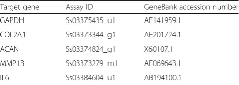
RT-qPCR was performed using an ABI PRISM 7900HT Sequence Detection System and Sequence Detection Software 2.2.2. The target genes were normalized to the glyceraldehyde-3-phosphate dehydrogenase (GAPDH). The relative mRNA expression of each target gene was determined using theΔΔCt method.
Total DNA quantification
After compression/H2O2 treatment, chondrocytes were collected and total DNA was purified using the DNeasy Blood and Tissue kit (69504, QIAGEN, Germany), fol-lowing the instructions provided by the manufacturer. Total DNA yield was quantified by an ultra violet/vis-ible/near infrared (UV/VIS/NIR) spectrophotometer (DU 7500, Beckman, USA) at the wavelength of 260 and 280 nm. The ratio of 260 to 280 nm was between 1.8 and 2.0.
Analysis of sGAG production
The sGAG production was evaluated in a DMMB (341088, Sigma, USA) assay, as previously described [29]. At the end of compression/H2O2 treatment, cells were reseeded in 6-well cell culture plates and incubated for 3 days. The culture medium of each sample was col-lected and 40μl of the supernatant of each sample trans-ferred to a 96-well microplate, after which 250 μl of DMMB solution was added. The DMMB-sGAG com-plex product was examined by an ELISA reader (Sunrise, TECAN, Switzerland) at the wavelength of 595 nm. The sGAG production activity of each sample was deter-mined using a calibration curve of condroitin-6-sulfate (C4384, Sigma, USA). The sGAG production was nor-malized to cell numbers by a total DNA assay (sGAG to DNA ratio). Chondrocytes derived from more than three porcine stifles were used for the experiments. Five re-peated measurements were performed for all samples.
Alcian blue staining
After compression/H2O2 treatment, cells were reseeded into 4-well chamber-slides and cultured for 3 days. Chondrocytes were washed twice with PBS and fixed in 10% neutral buffered formalin (H121-08, Mallinckrodt Analytical, USA) for 30 min and then washed twice with PBS. Alcian blue (pH 1.0, Muto pure chemicals, Japan) was added for 30 min and cells were then washed in
running water for 1 min. Nuclear fast red (1001210500, Merck, Germany) was added for 5 min and then washed in running water for 1 min. The cells were dehydrated in 2 changes of 95% alcohol and absolute alcohol (459844, Sigma, USA) for 1 min each. The sGAG con-tent images were taken by using an IX71 inverted micro-scope equipped with a DP30BW digital camera system (Olympus, Japan). Chondrocytes derived from more than three porcine stifles were used for the experiments. Five repeated measurements were performed for all samples.
Statistical analysis
The normality of variance of data was tested before stat-istical analysis. Statstat-istically significant differences be-tween the groups were determined by one-way ANOVA with Tukey’s post-hoc test. The results were expressed as mean ± standard deviation of the mean (SD) and consid-ered significant when the P-value was <0.05. Statistical analysis was performed using the SigmaPlot version 12.3 software (Systat Software Inc., San Jose, USA).
Results
Aucubin shows no cytotoxic effects on chondrocytes
As shown in Fig. 1, chondrocytes treated with 3, 10, 30, and 100 μM of aucubin showed no significant differ-ences in cytotoxicity or cell proliferation activity com-pared to controls at 1 or 3 days, indicating that aucubin within the concentration range of 3–100 μM shows no significant cytotoxic effects on chondrocytes at either 1 or 3 days.
Aucubin inhibits H2O2-induced ROS production in
chondrocytes
As shown in Fig. 2, the ROS production was significantly increased in response to H2O2 stimulation. Chondro-cytes treated with aucubin showed significant ROS scav-enging effects after 1, 4 and 24 h of incubation with 10 to 100 μM aucubin in a dose-dependent manner. The ROS scavenge effect of aucubin was further confirmed through fluorescence imaging. As shown in Fig. 3, both the green and red fluorescence emissions, representing general ROS and superoxide induced by H2O2, respect-ively, were markedly reduced in fluorescence intensity and area after 24 h of 100 μM aucubin pretreatment. Since 100 μM aucubin showed the greatest scavenging effect with limited cytotoxicity among other aucubin concentrations, we used 100 μM aucubin in the subse-quent experiments.
Aucubin inhibits H2O2-induced caspase-3 activity on
chondrocytes
[image:4.595.57.291.99.182.2]After H2O2 treatment, the caspase-3 activity of chon-drocytes showed a significant increase. However, chondrocytes treated with aucubin and caspase inhibitor
Table 1Primers used in this study
Target gene Assay ID GeneBank accession number
GAPDH Ss03375435_u1 AF141959.1
COL2A1 Ss03373344_g1 AF201724.1
ACAN Ss03374824_g1 X60107.1
MMP13 Ss03373279_m1 AF069643.1
Z-VAD-FMK markedly decreased the caspase-3 activity caused by H2O2stimulation (Fig. 4).
Aucubin inhibits H2O2-induced apoptosis and necrosis in
chondrocytes
After H2O2 treatment, the percentage of apoptotic and necrotic chondrocytes were significantly increased compared to the control group. However, in the aucu-bin pretreated group, the percentages of chondrocytes in
both early and late apoptosis states were significantly reduced compared to the H2O2 stimulated group (Fig. 5, Table 2).
Aucubin reverses H2O2-mediated and compression-mediated
gene expression of ACAN and COL2A1 in chondrocytes
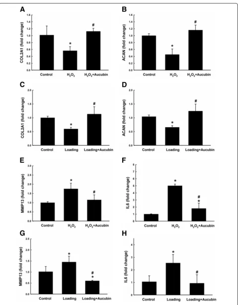
ACAN and COL2A1 are ECM related genes. As shown in the H2O2model of Fig. 6a and b, the expression of both ACAN and COL2A1 was significantly
down-Fig. 2ROS scavenge test of aucubin. Chondrocytes pretreated with 0, 3, 10, 30 and 100μM of aucubin for 0.5, 1, 4 and 24 h were stimulated with 1 mM H2O2for 30 min. ROS production was measured by the intensity of the dye fluorescence and represented as
relative fluorescence unit (RFU). Results shown represent mean ± SD obtained from five repeated measurements. *p< 0.05 compared with the control group.#p< 0.05 compared with the H
2O2group
[image:5.595.62.539.87.287.2] [image:5.595.60.538.462.684.2]regulated in the H2O2-treated group compared to the control group. However, pretreatment with aucubin re-versed the expression of these genes back to the level of the control group and showed marked up-regulation compared to the H2O2-treated group. Similar results were shown in the compression model. The expression of both ACAN and COL2A1 was also markedly down-regulated in the loading group, though the aucubin pre-treated chondrocytes had a significant up-regulation of both ECM component genes after compression stimula-tion compared to the control group (Fig. 6c and d).
Aucubin reduces H2O2-mediated and compression-mediated
gene expression of MMP13 and IL6 in chondrocytes
MMP13 is a catabolic gene in cartilage ECM homeo-stasis. As shown in Fig. 6e and g, in both H2O2 and compression models, the expression of MMP13 was up-regulated compared to the control group after stimulation. However, treatment with aucubin before H2O2 and compression stimulation maintained the expression of MMP13 at a comparable level to the control group. Similar results were shown in the
Fig. 3Fluorescence imaging of ROS production. Chondrocytes pretreated with 100μM of aucubin for 24 h were then stimulated with 1 mM H2O2for 30 min. Thegreenandredcolors are the fluorescence of general ROS (hydrogen peroxide, peroxynitrite, hydroxyl radicals, nitric oxide,
peroxy radical) and superoxide, respectively. Chondrocytes without aucubin nor H2O2treatment were set as the control group.Scale bar= 100μm.
(5 repeated measurements for each group)
Fig. 4Effect of aucubin on caspase-3 activity. Chondrocytes pretreated with 100μM of aucubin or Z-VAD-FMK as caspase inhibitor for 24 h were then stimulated with 1 mM H2O2for 30 min. Chondrocytes
without aucubin nor H2O2treatment were set as the control group.
Results shown represent mean ± SD obtained from five repeated measurements. *p< 0.05 compared with the control group.#p< 0.05
[image:6.595.57.540.88.406.2] [image:6.595.59.291.472.656.2]expression of the proinflammatory gene IL6. As shown in Fig. 6f and h, IL6 was up-regulated compared to the con-trol group in both models after stimulation. Moreover, the up-regulated IL6 expression was reversed to a similar level as the control group with the pretreatment of aucubin.
Aucubin increases sGAG production in compression model but not after H2O2stimulation
As shown in Fig. 7a, the sGAG to DNA ratio for the H2O2 treated group was significantly decreased com-pared to the control group. Treatment with aucubin showed no significant difference for the H2O2 treated group. However, the decreased sGAG to DNA ratio in-duced by compressive stress was significantly increased compared to the compression group (Fig. 7b).
Aucubin increases the sGAG content after both H2O2and
compression stimulation
As shown in Fig. 7c and d, the alcian blue staining was positive in the control groups for both H2O2 and com-pression models. The H2O2 treated group, on the con-trary, showed few areas of blue color with shrinking cell morphology. With aucubin pretreatment, some of the chondrocytes could maintain sGAG content. Similar re-sults were also evident in the compression model.
Discussion
Aucubin has significant antioxidant and radical scaven-ging properties in both in vivo and in vitro models [30],
making it a potential therapeutic agent in oxidative stress-induced diseases. Aucubin has been shown to re-duce ROS formation, malondialdehyde levels and β -galactosidase activity, and increase glutathione levels in UVB-irradiated human skin fibroblasts [31]. To evalu-ate the protective effects of aucubin in regards to OA, first we generated an osteoarthritic cell model with por-cine chondrocytes by using 1 mM H2O2, which de-creased 60% of chondrocytes proliferation activity but showed no significant or immediate cytotoxicity to un-treated control (data not shown). In this study, 10– 100μM of aucubin caused no significant cytotoxicity or differences in cell proliferation activity after incubation with chondrocytes at 1 or 3 days (Fig. 1). After H2O2 stimulation, 10–100 μM of aucubin was a sufficient range to reduce ROS production dose-dependently after at least 1 h of pretreatment (Fig. 2). Pretreatment with 100 μM of aucubin also inhibited H2O2-induced caspase-3 activity (Fig. 4). Xue et al. reported that aucu-bin inhibited H2O2‐induced apoptosis in PC12 cells through regulation of the endogenous oxidant– antioxi-dant balance [27]. In this study, we confirmed the apoptosis inhibition properties of aucubin via Annexin-V/PI flow cytometry (Fig. 5). The percentage of apop-totic cells was significantly reduced when 100 μM of aucubin were used, which is consistent with the Xue et al. study and suggests that aucubin protects chondro-cytes from cell apoptosis which is often observed dur-ing OA progression.
Fig. 5Annexin V/PI test of aucubin. Chondrocytes pretreated with 100μM of aucubin for 24 h were then stimulated with 1 mM H2O2for 30 min.
[image:7.595.61.539.88.231.2]Chondrocytes without aucubin nor H2O2treatment were set as the control group. (5 repeated measurements for each group)
Table 2Quantitative results of Annexin V/PI test of aucubin
Percentage of cell (%)
Normal Early apoptosis Late apoptosisi Necrosis
Control 89.7 ± 9.5 2.5 ± 4.6 5.0 ± 3.8 3.1 ± 1.8
H2O2 25.5 ± 7.3a 29.4 ± 4.2a 32.2 ± 6.8a 14.3 ± 7.6a
H2O2+ aucubin 60.1 ± 10.4a,b 15.4 ± 5.3a,b 15.8 ± 4.2a,b 10.7 ± 5.3
Results shown represent mean ± SD obtained from 5 repeated measurements.a
p<0.05 compared with the control group.b
p< 0.05 compared with the
[image:7.595.55.542.647.715.2]Aggrecan and type II collagen are the main components of chondrocyte ECM and provide cartilage with compres-sive and tensile resistance, respectively. In the OA cartilage, a loss of proteoglycans and decrease in type II collagen has been observed, leading to defective integrity of the cartilage [29]. In this study, both H2O2 and compressive loading caused significant down-regulations of ACAN and
COL2A1, suggesting the stimulated chondrocytes were undergoing a degeneration process with weakened anabolic activity (Fig. 6). However, chondrocytes pretreated with aucubin showed a protective effect by reversing the H2O2/ compression induced down-regulation of both ACAN and COL2A1 genes, which may implicate that aucubin has a beneficial effect on chondrocyte ECM production.
(See figure on previous page.)
Fig. 6Gene expressions after stimulation. Gene expressions of COL2A1 (a,c), ACAN (b,d), MMP13 (e,g) and IL6 (f,h) were examined by RT-qPCR. Chondrocytes pretreated with 100μM of aucubin for 24 h were then stimulated with 1 mM H2O2(a,b,eandf) for 30 min, or 60 psi compression
(c,d,gandh) for 24 h. Data from five independent biological replicates each with three technical replicates are expressed as fold change compared to untreated control and shown represent mean ± SD. Each target gene was normalized to GAPDH. *p< 0.05 compared with the control group.#p< 0.05 compared with the H2O2or loading group
Fig. 7sGAG production and Alcian blue staining of GAG contents after stimulation. The ratio of sulfated-GAG to DNA of chondrocytes after stimulated with 1 mM H2O2(a) for 30 min or compression (b) with 60 psi for 24 h. Alcian blue and nuclear fast red staining of the chondrocytes was performed
after stimulated with (c) 1 mM H2O2for 30 min or (d) compression with 60 psi for 24 h.Scale bar= 50μm. Results shown represent mean ± SD
[image:9.595.58.540.262.683.2]The expression of pro-inflammatory IL-6 inhibits the synthesis of aggrecan and is highly elevated in OA car-tilage [32]. IL-6 expression directly contributes to the inhibitory effect of aggrecan through the Notch recep-tor [33], mediating induction of MMP-13 expression [34]. MMP-13 is a catabolic factor for cartilage ECM metabolism, degrading proteoglycans and type II colla-gen [35, 36]. Aucubin has been shown to inhibit TNF-alpha and IL-6 production in antigen-stimulated rat basophilic leukemia-2H3 mast cells [37]. Aucubin also has been demonstrated to reverse the increased gene and protein expression of MMP13, iNOS and COX-2 induced by IL-1β stimulation in rat chondrocytes [38]. In this study, aucubin significantly reversed the elevated gene expression of IL6 and MMP13 genes in the H2O2 and compression stimulated chondrocytes, consistently demonstrating the anti-inflammatory and anti-catabolic properties of aucubin in chondrocytes (Fig. 6).
About the single reference gene of GAPDH, in the previous study we had examined the gene expression of porcine chondrocytes and used GAPDH as reference gene for RT-qPCR [28]. After analysis the GAPDH gene showed a consistent Ct value among groups with differ-ent treatmdiffer-ent. In the presdiffer-ent study, we used the same method to isolate the porcine chondrocytes and ob-served the same phenomenon of GAPDH expressions in RT-qPCR results. The Ct values of GAPDH did not show a noticeable difference among chondrocytes stimu-lated with compression, H2O2, Aucubin or in control group. We suggest that treatments used in the present study did not affect the expression of GAPDH gene, thus, we used GAPDH as the house keeping gene in this study.
In the degenerated cartilage, a decrease in the content of chondroitin sulfate results in a decrease of water con-tent that affects the capability of cartilage to absorb the external stress [39]. In this study, H2O2and compression stimulation significantly reduced the sGAG production activity and decreased sGAG content. Chondrocytes pre-treated with aucubin succeeded to maintain its sGAG content after both H2O2 and compression stimulation, and recover its sGAG production activity after compres-sion stimulation. However, aucubin failed to maintain the sGAG production activity after H2O2 stimulation (Fig. 7). According to these data, we suggest that the protective effects of aucubin may be overwhelmed by the oxidative stress of H2O2. The oxidative stress may lead to decrease the sGAG production activity of chon-drocytes. Nevertheless, aucubin still has beneficial ability to maintain sGAG content of chondrocytes after H2O2 and compression stimulation.
Conclusions
This study demonstrated that aucubin can reduces ROS production, caspase-3 activity, and cell apoptosis.
Aucubin can protect porcine chondrocytes from H2O2 and compression-induced dysregulation of COL2A1, ACAN, IL6 and MMP13 gene expressions. Addition-ally, aucubin helps porcine chondrocytes maintain sGAG contents after H2O2 and compressive stimulation. These results indicated that aucubin has protective effects in an osteoarthritic chondrocyte model induced by H2O2 and mechanical stimulus.
Abbreviations
ADAMTS:A disintegrin and metalloproteinase with thrombospondin motifs; DMEM: Dulbecco’s modified eagle’s medium; ECM: Extracellular matrix; GAPDH: Glyceraldehyde-3-phosphate dehydrogenase; IL-6: Interlukin-6; LDH: Lactate dehydrogenase; MMP13: Matrix metalloproteinase 13; OA: Osteoarthritis; PBS: Phosphate buffered saline; ROS: Reactive oxygen species; RT-qPCR: Reverse transcription quantitative polymerase chain reaction; sGAG: Sulfated-glycosaminoglycan
Acknowledgements
The authors wish to thank National Taiwan University academic writing education center for professional language editing.
Funding
This study was financially supported by National Science Council grant No. 100-2313-B-002-049-MY2. The funders had no role in study design, data col-lection and analysis, decision to publish, or preparation of the manuscript.
Availability of data and materials
The datasets during and/or analysed during the current study available from the corresponding author on reasonable request.
Authors’contributions
ICY and FHL conceived the research. ICY and FHL designed this work. FHL and CHH contributed new reagents and analytical tools. ICY performed the experiment. ICY, STC and YJS analyzed results. ICY, STC, HCL, YSC and FHL wrote the manuscript and revising it critically for important intellectual content. All authors read and approved the final manuscript.
Competing interests
The authors declare that they have no competing interests.
Consent for publication Not applicable.
Ethics approval and consent to participate
The isolation and use of porcine chondrocytes were approved by the Animal Experimentation Ethics Committee of National Taiwan University Hospital.
Author details
1Institute of Biomedical Engineering, National Taiwan University, No. 49,
Fanglan Rd, Taipei 10672, Taiwan.2Institute of Pharmacology, College of Medicine, National Taiwan University, No. 1, Sec. 1, Ren-Ai Rd, Taipei 10051, Taiwan.3Institute of Biomedical Engineering and Nanomedicine, National Health Research Institute, No. 35, Keyan Rd, Miaoli 35053, Taiwan. 4
Department of Orthopaedic Surgery, Taiwan Adventist Hospital, No. 424, Sec. 2, Bade Rd, Taipei 10556, Taiwan.5Department of Orthopaedic Surgery, National Taiwan University Hospital, No.7, Chung Shan S. Rd, Taipei 10002, Taiwan.
Received: 21 April 2016 Accepted: 13 January 2017
References
1. Dillon CF, Rasch EK, Gu Q, Hirsch R. Prevalence of knee osteoarthritis in the United States: arthritis data from the Third National Health and Nutrition Examination Survey 1991–94. J Rheumatol. 2006;33(1):2271–9.
3. Brosseau L, Rahman P, Toupin-April K, Poitras S, King J, De Angelis G, Loew L, et al. A systematic critical appraisal for non-pharmacological management of osteoarthritis using the appraisal of guidelines research and evaluation II instrument. PLoS One. 2014;9(1):e82986.
4. Jeong YJ, Kim I, Cho JH, Park DW, Kwon JE, Jung MW, et al. Anti-Osteoarthritic Effects of the Litsea japonica fruit in a rat model of osteoarthritis induced by monosodium iodoacetate. PLoS One. 2015;10(8):e0134856.
5. Heidari B. Knee osteoarthritis prevalence, risk factors, pathogenesis and features: part I. Caspian J Intern Med. 2011;2(2):205–12.
6. Felson DT. Developments in the clinical understanding of osteoarthritis. Arthritis Res Ther. 2009;11(1):203.
7. Li Y, Xu L. Advances in understanding cartilage remodeling. F1000Res. 2015; 4(F1000 Faculty Rev):642.
8. Kim H, Kang D, Cho Y, Kim JH. Epigenetic regulation of chondrocyte catabolism and anabolism in osteoarthritis. Mol Cells. 2015;38(8):677–84. 9. Kapoor M, Martel-Pelletier J, Lajeunesse D, Pelletier JP, Fahmi H. Role of
proinflammatory cytokines in the pathophysiology of osteoarthritis. Nat Rev Rheumatol. 2011;7(1):33–42.
10. Clouet J, Vinatier C, Merceron C, Pot-vaucel M, Maugars Y, Weiss P, et al. From osteoarthritis treatments to future regenerative therapies for cartilage. Drug Discov Today. 2009;14(19–20):913–25.
11. Musumeci G, Castrogiovanni P, Trovato FM, Weinberg AM, Al-Wasiyah MK, Alqahtani MH, et al. Biomarkers of chondrocyte apoptosis and autophagy in osteoarthritis. Int J Mol Sci. 2015;16(9):20560–75.
12. Yamazaki K, Fukuda K, Matsukawa M, Hara F, Matsushita T, Yamamoto N, et al. Cyclic tensile stretch loaded on bovine chondrocytes causes depolymerization of hyaluronan: involvement of reactive oxygen species. Arthritis Rheum. 2003;48(11):3151–8.
13. Kurz B, Lemke A, Kehn M, Domm C, Patwari P, Frank EH, et al. Influence of tissue maturation and antioxidants on the apoptotic response of articular cartilage after injurious compression. Arthritis Rheum. 2004;50(1):123–30. 14. Gabay O, Hall DJ, Berenbaum F, Henrotin Y, Sanchez C. Osteoarthritis and
obesity: experimental models. Joint Bone Spine. 2008;75(6):675–9. 15. Liu SC, Lee HP, Hung CY, Tsai CH, Li TM, Tang CH. Berberine attenuates
CCN2-induced IL-1beta expression and prevents cartilage degradation in a rat model of osteoarthritis. Toxicol Appl Pharmacol. 2015;289(1):20–9. 16. Shen C, Cai GQ, Peng JP, Chen XD. Autophagy protects chondrocytes from
glucocorticoids-induced apoptosis via ROS/Akt/FOXO3 signaling. Osteoarthritis Cartilage. 2015;23(12):2279–87.
17. Courties A, Gualillo O, Berenbaum F, Sellam J. Metabolic stress-induced joint inflammation and osteoarthritis. Osteoarthritis Cartilage. 2015; 23(11):1955–65.
18. Riedl SJ, Shi Y. Molecular mechanisms of caspase regulation during apoptosis. Nat Rev Mol Cell Biol. 2004;5(11):897–907.
19. Farooq SM, Boppana NB, Devarajan AC, Sekaran SD, Shankar EM, Li C, et al. C-phycocyanin confers protection against oxalate-mediated oxidative stress and mitochondrial dysfunctions in MDCK cells. PLoS One. 2014;9(4):e93056. 20. Cheng YH, Yang SH, Liu CC, Gefen A, Lin FH. Thermosensitive hydrogel
made of ferulic acid-gelatin and chitosan glycerophosphate. Carbohydr Polym. 2013;92(2):1512–9.
21. Chang LM, Yun HS, Kim YS, Ahn JW. Aucubin: potential antidote for alpha-amanitin poisoning. J Toxicol Clin Toxicol. 1984;22(1):77–85.
22. Li Y, Sato T, Metori K, Koike K, Che QM, Takahashi S. The promoting effects of geniposidic acid and aucubin in Eucommia ulmoides Oliver leaves on collagen synthesis. Biol Pharm Bull. 1998;21(12):1306–10.
23. Chang IM, Ryu JC, Park YC, Yun HS, Yang KH. Protective activities of aucubin against carbon tetrachloride-induced liver damage in mice. Drug Chem Toxicol. 1983;6(5):443–53.
24. Chang IM. Liver-protective activities of aucubin derived from traditional oriental medicine. Res Commun Mol Pathol Pharmacol. 1998;102(2):189–204. 25. Jin L, Xue HY, Jin LJ, Li SY, Xu YP. Antioxidant and pancreas-protective effect of aucubin on rats with streptozotocin-induced diabetes. Eur J Pharmacol. 2008;582(1–3):162–7.
26. Xue HY, Gao GZ, Lin QY, Jin LJ, Xu YP. Protective effects of aucubin on H2O2-induced apoptosis in PC12 cells. Phytother Res. 2012;26(3):369–74.
27. Xue H, Jin L, Jin L, Zhang P, Li D, Xia Y, et al. Neuroprotection of aucubin in primary diabetic encephalopathy. Sci China C Life Sci. 2008;51(6):495–502. 28. Chang CH, Hsu YM, Chen YC, Lin FH, Sadhasivam S, Loo ST, et al.
Anti-inflammatory effects of hydrophilic and lipophilic statins with hyaluronic acid against LPS-induced inflammation in porcine articular chondrocytes. J Orthop Res. 2014;32(4):557–65.
29. Cheng YH, Yang SH, Lin FH. Thermosensitive chitosan-gelatin-glycerol phosphate hydrogel as a controlled release system of ferulic acid for nucleus pulposus regeneration. Biomaterials. 2011;32(29):6953–61. 30. Park H, Temenoff JS, Holland TA, Tabata Y, Mikos AG. Delivery of TGF-beta1
and chondrocytes via injectable, biodegradable hydrogels for cartilage tissue engineering applications. Biomaterials. 2005;26(34):7095–103. 31. Ho JN, Lee YH, Park JS, Jun WJ, Kim HK, Hong BS, et al. Protective effects of
aucubin isolated from Eucommia ulmoides against UVB-induced oxidative stress in human skin fibroblasts. Biol Pharm Bull. 2005;28(7):1244–8. 32. Wang P, Guan PP, Guo C, Zhu F, Konstantopoulos K, Wang ZY. Fluid shear
stress-induced osteoarthritis: roles of cyclooxygenase-2 and its metabolic products in inducing the expression of proinflammatory cytokines and matrix metalloproteinases. FASEB J. 2013;27(12):4664–77.
33. Zanotti S, Canalis E. Interleukin 6 mediates selected effects of Notch in chondrocytes. Osteoarthritis Cartilage. 2013;21(11):1766–73. 34. Ashraf S, Mapp PI, Burston J, Bennett AJ, Chapman V, Walsh DA.
Augmented pain behavioural responses to intra-articular injection of nerve growth factor in two animal models of osteoarthritis. Ann Rheum Dis. 2014; 73(9):1710–8.
35. Halbwirth F, Niculescu-Morzsa E, Zwickl H, Bauer C, Nehrer S. Mechanostimulation changes the catabolic phenotype of human dedifferentiated osteoarthritic chondrocytes. Knee Surg Sports Traumatol Arthrosc. 2015;23(1):104–11.
36. Enochson L, Stenberg J, Brittberg M, Lindahl A. GDF5 reduces MMP13 expression in human chondrocytes via DKK1 mediated canonical Wnt signaling inhibition. Osteoarthritis Cartilage. 2014;22(4):566–77. 37. Jeong HJ, Koo HN, Na HJ, Kim MS, Hong SH, Eom JW, et al. Inhibition of
TNF-alpha and IL-6 production by Aucubin through blockade of NF-kappaB activation RBL-2H3 mast cells. Cytokine. 2002;18(5):252–9.
38. Wang SN, Xie GP, Qin CH, Chen YR, Zhang KR, Li X, et al. Aucubin prevents interleukin-1 beta induced inflammation and cartilage matrix degradation via inhibition of NF-κB signaling pathway in rat articular chondrocytes. Int Immunopharmacol. 2015;24(2):408–15.
39. Vynios DH. Metabolism of cartilage proteoglycans in health and disease. Biomed Res Int. 2014;2014:452315.
• We accept pre-submission inquiries
• Our selector tool helps you to find the most relevant journal
• We provide round the clock customer support
• Convenient online submission
• Thorough peer review
• Inclusion in PubMed and all major indexing services
• Maximum visibility for your research
Submit your manuscript at www.biomedcentral.com/submit