Neuroscience Institute Dissertations Neuroscience Institute
Summer 5-20-2013
Neural Regulation of Sexual Solicitation in Female Syrian
Neural Regulation of Sexual Solicitation in Female Syrian
Hamsters: Role of Oxytocin
Hamsters: Role of Oxytocin
Luis A. Martinez Georgia State University
Follow this and additional works at: https://scholarworks.gsu.edu/neurosci_diss
Recommended Citation Recommended Citation
Martinez, Luis A., "Neural Regulation of Sexual Solicitation in Female Syrian Hamsters: Role of Oxytocin." Dissertation, Georgia State University, 2013.
https://scholarworks.gsu.edu/neurosci_diss/8
by
LUIS A. MARTINEZ
Under the Direction of Aras Petrulis
ABSTRACT
guided precopulatory behaviors? (2) Is BNST or (3) MPOA required for the preferential expression of vaginal marking or investigation towards male odors?, and (4) Does OT interact with social odor pro-‐ cessing to regulate vaginal marking? We found that blockade of OT receptors (OTRs) in MPOA and BNST decreased vaginal marking to male odors. There was no effect of OTR blockade on sexual odor prefer-‐ ence. Selective lesions of BNST also disrupted preferential vaginal marking responses to male odors, without affecting sexual odor preference. In contrast, lesions of MPOA disrupted odor preference with-‐ out affecting vaginal marking responses. Finally, central blockade of OTRs eliminated the normal pattern of increased activation of neurons to male vs. female odors in BNST, but not MPOA. Considered togeth-‐ er, these results suggest that OT normally acts within BNST to drive preferential vaginal marking re-‐ sponses to male odors via selective facilitation of neural responses to these odors, and further, that there are separate and distinct neural circuits that regulate different forms of odor-‐guided precopulato-‐ ry behaviors in females.
INDEX WORDS: Olfaction, Reproduction, Sexual behavior, Sexual solicitation, Precopulatory, Appetitive, Oxytocin, Vasopressin, Medial preoptic area, Bed nucleus of the stria terminalis
by
LUIS A. MARTINEZ
A Dissertation Submitted in Partial Fulfillment of the Requirements for the Degree of Doctor of Philosophy
in the College of Arts and Sciences Georgia State University
Copyright by Luis Antonio Martinez
by
LUIS A. MARTINEZ
Committee Chair: Aras Petrulis Committee: Elliott Albers Walt Wilczynski Larry Young
Electronic Version Approved:
DEDICATION
ACKNOWLEDGEMENTS
I would like to take this opportunity to thank all the people that contributed to this process and made it possible for me to complete my dissertation.
First, I would like to thank Aras Petrulis, my graduate advisor and mentor. You have helped de-‐ velop me into the scientist I am today, and for that I am truly grateful. I am undoubtedly a better scien-‐ tific writer and thinker for my time spent in your lab. Although I acknowledge that the odds are infinites-‐ imally small, I hope to one day have a thought about hamsters that you have not already had.
Next I would like to thank my dissertation committee members, Elliott Albers (co-‐advisor), Walt Wilczynski, and Larry Young. I appreciate the training and support Elliott Albers provided me, particularly
during my first several years at Georgia State University. You encouraged me to think big and achieve more, and I have gotten as far as I have due in no small part to your influence. I thank Walt Wilczynski for his support throughout the Honeycutt Fellowship process, and for helping me see the broader con-‐ text wherein my research lies. Lastly, I thank Larry Young for providing critical insights into the method-‐ ology employed in my experiments, and for further refining my dissertation.
I would also like to thank the members of the Petrulis lab, past and present. Pam Maras, Laura Been, and Marc Badura, you guys were great labmates, and even better friends. Pam, Fridays are less
fun since you left. Laura, no one fits in a box like you do. Marc, may your ethanol always be RNase free. Marisa Levy, Nicole Peters, and Joe Terranova, I am glad to have gotten to know you over the past year.
Watch out for Ashleigh Burns on Thursdays. Many thanks as well to all the undergraduates I had the pleasure of mentoring during my time as a graduate student, including Rachel Atkinson, Johnny Garret-‐ son, Alica Helman, Preethy Kuriakose, Jamin Peters, Alix Pijeaux, Stephanie Sylvester, Manal Tabbaa, and
It has also been my pleasure to have a great set of friends here at Georgia State University. Thanks to Amanda Arnold, Marc Badura, Tim Balmer, Laura Been, Ashleigh Burns, Jenna Darling, James Doherty, Kelli Duncan, Leslie Dunham, Johnny Garretson, Marisa Levy, Cloe Luckett, Pam Maras, Camer-‐
on Miller, Devaleena Pradhan, Nicole Peters, Amy Ross, Mahin Shahbazi, Joseph Terranova, Jill Weath-‐
ington, and Nicole Victoria for brightening my days and being there for me.
I would like to thank the Neuroscience Institute faculty and staff for helping me to successfully navigate through the last seven years. In particular, I’d like to thank Don Edwards, Bill Walthall, Paul Katz, Sarah Pallas, Aras Petrulis, Tim Bartness, Anne Murphy, and Charles Derby for their instruction,
Andrew Clancy, Kim Huhman, and Charles Derby for serving on my qualifying exam committee, and
Mary Karom, Daniel Erwin, and Alisa Norvelle for their technical expertise and assistance.
I would also like to thank Ken and Georganne Honeycutt for their support during my last two years at GSU. Your enthusiasm for neuroscience and commitment to supporting NI graduate students mean a great deal to myself and the other Honeycutt Fellows.
Finally, I would like to thank my family for helping to see me through this process. Angel and Milagros Martinez, you made me realize the value of working hard and pursuing a higher education.
TABLE OF CONTENTS
ACKNOWLEDGEMENTS ... v
LIST OF TABLES ... x
LIST OF FIGURES ... xi
LIST OF ABBREVIATIONS ... xiii
CHAPTER 1: INTRODUCTION ... 1
1.1 Overview ... 1
1.2 Syrian Hamsters as a Model Species for Examining Female Precopulatory Behaviors ... 2
1.3 Neural Substrates of Female Precopulatory Behaviors ... 3
1.4 Oxytocin and Female Sexual Behaviors ... 5
1.5 Goal of Dissertation ... 7
CHAPTER 2: BLOCKING OXYTOCIN RECEPTORS INHIBITS VAGINAL MARKING TO MALE ODORS IN FEMALE SYRIAN HAMSTERS ... 8
2.1 Abstract ... 8
2.2 Introduction ... 9
2.3 Methods ... 11
2.4 Results ... 18
2.5 Discussion ... 20
2.6 Acknowledgements ... 25
Chapter 2 Tables ... 26
Chapter 2 Figures ... 29
3.1 Abstract ... 32
3.2 Introduction ... 33
3.3 Methods ... 35
3.4 Results ... 44
3.5 Discussion ... 47
3.6 Acknowledgements ... 52
Chapter 3 Tables ... 53
Chapter 3 Figures ... 55
CHAPTER 4: THE MEDIAL PREOPTIC AREA IS NECESSARY FOR SEXUAL ODOR PREFERENCE, BUT NOT SEXUAL SOLICITATION, IN FEMALE SYRIAN HAMSTERS ... 59
4.1 Abstract ... 59
4.2 Introduction ... 60
4.3 Methods ... 62
4.4 Results ... 72
4.5 Discussion ... 76
4.6 Acknowledgements ... 81
Chapter 4 Table ... 82
Chapter 4 Figures ... 83
CHAPTER 5: ENDOGENOUS OXYTOCIN IS NECESSARY FOR PREFERENTIAL FOS EXPRESSION TO MALE ODORS IN THE BED NUCLEUS OF THE STRIA TERMINALIS IN FEMALE SYRIAN HAMSTERS ... 88
5.1 Abstract ... 88
5.2 Introduction ... 89
5.4 Results ... 100
5.5 Discussion ... 103
5.6 Acknowledgements ... 109
Chapter 5 Tables ... 110
Chapter 5 Figures ... 112
CHAPTER 6: GENERAL DISCUSSION ... 123
6.1 Summary ... 123
6.2 Functional Regulation of Female Sexual Behaviors ... 124
6.3 Oxytocin and Social Behaviors ... 135
6.4 Conclusion ... 146
Chapter 6 Figures ... 148
REFERENCES ... 150
CURRICULUM VITAE ... 170
LIST OF TABLES
Table 2.1 Summary of behavioral measures from Experiment 1 ... 26
Table 2.2 Summary of behavioral measures from Experiment 2 ... 27
Table 2.3 Summary of behavioral measures from clean odor tests (Experiment 3) ... 28
Table 3.1 Odor preference and vaginal marking of partial lesion subjects ... 53
Table 3.2 Flank marking and lordosis of SHAM and BNST-‐X subjects ... 54
Table 4.1 Odor preference and scent-‐marking test results for partial lesion subjects ... 82
Table 5.1 Fos expression during stimulus exposures (Experiment 1) ... 110
Table 5.2 Flank marking responses during stimulus exposures (Experiment 2) ... 111
LIST OF FIGURES
Figure 2.1 Histological verification of injection sites ... 29
Figure 2.2 Number of vaginal marks during scent-‐marking tests ... 30
Figure 2.3 Odor box investigation during preference tests ... 31
Figure 3.1 Timeline and testing apparatus ... 55
Figure 3.2 Lesion reconstruction ... 56
Figure 3.3 Mean (± SEM) number of vaginal marks to male and female odors ... 57
Figure 3.4 Median (± IQR) duration of odor investigation ... 58
Figure 4.1 Lesion reconstruction ... 83
Figure 4.2 Mean (± SEM) duration of odor investigation (odor preference test) ... 84
Figure 4.3 Mean (± SEM) duration of odor investigation (odor discrimination test) ... 85
Figure 4.4 Mean (± SEM) number of vaginal marks ... 86
Figure 4.5 Mean (± SEM) number of flank marks ... 87
Figure 5.1 Timelines of experimental manipulations ... 112
Figure 5.2 Counting domains for quantifying oxytocin-‐ and Fos-‐positive cells ... 113
Figure 5.3 Immunohistochemical staining for oxytocin and Fos ... 115
Figure 5.4 Mean (± SEM) percentages of oxytocin/Fos double-‐labeled cells following stimulus exposures ... 116
Figure 5.5 Mean (± SEM) number of vaginal marks following ICV drug injections ... 117
Figure 5.6 Immunohistochemical staining for Fos in the bed nucleus of the stria terminalis and the medial preoptic area ... 118
Figure 5.7 Immunohistochemical staining for Fos in the anterior and posterior divisions of the medial amygdala ... 119
Figure 5.9 Relationships between the density of Fos-‐positive cells and the number of vaginal marks ... 121 Figure 5.10 Relationships between the density of Fos-‐positive cells and the number of vaginal marks ... 122 Figure 6.1 Functional pathways regulating odor-‐guided precopulatory behaviors ... 148 Figure 6.2 Regulation of preferential vaginal marking by oxytocin ... 149
LIST OF ABBREVIATIONS
ac, anterior commissure
ACo, anterior cortical amygdaloid nucleus AH, anterior hypothalamus
AOBs, accessory olfactory bulbs AOS, accessory olfactory system ASD, autism spectrum disorders AVP, arginine vasopressin
BLA, anterior division of the basolateral amygdaloid nucleus BLP, posterior division of the basolateral amygdaloid nucleus BMA, anterior division of the basomedial amygdaloid nucleus BMP, posterior division of the basomedial amygdaloid nucleus BNST, posterior division of the bed nucleus of the stria terminalis
BNSTpi, posterointermediate subdivision of the bed nucleus of the stria terminalis BNSTpl, posterolateral subdivision of the bed nucleus of the stria terminalis BNSTpm, posteromedial subdivision of the bed nucleus of the stria terminalis BNST-‐X, lesion of the posterior bed nucleus of the stria terminalis
Ce, central amygdaloid nucleus E, estrogen
ER, estrogen receptor f, fornix
FHVS, female hamster vaginal secretion
GDX+T, GDX plus testosterone replacement GP, globus pallidus
I, intercalated nuclei of the amygdala ic, internal capsule
IEG, immediate early gene IHC, immunohistochemistry IP, intraperitoneal
LH, lateral hypothalamus LPO, lateral preoptic area LS, lateral septum
LV, lateral ventricle
MA or ME, medial nucleus of the amygdala MeA, anterior division of the medial amygdala MeP, posterior division of the medial amygdala MLR, midbrain locomotor region
MOBs, main olfactory bulbs MOE, main olfactory epithelium MOS, main olfactory system MPOA, medial preoptic area
MPOA-‐AH, medial preoptic area-‐anterior hypothalamus continuum MPOA-‐X, lesions of the medial preoptic area
NAc, nucleus accumbens
OT, oxytocin
OTA, oxytocin receptor antagonist OTR, oxytocin receptor
OVX, ovariectomized ox, optic chiasm P, progesterone
PAG, midbrain periaqueductal gray PBS, phosphate buffered saline PE, proestrus
Pir, piriform cortex
PLCo, posterolateral cortical amygdaloid nucleus PMCo, posteromedial cortical amygdaloid nucleus PR, progesterone receptor
PVH, paraventricular nucleus of the hypothalamus SCN, superchiasmatic nucleus
SHAM, sham lesion of a specific brain area SI, substantia innominata
sm, stria medullaris SON, supraoptic nucleus sox, supraoptic decussation st, stria terminalis
V1aA, vasopressin 1a receptor antagonist V1aR, vasopressin 1a receptor
VMH, ventromedial nucleus of the hypothalamus VTA, ventral tegmental area
CHAPTER 1: INTRODUCTION
1.1 Overview
1.2 Syrian Hamsters as a Model Species for Examining Female Precopulatory Behaviors
Female Syrian hamsters are an excellent model for studying the neural regulation of precopula-‐ tory behaviors for several reasons. First, Syrian hamsters display a suite of precopulatory responses that are highly stereotyped and readily observed in a laboratory setting, including preferential investigation of opposite-‐sex odors and vaginal scent marking. Vaginal marking involves the female moving forward while maintaining contact between the perineum and the underlying substrate, thereby depositing vag-‐ inal secretion (Johnston, 1977). This behavior appears to be important for mate solicitation, as females will vaginal mark to form a ‘trail’ linking her nesting area with that of a male (Lisk et al., 1983). Vaginal secretion found within vaginal marks is highly attractive to males (Johnston, 1974), and males will vigor-‐ ously investigate and attempt to locate the source of the secretion (Johnston and Kwan, 1984; Johnston and Schmidt, 1979). In contrast to other animal models of female precopulatory behaviors, olfactory stimuli alone are sufficient to induce the expression of these behaviors in female hamsters. Females of this species preferentially investigate, and vaginal mark in response to, male vs. female odors, and this overall pattern of preferential responses to odors is maintained across multiple days of the reproductive cycle (Johnston, 1977; Petrulis and Johnston, 1999; Petrulis et al., 1999). Precopulatory behaviors in hamsters can therefore be assessed in the absence of males, thus allowing these behaviors to be as-‐ sessed independently other types of behaviors (i.e., copulation and aggression) that would otherwise be displayed by females when interacting with males.
a behavioral model system wherein to examine the specific neural mechanisms that underlie context-‐ appropriate precopulatory behaviors.
1.3 Neural Substrates of Female Precopulatory Behaviors
Olfactory systems
In hamsters as well as other vertebrates, social odors are initially detected and processed by the main (MOS) and accessory (AOS) olfactory systems (Keller et al., 2009). These two systems are special-‐ ized for detecting either volatile or non-‐volatile chemosignals (MOS and AOS, respectively). Low molecu-‐ lar weight, volatile chemosignals are dissolved in the olfactory mucosa and activate receptors on sensory cells in the main olfactory epithelium (MOE) (Keller et al., 2009). These sensory cells project to mitral cells within the main olfactory bulbs (MOBs), that then relay chemosensory information to downstream targets, including the medial amygdala (MA), the anterior cortical amygdala (ACo), the posterolateral cortical amygdala (PLCo), and olfactory cortex (e.g., piriform and entorhinal cortex) (Davis et al., 1978; Kang et al., 2009). Higher molecular weight, non-‐volatile chemosignals are detected by the vomeronasal organ (VNO), a blind-‐ending structure found at the base of the nasal septum (Meredith, 1991; Zufall et al., 2002). Chemosignals are actively pumped into the VNO, and thereby gain access to receptors pre-‐ sent on sensory neurons in the VNO epithelium. These neurons project to mitral cells in the accessory olfactory bulbs (AOBs), which relay chemosensory information to MA, the posteromedial cortical amyg-‐ dala (PMCo) and BNST (Davis et al., 1978; Kang et al., 2009).
Rather, VNO removal eliminates preferential vaginal marking to male vs. female odors (Petrulis et al., 1999). Together, these results demonstrate that the neural circuits regulating different forms of odor-‐ guided precopulatory behaviors in females are already distinct at the level of primary olfactory struc-‐ tures. This distinction may continue to be maintained within downstream targets of these systems, since lesions of MA eliminate opposite-‐sex odor preference and decrease overall levels of vaginal marking, without disrupting preferential vaginal marking to male odors (Petrulis and Johnston, 1999).
BNST
MPOA
Another brain area that functions downstream of MA to regulate precopulatory behaviors is MPOA. In contrast to MA and BNST, MPOA does not receive chemosensory information from the olfac-‐ tory systems directly; rather, this information reaches MPOA indirectly via BNST, MA, and other amyg-‐ daloid areas (Simerly and Swanson, 1986; Wang and Swann, 2006). Similar to BNST, MPOA is preferen-‐ tially activated by opposite-‐sex odors in male hamsters (Maras and Petrulis, 2010a), and lesions of MPOA eliminate opposite-‐sex odor preference in males (Been and Petrulis, 2010b). There is also sub-‐ stantial evidence implicating MPOA in the regulation of odor-‐guided precopulatory behaviors in females. In rats, lesions of MPOA decrease solicitational behaviors towards, and time spent with, a sexually-‐ experienced male, and disrupt the preference for intact male rat odors (Guarraci and Clark, 2006; Xiao et al., 2005). Although comparable data for the role of MPOA in sexual odor preference in female ham-‐ sters is not available, this area is involved in other precopulatory behaviors that can be induced by op-‐ posite-‐sex odors. Indeed, large, non-‐specific lesions of MPOA decrease vaginal marking during interac-‐ tions with males (Malsbury et al., 1977), and ultrasonic vocalizations by females following exposure to male hamsters (Floody, 1989). It is not known, however, if MPOA plays a unique and dissociable role from that of BNST in the regulation of precopulatory behaviors in females.
1.4 Oxytocin and Female Sexual Behaviors
form of oxytocin receptor (OTR) has been identified, a seven-‐transmembrane domain G-‐protein coupled receptor (Kimura et al., 1992). Binding of oxytocin to OTRs leads to increased release of calcium from intracellular stores, thereby increasing excitability of cells (Gimpl and Fahrenholz, 2001). The distribution of OTRs in the brain varies substantially across species (Barberis and Tribollet, 1996), and this variability has been proposed to underlie species-‐typical variations in sociality (Young et al., 1996).
OTRs are expressed in BNST and MPOA in a number of mammalian species (Campbell et al., 2009; Lee et al., 2008; Veinante and Freund-‐Mercier, 1997). In hamsters, however, OTRs are present in BNST but not in MPOA or other hypothalamic areas (Dubois-‐Dauphin et al., 1992). Despite this evidence, numerous studies have examined the effects of oxytocin acting within MPOA on sociosexual behaviors in this species. Injections of oxytocin into MPOA decrease aggressive responses towards a non-‐
1.5 Goal of Dissertation
CHAPTER 2: BLOCKING OXYTOCIN RECEPTORS INHIBITS VAGINAL MARKING TO MALE ODORS IN FE-‐ MALE SYRIAN HAMSTERS
Luis A. Martinez, H. Elliott Albers and Aras Petrulis Neuroscience Institute
Georgia State University, Atlanta, Georgia, USA 30302
Previously published in Physiology & Behavior 101(5): 685-‐692
2.1 Abstract
ing, or social odor investigation. Considered together, these results suggest that OT in MPOA-‐AH and/or BNST normally facilitates male odor-‐induced vaginal marking, providing further evidence that OT gener-‐ ally supports prosocial interactions among conspecifics.
2.2 Introduction
In female mammals, successful reproduction depends upon a suite of precopulatory behaviors that enhance the probability of locating and attracting a suitable mate (Petrulis, 2009; Takahashi, 1990). Precopulatory behaviors may be particularly important for reproductive success in species where adult members of the opposite sex live in isolation from each other, such as the Syrian hamster (Gattermann et al., 2001; Johnston, 1977). Vaginal marking is one form of precopulatory behavior in this species that serves to solicit potential mates (Huck et al., 1985; Lisk et al., 1983). This highly stereotyped behavior involves the female moving forward while maintaining contact between the perineum and the underly-‐ ing substrate, thereby depositing vaginal secretion (Johnston, 1977). In agreement with its role as a pre-‐ copulatory behavior, vaginal marks are highly attractive to male hamsters (Johnston and Schmidt, 1979; Johnston, 1974; Kwan and Johnston, 1980), and males may use vaginal marks to locate females for mat-‐ ing (Lisk et al., 1983). Furthermore, the expression of vaginal marking is critically dependent on internal hormonal state and external signals important for synchronizing mating activity (Johnston, 1977). Vagi-‐ nal marking levels are highest on the reproductive cycle day of proestrus, and are completely absent on the following cycle day (estrus) when females are sexually receptive and will engage in mating
(Johnston, 1977; Petrulis and Johnston, 1997). Vaginal marking is also preferentially directed towards males, as females will vaginal mark at higher levels in response to male odors than in response to female odors or clean bedding (Johnston, 1977; Petrulis and Johnston, 1999; Petrulis et al., 1998, 1999, 2000).
ropeptide oxytocin (OT) acting within the medial preoptic/anterior hypothalamus (MPOA-‐AH). OT is ex-‐ pressed in cell bodies and fibers within MPOA-‐AH (Whitman and Albers, 1998), and large, bilateral elec-‐ trolytic lesions of MPOA-‐AH inhibit vaginal marking (Malsbury et al., 1977). Injection of OT into MPOA-‐ AH of female hamsters (Whitman and Albers, 1995) or rats (Caldwell et al., 1989) enhances the expres-‐ sion of lordosis, a reflexive copulatory posture assumed by females (Beach, 1967). In contrast, blockade of oxytocin receptors (OTRs) in MPOA-‐AH via injection of a specific OTR antagonist (OTA) inhibits lordo-‐ sis in both these species (Caldwell et al., 1990; Pedersen and Boccia, 2002; Whitman and Albers, 1995). In addition to effects on lordosis, OT in MPOA-‐AH also increases the production of ultrasonic vocaliza-‐ tions (Floody et al., 1998), a pericopulatory behavior that induces approach in males (Floody, 1981).
2.3 Methods
Subjects
Female Syrian hamsters (Mesocricetus auratus) were purchased from Charles River Laboratories (Wilmington, MA, USA) at approximately 22 days of age. Subjects were individually housed upon arrival within the animal facility in an all-‐female room. In addition to experimental subjects, a separate group of male and female Syrian hamsters served as odor donors. Odor donors were also purchased from Charles River Laboratories as sexually mature adults (approximately 60 days of age). Odor donors were either individually housed or group housed (3-‐4 same sex animals per cage); in all cases, odor donors were un-‐ related to, and had no previous contact with, subjects prior to use in experiments. All animals were housed in solid-‐bottom Plexiglas cages (43 cm x 22 cm x 20 cm) containing corncob bedding and cotton nesting material (Nestlets; Ancare, Bellmore, NY) and maintained on a reversed light cycle (14:10 light:dark; lights out at 9 am). Behavior testing occurred during the first four hours of the dark portion of the light cycle. Food and water were available ad libitum. Animal procedures were carried out in accord-‐ ance with the National Institutes of Health Guide for the Care and Use of Laboratory Animals (NIH Publi-‐ cations No. 80-‐23; revised 1996) and approved by the Georgia State University Institutional Animal Care and Use Committee. All efforts were made to minimize the number of animals used and their suffering.
Estrous cycle monitoring
diately following behavioral estrus were defined as diestrous day 1, and diestrous day 2, respectively. In all cases, day refers to the dark phase of the light cycle.
Surgery
At two to three months of age, subjects were unilaterally implanted with guide cannulae. Sub-‐ jects were deeply anesthetized with sodium pentobarbital (90 mg/kg, i.p.; Nembutal, Ovation Pharma-‐ ceuticals, Deerfield, IL) and placed within a stereotaxic apparatus (Kopf Instruments, Tujunga, CA) with ear-‐ and incisor-‐bars positioned such that the top of the skull was level. Each subject was then fitted
with a single 4-‐mm 26-‐gauge guide cannula (Plastics One, Roanoke, VA), implanted at a 10° angle and directed at the following coordinates: 1.1 mm anterior to bregma, 1.7 mm lateral to the midline suture, and 4.0 mm ventral to dura. Injection cannulae were cut to extend the additional distance necessary to reach from the tip of the guide cannula to the target brain region. For Experiments 1 and 3, injection cannulae extended a total distance of 7.3 mm ventral to dura, effectively targeting the juncture between MPOA-‐AH and BNST. For experiment 2, injection cannulae extended either 6.8 mm or 7.8 mm ventral to dura, in order to more specifically target BNST or MPOA-‐AH, respectively. In all cases, stereotaxic coor-‐ dinates were derived from published neuroanatomical plates for the Syrian hamster brain (Morin and Wood, 2001). Guide cannulae were secured to the skull with dental cement and skull screws, and im-‐ mediately prior to the completion of the surgical procedure, subjects were injected subcutaneously with an analgesic agent (Ketofen; Fort Dodge Animal Health, Fort Dodge, IA). All subjects were allowed to recover for at least 10 days prior to behavioral testing.
Drug injections
OTA ([d(CH2)51,Tyr(Me)2,Thr4,Orn8, des-‐Gly-‐NH29]-‐vasotocin; Bachem, Torrance, CA) was diluted
compared to vasopressin 1a receptors (V1aRs) (Manning et al., 1989). Thirty minutes prior to behavioral
testing, subjects were injected via guide cannulae with either 200 nl of OTA (900 µM) or vehicle (saline). This concentration and volume of OTA is effective in eliciting behavioral effects in female hamsters when administered 30 minutes prior to testing (Harmon et al., 2002a). OTA or vehicle was delivered to
the target brain area through an injection cannula connected to a 1 µl Hamilton syringe. Subjects were lightly restrained for the 20 seconds while the drug was injected, and for an additional 20 seconds post-‐ injection to allow the drug to diffuse away from the tip of the injection cannula.
Scent-‐marking tests
Odor stimuli and apparatus
addition, the divisions on the plate provided a means for quantifying locomotor activity throughout the course of the behavioral tests.
Testing protocol
For each 15-‐minute scent-‐marking test, females were placed within a soiled stimulus cage and the number of vaginal and flank marks were scored live using a hand counter. Vaginal and flank marking are discrete, stereotyped events that are easily recognized (Johnston, 1977). A vaginal mark was scored each time the female moved forward with tail deflected upwards while maintaining contact between the perineum and the underlying substrate. A flank mark was scored each time the female moved for-‐ ward while maintaining contact between the flank region and the side of the stimulus cage. Tests were recorded using an overhead camera connected to a VHS video recorder. Videos were scored for the number of quadrant entries by researchers blind to the experimental conditions of the subjects, with inter-‐rater reliability of 90% or greater. Entry into a quadrant was scored whenever greater than 50% of the body mass of the subject crossed from one quadrant into another.
Odor investigation tests
Apparatus
For odor investigation tests (Experiment 3), the apparatus consisted of a modified 51 cm x 25.5 cm x 30.5 cm glass aquarium with opaque paper lining the exterior of the four vertical glass walls and the glass floor. A black line drawn parallel to the short axis of the apparatus bisected the available floor space, allowing activity levels to be quantified. Three 8 cm square acrylic odor containers were attached along one short wall of the apparatus. Each odor container had a perforated front door to allow subjects to investigate the volatile components of the stimuli without allowing direct access to the contents of the container. The top of the apparatus was secured with a clear Plexiglas lid, allowing for overhead vid-‐ eo recording of the subject throughout the behavioral test.
Stimuli
Odor stimuli were collected from group-‐housed odor donor hamsters (3-‐4 same-‐sex animals per cage, bi-‐weekly cage changes). During collection, approximately 50 ml of soiled corncob bedding and 12 g of soiled cotton nesting material were placed in a one-‐quart re-‐sealable plastic collection bag. In addi-‐ tion, separate damp gauze pads were used to wipe down the walls of the cage, the anogenital region, and the bilateral flank glands of odor donors, and these pads were included in the collection bag. Vagi-‐ nal secretion from odor donor females was collected onto an additional gauze pad by gently palpating the vaginal area with a disposable probe, and included in female odor stimuli collection bags. Hamsters investigate female odors collected on different days of the estrous cycle at relatively equivalent levels (Johnston, 1980); therefore, female stimuli (vaginal secretion, cage stimuli, and body odors) were col-‐ lected irrespective of cycle day of odor donors and were likely representative of multiple cycle days. Clean odor stimuli consisted of 10 ml of clean corncob bedding, four grams of clean cotton nesting ma-‐
minutes prior to use. Male and female odor stimuli were stored for up to one month and discarded if not used within that time.
Testing protocol
Subjects were tested in the apparatus on two consecutive proestrous days. Following recovery from cannula implant surgery, subjects were first tested with clean odor stimuli in each of the three odor containers (clean odor test). This test served to habituate the subjects to the testing protocol. Sub-‐ jects were then divided into two groups based on drug condition (OTA or vehicle). On the subsequent proestrous day, subjects were injected according to assigned drug condition and 30 minutes later placed into the choice apparatus (odor preference test). For this test, one of the two outer odor containers con-‐ tained male odor stimuli, the other outer container contained female odor stimuli, and the center con-‐ tainer contained clean odor stimuli. This pattern of odor box placement was designed to maximize the discriminability of male and female odors.
Subjects were allowed to freely explore the apparatus for 10 minutes and upon completion of the test, subjects were removed and the apparatus was cleaned thoroughly with 70% ethanol. Odor containers were emptied and cleaned with 70% ethanol. Prior to testing with another subject, fresh odor stimuli were added to the odor containers and the containers were replaced within the apparatus. The left/right positioning of the male and female odor stimuli containers was counterbalanced across subjects.
snout of a subject came within one cm of the perforated front panel of an odor box. Entry into a com-‐ partment was scored whenever greater than 50% of the body mass of the subject crossed from one compartment into another.
Histology and brain injection site verification
Upon completion of behavioral testing, subjects were administered a lethal dose of sodium pen-‐ tobarbital (0.2 ml, i.p.; SleepAway, Fort Dodge Animal Health, Fort Dodge, IA), and 200 nl india ink was infused via cannulae. Brains were removed, post-‐fixed in 10% neutral buffered formalin and 50 μm sec-‐ tions were taken using a vibrating microtome (Vibratome, Richmond, IL). Sections were examined under light microscopy for ink penetration and compared against published neuroanatomical plates for the Syrian hamster brain (Morin and Wood, 2001). The microinjection site was considered to be within the section of tissue containing the most ventral location of deposited ink.
Data analysis
All data were analyzed using SPSS for Windows, version 17.0 (SPSS Inc., Chicago, IL). Data were first examined to determine if the assumptions of parametric statistical tests were met. When assump-‐ tions were violated, an applicable non-‐parametric alternative was employed. For all statistical tests, re-‐ sults were considered to be statistically significant if p < .05.
subjects factor for all behavioral measures (number of vaginal/flank marks, number of quadrant entries and grooming duration).
For Experiment 3, odor investigation durations (clean odor tests) were examined using mixed-‐ design 2 X 3 ANOVAs with drug (OTA or vehicle) as a between-‐subjects factor and odor stimulus box lo-‐ cation (left, center, or right) as a within-‐subjects factor. Friedman tests followed by Wilcoxon sign-‐ranks tests with Bonferroni correction were used to examine the main effect of odor stimulus and the simple main effect of odor stimulus within each drug condition, respectively. For odor preference tests, the du-‐ ration of odor box investigation was adjusted, such that the time each subject spent investigating the left, center, and right odor boxes during clean odor tests was subtracted from the time spent investigat-‐ ing odor boxes in corresponding positions during odor preference tests. This adjustment corrected for any innate biases in investigation due to odor box positioning. Data were then analyzed as described for clean odor tests. For both clean odor tests and odor preference tests, the number of compartment en-‐ tries was analyzed via Mann-‐Whitney U tests.
2.4 Results
Histology
Experiment 1: Effects of OTA in MPOA-‐AH on male, female, and clean cage odor-‐induced vaginal
marking
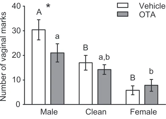
Microinjections of OTA into MPOA-‐AH decreased vaginal marking in response to male, but not in response to female or clean, odors (Figure 2.2). The interaction of drug by odor stimulus on the number of vaginal marks was statistically significant, F(2,27) = 5.485, p = .010. In the male odor condition (n = 11), OTA injections into MPOA-‐AH significantly decreased the number of vaginal marks compared to ve-‐ hicle injections, t(10) = -‐3.420, p = .007. In contrast, OTA did not impair vaginal marking in either the fe-‐ male odor condition (n = 10), t(9) = .805, p = .442, or the clean odor condition (n = 9), t(8) = -‐1.348, p = .214. Although there was a statistically significant effect of odor condition on the number of vaginal marks for both OTA-‐ and vehicle-‐injected subjects (Vehicle: F (2,27) = 15.334, p < .001; OTA: F (2,27) = 5.391, p = .011), post-‐hoc comparisons revealed that marking was significantly higher in the male odor condition compared to the female odor condition for OTA-‐injected subjects, whereas for vehicle-‐
injected subjects, marking was higher in the male odor condition compared to either the clean or female odor conditions. No significant effects of drug, odor condition, or drug by odor condition interaction, were observed for the number of flank marks or the number of quadrant entries (Table 2.1).
Experiment 2: Effects of OTA in MPOA-‐AH or BNST on male odor-‐induced vaginal marking
Experiment 3: Effects of OTA in MPOA-‐AH on social odor investigation
Clean odor tests
When tested prior to group assignment, subjects differentially investigated the three odor boxes when the same stimuli (clean cage materials) were presented in each box, χ2(2) = 20.222, p < .001. Spe-‐
cifically, subjects investigated the center box less than either the left box, z = -‐4.060, p < .05, or the right box, z = -‐3.820, p < .05. The investigation times for the two outer boxes, however, were not significantly different, z = -‐.793, p > .05. In addition, there were no pre-‐existing differences between the OTA (n = 16) and vehicle (n = 11) groups for odor box investigation or number of midline crosses (Table 2.3).
Odor preference tests
Irrespective of OTA (n = 16) or vehicle (n = 11) injection, subjects differentially investigated male, clean and female cage stimuli, χ2(2) = 21.308, p < .001 (Figure 2.3). Subjects investigated male odors
more than either clean odors, z = -‐4.178, p < .05, or female odors, z = -‐3.695, p < .05. The main effect of drug and the drug by odor stimulus interaction on the duration of odor investigation were not signifi-‐ cant. Furthermore, the effect of drug on the number of midline crosses was not significant (Table 2.3).
2.5 Discussion
sults support our hypothesis that OT normally facilitates the elevated levels of vaginal marking induced by male odors, through its actions within MPOA-‐AH and/or BNST.
Role of OT within MPOA-‐AH or BNST in scent marking
Vaginal marking behavior
The underlying neural mechanisms whereby OTA inhibits male-‐odor induced vaginal marking are not clear. Failure of antibodies generated against OTR to consistently label neurons in rodent brain tis-‐ sue has limited the characterization of OTR-‐bearing neurons in MPOA-‐AH or BNST (unpublished obser-‐ vations). Although the neural mechanisms whereby OT regulates the processing of vaginal marking-‐ relevant signals within these brain areas has not been examined, evidence from behavioral studies sug-‐ gests that OT may interact with the progesterone system to regulate reproductive behaviors. In ham-‐ sters and rats, OT can substitute for progesterone treatment in inducing lordosis in ovariectomized, es-‐ tradiol-‐primed females, and OTA can block the facilitation of lordosis by progesterone treatment (Caldwell et al., 1989, 1994; Whitman and Albers, 1995). These results raise the possibility that in the current study, OTA may have inhibited vaginal marking by blocking the downstream effects of proges-‐ terone. This seems unlikely, as proestrous female hamsters have relatively low levels of serum proges-‐ terone (Saidapur and Greenwald, 1978) and the surge in progesterone levels that occurs just prior to the onset of behavioral receptivity appears to inhibit vaginal marking (Johnston, 1977; Takahashi and Lisk, 1983; Takahashi et al., 1985). In contrast, estradiol treatment facilitates vaginal marking in response to males, either when administered systemically (Floody, 2002; Lisk and Nachtigall, 1988; Malsbury et al., 1977) or when implanted directly into MPOA-‐AH (Takahashi and Lisk, 1987; Takahashi et al., 1985). Giv-‐ en that OT and OTR expression in MPOA-‐AH and BNST are enhanced by estradiol in rats (Jirikowski et al., 1988; Patchev et al., 1993), it seems likely that OT may mediate the effects of estradiol on vaginal mark-‐ ing. More research is required, however, to determine the mechanisms whereby OT acting downstream of estradiol specifically enhances the vaginal marking response to male odors.
injection. When administered unilaterally, this volume and concentration of OTA effectively stimulates aggression in female hamsters 30 minutes, but not immediately, following injection (Harmon et al., 2002a). OTA may be working through different mechanisms to modulate aggression and vaginal mark-‐ ing; therefore, bilateral injections or other alterations to the timing, dose, or volume of injections may be necessary for OTA in MPOA-‐AH or BNST to maximally inhibit vaginal marking behavior.
Flank marking behavior
In contrast to vaginal marking, injections of OTA in MPOA or BNST had no effect on the number of flank marks in response to male, clean, or female odors. Although it has been reported that OT injec-‐ tions in MPOA-‐AH can induce flank marking in male hamsters (Albers et al., 1986; Ferris et al., 1984; Harmon et al., 2002b), in females this effect is dependent on the presence of a familiar, subordinate partner (Harmon et al., 2002b). In the current study, however, subjects were only tested with odors from unfamiliar stimulus animals. Interestingly, there were also no differences in flank marking to male, female, or clean odors, irrespective of drug condition. Female hamsters flank mark at higher levels in response to same-‐, rather than opposite-‐sex stimuli (Johnston, 1977); however, this effect is not con-‐ sistently observed in proestrous female hamsters (Petrulis and Johnston, 1999; Petrulis et al., 1999). Therefore, it is likely that a significant effect of odor condition on flank marking was not observed in the current study due to testing females only on the cycle day of proestrus.
prevalent in response to males or their odors, whereas flank marking appears to function in the estab-‐ lishment and maintenance of social hierarchies and territorial advertisement, and is most strongly in-‐ duced by same-‐sex odors (Petrulis, 2009). There is a high degree of overlap, however, both in the brain areas that regulate vaginal and flank marking, as well as in the expression patterns of OT and AVP within these areas (Albers and Bamshad, 1998; Keverne and Curley, 2004; Petrulis, 2009). Given that OTA ex-‐ hibits some level of V1aR antagonism (Manning et al., 1995), and V1aR expression has been confirmed in both MPOA-‐AH and BNST of Syrian hamsters (Caldwell and Albers, 2004), the possibility exists that the effects observed in the present study are the result of V1aR antagonism by OTA. This seems unlikely given that OTA had no effect on flank marking, a behavior that depends critically on V1aR signaling. Fur-‐ thermore, preliminary studies examining the effects of a specific V1aR antagonist, Manning compound, in MPOA-‐AH or BNST found no effects of this drug on vaginal marking (unpublished observations), sug-‐ gesting that the effects of OTA on vaginal marking are likely mediated by OTRs.
Role of OT within MPOA-‐AH or BNST in social odor investigation
in MPOA-‐AH or BNST may specifically target the male odor-‐induced component of vaginal marking with-‐ out generally affecting male odor investigation, suggesting that separate neural pathways regulate these two behaviors. This is supported by previous work in hamsters, as females with lesions of the vomer-‐ onasal organ fail to vaginal mark more to male vs. female odors but show normal patterns of preferen-‐ tial investigation of male odors, whereas ME lesions eliminate opposite-‐sex odor preference without affecting preferential marking in response to male odors (Johnston, 1992; Petrulis and Johnston, 1999; Petrulis et al., 1999).
Conclusion
Together, these results suggest that OT within MPOA-‐AH and/or BNST facilitates vaginal marking in female Syrian hamsters. This is in agreement with the role of OT in other sociosexual behaviors, and suggests that the general role of OT is to facilitate sociosexual interactions (Insel, 1992). Importantly, the present study found that the effects of OTA are specific to the male odor-‐induced component of vaginal marking, indicating that OT in these areas can modulate the behavioral response to chemosensory cues important for social behaviors.
2.6 Acknowledgements
We would like to thank Rachel Atkinson, Amelia Davies, Johnny Garretson, Mary Karom, and Preethy Kuriakose for their technical assistance in completing this project. This work was supported by NIH grant MH072930 to A. P., NSF grant IOS-‐0923301 to H. E. A., and in part by the Center for Behavioral Neuroscience under the STC program of the NSF, under agreement IBN 9876754.
Chapter 2 Tables
Table 2.1 Summary of behavioral measures from Experiment 1
Mean (± SEM) number of flank marks and quadrant entries by female hamsters during 15-‐minute scent-‐ marking tests to male (n = 11), clean (n = 10), or female (n = 9) cage odors, following injection of OTA or vehicle. There were no significant effects of drug or odor condition on flank marks or quadrant entries, all p > .05.
Number of flank marks Number of quadrant entries
Male Clean Female Male Clean Female
Drug
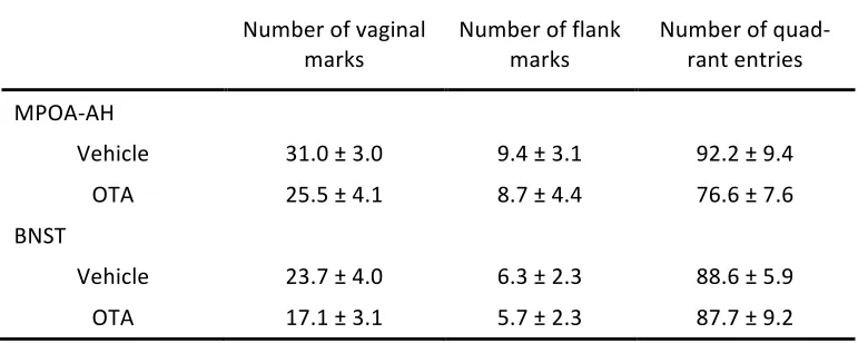
Table 2.2 Summary of behavioral measures from Experiment 2
Mean (± SEM) number of flank marks and quadrant entries by female hamsters during 15-‐minute scent-‐ marking tests to male odors, following injection of OTA or vehicle into either MPOA-‐AH (n = 10) or BNST (n = 10). OTA injections significantly decreased the number of vaginal marks compared to vehicle injec-‐ tions irrespective of brain area, p = .023. There were no significant effects of drug or brain area on the number of flank marks or quadrant entries, all p > .05.
Number of vaginal marks Number of flank marks Number of quad-‐rant entries
MPOA-‐AH
Vehicle 31.0 ± 3.0 9.4 ± 3.1 92.2 ± 9.4 OTA 25.5 ± 4.1 8.7 ± 4.4 76.6 ± 7.6
BNST
Vehicle 23.7 ± 4.0 6.3 ± 2.3 88.6 ± 5.9 OTA 17.1 ± 3.1 5.7 ± 2.3 87.7 ± 9.2
[image:46.612.66.456.168.322.2]Table 2.3 Summary of behavioral measures from clean odor tests (Experiment 3)
Median (± IQR) number of midline crosses and duration (in seconds) of odor box investigation by female hamsters during 10-‐minute clean odor tests preceding assignment to either OTA (n = 16) or vehicle (n = 11) groups. Subjects investigated the left and right boxes more than the center box, p < .001. There were no significant effects of drug or odor condition on the number of midline crosses, p > .05.
Number of midline crosses Investigation duration
Left Center Right
Group assignment
Vehicle 33.0 ± 13.0 29.0 ± 17.2 13.3 ± 12.7 22.3 ± 7.6 OTA 35.5 ± 8.8 23.6 ± 17.3 13.4 ± 12.4 29.0 ± 9.2
[image:47.612.69.514.156.253.2]Chapter 2 Figures
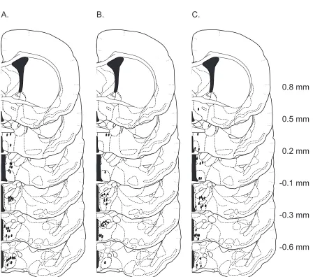
Figure 2.1 Histological verification of injection sites
Coronal sections through the rostral-‐caudal extent of the hamster brain of microinjection sites for Exper-‐ iment 1 (A), Experiment 2 (B), and Experiment 3 (C), located within MPOA-‐AH and BNST. Ovals represent microinjections that terminated within MPOA-‐AH, whereas triangles represent microinjections that ter-‐ minated within BNST.
A. B. C.
ox ox
-0.6 mm -0.3 mm -0.1 mm 0.2 mm 0.5 mm 0.8 mm
[image:48.612.84.532.96.492.2]Figure 2.2 Number of vaginal marks during scent-‐marking tests
OTA injected into MPOA-‐AH decreased vaginal marks to male, but not female or clean, odors, compared to vehicle injections. Following vehicle injections, subjects vaginal marked more to male than to female or clean odors. In contrast, OTA-‐injected subjects only marked more to male odors than to female odors, as marking to clean odors was intermediate to both male and female odors. * represents signifi-‐ cant difference between OTA and vehicle injected subjects in the male odor condition, p = .007. Dissimi-‐ lar letters within each letter type (upper-‐ and lowercase) represent significant mean differences follow-‐ ing Tukey’s post-‐hoc comparisons examining the simple main effect of odor within OTA and vehicle con-‐ ditions.
Male
Clean
Female
Number of vaginal marks
0
10
20
30
40
Vehicle
OTA
*
A
B
a
b
a,b
[image:49.612.81.352.94.289.2]Figure 2.3 Odor box investigation during preference tests
Subjects investigated male odors more than either female or clean odors, irrespective of drug (OTA or vehicle) injected into MPOA-‐AH (p < .05 with Bonferroni correction). Investigation duration times were adjusted to account for pre-‐existing biases in investigation due to odor box positioning, as described in
the Data Analysis section. Graphed data represent unadjusted investigation times.