ScholarWorks @ Georgia State University
ScholarWorks @ Georgia State University
Chemistry Dissertations Department of Chemistry
Spring 5-1-2012
Structure and Function Studies of Selenium Substituted Nucleic
Structure and Function Studies of Selenium Substituted Nucleic
Acids
Acids
Wen Zhang
Follow this and additional works at: https://scholarworks.gsu.edu/chemistry_diss
Recommended Citation Recommended Citation
Zhang, Wen, "Structure and Function Studies of Selenium Substituted Nucleic Acids." Dissertation, Georgia State University, 2012.
https://scholarworks.gsu.edu/chemistry_diss/63
This Dissertation is brought to you for free and open access by the Department of Chemistry at ScholarWorks @ Georgia State University. It has been accepted for inclusion in Chemistry Dissertations by an authorized
administrator of ScholarWorks @ Georgia State University. For more information, please contact
by
WEN ZHANG
Under the Direction of Dr. Zhen Huang
ABSTRACT
Nucleic acids are responsible for the storage of genetic information and directly participate in
gene replication, transcription and expression, and thereby the control of nucleic acids leads to the
regulation of genetic information flow and gene expression. Meanwhile, many non-coding RNAs are
in-volved in signal transduction directly. Moreover, nucleic acid-based therapeutic strategies have been
lead to drug candidates and are effective tools in drug discovery and disease study at the molecular level
as well as the genetic level. Consequently, the 3D crystal structure study and related functional research
on natural and unnatural nucleic acids have become very popular area, expanding their potential
appli-cation in medicinal and biological chemistry.
Since oxygen, sulfur, selenium and tellurium are in the same elemental family (VIA) in the
peri-odic table, we anticipate that oxygen atoms in nucleic acids can most likely be replaced with the other
chalcogen atoms without causing significant perturbations. Owing to the special K edge and unique
nucle-sential for nucleic acids’ structural determination at the atomic level. Additionally this novel elemental
feature (atomic size and electronic nature) provides nucleic acids with unique properties. In addition,
the selenium derivatization can facilitate crystal growth. Other chalcogen elements are applicable as
well to modify nucleic acid, generating some special biofunctions, like the application of
phosphorthioate oligonucleotide in gene therapy. This dissertation will outline the chalcogen elements
(especially selenium) modifications of nucleic acids, including syntheses strategies, structure studies and
potential therapeutic applications. Our research work here tries to show that (1) Selenium functionality
is able to facilitate the crystal structure determination, by both helping solve phase problem and
accel-erating crystal growth; (2) Selenium functionality can generate special capability to nucleic acids, like
improved base pair fidelity, novel atomic interactions and feasibility to be biological chemistry probe; (3)
Selenium derivatized oligonucleotides are extraordinary good candidates for gene therapy discovery,
considering its stability under nuclease environment. In general, these atom-specific replacements
gen-erate a new paradigm of nucleic acids.
by
WEN ZHANG
A Dissertation Submitted in Partial Fulfillment of the Requirements for the Degree of
Doctor of Philosophy
in the College of Arts and Sciences
Georgia State University
Copyright by Wen Zhang
by
WEN ZHANG
Committee Chair: Dr. Zhen Huang
Committee: Dr. David W. Boykin
Dr. Gangli Wang
Electronic Version Approved:
Office of Graduate Studies
College of Arts and Sciences
Georgia State University
ACKNOWLEDGEMENTS
The work described here was all under the direction of my supervisor, Prof. Zhen Huang. I
deep-ly appreciate Dr Huang for offering me this opportunity to work on this attractive project, giving me
val-uable suggestion during the research and educating me to be a qualified scientist. Both encouragement
and criticism from him will make me benefited. His dedication to science and distinguished
accomplish-ment also set up a perfect example and standard for me. I would like to thank Prof. David W. Boykin and
Prof. Gangli Wang, who have provided me their supportive guidance and opinion during my PhD career.
It is their help that make my research work more complete and smooth. Also I have to thank my
lab-mates for their support and concern in these more than 5 years. I will thank Dr Abdalla E.A. Hassan for
his teaching me organic synthesis skill, directing me with many projects and discussing with me about
experimental results. I will thank Dr Jia Sheng for his teaching me in many aspects, especially on
crystal-lography study, including crystal growth, fishing, and data collection and processing. It would be much
more difficult for me to set about structure determination without him. I will also show my appreciation
to Dr Julianne Caton-Williams, Dr Lina Lin, Dr Abdur Rob, Dr Sarah Spencer, Dr Jianhua Gan, Dr Hehua
Liu, Sibo Jiang, Manindar Kaur, Huiyan Sun, and Lilian Kamau for their discussions and helps in the lab.
Thanks also should be sent to Dr Sekar Chandrasekaran from NMR facility, Dr Siming Wang from MS
fa-cility and Dr Alexei Soares from Brookhaven National Laboratory for their great help. I will especially
thank my wife, Ying Zhang, who has always supported and encouraged me, in both academic and daily
life, to complete this PhD project. I also need to thank Molecualr Basis of Disease (MBD) program to
support me in recent years. This work is supported financially by Georgia Cancer Coalition (GCC)
TABLE OF CONTENTS
ACKNOWLEDGEMENTS ... iv
LIST OF TABLES ... vii
LIST OF FIGURES ... viii
LIST OF SCHEMES ... x
1. INTRODUCTION ... 1
1.1 Nucleic Acids--- Important Molecules for Biofunction Studies and Drug Discovery ... 1
1.2 Synthesis of Selenium Derivatized Nucleic Acids ... 3
1.3 Novel Structures and Functions of Selenium Derivatized Nucleic Acids ... 14
2. MATERIAL AND EXPERIMENTAL PART ... 22
2.1 Synthesis of 5-Selenium-Thymidine DNA for Structural and Enzymatic Study ... 22
2.1.1 Introduction ... 22
2.1.2 Synthesis Strategy and Structure Determination ... 26
2.1.3 Chalcogen Modified Oligonucleotides' Stability to Nuclease ... 37
2.2 Synthesis of 2-Selenium-Thymidine DNA and Improved Base Fidelity ... 39
2.2.1 Introduction ... 39
2.2.2 Synthesis Strategy, Structure Determination and Thermostability Study ... 41
2.3 Synthesis of 5'-Selenium-Thymidine DNA and Convenient Fluorescence Labeling ... 47
2.3.1 Introduction ... 47
2.3.2 Synthesis Strategy and Biofunctional Study ... 49
2.4 Synthesis of 5-Selenium-Cytidine DNA for Structural and Enzymatic Study ... 53
2.4.2 Synthesis Strategy and Structure Determination ... 55
2.4.3 Enzymatic Inhibition to methyltransferase and endonuclease ... 61
2.5 Synthesis of 4,5-Diselenium-Thymidine Nucleoside and DNA ... 64
2.5.1 Introduction ... 64
2.5.2 Synthesis Strategy ... 66
2.6 Facilitation of Selenium Modification on Nucleic Acid Crystallization ... 71
2.6.1 Introduction ... 71
2.6.2 Crystallizaiton Screening ... 72
3. RESULTS AND DISCUSSION ... 73
3.1 Synthesis of 5-Selenium-Thymidine DNA for Structural and Enzymatic Study ... 73
3.2 Synthesis of 2-Selenium-Thymidine DNA and Improved Base Fidelity ... 86
3.3 Synthesis of 5'-Selenium-Thymidine DNA and Convenient Fluorescence Labeling ... 92
3.4 Synthesis of 5-Selenium-Cytidine DNA for Structural and Enzymatic Study ... 101
3.5 Synthesis of 4,5-Diselenium-Thymidine Nucleoside and DNA ... 110
3.6 Facilitation of Selenium Modification on Nucleic Acid Crystallization ... 113
4. CONCLUSIONS... 115
APPENDIX ... 117
LIST OF TABLES
Table 2.1 Transient N3 protection and lithium/halogen exchange of compound 2 in the presence of
dimethyldiselenide………..………32
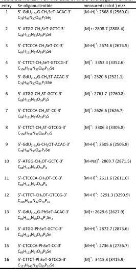
Table 3.1 MALDI-TOF MS Data of the 5-Se-T DNAs……….……….………75
Table 3.2 UV Melting Temperatures of the 5-Se-T DNAs……….……..75
Table 3.3 MALDI-TOF MS data of the 5-chalcogen-T DNAs……….……..79
Table 3.4 UV melting temperatures of the 5-chalcogen-T DNAs………..…………..80
Table 3.5 Relative inhibition efficiency of different modified DNA under different nuclease environ-ment………..84
Table 3.6 MS values of the 2-Se-T-containing DNAs………...………. 86
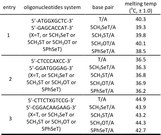
Table 3.7 Melting temperatures of native and SeT-DNA duplexes A………..………..88
Table 3.8 Melting temperatures of native and SeT-DNA duplexes B………..………..88
Table 3.9 MALDI-TOF MS Data of the 5-Se-C DNAs……….…102
Table 3.10 UV Melting Temperatures of the SeC-DNAs……….…102
Table 3.11 Data collection and Statistics……….……….……103
Table 3.12 Structure Refinement and Model Statistics………..……….……104
Table 3.13 MS values of the 4,5-Se-T-containing DNAs………..…….………..110
Table 3.14 Comparison of DNA crystallization process. A: Se-DNA; B: native DNA……….………….…….113
LIST OF FIGURES
Figure 1.1 Se-derivatized nucleic acids (SeNA)……….………..4
Figure 1.2 5’-selenium modified nucleoside and TT dimer……….…………5
Figure 1.3 Enzymatic incorporation of backbone selenium into DNAs……….. 11
Figure 1.4 Enzymatic incorporation of ATPaSe into RNA by T7 RNA polymerase……….. 11
Figure 1.5 Nucleic acids modified with potential selenium by atom-specific replacement………... 13
Figure 1.6 The global and local structures of the 4-Se-T DNA [(5’-GdUSe-G-SeT-ACAC-3’)2]……….. 15
Figure 1.7 The superimposed global and local structures of 6-Se-G containing DNA/RNA duplexes of the nucleic acid–protein complex………. 16
Figure 1.8 Photos of crystals of the native and Se-derivatized octamers……….. 17
Figure 1.9 Time-course enzymatic digestion of phosphoroselenoate RNA with snake venom phosphodiesterase I……….18
Figure 1.10 The time-course experiment of incorporating TTP and 4-SeTTP into DNA………19
Figure 1.11 Yellow-colored DNA generated by DNA polymerization reaction from 4-Se-TTP………19
Figure 1.12 Structures of some FDA-approved anti-HIV drugs………..20
Figure 2.1 UV spectra of 5-Se-T (red) and thymidine (blue) in MeOH……….28
Figure 2.2 Sequences those endonucleases recognize and cleave………...37
Figure 2.3 Native and Se-modified T/A base pair and T/G wobble pair………..39
Figure 2.4 Potential DNA methyltransferase inhibitors………61
Figure 2.5 Sequences those methyltransferase and endonuclease recognize……….62
Figure 3.1 HPLC analysis of crude 5′-DMTr-TT-5-SeT-T-3′ (15.3 min)……….74
Figure 3.2 HPLC analysis of the native and modified DNAs………..75
Figure 3.3 Global and local structures of the 5-Se-T-DNA [(5′-GdU2′-Se-G-5-SeT-ACAC-3′)2]………..76
Figure 3.5 Resistance study of 5-chalcogen-T DNAs to endonuclease AseI………..…….82
Figure 3.6 Comparison of inhibition of different modifications in DNA5 to exonuclease III…………..………83
Figure 3.7 Comparison of different 5-CH3Se-T DNAs’ inhibition to endonuclease AseI……….84
Figure 3.8 Global and local structures of the 2-Se-T-DNA [(5’-GdU2’-Se-G-SeT-ACAC-3’)2]……….…90
Figure 3.9 HPLC analysis of 5’-Se-T containing DNA (5’-dSeTGCGTAATACGACTCACTATAG-3’)……….92
Figure 3.10 MALDI-TOF MS spectrum of 5’-Selenium 22mer DNA………93
Figure 3.11 HPLC analysis of labeling efficiency of native DNA (5’-dGCGTAATACGACTCACTATAG-3’) and Se-DNA (5’-dSeTGCGTAATACGACTCACTATAG-3’)……….………….94
Figure 3.12 MS spectrum of 5’-fluorescein-labeled DNA-22mer……….…………95
Figure 3.13 HPLC analysis of fluorescent dye-labeled DNA-22mer……….………..95
Figure 3.14 UV analysis of the dye-labeled DNA-22mer……….………..95
Figure 3.15 HPLC analysis of fluorescein compound 5-IAF, dye-labeled DNA, and their co-injection……..96
Figure 3.16 DNA polymerization reaction monitored with the fluorescent dye-labeled DNA-22mer..….99
Figure 3.17 MS spectrum of full-length product of DNA polymerization reaction……….99
Figure 3.18 Reversed-phase HPLC analysis of crude DMTr-on Se-DNA (5’-DMTr-TC5SeT-3’)………101
Figure 3.19 SeC-DNA crystal structures (Se in gold)………105
Figure 3.20 Selenium modification effects on DNA methyltransferase interaction……….106
Figure 3.21 Endonuclease Hinp1I’s digestion to oligonucleotides with different modifications……….108
Figure 3.22 Structure change of 4,5-Se-T nucleoside under basic pH condition………112
Figure 3.23 UV-visible spectrum of 4,5-Se-thymidine under pH change……….….………112
Figure 3.24 Se-DNA crystals using nucleic acid mini-kit condition……….………..……114
LIST OF SCHEMES
Scheme 1.1 Synthesis of 2’-methylseleno containing uridine and cystidine phosphoramidite and DNAs.. 6
Scheme 1.2 Synthesis of the 2’-selenomethyl-2’-deoxyguanosine phosphoramidite……….7
Scheme 1.3 Synthesis of the 4-Se-thymidine phosphoramidite and Se-DNAs………..…..8
Scheme 1.4 Synthesis of the 6-(2-cyanoethyl)seleno deoxyguanosine phosphoramidite and oligonucleo-tides………..…………...9
Scheme 1.5 Synthesis of phosphoroselenoate dimer by selenium-cyanoethylphthalimide………10
Scheme 1.6 Chemical synthesis of 4-SeTTP and enzymatic synthesis of SeT containing DNAs…..…………..12
Scheme 2.1Synthesis of 5-Se-T phosphoramiditeand oligonucleotides………..27
Scheme 2.2 Synthesis of 5-chalcogen-T DNAs………..32
Scheme 2.3 Synthesis of 2-Se-T phosphoramidite and Se-DNA…………..………42
Scheme 2.4 Synthesis of the 5’-Se-DNA and labeled-DNA……….……….48
Scheme 2.5 Synthesis of the 2’-deoxy-5-(methylselenyl)cytidine phosphoramidite and its oligonucleo-tide………..……59
1. INTRODUCTION
1.1 Nucleic Acids—Important Molecules for Biofunction Studies and Drug Discovery
Nucleic acids are the storage of genetic information and directly participate in gene replication,
transcription and expression, and the control of nucleic acids can lead to modulation and regulation of
genetic information flow and gene expression1,2. In addition, many non-coding RNAs, like ribosomal
RNA, riboswitch and microRNA, are participated in signal transduction directly by regulatory functions3,4.
Moreover, nucleic acid-based therapeutic strategies, where the natural gene, instead of protein, is
rec-ognized as drug targets and the modified oligonucleotides (DNAs and RNAs) are applied as drug
candi-dates, are effective tools in drug discovery and disease study at the molecular level as well as the
genet-ic level5,6. Therefore, nucleic acids related research is essential for better understanding diseases and
identifying novel drug candidate and target for development of therapeutics with high efficiency to treat
some world-wide diseases. Because of the advantages of low toxicity, notable efficacy and high
specifici-ty, numerous nucleic acid-based potential therapeutics are under development or in clinical trials,
in-cluding antisense oligonucleotides, siRNAs, miRNAs, ribozymes and DNazymes and nucleic acid
aptamers7-9. Instead of the traditional attacking of aberrant proteins, modulating expression of specific
diseases’ gene by these nucleic acids therapies can lead to better treatment. Research activities in this
area require further development of new nucleic acid analogs with improved properties.
On the other hand, the unique structures of DNA and RNA provide nucleic acids multiple
func-tions and properties. The negative charges on phosphate groups make nucleic acids soluble in water,
and the hydrophobic nucleobases in the center can form Watson-Crick hydrogen bonds. Additionally,
the complicated RNA secondary and tertarial structures generate enzyme-like catalytic functions.
Re-cently, X-ray crystallography has been the most powerful technical tool to provide researchers detailed
candidate and understand nucleic acids related biological processes mechanism, such as DNA
replica-tion, ribozyme catalysis, DNA transcription and RNA translation10,11. Despite the bottle-necks of nucleic
acid crystallography and phase problem, it is greatly promising for researchers to rely on it for nucleic
1.2 Synthesis of Selenium Derivatized Nucleic Acids (SeNA)
Since oxygen atom is one of essential elements in nucleic acids, it is anticipated that the
atom-specific substitution of oxygen atoms with the other chalcogen atoms is a potential useful chemical
strategy to generate tremendous improvements of nucleic acids without causing significant alterations
in native structures, functions and protein interaction (such as higher bioavailability and stability). Sulfur
(S), selenium (Se)and tellurium (Te) are the elements following oxygen in the VIA group of the periodic
table. Selenium element was discovered in 19th century, and little systematic research was conducted at
that time owing to the toxicity. With the modern chemical and biological technologies, the existence of
selenium in natural organisms was demonstrated14. Nowadays selenium is considered as an essential
element, i.e. the trace micronutrient that functions as cofactor for reduction of antioxidant enzymes15.
Two Se-containing special amino acids, selenomethionine and selenocysteine, have been found in
na-ture16,17, and several studies have suggested there is a possible link between selenium deficiency and
many types of diseases, like cancer, HIV/AIDS and diabetes. Overall, its functions in biomolecules have
attracted increasing attention of scientific communities. Like sulfur, selenium was also found to exist in
natural tRNA in the form of 2-selenouridine and 5-[(methylamino)methyl]-2-selenouridine (mnm5se2u)
with the corresponding natural 2-thiouridine as a precursor14. The reaction was catalyzed by
2-selenouridine synthase with the selenophosphate as the selenium donor, and the S atom at 2-position
was replaced by Se18,19. This selenium functionality is usually found at the anticodon wobble positions of
several bacterial tRNAs, and interestingly may provide the translation process with higher accuracy and
efficiency20. However, compared with Se-derivatized proteins, research on selenium functionalized
nu-cleic acids is relatively limited owing to the stability, availability and other challenges. This fact
stimu-lates our lab to study the novel synthesis and property of selenium modified unnatural nucleic acids.
Because of its larger atomic size (atomic radius, O: 0.73 Å, S: 1.02 Å, Se: 1.16 Å) and its higher electron
nu-cleic acids with many unique and novel properties. As they are in the same elemental group, oxygen and
selenium and have closely-related chemical and electronic properties. This allows almost complete
re-tention of the native structures, functions, and protein recognitions after the nucleic acid modification
by replacing oxygen atoms with selenium. This atom-specific replacement allows many modification
choices in introducing the selenium elements, because of the multiple oxygen atoms in nucleic acids. By
screening for the ideal substitution sites, the multiple choices allow minimization of the potential
per-turbations. Furthermore, because of their larger atomic sizes, compared to oxygen (1.16 Å vs 0.73 Å),
and their higher electron densities, Se- derivatized nucleic acids may possess many unique properties.
NH N N O NH2 N O
OH or (H) O O P O O O -3'
Se
5'(Or other nucleobases)
Figure 1.1 Se-derivatized nucleic acids (SeNA).
Recently the synthesis, structure and function studies of selenium-derivatized nucleic acids
(SeNA) have been successfully pioneered by Huang and co-workers21-23. The Se-functionalities at various
positions, including the sugar 2’, 4’ and 5’ positions, nucleobases, and phosphate groups, have been
ac-complished by Huang’s and other research groups. SeNA provides extra research advantages in
chemis-try and biology areas, like chemical biology, molecular biology, and structural biology, i.e. X-ray
crystal-lography. This atomic Se-substitution also provides novel insights into structure and function of nucleic
acids, including base pair recognition, duplex stability and fidelity, catalysis, and non-coding RNA
In the sugar modification with selenium, the 5’-MeSe-derivatized phosphoramidites, including A,
C, G, T and U, were first synthesized by Huang’s group27. This route was achieved by activation of the
5’-position by good leaving groups, like Ts, Ms or Br, followed by an SN2 substitution with the selenide
rea-gents in presence of phase transfer catalyst. Experiments showed that these 5’-Se-modified
phosphoramidites are stable and can be incorporated into oligonucleotides with high yields. It was also
interestingly found by other researchers that the nucleic acid dimer with selenium functionality at 5’
bridging position possessed some toxicity28, which was worthy for biological application and drug
dis-covery. (Figure 1.2)
O OH
HSe B
OH (or H)
O O
RO T
P O R
O OH
Se T
Figure 1.2 5’-selenium modified nucleoside and TT dimer. (B=nucleobase)
After the demonstration of the Se-functionality stability, a novel phosphoramidite compound
(2’-α-methylseleno uridine phosphoramidite) was first synthesized by Huang’ laboratory, with the
3’,5’-protected-2,2’-anhydrous uridine as the intermediate29. It showed fine stability in solid-phase synthesis
and offered a high coupling efficiency (99% yield). In this work, the structure of a selenium-functionality
containing DNA decamer 5’-GCGTAU2’-SeMeACGC-3’ was determined at high resolution (1.3 Å), and the
structure was successfully solved based on the selenium anomalous signal. Later, the strategy to
intro-duce 2’-selenium functionality was improved to get higher yield30. In the experiment both U and C
nu-cleoside containing 2’-selenium modification were synthesized. The synthetic strategy was shown in
Scheme 1.1 Synthesis of 2’-methylseleno containing uridine and cystidine phosphoramidite and DNAs. (a) (Ph)2CO3, Na2CO3, DMF; (b) DMTr-Cl, Py; (c) NaSeCH3, EtOH-THF; (d) 2- cyanoethyl N,N
-diisopropyl-chlorophosphoramidite and N,N-diisopropylethylamine in CH2Cl2; (e) synthesis of oligonucleotides on
solid phase; (f) Ms-Cl, TEA, THF; (g) toluene/ tetrahexylammonium hydrogen sulfate, Na2CO3 (satd); (h)
(Bu)4N+ F-, THF; (i) TMS-Im, then Ac2O, TEA and DMAP in THF; (j) TMS-Im, then POCl3-triazole-TEA in
CH3CN; (k) NH4OH.
easily and the structure was determined using selenium to solve phase problem. In different to the
2’-Se-modified pyrimidine compound synthesis, the same strategy was not working for purine nucleoside
synthesis. This fact forced scientists to pioneer novel chemical approach to introduce selenium
function-ality into 2’-position. In Micura’s group, trifluoromethanesulfonyl (Tf) group was applied as good leaving
group at 2’-β-position of RNA A nucleoside, followed by the replacement of selenium element in a SN2
reaction31. Later guanosine RNA nucleoside with selenium at 2’-position was synthesized as well using
similar strategy. It should be noted that DTT is necessary in the 2’-selenium-modified purine synthesis,
Scheme 1.2 Synthesis of the 2’-selenomethyl-2’-deoxyguanosine phosphoramidite.
DNA purine nucleoside analogues with 2’-selenium-derivatization were synthesized by a novel
strategy32,33. In the synthesis, no DTT reducing reagent was needed, and 2’-position was also activated
by Tf group. By this novel and convenient strategy, modified DNA nucleosides were synthesized and
in-corporated into oligonucleotides for structural studies. The synthesis of 2’-Se-G was shown in scheme
1.2.
4’-Thio-modification has attracted a lot of interest because of some special properties, such as
anti-virus stability. Similarly, the replacement of the sugar ring oxygen with the selenium generates the
4’-seleno nucleoside. The syntheses of the 4’-selenocytidine and 4’-selenouridine were first achieved by
Matsuda’s group using stereoselective seleno-Pummerer reaction34. Later, a modified strategy was
ap-plied by Jeong’s group to achieve 4’-seleno nucleoside synthesis, and the crystal structure of the
4’-Se-nucleosides was also determined35. Their results indicated that the replacement of oxygen at 4’-position
rear-rangement to synthesize both pyrimidine and purine nucleosides containing the 4’-seleno
functionali-ty36. Basically all the four types of 4’-selenium-modified nucleosides can be accomplished now.
O OH O N NH O O DMTr O OH O N N N O DMTr O OH O N N Se O DMTr N N NC O O O N N Se O DMTr NC P
OCH2CH2CN
N O O O N NH Se O P O
O Oligonucleotide 3' P O O 5'-Oligonucleotide a b c d e f
Scheme 1.3 Synthesis of the 4-Se-thymidine phosphoramidite and Se-DNAs. a) TMS-Im and MeCN. b) 1H-Triazole, POCl3, Et3N. c) (NCCH2CH2Se)2, NaBH4, EtOH. d) 10% Et3N in MeOH. e) 2-Cyanoethyl N,N
diisopropyl (chloro) phosphoramidite and EtN(i-Pr)2 in CH2Cl2. f) Solid-phase synthesis.
The Se-nucleobase modifications of nucleic acids were first accomplished by Huang’s laboratory
recently22,23. The Se-functionality was introduced to the 4-position of thymidine (Scheme 1.3) by
selec-tive activation of the 4-position with triazoyl group, followed by the introduction of 2-cyanoethyl
selenide using the Huang’s reagent [(CNCH2CH2Se-)2] to protect selenium functionality24. The
corre-sponding 4-Se-T phosphoramidite was successfully synthesized and then incorporated into
oligonucleo-tides in a high yield. In this substitution reaction the selenium atom was introduced into nucleobase in
high yield, and it encouraged us to explore more substitution methods. Later, the 6-Se-deoxyguanosine
phosphoramidite was also synthesized for the first time37. The 6-position of guanosine was activated by
2,4,6-triisopropylbenzene-1-sulfonyl group, followed by the introduction of the Se- functionality. This
novel 6-Se-G-phosphoramidite was successfully incorporated into oligonucleotides by solid-phase
Scheme 1.4 Synthesis of the 6-(2-cyanoethyl)seleno deoxyguanosine phosphoramidite (3) and oligonu-cleotides (4). a) TIBS, DMAP, TEA, CH2Cl2, room temperature; b) diselenide, NaBH4/EtOH, -5 °C, 80% yield
in two steps; c) phosphoramidite, BTT, CH2Cl2, 75% yield; d) solidphase synthesis.
TIBS=2,4,6-triisopropylbenzene-1-sulfonyl chloride, DMAP=4-dimethylaminopyridine, diselenide = (NCCH2CH2Se)2,
phosphoramidite=2-cyanoethyl tetraisopropyl-phosphorodiamidite, BTT = 5-(benzylthio)-1H-tetrazole.
The phosphoroselenoates were synthesized via oxidative selenium incorporation38, and the
longer phosphoroselenoates were prepared for the antisense study39. Later, the phosphoroselenoates
were used for the structural study by Egli’s group26. This Se-modification is achieved during the
solid-phase synthesis by a selenizing reagent (such as potassium selenocyanate, KSeCN), replacing iodine. The
synthesis of PSe- modification oligonucleotides was in quantitative yield, especially when the
modifica-tion site was closed to 5’- end. This type of derivatizamodifica-tion also provided oligonucleotide high cytotoxicity.
In their work the DNA crystal structures with phosphoroselenoate modification were determined in high
resolution, which indicated that the selenium modification brought obvious perturbation to the
oligonu-cleotides. Subsequently, several other reagents were also developed to introduce the Se-functionality
O O DMTrO N NH O O O OAcOPh O N NH O O P O H
N Se CN
O O DMTrO N NH O O O OAcOPh O N NH O O P O Se NC O O
Scheme 1.5 Synthesis of phosphoroselenoate dimer by selenium-cyanoethylphthalimide.
(iPrC5H4)2TiSe541 and selenium-cyanoethylphthalimide42. It is worthy to mention that the
selenium-cyanoethylphthalimide was an efficient selenizing reagent with the selenium protected by cyan-oethyl
group. In this way the potential selenium oxidation was prevented (scheme 1.5). Though chemical
syn-thesis can successfully prepare phosphoroselenoates, the synthesized phosphoroselenoates are
diastereomer mixtures. Separation of diastereomers is very challenging and impractical for
oligonucleo-tides containing more than one phosphoroselenoate group. The enzymatic synthesis of
phosphoroselenoates was first developed by Huang’s group, and the diastereomerically pure
phosphoroselenoate oligonucleotides were synthesized enzymatically. Se-dNTP series was chemically
synthesized and the diastereomers were easily separated43. Interestingly, DNA polymerase could well
recognize and incorporate both dNTP diastereomers, thereby synthesizing diastereomerically pure
phosphoroselenoate DNAs. Later, phosphoroselenoate RNAs were also synthesized enzymatically with
diastereomeric Se-NTPs, -Se- -Se- -Se- -Se-UTP. They were
success-fully incorporated into the hammerhead ribozyme to generate diastereomerically pure RNAs for the
function study44,45. Interesting fact was that, on RNA version, the polymerase could recognize one
Se-moiety at the 4-position was synthesized as well, and it was recognized by the DNA polymerase as
well as the native TTP46 (scheme 1.6).
Figure 1.3 Enzymatic incorporation of backbone selenium into DNAs. (A) Primer and template sequenc-es. (B) Time-course primer extension using R-Se-TTP I and II (Sp and Rp) on 19% polyacrylamide gel. (C) Exonuclease III digestion after primer extension.
Scheme 1.6 Chemical synthesis of 4-SeTTP and enzymatic synthesis of SeT containing DNAs (5). (a) TMS-Im; 1,2,4-triazole-POCl3-Et3N; (b) (NCCH2CH2Se)2/NaBH4, EtOH; (c) 80% acetic acid; (d) POCl3, Me3PO4;
tributylamine (TBA), pyrophosphate, DMF; H2O; (e) 0.05 M K2CO3; (f) template-dependent DNA
polymer-ization.
As talked about above, various types of modified nucleic acids with potential selenium
O O O
B
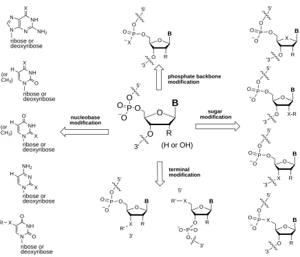
phosphate backbone modification sugar modification nucleobase modification NH N N N X NH2 NH N X O H NH N O O X ribose or deoxyribose ribose or deoxyribose ribose or deoxyribose R NH N O X H ribose or deoxyribose R (or CH3)O O X B P O O O 3' terminal modification (H or OH)
P O O 3' X O O B P O O O O 5' 5' '3 R O O O B P O O O 5' '3 X-R O X O B P O O O 5' '3 R O O X B P O O O 5' '3 R O O O B P O O X 5' '3 R (or CH3)
[image:26.612.75.511.75.449.2]R R' O X O B P O O O 5' R R' 5' 3' N N NH2 X H ribose or deoxyribose
1.3 Novel Structures and Functions of Selenium Derivatized Nucleic Acids
Since the discovery of the naturally Se-containing tRNAs14, the Se-modification has attracted
sci-entists’ enthusiasm to study its unique properties in biochemical and biological systems. Owing to its K
edge of 0.9795 Å and its signal for multiple wavelength anomalous dispersion (MAD) and single
wave-length anomalous dispersion (SAD) analysis, selenium is considered as an ideal anomalous scattering
center in crystal X-ray diffraction, which significantly facilitates crystallographic studies on biological
macromolecules. Since the first successful structural determination of a protein with selenomethionine
(replacing sulfur with selenium) and MAD technique developed by Hendrickson’s laboratory 20 years
ago47, more and more novel protein structures have been determined with the selenomethionine
derivatization48-50. Fortunately, the sulfur substitution with selenium does not cause significant
perturba-tions inprotein structures and functions. Thus, it is not surprising that currently over two thirds of the
novel protein structures are determined by the selenomethionine strategy and MAD phasing (RCSB
Pro-tein DataBank)50. Clearly, the selenomethionine and MAD strategies have revolutionized the protein
X-ray crystallography.
However, it was surprising that oxygen atoms in nucleic acids can also be stably replaced with
selenium without causing significant perturbations in structures and functions. This novel strategy with
selenium-derivatized nucleic acids (SeNA) is changing the X-ray crystallography of nucleic acids and
pro-tein-nucleic acid complexes. The classical strategy in nucleic acid crystallography relies on the halogen
derivatization, such as Br or I derivatization51. Unfortunately, the I- or Br-derivatized nucleic acids are
sensitive to light, such as UV and X-ray light. In addition, the modification sites for the halogen
incorpo-rations are very limited: primarily the 5-position of deoxyuridine, which may cause unavoidable
pertur-bations in structures and functions52. In comparison, the Se-derivatization of nucleic acids offers better
stability and much more diversities than the halogen-derivatization, since in nucleic acids, there are
Se-containing nucleoside phosphoramidites, and pioneered the chemical and enzymatic syntheses and the
selenium-derivatization of nucleic acids for structure and function studies. Through the collaboration
among Huang, Egli and their co-workers, the first structure of the Se-derivatized nucleic acids was
de-termined via the direct selenium derivatization and MADphasing53. Over the past several years, many
crystal structures of DNAs, RNAs and protein-nucleic acid complexes have been successfully determined.
This novel Se-derivatization strategy has received more and more attention. It will revolutionize X-ray
crystallography of nucleic acids, protein-nucleic acid complexes, as well as small molecule ligand-nucleic
[image:28.612.74.379.279.514.2]acid complexes.
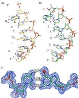
Figure 1.6 The global and local structures of the 4-Se-T DNA [(5’-GdUSe-G-SeT-ACAC-3’)2]. (A) The duplex
Figure 1.7 The superimposed global and local structures of 6-Se-G containing DNA/RNA duplexes of the nucleic acid–protein complex. a) The structure of the Se-DNA sequence (2R7Y, in yellow) is superim-posed over the corresponding native DNA (2G8U, in grey). b) The structure of the RNA sequence (2R7Y, in green) is superimposed over the corresponding native (2G8U, in grey). c) The Se-G3/C5 base pair (2R7Y) with the experimental electron density shows three H-bonds (exo-6-Se/exo-4-NH2, 1-NH/N(3),
and exo-2-NH2/exo-2-O) with bond lengths of 3.48, 3.16, and 2.59Å, respectively.
The crystal structures of the selenium-modified nucleic acids provide lots of useful structural
in-formation on nucleic acids. The DNA crystal structures containing 4-Se-T24 and 6-Se-G37 indicated that
the selenium modifications generated no obvious structural perturbation comparing with the native
DNA structures (figure 1.6 and figure 1.7), and that a novel hydrogen bond between the selenium atom
and the amino group (Se…H-N) was formed. In addition, unique structural and functional properties of
the selenium-modified nucleic acids were discovered. The corresponding melting temperature studies
Unexpectedly, we found another useful advantage of the selenium modifications: the
crystalliza-tion facilitacrystalliza-tion (figure 1.8), especially by the 2’-Se derivatizacrystalliza-tion. The exciting fact is that with the
sele-nium functionality at the 2’-position of sugar, nucleic acids (especially DNAs) are able to crystallize much
faster under broader buffer conditions and with higher diffraction qualities52. One example is the
appli-cation of 2’-SeMe-dU. The DNA oligonucleotide (GdU2’-SeGTACAC)2 crystallized in a few days from 20 out
of the 24 buffer conditions, while the corresponding native DNA (GTGTACAC)2 did not crystallize over 2-3
months53,54. The crystallization condition of the native DNA is quite narrow. Obviously, the selenium
derivatization is advantageous.
Figure 1.9 Time-course enzymatic digestion of phosphoroselenoate RNA with snake venom phosphodiesterase I. (a) gel electrophoresis autoradiography; (b) the digestion Vs time plot.
Selenium modifications result in nucleic acids with many additional unique properties,
particu-larly useful in biochemical and biological investigations. For instance, the nucleoside triphosphates
derivatized with selenium at non-bridging α-position of phosphate were synthesized, and their two
diastereomers were separated45. It was demonstrated that only one of these two Se-modified
triphos-phate diastereomers is a good substrate for T7 RNA polymerase in vitro transcription, while the other
diastereomer is not recognized by the enzyme: neither as a substrate nor an inhibitor44. On contrary,
DNA polymerase recognizes both diastereomers43. The recognition differences of the NTPs and
Se-dNTPs can be used to study the catalysis and substrate interactions of DNA and RNA polymerases.
An-other discovery is that the Se-hammerhead ribozymes transcribed with the nucleoside
Figure 1.10 The time-course experiment of incorporating TTP and 4-SeTTP into DNA. (A): the gel electro-phoresis; (B): the plot of the TTP and SeTTP incorporation into DNA.
display a low or no activity depending on the Se-modified nucleotides, indicating their participation in
the catalysis. These Se-NTPs offer auseful methodology for rapid screening of ribozymes and other
non-coding RNAs. Furthermore, it is found that both phosphoroselenoate DNAs and RNAs resist nuclease
digestion, which is especially useful in developing Se-oligonucleotide therapeutics (figure 1.9).
Moreo-ver, the triphosphate with the Se-derivatization at the thymidine 4-postion was also synthesized,
recog-nized by DNA polymerase, and incorporated into DNAs (figure 1.10). The enzymatic incorporation
effi-ciency was as good as native TTP. Interestingly, this replacement of a single oxygen atom with selenium
in the nucleobase generates yellow-colored DNA, which will be used as a unique probe for the
enzymat-ic assay46 (figure 1.11).
Due to the selenium toxicity and limitation of investigation strategies, in the past, the selenium
modification was not significantly explored in drug discovery research, compared to the sulfur
modifica-tion and gene therapy. Since recently selenium has been recognized as an essential element, drug
dis-covery with the selenium modifications becomes an opportunity. There is no reason that all the
ap-proaches applied in the S-derivatized gene therapy cannot be applied in the Se-derivatized gene
thera-py. As an element from the same elemental family as sulfur, selenium-functionalized nucleosides,
nu-cleotides and nucleic acids may possess ideal stabilities, and chemical and biological functions.
Further-more, the special electronic and dimensional characteristics of selenium generate unique advantages,
making SeNA an advanced approach for exploring disease mechanisms and treatments at the atomic
level. As discussed previously, the Se-derivatization of nucleic acids is an advanced tool for structure
de-termination. In Huang’s lab, a ternary complex with 6-Se-G containing DNA, RNA and RNase H protein
was crystallized, and their crystal structure was determined37. This is the first example to solve the
pro-tein structure with SeNA facilitation, and it opened a gate for the application of selenium-derivatized
nucleic acids in drug design and discovery. Another advantage of SeNA is its resistance to nuclease in
vivo or in vitro, which is the essential requirement for the oligonucleotide therapy. Similar to the
thio-modified antisense oligonucleotides, we have demonstrated recently that the Se-thio-modified nucleic acids,
including the modifications on the backbone, sugar and nucleobase, resist nuclease degradation. It is
believed that this novel SeNA can also play important roles in drug discovery.
O HO N N NH2 O O HO N NH O O O HO N NH O O N3
ddC d4T AZT
Another important application of selenium in therapeutic development is the synthesis of
cleoside analogues as potential anticancer, antimicrobial or antiviral drugs. Most of the therapeutic
nu-cleosides contain chemical modifications disrupting nucleic acid synthesis or damage repair. Successful
examples are the compounds used to block the human immunodeficiency virus replication in order to
cure AIDS. The best-known approved drugs (figure 1.12) include 3’-azido-3’-deoxythymidine (AZT),
2’,3’-dideoxycytidine (ddC), 2’,3’-dideoxyadenosine (ddA), and 2’,3’-didehydro-3’-deoxythymidine (d4T). Their
efficient synthesis is critical in medicinal chemistry and drug manufacture. Researchers have developed
some seleno-mediated synthesis routes to make reactions simple and efficient. Selenium functionalities
were introduced to the nucleosides, in order to generate 2’, 3’-unsaturated nucleosides. The
phenylselenyl group was the most frequently used. Several laboratories have achieved thesyntheses of
the 2’,3’-dideoxy- and 2’,3’-didehydro-2’,3’-dideoxynucleosides55-57. After the synthesis of the
2’-phenylselenenyl nucleosides, 2’,3’-unsaturated nucleosides were created by treatment with H2O2 or n
2. MATERIAL AND EXPERIMENTAL PART
2.1 Synthesis of 5-Selenium-Thymidine Nucleoside and DNA for Structural and Enzymatic Study
2.1.1 Introduction
Hydrogen bonds are formed between hydrogen-bond acceptors and donors (X-H). In a classical
hydrogen bond, X is an atom with strong electron-negativity (oxygen, nitrogen, etc.). However, recently
hydrogen bonds where X is an atom with weak electron-negativity (e.g., carbon) are gaining more
ac-ceptance and importance, for instance carbon in C-H···O=C hydrogen bond58-61. The interactions
between C-H and hydrogen-bond acceptors (electron donors), such as C-H···O=C hydrogen bond in
proteins58, C-H···O=C in uracil crystal59, and C-H···Cl in a guest-host system62, and other
non-conventional interactions (such as H···πinteraction in RNA)63have played critical roles in molecular
recognition, catalysis, and DNA duplex stability within chemical and biological systems64-67. Since a
nega-tively charged phosphate group is an excellent electron donor and the phosphorylation and
dephosphorylation are common cellular regulation mechanisms, we investigated whether a C-H (or CH3)
group is capable of forming a hydrogen bond with a phosphate group.
In DNA duplexes52,68,69, the 5-methyl group of thymidine is normally 4-5 Å away from the closest
oxygen (pro-Sp oxygen) of the 5′-phosphate group (O--PO3). In order to extend the CH3 group closer to
the phosphate and to give the CH3 more rotational flexibility at the same time, we inserted an atomic
linker between the methyl group and the C5 carbon of thymidine. If the CH3 and phosphate (O--PO3)
groups are able to form a genuine hydrogen bond, they willbe observed in a closer proximity and with
strong electron density between them in a crystal structure. If there is no such stabilizing interaction
between them, the methyl group would prefer to turn away from the phosphate due to the
steric-hindrance and point into the major groove. Since the size and geometry are essential requirements for
and C5 carbon. Herein we report the first synthesis of 5-Se-thymidine phosphoramidite (5, Scheme 2.1),
its chemical incorporation into DNAs,and the biophysical and structural studies of the DNAs containing
the Se-extended 5-CH3. Excitingly, we have discovered a novel methyl/phosphate hydrogen bond
(CH···O--PO3) for the first time.
On the other hand, gene therapies are kinds of important drug molecules, which generate low
toxicity, own high specificity and efficiency. The oligonucleotide drug molecule is supposed to exhibit
some advantages, like specific binding affinity to the target nucleic acid and high stability to nucleases in
vivo5,52. Nucleases, including DNases and RNases, are a class of enzymes that catalyze the hydrolysis of
phosphodiester linkage of nucleic acids, resulting in the degradation of single or double stranded nucleic
acid70. Nucleases’ activity is involved in many biological pathways, such as gene expression regulation in
siRNA and micro-RNA mechanisms71-73 or Okazaki fragment synthesis in genomic DNA replication74. A
crucial cellular role of DNases and RNases is to protect cells from foreign pathogenic DNAs and RNAs.
Thus, stability against endogenous nucleases is a prerequisite property for oligonucleotides based
ther-apeutics, such as antisense-DNA, siRNA, and micro-RNA. Currently the chemical modification is one of
the effective strategies to obtain the high stability under nuclease environment75. Numerous efforts
have been devoted to achieve chemically modified nucleic acid drug molecules, mainly through
derivatizing the sugar and the phosphate backbone moieties, without adversely affecting DNA/DNA and
DNA/RNA duplex formation. For instance, a number of DNA modifications, such as phosphorthioate76,
borano-phosphate77, PNAs78, LNAs79, and certain 2’-sugar modifications, including 2’-OMe80, 2’-ara-F81
and 2’-NH282, are among chemical approaches that showed enhanced DNA or RNA duplex stability
against nuclease digestion. The most successful example is the application of phosphorthioate
contain-ing gene therapy, which were approved by FDA as an antiviral drug in 199883,84. However, fewer reports
appear in the literatures that address the correlation of DNAs containing modified nucleobases and the
groove, a plethora of substituents of different sizes have been tethered at the 5-position of
2’-deoxyuridine, and the modified building block has been incorporated into DNA by solid phase synthesis
or enzymatically via the corresponding triphosphate derivatives86-88. In most cases, the wide open space
available in the major groove for the C5 substituent on 2’-deoxyuridine results in DNA/DNA and
DNA/RNA duplexes with higher or comparable thermal stability to the natural DNAs89. Despite of the
broad applications of 5-modified 2’-deoxyuridine containing nucleic acids and the spatial proximity of
the C5-substituents to the phosphodiester linkage90, few studies have reported on their stability against
nuclease digestion. Generally, nucleases use one or more divalent metal cation to activate the scissile
phosphate group and nucleophilic attack by a water molecule. For instance, HincII, a type II
endonucle-ase, utilizes a network of hydrogen bonds between the divalent metal cofactor, glutamate, aspartate
residues, water, and the phosphate group at the enzyme’s active site and thereby facilitate the
hydroly-sis process of the phosphordiester linkage91. We envisioned that an insertion of a single atom, selenium
(atomic radius 1.16 Å), at the 5-position of thymidine would extend the 5-CH3 group further toward the
scissile phosphate group and disturb the metal cation activation/H2O nucleophilic attack, thereby
allow-ing DNA duplexes with nuclease resistance. The geometry and the free rotation around the 5-Se-CH3
might retard the enzyme–duplex binding, the hydration pattern at the enzyme’s active site and thus
in-terrupt the phosphate hydrolysis. Moreover, the electron donating property of the selenium atom
would potentially enhance the DNA duplex stability by increasing the stacking interaction. These
fea-tures are important for oligonucleotides based therapies. In addition, our lab’s pioneering research has
shown that the Se-derivatization has a great potential for crystal structure determination of nucleic
ac-ids or nucleic acid-protein complex via MAD or SAD phasing9,92. Different from the synthesis above, we
report on the novel synthesis 5-MeSe-2’-deoxyuridine phosphoramidite using lithiating reagent in high
yield (Scheme 2.2), its chemical incorporation into DNAs, and as a proof of principle we demonstrated
and structural studies also showed no perturbation was generated by the selenium atom, which is
im-portant for the nucleic acid therapy. In order to compare the selenium functionality with other
chalcogen elements, we accomplished the synthesis of DNA with other modifications at 5-position of
thymidine as well, such as MeO-, MeS-, and PhSe-, applying similar strategy. We anticipate that the
thermostability, enzymatic and structural studies of the 5-chalcogen-modified thymidine containing
2.1.2 Synthesis Strategy and Structure Determination
The first trial to synthesize the 5-Se-thymidine phosphoramidite (5) started from 3′,5′-di-O
-benzyol-2′-deoxyuridine (1)93. Though the arylselenylation at the 5-position of pyrimidines was reported
over a decade ago29, the incorporation of alkylselenyl substitutions (such as CH3-Se) at the 5-position has
not been reported in the literature, probably due to the instability of the alkylselenyl intermediate. After
several attempts of screening Lewis acids as electrophile activators, Mn(OAc)3 was found effective to
promote the methylselenation at the 5-position of 1 with dimethyldiselenide CH3SeSeCH3. Treatment of
1 with CH3SeSeCH3 (in the presence of Mn(OAc)3 in AcOH at 90 °C) gave 5-Sethymidine derivative 2 in
good yield. NMR analysis of 2 showed the disappearance of the H-5 peak and the appearance of the H-6
singlet peak, and displayed the characteristic 1H and 13C chemical shifts of the 5-SeCH3 moiety at 2.06
and 7.24 ppm, respectively. Deprotection of 2 with NaOCH3 in MeOH gave 3 in a quantitative yield. UV
spectrum of 4shows λ max absorption at 309 nm, red-shifted by 44 nm when compared with thymidine
(Figure 3.1). This large red-shift is caused by the electron-donating effect of 5-SeCH3 on the nucleobase
π-system. Following the tritylation of 3, compound 4 was converted to 5 by the standard
O OBz BzO N NH O O
(CH3)2Se2 Mn(OAc)3
AcOH, 90oC, 36h 56% O OBz BzO N NH O O Se
H3C
O OH HO N NH O O Se
H3C NaOCH3 MeOH 93% O OH DMTrO N NH O O Se
H3C
DMTrCl, Py. 82% O O DMTrO N NH O O Se
H3C
P N
O
CN
i-Pr2NP(Cl)CH2CH2CN 5-BTT
DIEA CH2Cl2
81% O O O N NH O O Se
H3C
P O O O DNA P O
O O DNA
solid phase synthesis
1 2 3
4
5 6
Scheme 2.1Synthesis of 5-Se-T phosphoramiditeand oligonucleotides.
Syntheses strategies: a mixture of 3’,5’-di-O-benzoyl-2’-deoxyuridine (1) (1.89 g, 4.35 mmol),
Mn(OAc)3 (3.45 g, 13.0 mmol), and CH3SeSeCH3 (1.23 mL, 13.0 mmol) in glacial AcOH (30 mL) was heated
for 36 h at 90 oC. The mixture was cooled down to room temperature and the insoluble material was
filtered off on a Celitepad, and washed with EtOAc. The filtrate was evaporated, and co-evaporated with
toluene (25mL x 3 times). The residue was purified by flash column chromatography (SiO2: 15 % EtOAc in
CHCl3) to give 2 (1.3 g, 56%) as a white solid, (eluate: 25 % EtOAc in CHCl3) to recover 1 (0.75 g, 40%) as a
white solid. Our NMR analysis showed the disappearance of H-5 peak and appearance of H-6 singlet
peak, and displayed the characteristic 1H and 13C resonance signals 5-SeCH3 moiety of 2 at 2.06 and 7.24
ppm, respectively. Spectral Data for 2: UV λmin= 307 nm (ε = 4700 M-1cm-1 in MeOH), and λmax= 263 nm
(ε = 9650 M-1cm-1 in MeOH); 1H-NMR (CDCl3) δ: 9.29 (1H, s, NH, exchanged with D2O), 8.11-8.03 (4H, m,
Bz), 7.76 (1H, s, H-6), 7.61-7.58 (2H, m, Bz), 7.50-7.45 (4H, m, Bz), 6.42 (1H, dd, H-1’, J = 5.6, J = 8.8 Hz),
14.4 Hz), 2.36 (1H, m, H-2’b), 2.06 (3H, s, SeCH3); 13C-NMR (CDCl3) δ: 166.12 (C=O, Bz), 166.15 (C=O,
Bz), 161.64 (C4), 150.35 (C2), 140.05 (C-6), 133.90 (Ph, Bz), 133.78 (Ph, Bz), 129.97 (Ph, Bz), 129.78 (Ph,
Bz), 129.38 (Ph, Bz), 129.11 (Ph, Bz), 128.90 (Ph, Bz), 128.76 (Ph, Bz), 104.69 5), 85.71 (C4’), 83.16
(C-1’), 75.13 (C-3’), 64.51 (C-5’), 38.55 (C2’), 7.24 (SeCH3); HRMS (ESI-TOF): Molecular formula,
[image:41.612.88.396.208.447.2]C24H22N2O7Se; [M-H]+: 529.0505 (calc. 529.0514).
Figure 2.1 UV spectra of 5-Se-T (red) and thymidine (blue) in MeOH.
To a solution of 2 (0.827 g, 1.56 mmol) in MeOH (10 mL) was added 1MNaOMe in MeOH (1.6
mL). The mixture was stirred for 3 h at room temperature. The mixture was neutralized with Dowex 50
(H+) and the resin was filtered off. The filtrate was evaporated and the residual solid was purified by
flash silica gel column (eluate: 7 % MeOH in CH2Cl2) to give 3 (0.466 g, 93%) as a white solid, which was
crystallized from EtOH: Spectral Data for 3: UV λmax= 309 (ε =2000 M-1cm-1 in MeOH); 1HNMR (DMSO-d6)
δ: 11.52 (1H, s, NH, exchanged with D2O), 7.83 (1H, s, H-6), 6.18 (1H, t, H-1’, J = 6.4 Hz), 5.23 (1H, brd,
3’-OH, exchanged with D2O), 5.10 (1H, brt, 5’-OH),4.26 (1H, m, H-3’), 3.80 (1H, m, H-4’), 3.60 (2H, m, H5’a,
104.03 (C-5), 87.53 (C4’), 84.56 (C-1’), 70.40 (C-3’), 61.08 (C-5’), 38.90 (C2’), 5.25 (SeCH3); HRMS
(ESI-TOF): Molecular formula, C10H13N2O5Se; [M]+: 320.9996 (calc. 320.9990).
2’-deoxy-5-methylselenyluridine (3) (0.239 g, 0.74 mmol) was co-evaporated with dry pyridine
(5 mL x 3 times). The dried residue was dissolved in dry pyridine (3 mL) and cooled to 0 oC and treated
with a solution of 4,4’-dimethoxytrityl chloride (0.277 g, 0.82 mmol) in dry pyridine (3 mL). The mixture
was stirred for 3 h at room temperature. The solvent was evaporated and the residue was partitioned
between EtOAc and H2O. The organic phase was dried over MgSO4 and evaporated. The residue was
pu-rified by silica gel column chromatography (SiO2 was preequalized with 1% Et3N in CH2Cl2, eluate 4%
MeOH in CH2Cl2) to give (0.380 g, 82%) of 4 as pale yellow foam: 1H-NMR (CD2Cl2) δ: 9.39 (1H, s, NH,
ex-changed with D2O), 7.90 (1H, s, 6), 7.46-7.21 (9H, m, DMTr), 6.87-6.84(4H, m, DMTr), 6.29 (1H, dd,
H-1’, J = 6.4, J = 7.6 Hz), 4.43 (1H, m, H-3’), 4.05 (1H, m, H-4’), 3.68 (6H, 2 s, OMe), 3.30 (1H, dd, H5’a, J =
3.8, J = 10.5 Hz), 3.24 (1H, dd, H5’b, J = 3.6, J = 10.5 Hz), 2.58 (1H, d, 3’-OH), 2.42 (1H, ddd, H- 2’a, J = 3.8,
J = 7.7, J = 10.8 Hz), 2.36 (1H, m, H-2’b), 1.90 (3H, s, SeCH3); 13C-NMR (CD2Cl2) δ: 162.48 (C4), 159.29 (Ar),
150.94 (C2), 145.27 (Ar), 141.81(C-6), 136.21 (Ar), 136.06 (Ar), 130.64 (Ar), 130.62 (Ar), 128.57 (Ar),
128.50 (Ar), 127.47 (Ar), 113.76 (Ar), 104.21 (C-5), 87.33 (CAr3), 86.82 (C4’), 85.89 (C-1’), 72.82 (C-3’),
64.24 (C-5’), 55.78 (OCH3), 41.55 (C2’), 7.44 (SeCH3); HRMS (ESI-TOF): Molecular formula, C31H32N2O7Se;
[M+Na+]+: 647.1274 (calc. 647.1272).
Disopropylethylamine (20 μL, 0.12 mmol) was added to a solution of 4 (0.1 g, 0.16 mmol),
5-(sbenzylthio)-1H-tetrazole (0.002 g, 0.008 mmol) and 2-cyanoethyl-N,N,N,N-tetraisopropyl phosphane
(96 mg, 0.32 mmol) in dry CH2Cl2 (5 mL) at 0 oC. The mixture was stirred for 2 h at room temperature
then slowly poured into pentane (100 mL). The produced white precipitate was filtered off, dissolved in
CH2Cl2 (1 mL), and precipitated in pentane. The collected fine powdered white solid was dissolved in
CH2Cl2 and dried under reduced pressure to give 5 (108 mg, 82%) as a mixture of two diasteromers and
(eluate: 30% EtOAc in CH2Cl2) to give a mixture of two diasteromers: 1H-NMR (CD3CN, two sets of signals
for a mixture of two diasteromers) δ: 9.16 (1H, s, NH, exchanged with D2O), 7.76 and 7.17 (1H each, s,
H-6), 7.52-7.22 (9H, m, DMTr), 6.95-6.82 (4H, m, DMTr), 6.19 (1H, dd, H-1’), 4.57 (1H, m, H-3’), 4.10 and
4.05 (1H, m, H-4’), 3.75 (6H, 2 s, OMe), 3.65 and 3.55 (m, CH-ipr), 3.30 (2H, m, H5’a and H5’b), 2.63-2.51
(2H, dd, CH2), 2.47-2.31 (1H, H-2’a and H-2’b), 1.96 (3H, s, SeCH3), 1.17-1.03 (2x 24H, m, CH3-iPr);
13C-NMR (CD3CN, two sets of signals for a mixture of two diasteromers) δ: 162.63 (C4), 159.82 (Ar), 151.21
(C2), 146.01 (Ar), 141.65 and 141.57 (C-6), 136.84 (Ar), 136.79 (Ar), 136.73 (Ar), 132.30 (Ar), 131.20
(Ar),131.17 (Ar), 131.15 (Ar), 129.76 (Ar), 129.12 (Ar), 127.98 (Ar), 114.20 (Ar), 104.44 and 104.32 (C-5),
118.80 and 118.38 (CN), 87.52 (CAr3), 86.38 and 86.34 (C4’), 86.14 and 86.08 1’), 74.54 and 74.37
(C-3’), 64.41 and 64.23 (C-5’), 59.64 and 59.45 (OMe), 44.15 and 44.03 (C2’), 40.47,40.43, 40.37, 40.32
(CH-iPr), 24.99, 24.93 and 24.86 (CH3-iPr), 21.13, 21.06, 20.99 (CH2), 7.09 and 7.05 (SeCH3); 31P-NMR
(CD3CN) δ: 147.95, 147.92; HRMS (ESI-TOF): Molecular formula, C40H49N4O8PSe; [M-H]+: 823.2374 (calc.
823.2375).
Another novel incorporation of selenium derivatization into 5-position of 2’-deoxyuridine was by
using lithiating reagent in high yield (Scheme 2.2), and as a proof of principle we demonstrated their
stability against nucleases such as AseI and SalI-endonucleases, and exonuclease III. The synthesis
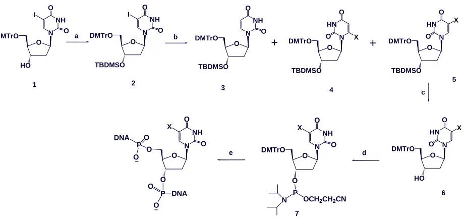
start-ed from the protection of 3’-hydroxyl group of 5-iodo-5’-DMTr-2’-deoxyuridine (1). After the protection
by TBDMS group, selenyl or thio functionality was introduced into 5-position. Methods available in the
literature for the introduction of non-carbon substitiuents at the 5-position of uridine or 2’-deoxyuridine
include electrophillic addition-elimination to the C5-C6 double bond94, palladium catalyzed nucleophillic
substitution of 5-mercururio derivatives89, and lithiation of 5-halo derivatives in the presence of the
electrophilic species95. The milder reaction condition of the later procedure led us to examine its
versa-tility for the synthesis of the novel 5-methylselenyl-2’-deoxyuridine derivative (5, X=CH3Se,Scheme 2.2).
reported for the synthesis of variety of 5-substituted pyrimidine nucleosides. However, a common
drawback for these reactions is the formation of the reduction byproduct, deoxyuridine derivatives. A
high C-5/C-6 regioselectivity is observed in the case of uridine derivatives due in part to the steric effects
induced by the 2’-hydroxyl’s protecting group96. For deoxyuridine derivatives which lacks the
2’-hydroxyl group, lithium/halogen exchange of 5-halo-2’-deoxyuridine usually produces high C-5
regioselectivity97. A protection of the N3-imido moiety 5-halo-2’-deoxyuridine is required to eliminate
the N3-H competition for the lithiation agent and thus results in high C5 lithiation yield95,98. We
envi-sioned that applying a transient N3-imido moiety protection of 5-halo-2’-deoxyuridine derivative (2)
fol-lowed by lithium-halogen exchange and quenching with (CH3)2Se2 would provide efficient access to 5.
Treatment of 5-iodo-2’-deoxyuridine derivative 2 with NaH (1.5 equiv), n-BuLi (2.2 equiv) and excess of
(CH3)2Se2 gave, however, an inseparable mixture of 5-methylselenyl derivative 5 and the corresponding
6-methylselenyl derivative 4 in 7:1 ratio (entry 1, Table 2.1) along with the reduced byproduct derivative
3 (Scheme 2.2). In attempt to improve the C5/C6 regioselectivity, a bulkier lithiating agent, LiHMDS was
utilized (entry 5, Table 2.1), however no lithaition occurred under these conditions. Lithiation using sec
-BuLi, or tert-BuLi gave similar results to n-BuLi in terms of C-5/C-6 regioselectivity (entries 3 and 4, Table
2.1). We next examined the effect of the reaction concentration on the C-5/C-6 regioselectivity, at
high-er concentration (0.15 M and 0.2 M) with n-BuLi as the lithiating agent (entries 6 and 7, Table 2.1), the
methylselenylation occurred exclusively at the C-5 position, yet the reduced byproduct 3 was isolated in
about 20% yield. Apparently, at higher concentration, the rate of lithium/halogen exchange at the
5-position is faster, discouraging the formation of the 6-lithio intermediate and thus results in the
ob-served high regioselectivity. We also found that decreasing the molar equivalence of NaH to (ca. 1.05
equiv) substantially eliminates the formation of the reduced product 3 (entry 8, Table 2.1).
pro-vided the building block 7 for solid phase synthesis (Scheme 2.2). Same reaction conditions were applied
as well to introduce CH3S and PhSe groups into 5-position of compound 2.
O HO DMTrO N NH O I O O TBDMSO DMTrO N NH O O O TBDMSO DMTrO HN N O O 3 O O DMTrO N NH O X O P OCH2CH2CN N 7 c O O O P DNA O O P O O DNA b d e 4 N NH O X O a O TBDMSO DMTrO N NH O I O + O TBDMSO DMTrO HN N O O X + X O HO DMTrO HN N O O X
1 2 5
6
Scheme 2.2 Synthesis of 5-chalcogen-T DNAs. Reagents and conditions: a) TBDMSCl, Im., DMF, 4 h, rt, 92 %; b) i) NaH, THF, 15 min, r.t., ii) lithiating agent (table 1), THF, 30 min., -78 °C, iii) X2, 1 h, -78 °C; c)
TBAF, THF, 4 h , rt, 92%; d) i-Pr2NP(Cl)CH2CH2CN, DIEA, CH2Cl2, 1 h, rt, 82%; e) solid phase synthesis.
X=CH3Se, CH3S or PhSe.
Table 2.1 Transient N3 protection and lithium/halogen exchange of 2 in the presence of
dimethyldiselenide.a
entry NaHb (eq.)
lithiating agentc(eq.)
concen-tration (M)
ratiod 5:4
yielde % 5+4 3
1 1.5 n-BuLi (3) 0.1 7:1 71 20 2 1.5 n-BuLi (1.2) 0.1 10:1 73 15 3 1.5 sec-BuLi (2.2) 0.1 11:1 68 23 4 1.5 tert-BuLi (2.2) 0.1 10:1 65 21 5 1.5 LiHMDS (3) 0.1 -f - - 6 1.5 n-BuLi (2.2) 0.15 35:1 75 20 7 1.5 n-BuLi (2.2) 0.2 only 5 71 19 8 1.05 n-BuLi (2.2) 0.2 only 5 85 5
a) All reactions were performed in the presence of 4 equivalents of (CH3)2Se2; b) Deprotonation of the
N3-H was performed at ambient temperature for 30 min; c) Lithium/halogen exchange was performed at
-78 ºC for 30 min. d) The ratio was determined by the integration of the H-1’ of the 1H-NMR of the mix-ture. e) Isolated yields after silica gel chromatography. f) The starting material was recovered.
Syntheses strategies: the new strategy to synthesize 5-Se-T started from
[image:45.612.78.538.123.341.2](35.5 mg, 1.4 mmol) was added portion wise to a solution of 2 (1.03 g, 1.33 mmol) in dry THF (7 mL) at
room temperature in dry glove box. The mixture was stirred for 30 min until complete cease of
hydro-gen gas evolution, then cooled down to -78 oC and treated with 1.4 M solution of n-BuLi in hexanes (2.1
mL, 2.93 mmol) was added dropwise over 10 min. The mixture was stirred for 30 min then treated with
(CH3)2Se2 (0.5 mL, 5.32 mmol) and the mixture was further stirred for 1 h at the same temperature.
Sat-urated solution of NH4Cl (5 mL) was added and the mixture was warmed to room temperature.
Ethylacetate was added to the mixture and the whole was washed with H2O, brine, dried over MgSO4,
and evaporated under reduce pressure. The residue was purified by flash silica gel chromatography
(eluate: 20% EtOAc in hexanes) gave 5 (0.83 g, 85%) as a colorless foam. Elution with 25% EtOAc in
hex-anes gave 3 (45 mg, 5%) as a colorless foam. Spectral Data for 5: 1H-NMR (CDCl3
exchanged with D2O), 7.99 (1H, s, H-6), 7.47-7.22 (9 H, m, Ar), 6.87-6.84 (4H, m, Ar), 6.31 (1H, dd, H-1’, J
= 5.9, J = 7.6 Hz), 4.46 (1H, m, H-3’), 4.02 (1H, m, H-4’), 3.81 (6H, s, CH3O), 3.41 (1H, dd, H-5’a, J = 3.2, J =
10.7 Hz), 3.31 (1H, dd, H-5’b, J = 3.4, J = 10.7 Hz), 2.39 (1H, ddd, H-2’a, J = 2.6, J = 5.8, J = 13.2 Hz), 2.19
(1H, m, H-2’b), 2.05 (9 H, s, tert-Butyl), 0.05 (3H, s, CH3), 0.01 (3H, s, CH3); 13C-NMR (CDCl3
(C4), 150.09 (C2), 141.57 (C-6), 135.60 (Ar), 135.54 (Ar), 130.11 (Ar), 128.12 (Ar), 127.95 (Ar), 127.00
(Ar), 113.28 (Ar), 103.56 (C-5), 86.86 (C4’), 85.55 (C-1’), 72.54 (C-3’), 63.13 (C-5’), 55.25 (OMe), 41.72
(C2’), 25.74 (CMe3),17.95 (CMe3), 7.30 (SeCH3), -4.69 (SiMe2), -4.86 (SiMe2); HRMS (ESI-TOF): Molecular
formula C37H45N2O7SeSi [M-H]+: 737.2147 (calc.737.2161).
A 1 M solution of TBAF in THF (1.55 mL) was added to a solution of 5 (0.75 g, 1.02 mmol) in THF
(15 mL) at 0 oC. The mixture was stirred for 6 h at room temperature. The solvent was evaporated and
the residue partitioned between EtOAc and H2O. The organic phase was dried over MgSO4 and
evapo-rated the residue was purified by silica gel column chromatography (the silica gel was pre-equalized with
1% Et3N in CH2Cl2, eluate 4% MeOH in CH2Cl2) to give (0.58 mg, 92%) of 6 as pale yellow foam: 1H-NMR
![Figure 1.6 The global and local structures of the 4-Se-T DNA [(5’-GdUSe-G-SeT-ACAC-3’)2]](https://thumb-us.123doks.com/thumbv2/123dok_us/9222749.990214/28.612.74.379.279.514/figure-global-local-structures-dna-gduse-set-acac.webp)


![Figure 3.4 Global and local structures of the 5-Se-T-DNA [(5′-GdU2′-Se-G-5-SeT-ACAC-3′)2]](https://thumb-us.123doks.com/thumbv2/123dok_us/9222749.990214/90.612.88.355.272.504/figure-global-local-structures-dna-gdu-set-acac.webp)