0095-1137/06/$08.00⫹0 doi:10.1128/JCM.00732-06
Copyright © 2006, American Society for Microbiology. All Rights Reserved.
Expertise of Laboratories in Viral Load Quantification, Genotyping,
and Precore Mutant Determination for Hepatitis B Virus
in a Multicenter Study
Syria Laperche,
1* Vincent Thibault,
2Franc¸oise Bouchardeau,
1Sophie Alain,
3Sandrine Castelain,
4Michelle Gassin,
5Marie Gueudin,
6Philippe Halfon,
7Sylvie Larrat,
8Franc¸oise Lunel,
9Miche
`le Martinot-Peignoux,
10Bernard Mercier,
11Jean-Michel Pawlotsky,
12Bruno Pozzetto,
13Anne-Marie Roque-Afonso,
14Franc¸oise Roudot-Thoraval,
15Karine Saune
´,
16and Jean-Jacques Lefre
`re
17,18Centre National de Re´fe´rence pour les He´patites B et C en Transfusion, De´partement des Agents Transmissibles par le Sang,
Institut National de la Transfusion Sanguine, Paris,1Laboratoire de Virologie, Centre Hospitalo-Universitaire Pitie´-Salpeˆtrie`re,
Paris,2Laboratoire de Virologie-Bacte´riologie, Centre Hospitalo-Universitaire, Limoges,3Laboratoire de Virologie,
Centre Hospitalo-Universitaire, Amiens,4Laboratoire de Virologie, Centre Hospitalo-Universitaire, Nantes,5
Laboratoire de Virologie, Centre Hospitalo-Universitaire, Rouen,6 Laboratoire Alphabio, Marseille,7
Laboratoire de Virologie, Centre Hospitalo-Universitaire, Grenoble,8Laboratoire de Virologie-Bacte´riologie,
Centre Hospitalo-Universitaire, Angers,9 Unite´ de Virologie, Inserm U 481 Centre Abrami,
Hoˆpital Beaujon, Clichy,10Laboratoire de Virologie Mole´culaire, Etablissement Franc¸ais du Sang,
Bretagne, Brest,11Laboratoire de Virologie-Bacte´riologie, Centre Hospitalo-Universitaire Henri-Mondor,
Cre´teil,12Laboratoire de Virologie-Bacte´riologie, Centre Hospitalo-Universitaire, Saint-Etienne,13
Laboratoire de Virologie, Hoˆpital Paul Brousse, Villejuif,14Service de Sante´ Publique,
Centre Hospitalo-Universitaire Henri-Mondor, Cre´teil,15Laboratoire de Virologie,
Centre Hospitalo-Universitaire Purpan, Toulouse,16Laboratoire d’He´matologie,
Centre Hospitalo-Universitaire, Amiens,17and De´partement des Agents Transmissibles
par le Sang, Institut National de la Transfusion Sanguine, Paris,18France
Received 6 April 2006/Returned for modification 16 May 2006/Accepted 22 June 2006
A national evaluation study was performed in 14 specialized laboratories with the objective of assessing their capacities to provide (i) hepatitis B virus (HBV) viral loads (VL), (ii) HBV genotypes, and(iii) identification of precore/core mutants. The panel consisted of 12 HBV DNA-positive samples with VLs from 2.8 to 9.1 log10 copies/ml, different HBV genotypes (A to F), and 3 mutant and 9 wild-type samples at nucleotide 1896. The coefficients of variation of the mean VLs ranged from 2.4% to 10.4% with the Cobas HBV Monitor assay, from 1.8% to 5.5% with the CobasTaqMan 48, from 1.5 to 26.2% with RealArt HBV PCR, and from 0 to 7% with branched DNA (bDNA). The Cobas Monitor assay underestimated the VLs of genotype F samples, with differences ranging from 1.4 to 2.4 log10copies/ml. The accuracies of genotype determinations ranged from 33% to 100%, and those of precore mutant determinations ranged from 25 to 100%. This study showed some drawbacks of two widely used assays: (i) Cobas Monitor has a narrow dynamic range and underestimates genotype F sample VLs and (ii) bDNA shows poor sensitivity and may fail to identify patients with low VLs. With higher performance in terms of analytical sensitivity combined with a larger dynamic range and an ability to quantify the main genotypes equally, real-time PCR methods appear more appropriate for accurate mon-itoring of HBV DNA quantification. Furthermore, the clinical implications of HBV genotyping and the determination of precore/core mutants need to be clearly stated to justify the standardization of these methods.
Hepatitis B virus (HBV) is responsible for chronic liver disease, with a risk of evolution toward severe complications, such as cirrhosis and hepatocellular carcinoma. The reservoir of chronically infected patients is estimated at 350 million worldwide. An increasing number of studies demonstrate the roles of several biological markers, together with biochemical indices (especially the alanine aminotransferase level), the HBV viral load (VL), the presence of precore/core promoter mutations (20) and drug resistance mutations (38), and
geno-type (31), in the individual’s response to treatment (15). It is expected that HBV VL measurement and HBV sequence poly-morphism determination will soon provide clinicians with im-portant information to adapt HBV-specific treatments.
Furthermore, the availability of molecular diagnostic tests for quantification of HBV DNA, especially by real-time PCR methods, is expanding. The main purpose of HBV VL deter-mination is to evaluate the activity of infection and to screen and monitor patients for antiviral treatments. These tools are also useful in diagnosing an HBV infection in some specific situations, such as the screening of blood donations to ensure blood safety with regard to the HBsAg silent phase (window period) (6) and occult HBV infection (37), as well as in cases of atypical serologic patterns involving an envelope mutant (57).
* Corresponding author. Mailing address: Centre National de Re ´f-e
´rence pour les He´patites B et C en Transfusion, De´partement des Agents Transmissibles par le Sang, Institut National de la Transfusion Sanguine, Paris, France. Phone: (33) 1 44 49 30 52. Fax: 1 44 49 30 59. E-mail: [email protected].
3600
on May 16, 2020 by guest
http://jcm.asm.org/
HBV replication kinetics and mechanisms, through a pre-genomic RNA intermediate and a reverse transcription step, are important sources of viral diversification. The spontaneous tendency of HBV to accumulate mutations, combined with natural selection pressure, have led to the emergence of eight described genotypes (A to H) defined by a divergence of at least 8% with the complete reference genome sequence (51, 53). It has also been demonstrated that these genotypes show distinct geographic distributions (51, 57). Furthermore, the occurrence of mixed-genotype infections does not seem un-common (14). To date, HBV genotype determination is mainly useful for epidemiological studies or for tracing a source of contamination. However, differences in the natural history of the disease according to genotype are being explored, and the clinical relevance of genotyping is emerging. Indeed, there is evidence that HBV genotypes influence the HBeAg serocon-version rate: Asian studies have especially demonstrated that genotype B-infected patients seroconvert more rapidly to anti-HBe than those infected with genotype C (33, 59). Further-more, genotype C seems to be linked to a more severe liver disease than genotype B in Far Eastern studies (13, 19), and the same observation was reported for genotype D versus ge-notype A in India (55). There are also indications in Europe that the infection outcome is more severe with genotype F than with genotype A or D (50). In addition, the genotype might influence the response to interferon-based treatments (31), with a globally better response for genotype B than C, espe-cially in Asian studies (17, 33, 52). The influence of genotype in the response to nucleoside or nucleotide analogues is probably less important. The clinical significance of precore and core promoter variants associated with HBeAg-negative liver dis-ease is more controversial due to the many host and viral factors. However, HBeAg-negative chronic hepatitis B seems to be associated with a more severe liver disease with a very low rate of spontaneous disease remission and a low sustained response rate to antiviral therapy (40).
Considering the emerging clinical relevance of all of these markers, and before initiating large-scale studies, the Action Coordonne´e 11 group of the Agence Nationale de Recherches sur le SIDA et les he´patites B et C initiated an evaluation of HBV VL determination and HBV-genotyping and precore mutation determination methods used in specialized laborato-ries involved in multicenter clinical trials. The aim of this study was to assess the capacities of these specialized laboratories to provide data on HBV infection through molecular investiga-tions.
MATERIALS AND METHODS
Constitution of the panel and assays used for its selection.The panel included 12 plasma samples collected from HBV-infected untreated blood donors se-lected as a subset of subtypes with several ranges of viral loads. The character-istics of these samples are given in Table 1. The VL, the HBV genotype, and the presence of precore/core mutations were determined for each sample.
VL determination was performed twice in two separate runs, with two proce-dures used according to the manufacturers’ instructions: Cobas Amplicor HBV Monitor and the CobasTaqMan 48 Real-Time PCR System (Roche Molecular Systems, Meylan, France) (limits of detection [LOD], 200 and 35 copies/ml, respectively).
The genotype was determined twice in two separate runs with an in-house method based on the sequence analysis of HBV gene S, using a QIAamp DNA Minikit (QIAGEN, Courtaboeuf, France) for HBV DNA extraction, and a consensus PCR assay amplifying an S gene fragment comprised of nucleotides 256 to 725. Each PCR experiment included negative and positive controls. Double-strand direct sequencing was carried out by the dideoxynucleotide chain terminator method by MWG Biotech (Roissy, France). Several reference se-quences of HBV genotypes (A to H) drawn from GenBank were included in the data bank. All sequences were aligned using ClustalW software (56). Phyloge-netic analysis was performed using the Phylip package. Distances between se-quences were analyzed using the neighbor-joining algorithm based on the Kimura 2 parameter distance estimation method for the nucleotide sequences and on the Dayhoff PAM matrix for the amino acid sequences (48).
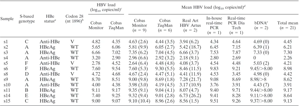
[image:2.585.43.542.80.257.2]Finally, the presence of precore/core mutations was studied twice in two separate runs using a commercially available line probe assay for identification of HBV precore variants (INNO-LiPA HBV Precore; Innogenetics, Gent, Bel-gium) according to the manufacturer’s instructions. For the purpose of the experiments, the recommendation that at least codon 28 (with the presence of TABLE 1. Characteristics of the 12 samples on the panel
Sample S-based genotype
HBe
statusa Codon 28
(nt 1896)b
HBV load (log10copies/ml)c
Mean HBV load (log10copies/ml)d
Cobas Monitor
Cobas
TaqMan
Cobas Monitor (n⫽9)
Cobas
TaqMan (n⫽6)
Real Art HBV Artus
(n⫽2)
In-house real-time PCR (n⫽1)
Real-time PCR Dia Tech (n⫽1)
bDNAe
(n⫽2)
Total mean (n⫽21)
s1 C Anti-HBe V 4.82 4.35 4.63 (2.6) 4.44 (3.5) 3.94 (6.2) 4.34 4.64 4.69 (0) 4.45 s2 A HBeAg WT 5.65 6.06 5.81 (9.9) 6.05 (2.7) 5.42 (18.7) 6.45 7.15 6.39 (1) 6.21 s3 A HBeAg WT 6.66 7.02 7.35 (6.2) 7.04 (4.5) 6.66 (3.7) 7.53 7.87 7.33 (0) 7.30
s4 A Anti-HBe WT 3.20 2.90 2.96 (6.6) 2.92 (3.2) 2.18 (9.1) 2.80 2.69 0 2.26
s5 F Anti-HBe V 2.78 4.52 2.64 (6.4) 4.48 (4.8) 4.08 (3.7) 4.54 4.48 5.03 (2) 4.21 s6 F HBeAg WT 7.60 9.54 7.60 (5.3) 9.30 (5.5) 8.46 (11.5) 9.83 9.21 9.45/⬎8.00 8.98 s8 D Anti-HBe V 4.72 4.68 4.67 (2.4) 4.47 (5.1) 4.41 (11.9) 4.53 3.45 4.98 (0) 4.42 s9 A HBeAg WT 8.70 8.51 9.00 (9.8) 8.69 (1.8) 7.28 (21.7) 9.08 8.69 8.98/⬎8 8.62 s10 A Anti-HBe WT 4.00 4.38 3.98 (3.0) 4.19 (4.5) 3.17 (10.9) 3.76 3.91 4.43 (7) 3.91 s11 B HBeAg WT 9.11 9.17 9.35 (9.1) 9.04 (4.1) 8.07 (4.7) 9.40 9.71 9.44/⬎8.00 9.17 s14 E HBeAg WT 7.40 9.25 9.32 (9.4) 9.01 (2.8) 6.73 (26.2) 9.41 8.28 9.11/⬎8.00 8.64 s15 C HBeAg WT 9.00 9.07 9.10 (10.4) 8.96 (2.6) 8.56 (1.5) 9.51 9.26 9.37/⬎8.00 9.13
a
Anti-HBe and HBeAg assays were performed with ETI-EBK antibody and ETI-EBK plus (Dia Sorin, Saluggia, Italy), respectively.
b
V, precore stop codon mutation (G to A at nucleotide关nt兴1896); WT, wild type as defined by Inno-Lipa.
c
VL was obtained in laboratory P, which prepared the panel.
d
Mean VLs were obtained in all participating laboratories (including laboratory P). % CV values are given in parentheses.
e
For samples for which the mean could not be determined, the VL values are presented separated by a slash.
on May 16, 2020 by guest
http://jcm.asm.org/
the G1896A point mutation) should be analyzed was made in order to cover this most prevalent mutant.
Participating laboratories.Fourteen laboratories (coded from A to N) par-ticipated in the study. All had to perform HBV VL determinations, while HBV genotyping and precore/core mutation determinations were optional.
For HBV VL determination, 9 of the 14 laboratories used one method, three used two methods, and one used three methods. Including the laboratory that had prepared the panel (laboratory “P”) and that retested the panel under the same conditions as the other participants, a total of 21 results of HBV VL determination were given and analyzed. The quantification methods consisted of Cobas HBV Monitor (Roche Molecular Systems) (LOD, 200 copies/ml; linear range, 200 to 2⫻105copies/ml; laboratories P, A, C, D, F, G, H, I, and M),
CobasTaqMan 48 (Roche Molecular Systems) (LOD, 35 copies/ml; linear range, 170 to 6.4⫻108copies/ml; laboratories P, B, J, K, L, and N), the RealArt HBV
PCR kit (Artus Abbott, Rungis, France) (LOD, 30 copies/ml; linear range, 30 to 3⫻1010copies/ml; laboratories H and I), HBV RealQuantTM PCR (DiaTech,
Jesi, Italy) (laboratory A), and a home-made HBV quantification real-time PCR method (LOD, 50 copies/ml; linear range, 50 to 5⫻108copies/ml; laboratory L).
Finally, two laboratories (E and L) used the branched-DNA (bDNA) signal amplification technology, the HBV bDNA assay (VERSANT HBV 3.0 Assay; Bayer HealthCare-Diagnostics, Tarrytown, New York) (LOD, 2,000 copies/ml; linear range, 2⫻103to 1⫻108copies/ml).
Ten laboratories provided HBV-genotyping results. Four used commercially available methods, three used the INNO-Lipa HBV-genotyping test (Innogenet-ics, Gent, Belgium), and one used the TrueGENE HBV-genotyping test (Bayer Diagnostics, Tarrytown, NY). The other six laboratories used an in-house S gene-sequencing method.
Nine laboratories provided results for the precore/core mutation determina-tion: three used the INNO-LiPA HBV precore assay (Innogenetics, Gent, Bel-gium), while six provided results obtained from a sequence analysis of the precore/core region of the genome.
Interpretation of the results.To compare the VL results, the data obtained from all of the laboratories were analyzed as a whole. For clarity of the report, all the results were converted into copies/ml (at the time of study, Roche Diag-nostics did not recommend expressing the results with Cobas Monitor as inter-national units) through the appropriate conversion factor provided by the man-ufacturers, with 1 international unit corresponding to 5.82 copies for the Roche
TaqMan assay, to 7.5 copies for the Artus method, to 7 copies for the Dia Tech method, and to 5.6 copies for the bDNA assay. Values converted into log10units
obtained for each panel sample were compared to the mean of all measurement values given by all participants using the same procedure. In addition, methods were compared by comparison of the mean log10VLs calculated for each sample.
A difference of more than 0.5 log10was considered significant. When a VL was
not determined with precision but was above the linear range of the assay, the result was excluded from the calculation of the mean values.
For the analysis of genotype and precore mutation results, the data from the participating laboratories were compared to those obtained in laboratory P.
RESULTS
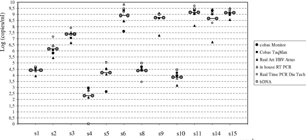
HBV viral load determination.For HBV VL determination, 10 of the 14 laboratories used a single method, 4 used two methods, and 1 used three methods. A total of 21 results of HBV DNA quantification were provided and analyzed. A sum-mary of the results obtained with the different assays is given in Table 1. Figure 1 represents the dispersion of mean HBV viral loads obtained with each assay in tested samples.
Nine measures of HBV VL obtained with Cobas Monitor were analyzed for all samples, except for samples s9, s11, s14, and s15, which were not diluted by one laboratory and there-fore were not precisely quantified (thus, eight measures were analyzed for these four samples). The coefficients of variation (CV) of the mean VL ranged from 2.4% to 10.4% (Table 1). There was no correlation between the mean VL and the CV. However, the highest CV tended to be observed for the highest VL. Compared with the other methods, the Cobas Monitor assay underestimated the VLs of genotype F samples, with differences ranging from 1.4 to 2.4 and from 0.86 to 2.2 log10
copies/ml for samples s5 and s6, respectively. The interlabora-tory comparison showed the tendency of laborainterlabora-tory F to give significantly higher VL values (from 0.63 to 2.14 log10copies/ml)
for samples with VLs above 7 log10copies/ml (n⫽7) (Fig. 1).
Significant differences were also observed for two samples quantified in laboratory A (the VL was overestimated, with differences of 0.68 [s6] and 0.59 [s11] log10copies/ml,
respec-tively) and in four samples (s9, s11, s14, and s15) from
labo-FIG. 1. Dispersion of HBV viral loads, expressed as means, obtained with each assay. The means of viral loads including all results for each sample are symbolized by linked circles.
on May 16, 2020 by guest
http://jcm.asm.org/
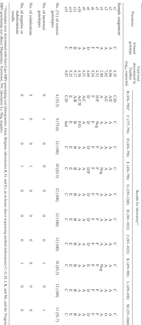
[image:3.585.43.541.69.297.2]TABLE 2. Performances of the laboratories in HBV genotyping Parameter S-based genotype Viral load determined by Taq Man (log 10 copies/ml) Results for laboratory a : B (456–798) b C (377–794) D (456–798) E (456–798) G (250–1280) H (301–1022) J (301–1022) K (459–988) L (458–830) M (253–1060) Sample assignment s1 C 4.35 C/D C C C C C C C C C s2 A 6.06 A/E A A A A A A A A A s3 A 7.02 A/C A A A A A A A A G s4 A 2.90 A A A Neg A A A Neg A A s5 F 4.52 D/F Neg F F F F F F F F s6 F 9.54 F F F F F F F F F F s8 D 4.68 D D D D/F D D D E D D s9 A 8.51 A/C A A A A A A A A A s10 A 4.38 A/C/E F / GA A A A A A A A s11 B 9.17 A/B B B B B B B B B B s14 E 9.25 E E E E E E E E E E s15 C 9.07 C/D Ind C C C C C C C C No. (%) of correct genotypes 4 (33.3) 9 (75.0) 12 (100) 10 (83.3) 12 (100) 12 (100) 12 (100) 10 (83.3) 12 (100) 11 (91.7) No. of incorrect genotypes 01 0 0 0 0 0 1 0 1 No. of coinfections 8 0 0 1 0 0 0 0 0 0 No. of negative or indeterminate results 02 0 1 0 0 0 1 0 0 a Genotypes were determined with Inno-Lipa HBV genotyping test (Innogenetics, Gent, Belgium; laboratories B, D, and E), an in-house direct sequencin g method (laboratories C, G, H, J, K, and M), and the Trugene HBV-genotyping test (Bayer Diagnostics, Tarrytown, NY; laboratory L). Neg, negative. b Position in the nucleotide sequence of the amplified segment in the S gene of HBV.
on May 16, 2020 by guest
http://jcm.asm.org/
[image:4.585.141.430.64.724.2]ratory I (underestimation, with differences from 0.54 to 0.70 log10copies/ml). The four samples involved in these
misclas-sifications had VLs above 7.6 log10copies/ml.
Three to six measures of VL determined with the Cobas
TaqMan were included in the calculation of the mean VL value, depending on the samples. Four and three measures were taken into consideration for samples s6 and s9 and for samples s11, s14, and s15, respectively. The CV of the mean VL ranged from 1.8% to 5.5%. Significant differences were observed for two samples (s6 and s11) that were diluted before being tested in laboratory J (the VL was overestimated, with differences of 0.69 and 0.77 log10copies/ml, respectively), and
two samples (s3 and s6) were underestimated by laboratories K and L, respectively (with differences of 0.62 and 0.54 log10
copies/ml). The four samples involved in these misclassifica-tions had VLs above 7.6 log10copies/ml.
The results obtained with the Artus procedure in two labo-ratories showed high CV values (from 1.5 to 26.2%). Six sam-ples were quantified with VL differences of more than 0.5 log10
(from 0.74 to 2.24 log10copies/ml). Moreover, the genotype E
sample was quantified with a significantly lower VL by com-parison to the other assays (1.9 log10-copy/ml difference from
the mean result).
Determinations of the VL performed with bDNA in two laboratories demonstrated very good reproducibility, with an extremely low CV (calculated for six samples) ranging from 0 to 7%, whatever the VL. However, s4, which had the lowest VL of the panel (2.9 log10copies/ml), was not detected by the two
laboratories using this method (Table 1).
Genotype determination.The accuracies of genotype deter-minations ranged from 33% to 100% (Table 2). Of the 10 laboratories that provided results, 5 correctly identified all samples; 3 gave an erroneous result for a unique sample; 2 gave the correct genotype for all samples but in association with one or two other genotypes in eight and one samples, respectively; 3 did not provide results for one or two samples due to a failure at the amplification or sequencing steps—twice for sample s4 (genotype A; 2.9 log10copies/ml) and once for
samples s5 and s15 (genotype F, 4.5 log10copies/ml, and
geno-type C, 9.0 log10copies/ml).
Of the 120 expected correct results, 104 (86.7%) were accu-rate. Among the 16 incorrect results, 9 involved mixed-geno-type results, 3 were erroneous genomixed-geno-types, and 4 were PCR amplification or sequencing failures. Only two samples (geno-types F and E, with VLs of 9.5 log10copies/ml in both cases)
were correctly identified by all of the laboratories.
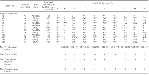
Precore mutants. The accuracies of precore mutant deter-minations ranged from 25 to 100% (Table 3). Of the nine laboratories that provided results, two correctly identified all of the tested samples (12/12 for laboratory L and 8/8 for labora-tory N), one gave erroneous results for 8 samples, one pro-vided a dual wild-type (WT) and 1896 variant (V) infection, and four missed 1 to 3 samples due to a failure in PCR am-plification, while two declared a failure in the identification of the 28 codon in 2 or 3 samples.
[image:5.585.43.543.80.333.2]Of the 108 expected correct results, 82 (76%) were accurate. Among the 26 incorrect results, 9 were erroneous results (V instead of WT in eight samples and one dual V-WT infection),
TABLE 3. Performances of the laboratories in the identification of precore mutants (position 1896)
Parameter S-based genotyping
HBe status
Viral load determined by
TaqMan (log10
copies/ml)
Results for laboratorya
:
Pb
Bb
Eb
Lb
Gc
Hc
Jc
Kc
Mc
Nc
Sample assignment
s1 C Anti-HBe 4.35 V V V V V V V V V V
s2 A HBeAg 6.06 WT V WT WT WT WT WT WT WT NT
s3 A HBeAg 7.02 WT V WT WT WT WT WT WT WT WT
s4 A Anti-HBe 2.90 WT WT Neg WT WT WT WT Neg Neg WT
s5 F Anti-HBe 4.52 V V V V Ind V Neg V V V
s6 F HBeAg 9.54 WT V WT WT Ind WT WT WT WT NT
s8 D Anti-HBe 4.68 V V NT V V V V V V V
s9 A HBeAg 8.51 WT V WT WT WT Ind WT WT WT WT
s10 A Anti-HBe 4.38 WT V WT WT Ind WT WT WT WT WT
s11 B HBeAg 9.17 WT V WT/V WT WT Ind WT WT WT WT
s14 E HBeAg 9.25 WT V WT WT WT WT WT WT Neg NT
s15 C HBeAg 9.07 WT V WT WT WT WT WT NT Neg NT
No. (%) of correct results
3/12 (25) 9/12 (75) 12/12 (100) 9/12 (75) 10/12 (83) 11/12 (92) 10/12 (83) 9/12 (75) 8/12 (67)
No. of false results 8 1 0 0 0 0 0 0 0
No. of negative or untested samples
0 2 0 0 0 1 2 3 4
No. of indeterminate results
0 0 0 3 2 0 0 0 0
aV, precore stop codon mutation (G to A at nucleotide 1896); Neg, negative; Ind, indeterminate; NT, not tested. bInnoLiPA HBV precore (Innogenetics, Gent, Belgium).
cIn-house direct sequencing method.
on May 16, 2020 by guest
http://jcm.asm.org/
12 were false-negative results or untested samples, and 5 were due to an indeterminate result.
DISCUSSION
Among the techniques described in recent years for quanti-fying HBV DNA by PCR (16, 24, 29), some have been based on semiautomatic assays and were described as providing good sensitivity for the detection of HBV DNA (39, 41). More recently, introduction of real-time PCR technology for HBV viral load measurement has offered even better characteristics, with an overall larger range of quantification and improved sensitivity (1, 5, 23, 26, 36, 58). Although these two technolo-gies have been assessed and compared within different single-center laboratories, an evaluation of such techniques through a multicenter quality control study was missing. The strength of such an evaluation is to provide information on what may happen to a patient who may be followed over time in different laboratories using in-house or commercially available tech-niques. In terms of the technical handling characteristics of the six studied HBV DNA quantification methods, one of the major inconveniences involved the Cobas Monitor PCR sys-tem, which has the narrowest dynamic range and requires dilution of samples in cases of high VL to artificially extend the test linearity. This drawback has been pointed out previously (43, 58). Due to the error rate linked to the dilution process, a weakness is introduced in the accuracy of the technique, espe-cially for active replicative HBV carriers with VLs that may reach over 9 log10copies/ml. It is likely that the highest CV in
interlaboratory comparison observed in our study with the Cobas Monitor for samples with a high VL reflects this phe-nomenon.
In the performances of the HBV DNA quantification meth-ods, we observed an underestimation of the VLs of genotype F samples with Cobas Monitor. Although genotype F is mainly present in Central and South America (4, 7), this failure of Cobas Monitor (58) may have an impact on the monitoring of HBV infection, particularly if the genotype is not determined before VL quantification. Thus, we recommend not using this assay for the monitoring of patients infected by a genotype F strain.
The Artus System tended to provide VL results lower than those of the other assays, especially for the genotype E sam-ples. However, the high CV observed between the results of the two laboratories that used this assay could suggest inter-laboratory variability. Further extended studies of the Artus Real Time PCR system (as well as the other real-time PCR methods used by only one participating laboratory) are needed to assess the performances of these new methods of HBV DNA quantification.
Although the observed differences were not significant com-pared with the other assays, the bDNA assay tended to give higher VL results, as previously reported (25). Moreover, the reproducibility rate was very high (46). However, due to its low sensitivity, linked to the principle of this assay (49), the bDNA assay had a high failure rate for the two samples of the panel with the lowest VLs.
The results obtained here and in other studies have demon-strated that the real-time PCR methods offer several advan-tages in terms of analytical sensitivity, with lower limits of
detection and larger dynamic ranges of quantification. The good performances of real-time PCR methods and the avail-ability of automatic platforms will justify the use of these tests for the diagnosis of HBV infection in the near future, espe-cially for atypical serological patterns, as in occult infection (2, 18), in HBsAg carriers with a low level of replication (22, 42), or for treatment monitoring in order to identify resistant mu-tants that could be difficult to demonstrate with methods ex-hibiting a low limit of detection.
Although correct results were obtained for 50% of the par-ticipants for the entire panel of HBV genotypes, the data analysis revealed a few discrepancies, essentially due to poor amplification sensitivities of the samples with the lowest VLs and sometimes due to misclassification. Indeed, the false-neg-ative results obtained with samples s4 and s5 were probably due to their low VLs rather than to their genotypes. The misclassification of samples s3 and s10, both containing a geno-type A strain and classified as G and F/G by two laboratories using an in-house method, may be explained by the close phy-logenic proximity of A and G genotypes in the amplified region (3, 54). However, the possibility that these two samples were really coinfected with the two genotypes cannot be excluded, since that seems to be the case for most subjects infected with genotype G. More surprisingly (and contrary to data provided by three other laboratories using the same technology), labo-ratory B reported 67% mixed infections with the Inno-Lipa method. This method has been shown to be very sensitive for identification of mixed-genotype infections, but the reported rate of mixed infections is usually between 0.1 and 10% (30, 45). Clearly, Inno-Lipa allows rapid detection of HBV geno-types in diagnostic laboratories, but extensive comparison stud-ies of this assay with cloning experiments are needed to estab-lish the reality of the mixed infections and to clearly exclude any issue of specificity.
Contrary to the results that we have obtained for HCV genotyping (35), the fact that no reference genome database was provided to all the participants did not seem to have influenced the genotyping results obtained by direct sequenc-ing. The reason for this finding is probably the absence of subtype definition for HBV, permitting a less precise phylo-genic analysis without consequence for the final genotyping classification.
In the precore/core region, the most common naturally oc-curring HBV mutations are G1896A, which creates a stop codon in the precore gene (9, 11), and an A1762T-G1764A dual mutation in the core promoter region, which is responsi-ble for a down-regulation of HBcAg production (9). Some of the participating laboratories did not routinely perform the determination of these mutations (only nine provided results). Besides laboratory B, which gave 75% incorrect results for an unknown reason (a nonspecific hybridization with the line probe assay could be involved), incorrect answers were mainly due to an absence of amplification or an indeterminate result independent of the method used. This is in accordance with the fact that the determination of precore/core mutations is not performed on a routine basis and that only a few laboratories are accustomed to the procedure. Indeed, the usefulness of identification of these HBV mutations is still being debated (10, 12, 32, 34). Moreover, the relationship between these mutants and the response to antiviral treatment remains
on May 16, 2020 by guest
http://jcm.asm.org/
troversial (9, 20, 28, 34) and could be due to other factors, such as genotypes or recruitment bias (21, 27, 44). Thus, it does not seem crucial to determine the existence of these mutations in order to evaluate the severity of the disease and to manage antiviral treatments.
One may conclude from this multicenter study that the large majority of expert laboratories routinely use commercial assays for HBV VL determinations (8, 47). The study also showed some drawbacks with two widely used assays: (i) Cobas Mon-itor has a narrow dynamic range and underestimates genotype F sample VLs and (ii) bDNA shows poor sensitivity and may therefore fail to identify patients with low VLs. With higher performance in terms of analytical sensitivity combined with a larger dynamic range and an ability to quantify the main ge-notypes equally, the real-time PCR methods appear more ap-propriate for accurate monitoring of HBV DNA quantification. Furthermore, the clinical implications of HBV genotyping, as well as the determination of precore/core mutants, need to be clearly stated to justify the standardization of these methods. A problem that could justify the intensive use of appropriate diagnostic tools would be the emergence of a high number of drug-resistant mu-tants.
ACKNOWLEDGMENTS
This work was supported by a grant from the Agence Nationale de Recherches sur le SIDA et les Hepatites B et C.
We thank Ve´ronique Descamps, Annie Girault, Joelle Lerable, Christine Portal, and Annabelle Servant-Delmas for their technical assistance.
REFERENCES
1.Aliyu, S. H., M. H. Aliyu, H. M. Salihu, S. Parmar, H. Jalal, and M. D. Curran.2004. Rapid detection and quantitation of hepatitis B virus DNA by real-time PCR using a new fluorescent (FRET) detection system. J. Clin. Virol.30:191–195.
2.Allain, J. P.2004. Occult hepatitis B virus infection: implications in trans-fusion. Vox. Sang.86:83–91.
3.Arauz-Ruiz, P., H. Norder, B. H. Robertson, and L. O. Magnius.2002. Genotype H: a new Amerindian genotype of hepatitis B virus revealed in Central America. J. Gen. Virol.83:2059–2073.
4.Arauz-Ruiz, P., H. Norder, K. A. Visona, and L. O. Magnius.1997. Genotype F prevails in HBV infected patients of Hispanic origin in Central America and may carry the precore stop mutant. J. Med. Virol.51:305–312. 5.Beuselinck, K., M. van Ranst, and J. van Eldere.2005. Automated extraction
of viral-pathogen RNA and DNA for high-throughput quantitative real-time PCR. J. Clin. Microbiol.43:5541–5546.
6.Biswas, R., E. Tabor, C. C. Hsia, D. J. Wright, M. E. Laycock, E. W. Fiebig, L. Peddada, R. Smith, G. B. Schreiber, J. S. Epstein, G. J. Nemo, and M. P. Busch.2003. Comparative sensitivity of HBV NATs and HBsAg assays for detection of acute HBV infection. Transfusion43:788–798.
7.Blitz, L., F. H. Pujol, P. D. Swenson, L. Porto, R. Atencio, M. Araujo, L. Costa, D. C. Monsalve, J. R. Torres, H. A. Fields, S. Lambert, C. Van Geyt, H. Norder, L. O. Magnius, J. M. Echevarria, and L. Stuyver.1998. Antigenic diversity of hepatitis B virus strains of genotype F in Amerindians and other population groups from Venezuela. J. Clin. Microbiol.36:648–651. 8.Brunetto, M. R., and F. Bonino.2004. Treatment of chronic hepatitis B: from
research to clinical practice via the consensus conferences. Curr. Pharm. Des.10:2063–2075.
9.Brunetto, M. R., M. Giarin, G. Saracco, F. Oliveri, P. Calvo, G. Capra, A. Randone, M. L. Abate, P. Manzini, M. Capalbo, et al.1993. Hepatitis B virus unable to secrete e antigen and response to interferon in chronic hepatitis B. Gastroenterology105:845–850.
10.Brunetto, M. R., M. M. Giarin, F. Oliveri, E. Chiaberge, M. Baldi, A. Alfarano, A. Serra, G. Saracco, G. Verme, H. Will, et al.1991. Wild-type and e antigen-minus hepatitis B viruses and course of chronic hepatitis. Proc. Natl. Acad. Sci. USA88:4186–4190.
11.Carman, W. F., M. R. Jacyna, S. Hadziyannis, P. Karayiannis, M. J. McGarvey, A. Makris, and H. C. Thomas.1989. Mutation preventing formation of hepatitis B e antigen in patients with chronic hepatitis B infection. Lancetii:588–591. 12.Chan, H. L., N. W. Leung, M. Hussain, M. L. Wong, and A. S. Lok.2000.
Hepatitis B e antigen-negative chronic hepatitis B in Hong Kong. Hepatol-ogy31:763–768.
13.Chan, H. L., M. L. Wong, A. Y. Hui, L. C. Hung, F. K. Chan, and J. J. Sung.
2003. Hepatitis B virus genotype C takes a more aggressive disease course than hepatitis B virus genotype B in hepatitis B e antigen-positive patients. J. Clin. Microbiol.41:1277–1279.
14.Chen, B. F., C. J. Liu, G. M. Jow, P. J. Chen, J. H. Kao, and D. S. Chen.2006. Evolution of hepatitis B virus in an acute hepatitis B patient co-infected with genotypes B and C. J. Gen. Virol.87:39–49.
15.Chen, C. J., H. I. Yang, J. Su, C. L. Jen, S. L. You, S. N. Lu, G. T. Huang, and U. H. Iloeje.2006. Risk of hepatocellular carcinoma across a biological gradient of serum hepatitis B virus DNA level. JAMA295:65–73. 16.Chen, T., J. M. Luk, S. T. Cheung, W. C. Yu, and S. T. Fan.2000. Evaluation
of quantitative PCR and branched-chain DNA assay for detection of hepa-titis B virus DNA in sera from hepatocellular carcinoma and liver transplant patients. J. Clin. Microbiol.38:1977–1980.
17.Chien, R. N., C. T. Yeh, S. L. Tsai, C. M. Chu, and Y. F. Liaw.2003. Determinants for sustained HBeAg response to lamivudine therapy. Hepa-tology38:1267–1273.
18.Conjeevaram, H. S., and A. S. Lok.2001. Occult hepatitis B virus infection: a hidden menace? Hepatology34:204–206.
19.Duong, T. N., N. Horiike, K. Michitaka, C. Yan, M. Mizokami, Y. Tanaka, K. Jyoko, K. Yamamoto, H. Miyaoka, Y. Yamashita, N. Ohno, and M. Onji.
2004. Comparison of genotypes C and D of the hepatitis B virus in Japan: a clinical and molecular biological study. J. Med. Virol.72:551–557. 20.Erhardt, A., U. Reineke, D. Blondin, W. H. Gerlich, O. Adams, T. Heintges,
C. Niederau, and D. Haussinger.2000. Mutations of the core promoter and response to interferon treatment in chronic replicative hepatitis B. Hepatol-ogy31:716–725.
21.Funk, M. L., D. M. Rosenberg, and A. S. Lok.2002. World-wide epidemi-ology of HBeAg-negative chronic hepatitis B and associated precore and core promoter variants. J. Viral Hepat.9:52–61.
22.Ganem, D., and A. M. Prince.2004. Hepatitis B virus infection—natural history and clinical consequences. N. Engl. J. Med.350:1118–1129. 23.Garson, J. A., P. R. Grant, U. Ayliffe, R. B. Ferns, and R. S. Tedder.2005.
Real-time PCR quantitation of hepatitis B virus DNA using automated sample preparation and murine cytomegalovirus internal control. J. Virol. Methods126:207–213.
24.Gerken, G., J. Gomes, P. Lampertico, M. Colombo, T. Rothaar, M. Trippler, and G. Colucci.1998. Clinical evaluation and applications of the Amplicor HBV Monitor test, a quantitative HBV DNA PCR assay. J. Virol. Methods
74:155–165.
25.Gilbert, T.2003. Exploring the dynamics of power: a Foucauldian analysis of care planning in learning disabilities services. Nurs. Inq.10:37–46. 26.Gordillo, R. M., J. Gutierrez, and M. Casal.2005. Evaluation of the COBAS
TaqMan 48 real-time PCR system for quantitation of hepatitis B virus DNA. J. Clin. Microbiol.43:3504–3507.
27.Grandjacques, C., P. Pradat, L. Stuyver, M. Chevallier, P. Chevallier, C. Pichoud, M. Maisonnas, C. Trepo, and F. Zoulim.2000. Rapid detection of genotypes and mutations in the pre-core promoter and the pre-core region of hepatitis B virus genome: correlation with viral persistence and disease severity. J. Hepatol.33:430–439.
28.Hadziyannis, S. J., G. V. Papatheodoridis, E. Dimou, A. Laras, and C. Papaioannou.2000. Efficacy of long-term lamivudine monotherapy in pa-tients with hepatitis B e antigen-negative chronic hepatitis B. Hepatology
32:847–851.
29.Hendricks, D. A., B. J. Stowe, B. S. Hoo, J. Kolberg, B. D. Irvine, P. D. Neuwald, M. S. Urdea, and R. P. Perrillo.1995. Quantitation of HBV DNA in human serum using a branched DNA (bDNA) signal amplification assay. Am. J. Clin. Pathol.104:537–546.
30.Hussain, M., C. J. Chu, E. Sablon, and A. S. Lok.2003. Rapid and sensitive assays for determination of hepatitis B virus (HBV) genotypes and detection of HBV precore and core promoter variants. J. Clin. Microbiol.41:3699– 3705.
31.Janssen, H. L., M. van Zonneveld, H. Senturk, S. Zeuzem, U. S. Akarca, Y. Cakaloglu, C. Simon, T. M. So, G. Gerken, R. A. de Man, H. G. Niesters, P. Zondervan, B. Hansen, and S. W. Schalm.2005. Pegylated interferon alfa-2b alone or in combination with lamivudine for HBeAg-positive chronic hepa-titis B: a randomised trial. Lancet365:123–129.
32.Kao, J. H., P. J. Chen, M. Y. Lai, and D. S. Chen.2003. Basal core promoter mutations of hepatitis B virus increase the risk of hepatocellular carcinoma in hepatitis B carriers. Gastroenterology124:327–334.
33.Kao, J. H., P. J. Chen, M. Y. Lai, and D. S. Chen.2004. Hepatitis B virus genotypes and spontaneous hepatitis B e antigen seroconversion in Taiwanese hepatitis B carriers. J. Med. Virol.72:363–369.
34.Kidd-Ljunggren, K., M. Oberg, and A. H. Kidd.1997. Hepatitis B virus X gene 1751 to 1764 mutations: implications for HBeAg status and disease. J. Gen. Virol.78:1469–1478.
35.Laperche, S., F. Lunel, J. Izopet, S. Alain, P. Deny, G. Duverlie, C. Gaudy, J. M. Pawlotsky, J. C. Plantier, B. Pozzetto, V. Thibault, F. Tosetti, and J. J. Lefrere.2005. Comparison of hepatitis C virus NS5b and 5⬘noncoding gene sequencing methods in a multicenter study. J. Clin. Microbiol.43:733–739. 36.Leb, V., M. Stocher, E. Valentine-Thon, G. Holzl, H. Kessler, H. Stekel, and J. Berg.2004. Fully automated, internally controlled quantification of
on May 16, 2020 by guest
http://jcm.asm.org/
atitis B virus DNA by real-time PCR by use of the MagNA Pure LC and LightCycler instruments. J. Clin. Microbiol.42:585–590.
37.Liu, C. J., S. C. Lo, J. H. Kao, P. T. Tseng, M. Y. Lai, Y. H. Ni, S. H. Yeh, P. J. Chen, and D. S. Chen.2006. Transmission of occult hepatitis B virus by transfusion to adult and pediatric recipients in Taiwan. J. Hepatol.44:39–46. 38.Locarnini, S., A. Hatzakis, J. Heathcote, E. B. Keeffe, T. J. Liang, D. Mutimer, J. M. Pawlotsky, and F. Zoulim.2004. Management of antiviral resistance in patients with chronic hepatitis B. Antivir. Ther.9:679–693.
39.Lopez, V. A., E. J. Bourne, M. W. Lutz, and L. D. Condreay.2002. Assess-ment of the COBAS Amplicor HBV Monitor Test for quantitation of serum hepatitis B virus DNA levels. J. Clin. Microbiol.40:1972–1976.
40.Marcellin, P., C. Castelnau, M. Martinot-Peignoux, and N. Boyer.2005. Natural history of hepatitis B. Minerva Gastroenterol. Dietol.51:63–75. 41.Marin, I. J., M. Poljak, K. Seme, J. Meglic-Volkar, M. Maticic, G. Lesnicar,
and V. Brinovec.2001. Comparative evaluation of semiautomated COBAS AMPLICOR hepatitis B virus (HBV) monitor test and manual microwell plate-based AMPLICOR HBV MONITOR test. J. Clin. Microbiol.39:758– 761.
42.Martinot-Peignoux, M., N. Boyer, M. Colombat, R. Akremi, B. N. Pham, S. Ollivier, C. Castelnau, D. Valla, C. Degott, and P. Marcellin.2002. Serum hepatitis B virus DNA levels and liver histology in inactive HBsAg carriers. J. Hepatol.36:543–546.
43.Noborg, U., A. Gusdal, E. K. Pisa, A. Hedrum, and M. Lindh.1999. Auto-mated quantitative analysis of hepatitis B virus DNA by using the Cobas Amplicor HBV monitor test. J. Clin. Microbiol.37:2793–2797.
44.Orito, E., M. Mizokami, H. Sakugawa, K. Michitaka, K. Ishikawa, T. Ichida, T. Okanoue, H. Yotsuyanagi, S. Iino, et al.2001. A case-control study for clinical and molecular biological differences between hepatitis B viruses of genotypes B and C. Hepatology33:218–223.
45.Osiowy, C., and E. Giles.2003. Evaluation of the INNO-LiPA HBV geno-typing assay for determination of hepatitis B virus genotype. J. Clin. Micro-biol.41:5473–5477.
46.Pawlotsky, J. M., A. Bastie, C. Hezode, I. Lonjon, F. Darthuy, J. Remire, and D. Dhumeaux.2000. Routine detection and quantification of hepatitis B virus DNA in clinical laboratories: performance of three commercial assays. J. Virol. Methods85:11–21.
47.Perrillo, R. P.2005. Current treatment of chronic hepatitis B: benefits and limitations. Semin. Liver Dis.25(Suppl. 1):20–28.
48.Phylip, F. J.1989. Phylogeny Interference Package. Cladistics5:164–166. 49.Pichoud, C., F. Berby, L. Stuyver, M. A. Petit, C. Trepo, and F. Zoulim.2000.
Persistence of viral replication after anti-HBe seroconversion during antivi-ral therapy for chronic hepatitis B. J. Hepatol.32:307–316.
50.Sanchez-Tapias, J. M., J. Costa, A. Mas, M. Bruguera, and J. Rodes.2002. Influence of hepatitis B virus genotype on the long-term outcome of chronic hepatitis B in western patients. Gastroenterology123:1848–1856. 51.Schaefer, S. 2005. Hepatitis B virus: significance of genotypes. J. Viral
Hepat.12:111–124.
52.Seo, Y., S. Yoon, K. Hamano, M. Nakaji, Y. Yano, M. Katayama, T. Ninomiya, Y. Hayashi, and M. Kasuga.2004. Early response to interferon alpha treatment and long-term clinical outcome in Japanese patients with chronic HBV geno-type C infection. Int. J. Mol. Med.13:75–79.
53.Simmonds, P., and S. Midgley.2005. Recombination in the genesis and evolution of hepatitis B virus genotypes. J. Virol.79:15467–15476. 54.Stuyver, L., S. De Gendt, C. Van Geyt, F. Zoulim, M. Fried, R. F. Schinazi,
and R. Rossau.2000. A new genotype of hepatitis B virus: complete genome and phylogenetic relatedness. J. Gen. Virol.81:67–74.
55.Thakur, V., R. C. Guptan, S. N. Kazim, V. Malhotra, and S. K. Sarin.2002. Profile, spectrum and significance of HBV genotypes in chronic liver disease patients in the Indian subcontinent. J. Gastroenterol. Hepatol.17:165–170. 56.Thompson, J. D., D. G. Higgins, and T. J. Gibson.1994. CLUSTAL W: improving the sensitivity of progressive multiple sequence alignment through sequence weighting, position-specific gap penalties and weight matrix choice. Nucleic Acids Res.22:4673–4680.
57.Weber, B.2005. Genetic variability of the S gene of hepatitis B virus: clinical and diagnostic impact. J. Clin. Virol.32:102–112.
58.Weiss, J., H. Wu, B. Farrenkopf, T. Schultz, G. Song, S. Shah, and J. Siegel.
2004. Real time TaqMan PCR detection and quantitation of HBV genotypes A-G with the use of an internal quantitation standard. J. Clin. Virol.30:
86–93.
59.Yuen, M. F., E. Sablon, Y. Tanaka, T. Kato, M. Mizokami, J. Doutreloigne, H. J. Yuan, D. K. Wong, S. M. Sum, and C. L. Lai.2004. Epidemiological study of hepatitis B virus genotypes, core promoter and precore mutations of chronic hepatitis B infection in Hong Kong. J. Hepatol.41:119–125.